Preprint
Review

Atomic Layer Deposition of Antibacterial Nanocoatings: A Review

Altmetrics

Downloads

136

Views

63

Comments

0

A peer-reviewed article of this preprint also exists.

Submitted:

18 October 2023

Posted:

19 October 2023

You are already at the latest version

Alerts
Abstract
In recent years, antibacterial coatings have become a significant approach in the global fight against bacterial pathogens. Developments in materials science, chemistry and biochemistry have led to a plethora of materials and chemical compounds that have potential for creating antibacterial coatings. Nonetheless, there has been insufficient consideration given to the analysis of the techniques and technologies used to apply these coatings. Among the various inorganic coating techniques, Atomic Layer Deposition (ALD) is noteworthy. It enables the successful synthesis of high-purity inorganic nanocoatings on surfaces of complex shape and topography, while also providing precise control over their thickness and composition. ALD has various industrial applications, but its practical application in medicine is still limited. In recent years, a substantial number of papers have been published on the suggested implementation of thin films and coatings produced through ALD in medicine, notably those with antibacterial properties. The aim of this paper is to carefully evaluate and analyse the relevant literature on this subject. Simple oxide coatings, including TiO2, ZnO, Fe2O3, MgO, and ZrO2, were examined, as well as coatings containing metal nanoparticles such as Ag, Cu, Pt, and Au, and mixed systems such as TiO2-ZnO, TiO2-ZrO2, ZnO-Al2O3, TiO2-Ag, and ZnO-Ag. Through comparative analysis, we have been able to draw conclusions on the effectiveness of various antibacterial coatings of different compositions, including key characteristics such as thickness, morphology, and crystal structure. ALD has been analysed in its use for developing antibacterial coatings for various applications. Furthermore, assumptions regarding the most promising development areas have been presented.
Keywords: 
Subject: Chemistry and Materials Science  -   Surfaces, Coatings and Films

1. Introduction

Different nanomaterials can provide strong bactericidal and bacteriostatic effect, i.e. reduce the adhesion of individual bacteria and the rate of colony formation, as well as inhibit their proliferation [1,2,3]. The global COVID-19 pandemic has dramatically increased interest in and demand for antimicrobial and antiviral nanocoatings [4,5]. It has also sparked a wave of innovation. Innovators are using a wide range of approaches, including traditional biocides, photocatalytic coatings and new nanocoatings.
To date, a wide variety of methods have been used to produce antibacterial nanocoatings. These deposition techniques use different physical and chemical processes and have been reviewed in a number of publications [5,6,7]. Among these technologies, Atomic Layer Deposition (ALD) is particularly noteworthy. ALD is a successfully developing industrial technology, but it is mainly used in microelectronics [8,9], Li-ion battery modification [10,11], sensors [12,13] and catalysis [14,15], and is not yet practically applied in the medical industry. At the same time, a significant number of papers have been published to date in which ALD has been successfully applied to antibacterial coatings. In recent years, there has been a crisis in the reproducibility of scientific publications [16,17], whereas ALD is a standardised and automated method and its results are usually well reproducible in most commercial and handmade equipment [18,19]. Therefore, a literature review on the synthesis of antibacterial coatings by ALD can provide a fairly reliable comparative analysis of the results obtained by different research groups and thus provide new reliable conclusions.
ALD is a method of synthesising inorganic thin films and coatings based on the reaction of chemical groups on the substrate surface with reagents in the gas phase [20,21]. A special feature of the method is the self-saturating nature of such reactions, as the reactive groups on the substrate surface are depleted. Once saturation is reached, these groups can be recovered by the next chemical reaction. The self-saturation and recovery processes can be carried out cyclically, allowing a controlled build-up of the coating with high thickness accuracy (Figure 1). Since chemical reactions and film growth occur only at the substrate-gas phase interface, the resulting coatings are characterised by high thickness uniformity, even on substrates with complex topography and porous substrates (conformality) (Figure 1) [22]. Because the process is based on chemical reactions with the substrate surface, the resulting coatings are chemically bonded to the substrate and therefore have high adhesion. Another important feature of ALD is the ability to run the process at relatively low temperatures, in some cases close to room temperature, which allows the coating of temperature sensitive substrates such as polymers, fabrics and various biological substrates.
Due to the nature of the chemical processes, ALD is best suited for the synthesis of oxides and their mixtures or nanolaminates. Metals such as Ag, Cu, Au, Ni, Pt can also be produced by ALD, but due to the peculiarities of nucleation, individual nanoparticles usually grow rather than a continuous film [23,24]. One of the disadvantages of this method is the low growth rate. Since only a fraction of a monomolecular layer grows during an ALD cycle of several seconds to several minutes, the normal growth rate for most ALD processes does not exceed tens of nanometres per hour. Therefore, ultrathin films with thicknesses ranging from a few nanometres to tens of nanometres, and in a few cases up to several hundred nanometres, are typically obtained by ALD.
Despite the low growth rate, nanocoatings of inorganic bactericidal materials obtained by ALD can find wide applications in a variety of products such as dental and orthopaedic implants [25,26,27,28], contact lenses [29] and touch screens [30,31,32], antibacterial textiles [33,34,35,36] and water purification systems [37,38,39].
Microbiocidal and microbiostatic effects are possible for inorganic oxide and metallic nanoscale coatings. The microbicidal effect is achieved by various mechanisms such as the generation of reactive oxygen species (ROS), ion permeation and the "contact killing" mechanism. ROS such as hydrogen peroxide (H2O2), superoxide (O2-), hydroxyl radical (OH), and singlet oxygen (1O2) cause oxidative stress by directly damaging the lipids of bacterial cell membranes, resulting in loss of membrane function [40]. Ions of many inorganic compounds can penetrate bacterial membranes and disrupt metabolic processes and RNA replication [41]. In addition, some nanocoatings, due to their special morphology and surface topography, can disrupt the integrity of bacterial cell walls and implement the "contact-killing" mechanism.
The influence of surface hydrophilicity should also be considered when analysing antibacterial properties. Unfortunately, it is not possible to say unequivocally which level of hydrophilicity/hydrophobicity is ideal for the best antibacterial properties. The optimum surface hydrophilicity depends on the application. For example, in the case of medical implant coatings, a more hydrophilic surface favours the sorption of various peptides and biomolecules, host cells and bone extracellular matrix, and thus microbial pathogens lose the "race for the surface" [42]. On the other hand, hydrogen bonding and weak hydrophobic interactions between bacterial peptidoglycan and the surface can easily facilitate bacterial adhesion on a moderately wettable surface [41]. Surface charge is also important, as bacterial surfaces are usually negatively charged and positive surface charge of coatings favours bacterial adhesion [41]. In addition, surface morphology and topography can significantly influence bacterial cell adhesion, as a surface with a different morphology provides different conditions in terms of size and number of contact sites for bacteria.
At very small thicknesses, typical for ALD coatings, the influence of the substrate on wettability, structure and defects is manifested. In addition, due to the significant share of the defective near-surface layer in the total volume of nanocoatings, their thickness largely determines their functional properties, including antibacterial activity.
Therefore, in order to analyse the antibacterial effect of different coatings, not only the chemical composition of the coating, but also its thickness [43], topography and morphology, hydrophilicity and surface charge [44], should be taken into account, and in the case of the photocatalytic mechanism of ROS generation, the crystalline structure of the coatings is fundamental [45].
In this review, we provide a detailed analysis on the use of various inorganic coatings prepared by the ALD as antibacterial coatings. The review is structured according to the chemical composition of the coatings, but a discussion of the results is also provided, with particular emphasis on fields of practical application, comparing the results for different compounds and analyzing the influence of the above factors on the antibacterial performance. An attempt is also made to evaluate the future prospects of the ALD.

2. Methods

2.1. Literature Search

The first comprehensive electronic search was carried out using the Scopus database on 18 August 2023. In addition, a global screening was performed using Google Scholar and ALD websites. Articles were searched using combinations of the following keywords: atomic layer deposition and antibacterial or bactericidal. Searches were restricted to articles and communications published in English. The last search date was 5 September 2023. The search and analysis of the data was partly done using PRISMA (Preferred Reporting Items for Systematic Review and Meta-Analyses) [46].

2.2. Inclusion and Exclusion Criteria

The inclusion criteria were—(1) ALD should be used to produce the entire coating or used as a separate production step; (2) Antibacterial test results must be contained; (3) the conference proceedings, reviews, books and book chapters were excluded.

2.3. Study Selection

Titles, abstracts and keywords were screened to assess the suitability of the search results. The full-text papers selected during screening were then downloaded and analysed to assess whether or not they met the inclusion criteria.

2.4. Data Extraction

Extracted information for initial analysis included—(1) The main purpose of the study; (2) The type of substrate; (3) The composition and thickness of the coatings; (4) ALD conditions (temperatures and precursors); (5) Type of bacteria and testing method; (6) The list of methods for studying samples; (7) The main results.

2.5. Analysis of the Extracted Data

The extracted data were summarised in tables and diagrams, and then analysed, including comparisons, systematisation, generalisation, and highlighting the most interesting and significant findings. On this basis, the text of this review was prepared.

2.6. Search Results

A total of 148 studies were identified using Scopus database (Figure 2). After screening the types of papers, 19 studies were excluded. After titles, abstracts and key words screening 73 papers were also excluded. As a result, 56 studies were selected for full-text analysis. 54 papers were accessible. After full-text analysis, only 46 articles remained, but 22 new relevant articles were identified based on related references and additional searches on websites and Google Scholar. Finally, results of 68 articles and communications were included in this review.

3. Results

3.1. Titanium oxide

3.1.1. Photocatalytic Generation of ROS

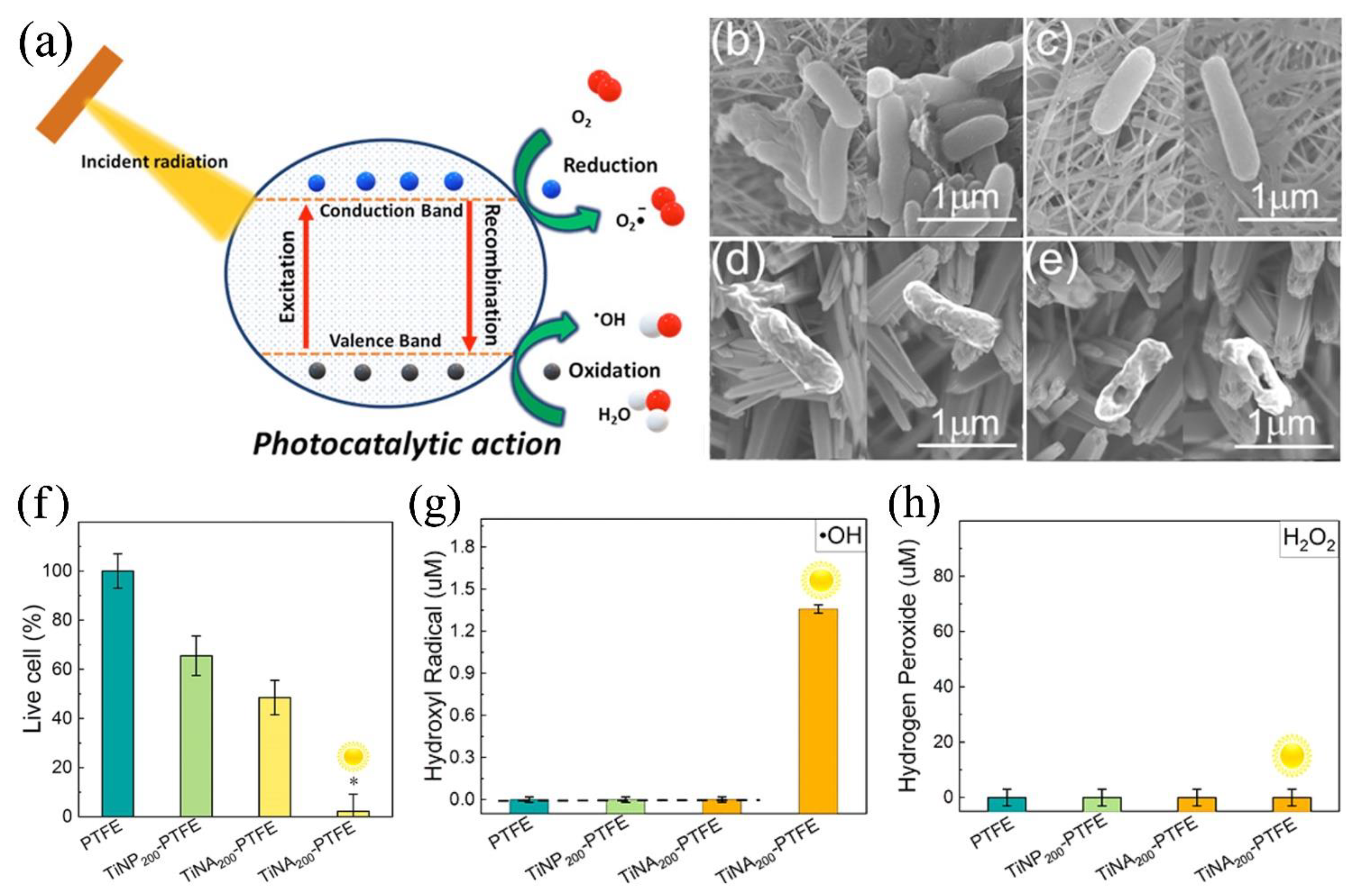
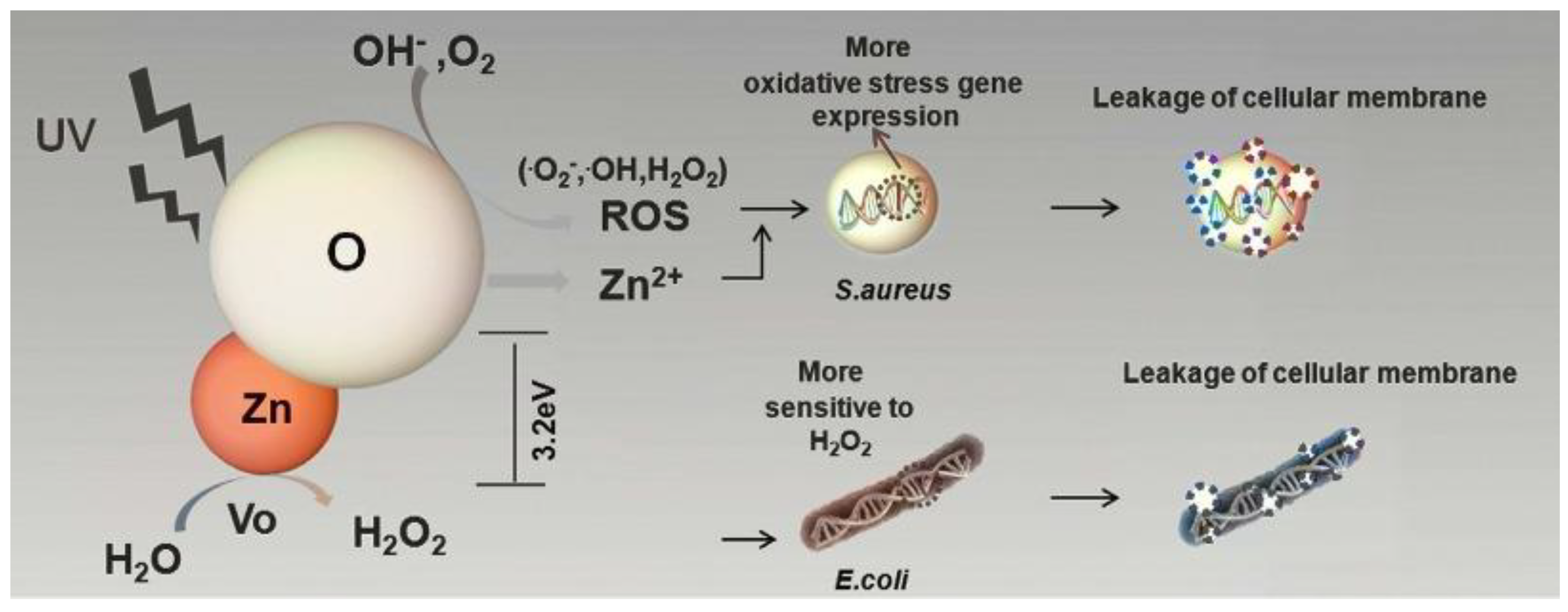
Despite the ease of obtaining titanium oxide by ALD, there are not many studies on its antibacterial properties (Table 1). The antibacterial activity of titanium oxide is strongly dependent on the influence of near UV light on the material. TiO2 is a semiconductor with a band gap of about 3.2 eV and therefore electron-hole pairs are formed under UV irradiation with a wavelength of less than 380 nm. In the presence of water, the conduction band electrons can react with molecular oxygen to form superoxide anions (O2) but the valence band holes oxidise water to hydroxyl radicals (OH) [47] (Figure 3a). These ROS can readily break down organic matter, disrupting the cell walls of bacteria and killing them.
The disruption of cell wall integrity due to ROS action can be observed by scanning electron microscopy. For example, Zhang et al. observed a significant bactericidal effect due to cell membrane damage of E. coli on the surface of TiO2 nanoarrays after sunlight irradiation (Figure 2b-e) [39]. In fact, according to a number of studies [29,30,39,54] significant antibacterial and antifungal activity is only shown when TiO2 is irradiated in the ultraviolet range. According to the results of Godlewski et al., who tested various ALD oxides on 16 different strains without additional illumination, TiO2 has significantly less bactericidal activity than not only ZnO, but also ZrO2, HfO2, Al2O3 [55]. The question of which types of ROS are the most active and play a more important role in the antibacterial effect of remains open, but Zhang et al. showed that hydroxyl radicals are predominantly formed and determine the activity against E. coli (Figure 3 f-h) for TiO2 nanoarrays obtained using combination ALD and solvothermal deposition [39].
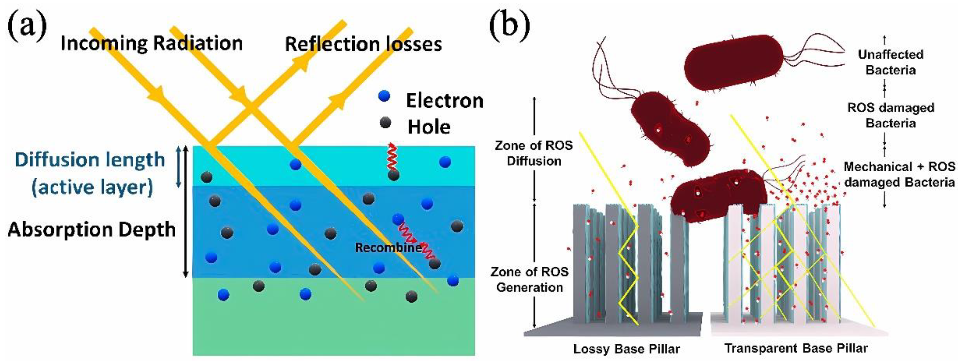
However, even when UV radiation is used, the results obtained by different authors vary considerably. The photocatalytic activity of TiO2 is strongly dependent on impurities and structural defects, which are particularly abundant in nanocoatings. Thermally generated defects of TiO2 could reduce the lifetime of the photogenerated electron-hole pairs, so the use of low temperature processes can help to reduce these defects [56]. In addition, the thickness of the coatings and the specific surface area can be important factors in increasing the number of ROS formed. Absorption of UV radiation extends to micron thicknesses but diffusion of ROS to the surface is only limited by the surface layer of a few nanometres (Figure 4) [47,50]. For ultrathin nanofilms, the substrate material also plays an important role, as the efficiency of photocatalytic antibacterial surfaces is limited by the absorption of light in it [47,50].
However, there is evidence that nanocoatings can generate ROS even without UV irradiation because they have many defects in their structure that add new levels to the forbidden band of the material. The antibacterial and antifungal activity of TiO2 under dark measurements has indeed been recorded in many studies [29,39,49,52,53,57,58]. However, the most likely cause of this phenomenon is a change in wettability or other surface properties after coating. In most of these studies, the authors observed an increase in the hydrophilicity of the surface, which leads to the realisation of the microbiostatic effect, i.e. a decrease in the adhesion of bacteria and their ability to form a biofilm. This effect is particularly evident for coatings applied to polymeric substrates such as polytetrafluoroethylene (PTFE) [39], polyamide 66 [58], PDMS [52] the initial surface of which is hydrophobic, and with the application of coatings is significantly hydrophilised.

3.1.2. Morphology and Topography of Surface

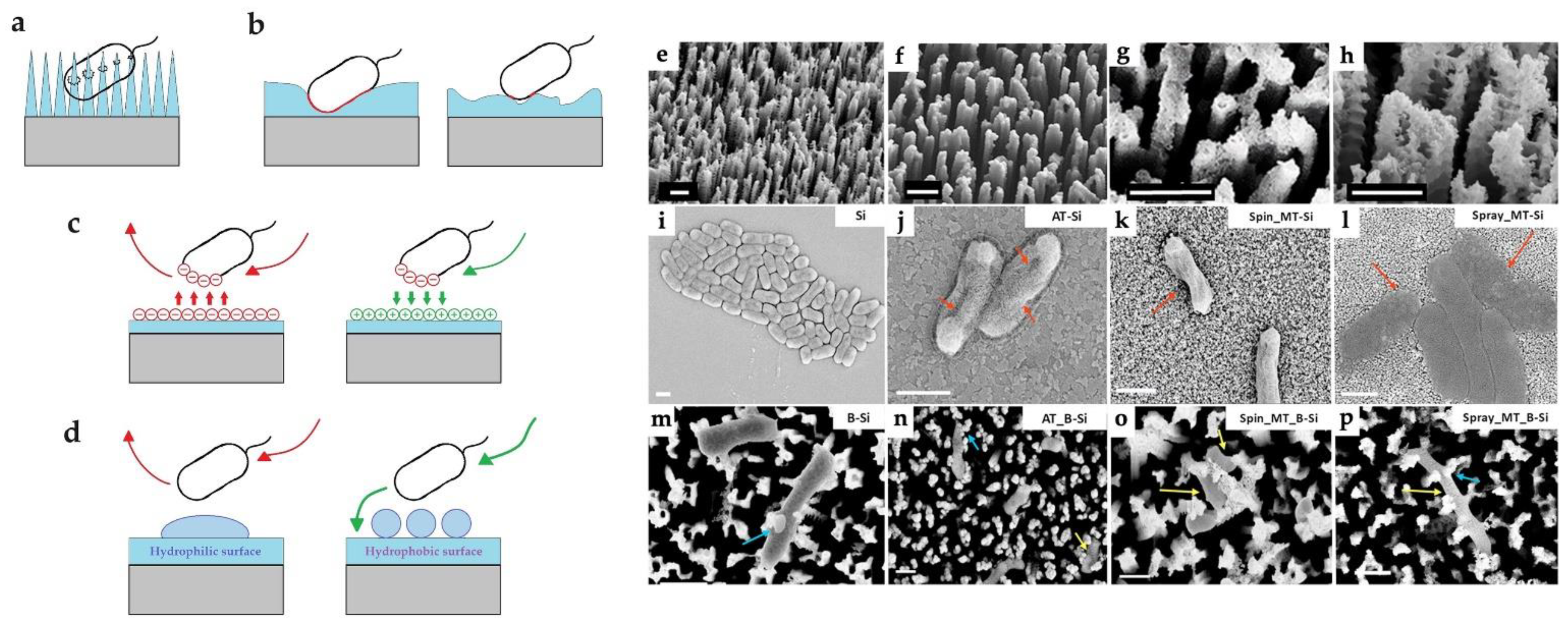
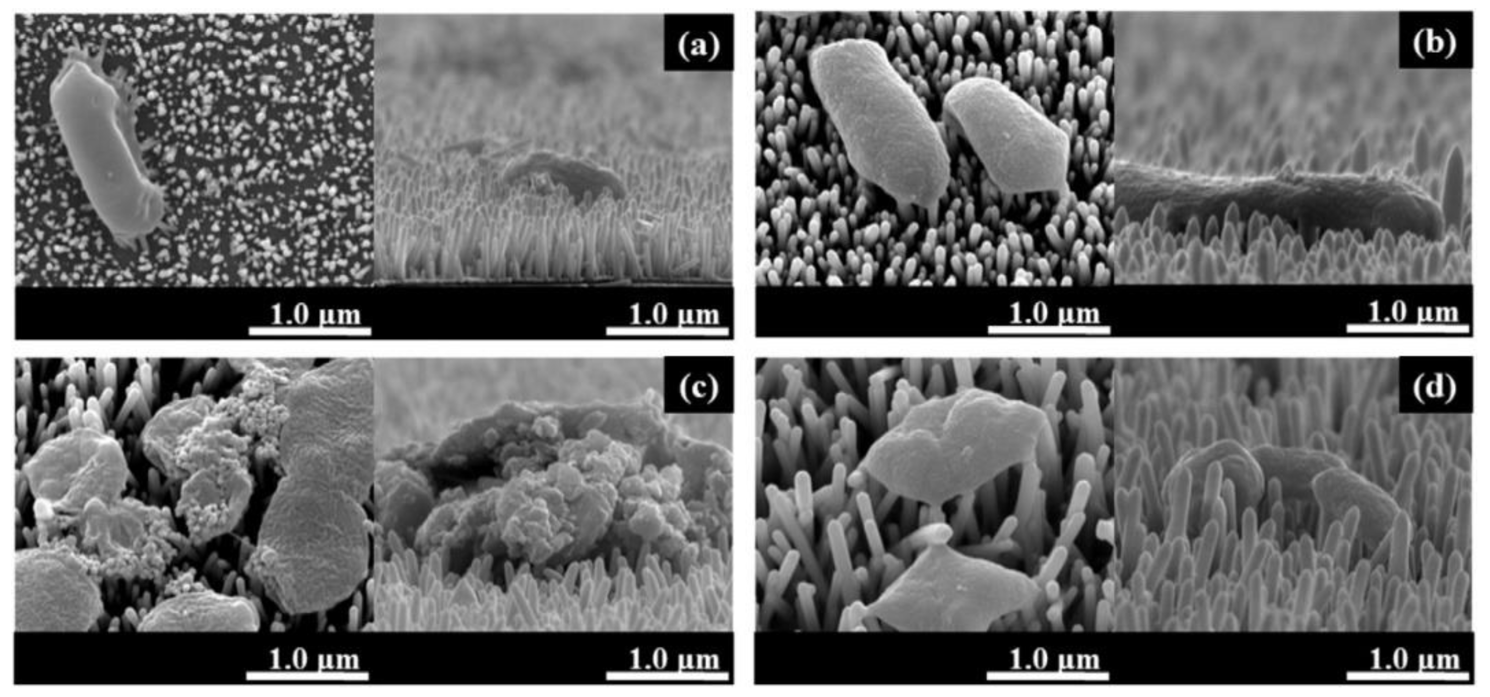
Surface morphology and topography can also be a critical factor in determining antibacterial properties. Surfaces with a certain morphology can cause significant stretching of the bacterial cell membrane, resulting in piercing (Figure 5a). Such disruption of cell membranes was observed by Singh et al. on samples of black silicon nanopillars coated with TiO2 using ALD, spin coating and spray coating (Figure 5e-p). In addition to the contact-killing effect, the surface morphology can significantly influence or even determine the type of bacterial adhesion by changing the area of the contact zones or the charge and wetting of the surface (Figure 5b-d). For example, the TiO2-coated nanoporous alumina membrane with a pore size of 20 nm inhibited the adhesion of S. aureus and E. coli, whereas the TiO2-coated membrane with a pore size of 100 nm did not [51]. A significant influence of surface morphology has also been reported in [37] where smooth ALD coatings, nanotubes and TiO2 nanoparticles were compared. Nanomaterials with more developed surface morphology showed better antibacterial activity against E. coli.

3.1.3. Thickness of the Coatings

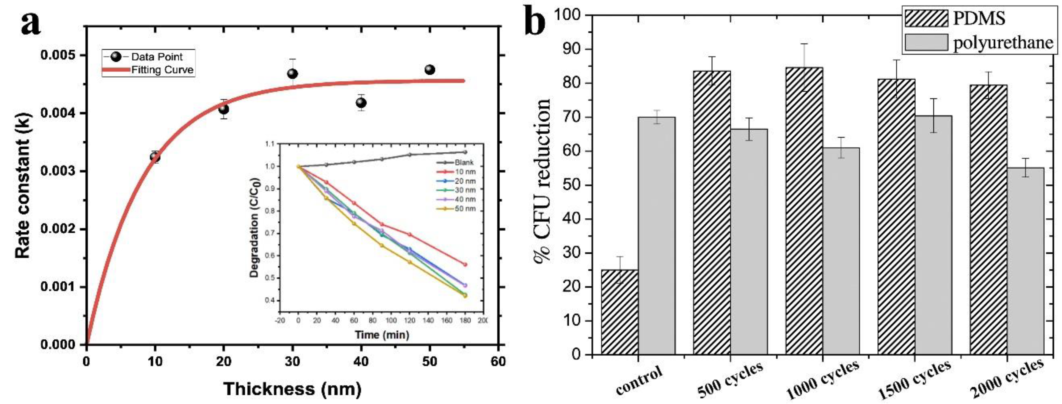
As discussed in Section 3.1.1, film thickness plays a significant role in ROS generation. At present, the literature presents results for ALD of TiO2 films with thicknesses ranging from 4 to 200 nm. It is difficult to compare results from different papers, but a number of researchers have considered the influence of thickness experimentally. It has been shown in [50] that the maximum degradation of methylene blue, which is an effective indicator of ROS generation, is achieved for titanium oxide on flat silicon at 40 nm and further increases in thickness have no effect (Figure 6a), as ROS are quite active and their diffusion is only possible from a thin near-surface layer (Figure 4a).
In the study Pessoa et al. TiO2 coatings of 50 to 200 nm thickness (500-2000 ALD cycles) on PU and PDMS surfaces were investigated against C. albicans yeasts and no noticeable difference was observed after UV exposure (Figure 6b) [29]. In the absence of light, the effect of thickness was recorded, but it was due to changes in surface free energy (SFE) and surface wettability. Comparative studies of lower thickness films were only carried out in one study, but did not provide any useful information on the thickness effect. 10 nm, 20 nm and 50 nm ALD TiO2 coated PA66 fabrics were investigated by Akyildiz et al. using the agar diffusion plate test with E. coli and S. aureus in both dark and sunlight exposure. All TiO2 coated fabrics showed no inhibition zone for both S. aureus and E. coli. However, the TiO2 coated samples were less covered with bacteria compared to the uncoated samples [58].

3.1.4. Crystalline Structure of the Coatings

Titanium oxide has three crystalline modifications (anatase, rutile, brukite), but rutile and brukite are only formed at relatively high temperatures. In ALD temperature regimes, only anatase is usually obtained, which is considered to be much more antibacterial than rutile [45]. However, crystallite formation is only possible from a thickness of about 10-12 nm [59], below which the film becomes amorphous regardless of the synthesis temperature. Lee et al. showed that increasing the synthesis temperature (from 70 to 300°C) and consequently the crystallinity improves the antibacterial properties against E. coli for TiO2 under UV exposure [54]. Molina-Reyes et al. also showed that a 10 nm TiO2 film with anatase structure deposited on amorphous TiO2 nanotubes significantly reduced the survival rate of E. coli [45]. However, measurements without additional illumination using S. aureus showed that crystalline coatings obtained at 190°C were even less antibacterial than amorphous coatings obtained at 160 and 120°C, and when tested on E. Coli there was no difference between the samples [49].
However, one of the most interesting effects of crystallinity on antibacterial properties is the synergistic effect of anatase and rutile, which significantly increases bactericidal activity due to the intrinsic alignment of the valence and conduction bands between the anatase and rutile phases. This alignment promotes the separation of photogenerated electrons and holes and increases their activity to generate ROS [45].

3.1.5. Precursors and ALD Temperature

Three main titanium precursors titanium (IV) isopropoxide (TTIP) [54,60], titanium chloride - TiCl4 [29,48,51] and tetrakis(dimethylamido)titanium - TDMAT [34,45,49,52,53] are used for the synthesis of TiO2 by ALD. The second reagent used is mainly water, but Darwish et al. used ozone [53] and Goldfinger et al. used oxygen plasma coupled to TDMAT to modify PMMA and PDMS respectively [52]. At the same time, coating with such a strong oxidant resulted in a significant decrease in the wetting angle (from 110 to <20), whereas modifying the PDMS surface with TiCl4/H2O, even with rather thick films of 100-200 nm thickness, practically does not change the wettability [29]. At the same time, the use of TiCl4/H2O has a significant disadvantage - chlorine contamination during synthesis at low temperatures (around 80°C). [29]. In the synthesis of complex oxide systems, there is also the possibility of chlorine contamination [61]. Titanium oxide synthesis temperatures can vary considerably. When using temperature sensitive substrates, temperatures from 65°C [53] can be used, and if required, crystalline coatings can be obtained up to 300°C [48,51] and higher.

3.1.6. Effect of Coatings on Different Strains

The vast majority of studies on ALD TiO2 coatings have been carried out on E. coli and S. aureus, including methicillin-resistant S. aureus (MRSA). Titanium oxide coatings of 4.3 and 8.6 nm thickness deposited on nanoporous alumina membranes are more effective against S. aureus than against E. coli [51], however Liu et al. showed that 100 nm thick films deposited on titanium at different temperatures (120 160, 190°C) have higher activity against E. coli than against S. aureus and MRSA [49]. However, most studies show no significant difference in activity against S. aureus and E. coli [30,55,58,62].
The fungal pathogen C. albicans has been used in several studies for the development of antibacterial materials in contact with the environment (dental implants, catheters, contact lenses [29,53,63,64]. TiO2 coatings are effective in UV treatment [29], but even in dark conditions the adhesion of C. albicans is significantly reduced due to the increased hydrophilicity of the surface [53]. Another study tested a range of oxide ALD coatings (ZrO2, ZnO, HfO2, Al2O3, TiO2) using 16 strains of Gram-positive and Gram-negative bacteria [55]. However, only the disc diffusion method was used and the study lacks depth and has conflicting results.

3.2. Doped TiO2 and Combined TiO2

The ALD technique permits employing various reagents and their arbitrary mixture at diverse synthesis stages, making ALD auspicious not just for acquiring pristine oxide coatings but also for doped oxide systems. Therefore, incorporating reagents besides the titanium-containing precursor and oxidising agent enables the doping of titanium oxide with diverse atoms [65]. Doping of titanium oxide can create further energy levels within its forbidden zone, generating a potent photocatalytic effect under both UV and visible radiation. This widens its potential usage as an antibacterial material given that only 4% of solar energy constitutes UV light [60]. The investigation of doped TiO2 by ALD has only been conducted in two works by a single scientific team [62,66]. The researchers studied thin films of TiO2, TiON, TiN, TiAlN, TiO2:V2O5 tested with E. coli and S. aureus. All the coatings showed antibacterial activity, even in daylight, but the results were more effective when exposed to UV radiation. The doped films generally showed superior performance compared to pure TiO2 [62]. The authors investigated vanadium-doped TiO2 nanofilms on polypropylene (PP) hernia meshes in vitro and in vivo. The coatings were found to be highly effective in preventing the adherence of S. aureus and E. coli, and showed excellent antibacterial activity. The research team of Takoudis et al. investigated mixed oxide systems consisting of TiO2 and ZrO2 on PMMA surfaces using C. albicans and S. oralis [63,64]. The TiO2-ZrO2 systems were found to be more effective than single oxides, despite conflicting results on bacterial adhesion and survival.
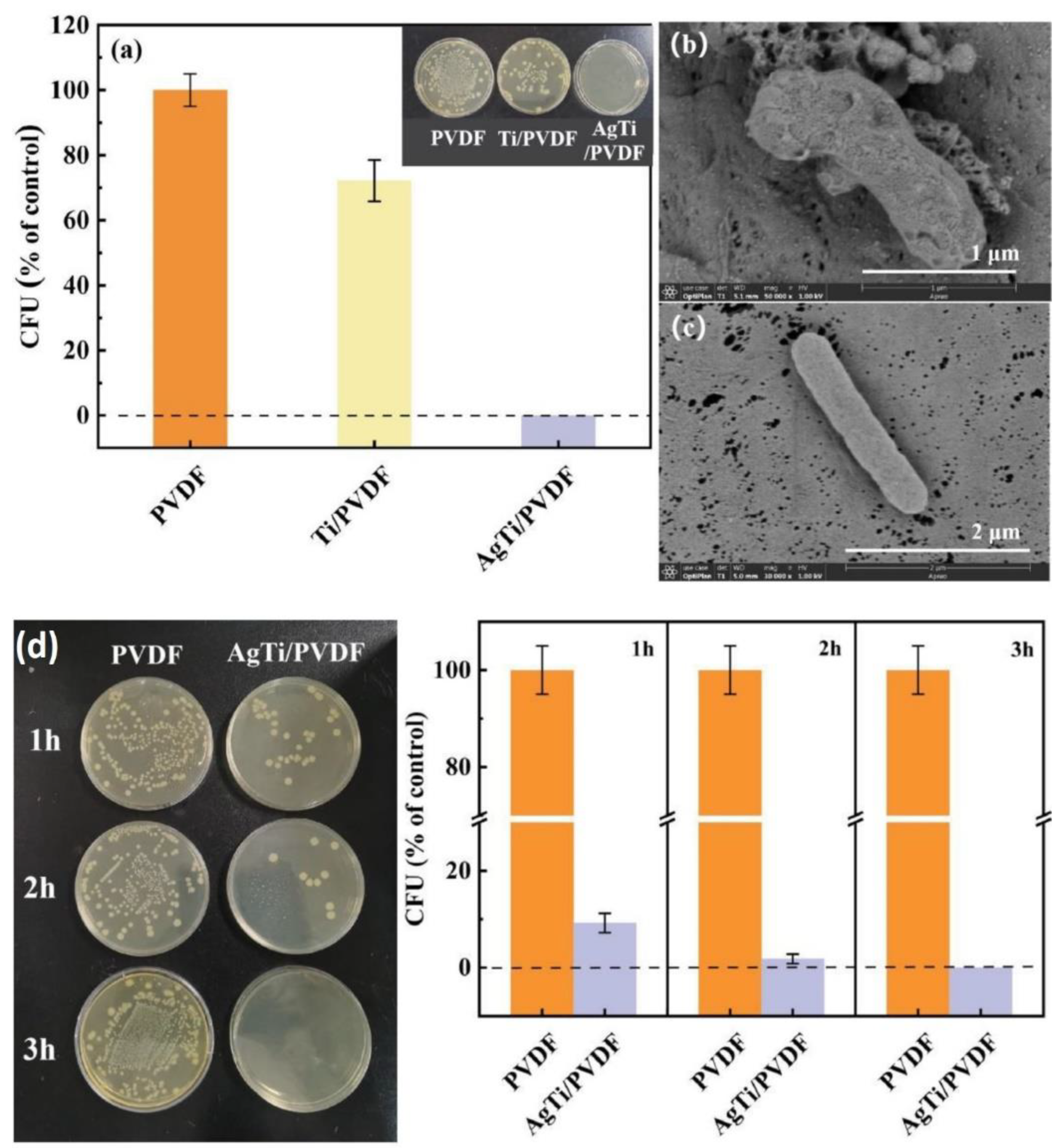
In recent years, noble metal sensitisation has emerged as an effective method for enhancing the photocatalytic and antibacterial activity of titanium oxide. This process reduces the recombination of photo-induced carriers at the noble metal/semiconductor interface [67]. Typically, noble metals such as silver [30,68,69] and gold [60] are deposited as nanoparticles on pre-synthesised ALD TiO2. While silver has been shown to possess powerful antibacterial properties due to Ag+ release, the formation of a Schottky heterojunction at the TiO2/Ag interface results in reduced electron-hole recombination rates. The consequence of this is the formation of an increased amount of bactericidal ROS when exposed to stimulated sunlight. Against S. aureus and E. coli, the antibacterial effectiveness of the TiO2/Ag coating was found to be 98.2% and 98.6% respectively [30]. Notably, TiO2/Ag samples demonstrated distinct deformation and cracking in both strains. The authors suggest that the rupture of membranes is attributed to ROS. However, they also observed that cell walls of numerous bacteria were destroyed when silver nanoparticles (NPs) were used without TiO2. In the other study the results of almost 100% killing efficiency of TiO2/Ag structures against E.coli was mainly achieved through Ag+ dissolution (Figure 7 a-d) [68].
Furthermore, Au nanoparticles (NPs) can be used as efficient sensitizers. As such, tiny amounts of Au NPs that efficiently absorb visible radiation are ideal materials to enhance the photocatalytic properties of TiO2 on the surface of polyvinylidene fluoride (PVDF) membranes, exhibiting potent antibacterial activity towards E. coli under direct solar irradiation [60].To understand such complex systems, it is necessary to continue to study and optimise properties such as the thickness and structure of the TiO2 layer, the size of the metal nanoparticles and the density of the distribution on the surface. For example, according to research by Zhang et al, the efficacy against E. coli in dark and sunlight conditions is significantly increased as the particle size of Au decreases, resulting in an improvement in the antibacterial properties. In addition, the thickness of the TiO2 film has a significant effect on the hydrophilicity, and efficiency of light radiation adsorption [60].

3.3. Zinc Oxide

3.3.1. Mechanisms of ZnO Bactericidal Effect and Their Role

ZnO, as well as TiO2, is an n-type semiconductor with a band gap around 3.3eV [70], meaning that absorption starts near the UV and it can generate oxidative ROS [71]. However, this is not the only factor that determines the antimicrobial activity of the coatings. Zn2+ ions can be released in the aqueous environment, easily transported across the cell membrane, interact with the active protease [72,73] and reduce intracellular ATP levels in bacteria [74]. It has also been reported that nanoscale ZnO can bind to proteins or carbohydrates on the cell surface, modify the amino acid side chains and cause cell death [75]. In addition, ZnO grows in the form of oriented crystallites, the morphology of which can lead to damage of the cell membrane during bacterial adhesion. Although the application of ZnO does not usually result in a significant increase in hydrophilicity, surface charge and surface energy factors may also be responsible to some extent for changes in the nature of bacterial adhesion. It is still debated which mechanism is most important and which is less important. To date, the influence of different mechanisms on the antibacterial properties of ALD ZnO has been investigated in several studies.
A number of studies show that the antibacterial activity of ZnO is either absent [54,70], or very low [76] under dark conditions (Figure 8). The importance of ROS is confirmed by data from Li et al. who investigated the effect of the active ROS scavenger vitamin E on reducing the antibacterial activity of 46 nm ZnO coatings against E. coli and S. aureus [77]. The question of exactly what kind of ROS are generated when ALD ZnO nanocoatings are exposed to UV radiation is still open. Park et al. recorded a significant amount of (a) O2-*, (b) *OH, and (c) 1O2 upon UV irradiation of 74 nm ZnO films on glass substrates [70]. The concentration of superoxide anions was found to be several orders of magnitude higher than that of the other ROS. Qian_et al. showed that 33 nm coatings deposited on PP nonwovens were active against E. coli even without light, but increased significantly under xenon lamp illumination. In this case, the main ROS species that are produced under the illumination is the singlet oxygen [78]. However, even in the absence of illumination, small amounts of ROS can form due to defect structures. The oxygen vacancies present on the ZnO surface can interact with moisture to form superoxide radicals and H2O2 [35,79].
Despite this, several other studies of ALD ZnO have shown that the influence of UV exposure is minimal or absent and that the main factor in the antibacterial activity is zinc ions [58,80,81]. This is also supported by the fact that the deposition of thin layers of Al2O3, acting as a diffusion barrier on the surface of ZnO, reduces the activity against E. coli [82]. Also, the thicker the Al2O3 coating, the lower the antibacterial activity. This result directly indicates the importance of dissolution of zinc ions from the surface.
The difference in the above results can hardly be explained by the influence of composition and structure of the obtained zinc oxide. The point is that in all the described works diethyl zinc is used as an ALD precursor and as a result polycrystalline coatings with similar morphology are formed irrespective of the synthesis temperature. The The coatings examined usually have a thickness of a few tens of nanometres, which is sufficient for significant ROS generation. The morphology of the substrate can have a small influence on the morphology of the ZnO, and indeed the substrates in the above-mentioned studies were different, but unfortunately no reliable conclusions about the influence of the substrate can be drawn from the data presented.
A detailed study of the influence of the contribution of different factors was carried out in the work where arrays of ZnO nanorods with different morphologies were coated with ALD Al2O3 [83]. It was shown that ZnO is effective even without light (96% inactivation of E. coli). At the same time, the application of a thin conformal layer of Al2O3 on the surface of ZnO reduces the antibacterial activity against E. coli, but it remains high (about 54%). This allowed us to conclude that the main factor in the activity is the relief and morphology of the surface, as ZnO nanorods can easily stabbed bacterial cells.

3.3.2. The Effect of Thickness

As zinc oxide is relatively soluble, the thickness of the coatings plays an important role in the duration of the antibacterial effect. However, excessively thick coatings can have adverse effects on certain applications, such as medical implants. For example, Wu et al. showed that relatively thick ZnO films (more than 50 cycles) on mesoporous TiO2 released too much zinc, which negatively affected the viability, proliferation and differentiation of MC3T3-E1 osteoblasts [84]. They also showed that the mesoporous structures overgrow with relatively thick coatings and cannot fulfil the role of drug carriers. A negative effect on MG-63 cell proliferation of 195-271 nm thick films was reported [26]. Cytotoxicity of 26 and 52 nm films on HS-27 fibroblasts was also observed [85]. Our recent work also showed the negative effect of 40 nm ZnO on the viability of MG-63 preosteoblasts and mesenchymal stem cells FetMSCs [61]. Li et al. found that 76 nm coatings did not significantly affect the adhesion and viability of MC3T3-E1, but had a negative effect on spreading [86]. However, in other study it was showed that very thin films around 5.3 nm conversely improved adhesion and spreading of MC3T3-E1 [87]. Indeed, research into the biocompatibility of ZnO coatings with varying thicknesses of less than 10 nm using MC3T3-E1 revealed a two-fold impact on cytological response. While cell differentiation significantly increased, cell proliferation decreased with increased coating thickness [43,84].
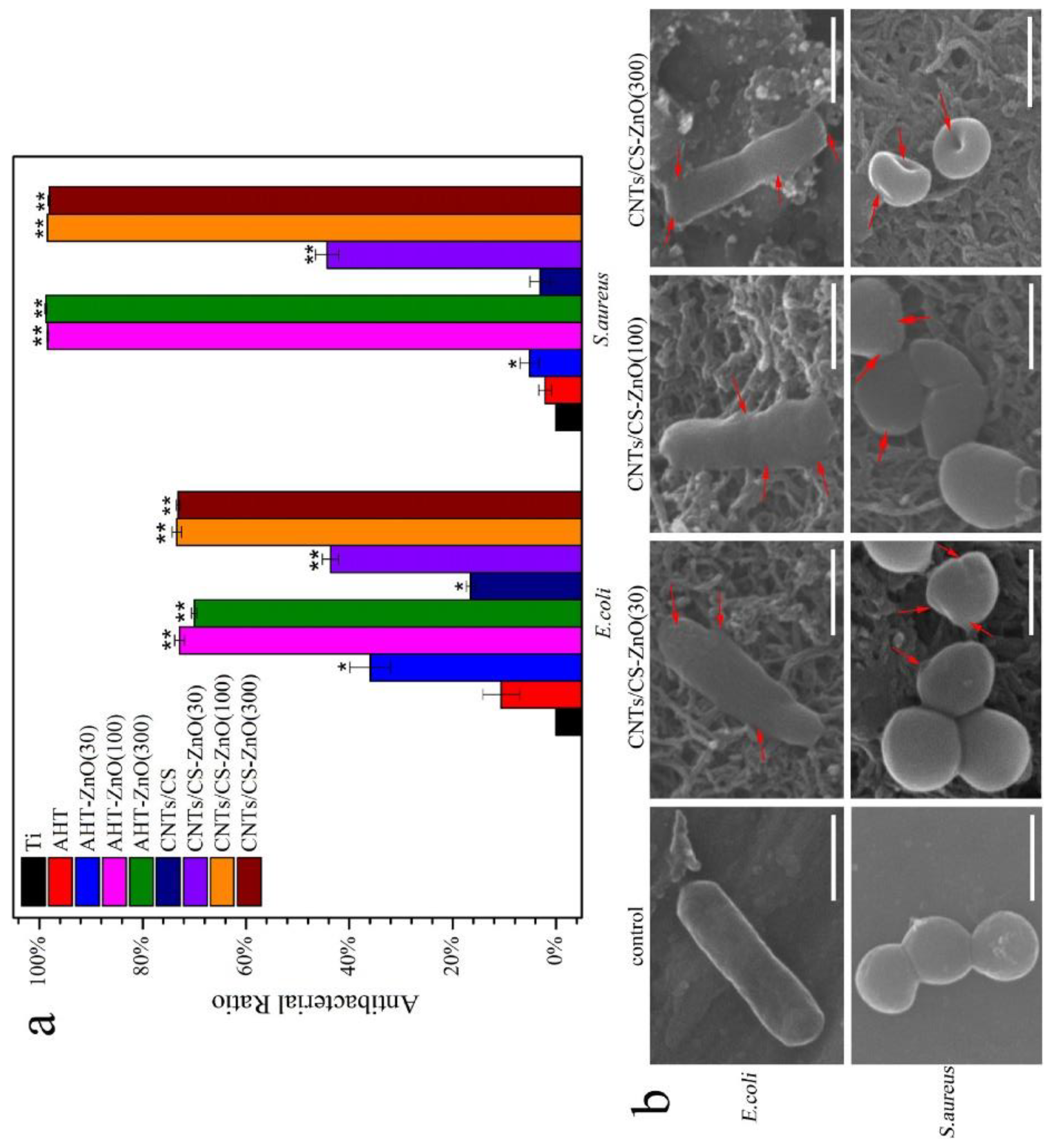
A similar situation is observed with bacterial cells. Puvvada et al. showed that ultra-thin zinc oxide films are not only ineffective, but actually promote bacterial growth, but from a thickness of about 2-10 nm (10-50 ALD cycles) the antibacterial effect begins to manifest itself sharply [36]. A similar threshold of several nanometers (30 ALD cycles) for E. coli and S. aureus is reported in another study [43] (Figure 9). At lower thicknesses, the opposite effect is observed and the authors suggest that Zn2+ at low concentrations acts as a nutrient for bacterial growth. In general, the vast majority of studies with films thicker than 5-10 nm confirm that increasing the thickness significantly improves the antibacterial effect [34,35,54,58,79,84].
However, there are several studies that found no effect of ZnO thickness on E. coli and S. aureus. For example, no difference in antibacterial activity was found between 26 and 52 nm ALD ZnO according to [85]. Kääriäinen et al. found no difference in antibacterial properties for 45, 95 and 285 nm thick films [71]. And Basiaga et al. states that no significant differences were found in the CFU adhering to the surface of 316LVM steel depending on the number (500 and 1500) of ALD cycles of ZnO [26]. It can therefore be concluded that a thickness of a few tens of nanometres is sufficient to achieve a significant antibacterial effect. The effect is observed even under dark conditions. Therefore, the elution of Zn2+ is probably responsible for the antibacterial properties of ZnO films.
As zinc oxide is soluble in an aqueous medium, it is crucial to focus on the changes in antibacterial properties that may occur. Lee et al. showed that the significant antibacterial activity of 55 nm coatings against E. coli was observed within the first few tens of minutes and reached a maximum after 1 hour [54], however, saturation of zinc dissolution is observed within the first few days for 100 nm coatings. [80]. According to Puvvada et al. the antibacterial effect increases with time and reaches a maximum at the end of 1 day [36]. Considering that the ALD coatings are very thin, it is important to know how long the antibacterial effect lasts. Yao et al. showed that thin coatings of about 5 nm on flat polished Y-ZrO2 are effective in the first days of presoaking, and the activity disappears on days 7 and 14 [87]. At the same time, it remains on the SLA-ZrO2 surface with an analogue coating. It can therefore be concluded that coatings of a few nanometres are sufficient for use in medical implants, as significant antibacterial activity is only required in the initial few days after surgery, whereas thicker coatings are required, for example, for water purification or antibacterial textiles.

3.3.3. The Effect of the Deposition Temperature

Several studies have investigated the effect of ALD temperature on the functional properties of ZnO coatings. According to the results of dynamic tests, Lee et al. showed that under UV exposure, the number of surviving E. coli decreases significantly in the transition from weakly crystalline ZnO obtained at 70 °C to fairly well crystallized ZnO obtained at 100 °C [54]. Increasing the synthesis temperature further to 125 and 200 °C slightly increases the number of surviving bacteria. Increased survival and improved adhesion of E. coli and S. aureus when the ALD temperature was increased from 100 to 300 °C has also been reported in [26]. Wang et al. showed that when ZnO was post-annealed between 225 and 275 °C, the change in survival of oral bacteria, including S. sanguinis, Bifidobacterium and Porphyromonas gingivalis, was non-monotonic and decreased between 200 and 250 °C, but increased at 275 °C [80]. This characteristic of the effect of annealing temperature on survival correlates very well with the release of Zn2+. Thus, although the synthesis temperature of ZnO has almost no effect on the structural and morphological characteristics, there is an influence on the antibacterial properties, but it is still poorly understood.

3.3.4. The Effect of the Morphology, Topography and Wettability

Surface morphology, topography, surface free energy, surface charge, and wettability are also important surface properties that influence antimicrobial activity. Furthermore, these characteristics are interrelated and must be considered together. Since the ALD process produces conformal and homogeneous coatings, the resulting topography and morphology are usually determined by the morphology and topography of the substrate. For example, Basiaga et al. showed that the polished surface of 316LVM steel with ZnO coatings was much less favourable for the adhesion of E. coli and S. aureus than the micro-rough surface obtained by SLA technology and subsequent ALD of ZnO [26]. Indeed, surface inhomogeneities comparable to the size of the bacteria can favour their adhesion (Figure 5b). However, nanoscale roughness should enhance the bactericidal properties, since in this case the specific surface area increases significantly and the release of Zn ions increases accordingly [80]. Experimental data show that even micro-roughened ZrO2 substrates obtained by SLA become extremely active against E. coli, S. aureus and P. gingivalis after ZnO deposition [87]. In addition, it has been shown that nanostructures with sharp edges or rods can pierce the cell membrane and lead to bacteria death [83] (Figure 10). Although ALD produces uniform films, their low thickness means that the influence of the substrate affects the wettability and surface energy. As a result, depending on the substrate type, ZnO films can exhibit both hydrophilic properties when grown on inorganic substrates [79,80,84], and hydrophobic properties when grown on polymeric substrates [43,77,78].

3.3.5. Effects on Various Types of Bacteria and Viruses

The issue of ZnO's antibacterial activity against different strains is very interesting. ZnO is very effective against the gram-negative E. coli and the gram-positive S. aureus strains. A comparative analysis of the results shows that in most cases the antibacterial activity against S. aureus and E. coli is either close [84,85,88] or higher for S. aureus [26,34,43,58,78]. However, in the dynamics, the antibacterial activity against E. coli was maintained for several hours, while the antibacterial activity against S. aureus decreased [77]. The authors explain this by the greater sensitivity of the E. coli membranes to ROS. Similar results were obtained for cellulose fibres coated with Al2O3/ZnO bilayer nanofilms [76]. E. coli were completely inactivated after only 30 min of UV irradiation, whereas S. aureus required at least 45 min of light exposure for 100% inactivation.
Of particular interest is the study where ZnO coatings were prepared with a surface enriched with either zinc or oxygen [79]. The oxygen-enriched surface was found to release more Zn2+ and ROS, resulting in greater efficacy against S. aureus. At the same time, the zinc-enriched surface releases more H2O2, which is more active against E. coli (Figure 11).
Despite the apparent efficacy against E. coli and S. aureus, ALD ZnO is not effective against all strains. For example, Skoog et al. showed lack of activity against Pseudomonas aeruginosa, Enterococcus faecalis and Candida albicans [89]. Vaha-Nisi et al reported lack of activity against the bacterium Bacillus subtilis and a fungus Aspergillus niger [82].
Antiviral activity has also been investigated in several papers, but the results are rather contradictory. Kumar et al. showed the efficacy of ZnO applied to fibrous silk against coronavirus and rhinovirus and Dicastillo et al. with high antibacterial activity against E. coli and S. aureus found no activity against norovirus [90].

3.4. ZnO Based Nanocomposites and Nanolaminates

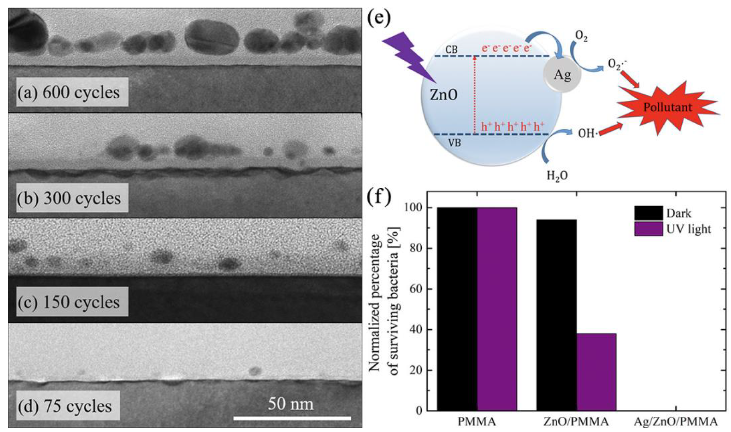
As in the case of titanium oxide, noble metal sensitisation can also be effective for zinc oxide. In the work of Di Mauro et al. ALD Ag NPs with sizes ranging from 4 to 25 were synthesised on a previously prepared ALD ZnO ultrathin film (Figure 12a-d) [38]. Such heterogeneous photocatalysis showed high efficiency in the decomposition of various organic pollutants in aqueous solution: methylene blue (MB), paracetamol, and sodium lauryl sulfate, but the authors paid very little attention to antibacterial properties. The obtained ZnO-Ag systems were very effective against E. Coli in dark and UV light conditions (Figure 12f), but unfortunately the authors did not consider the effect of silver particle size on the bactericidal properties. Akyildiz et al. synthesised photocatalytic systems by depositing a 20 nm layer of ZnO on cotton fibers followed by photochemical deposition of Ag NPs [33]. However, the antibacterial effect of silver was too high to draw any conclusions about metal sensitisation of ZnO. ZnO nanorods decorated with Ag NPs were very effective against E. coli according to another study [91].
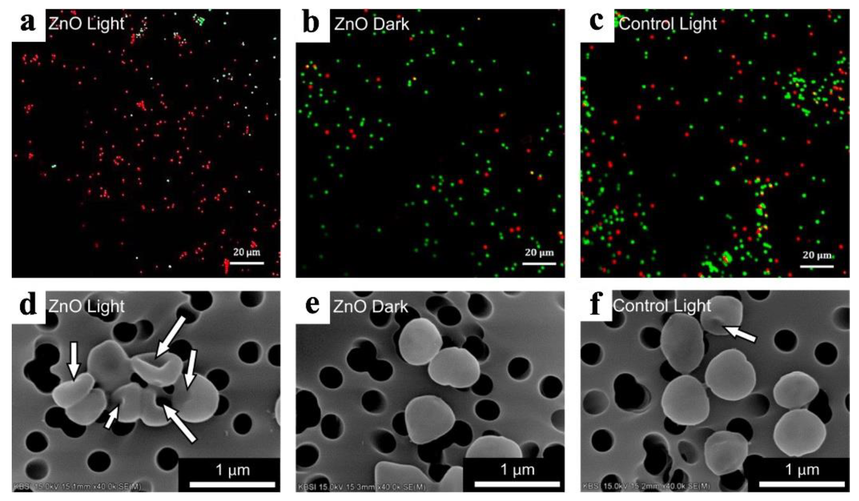
Liu et al. synthesised cubic Cu2O particles on the surface conductive and transparent ITO glass [31]. Then ZnO coatings were applied for the formation of p-n heterojunction which limit recombination of electrons and holes and improve photocatalytic efficiency. This heterojunction was effective against S. aureus both at dark and simulated sunlight condition (Figure 13). The results showed that bactericidal effect achieved due to both high effective ROS formation and Cu/Zn ions elution. Sr doped titania nanotubes were coated with 30 cycles of ZnO and then grafted with octadecylphosphonic acid (OPDA) [44]. Sr-ZnO composites showed high activity against S. aureus and E. coli, but the OPDA coating decreased activity due to shielding and reducing the dissolution of Zn ions.
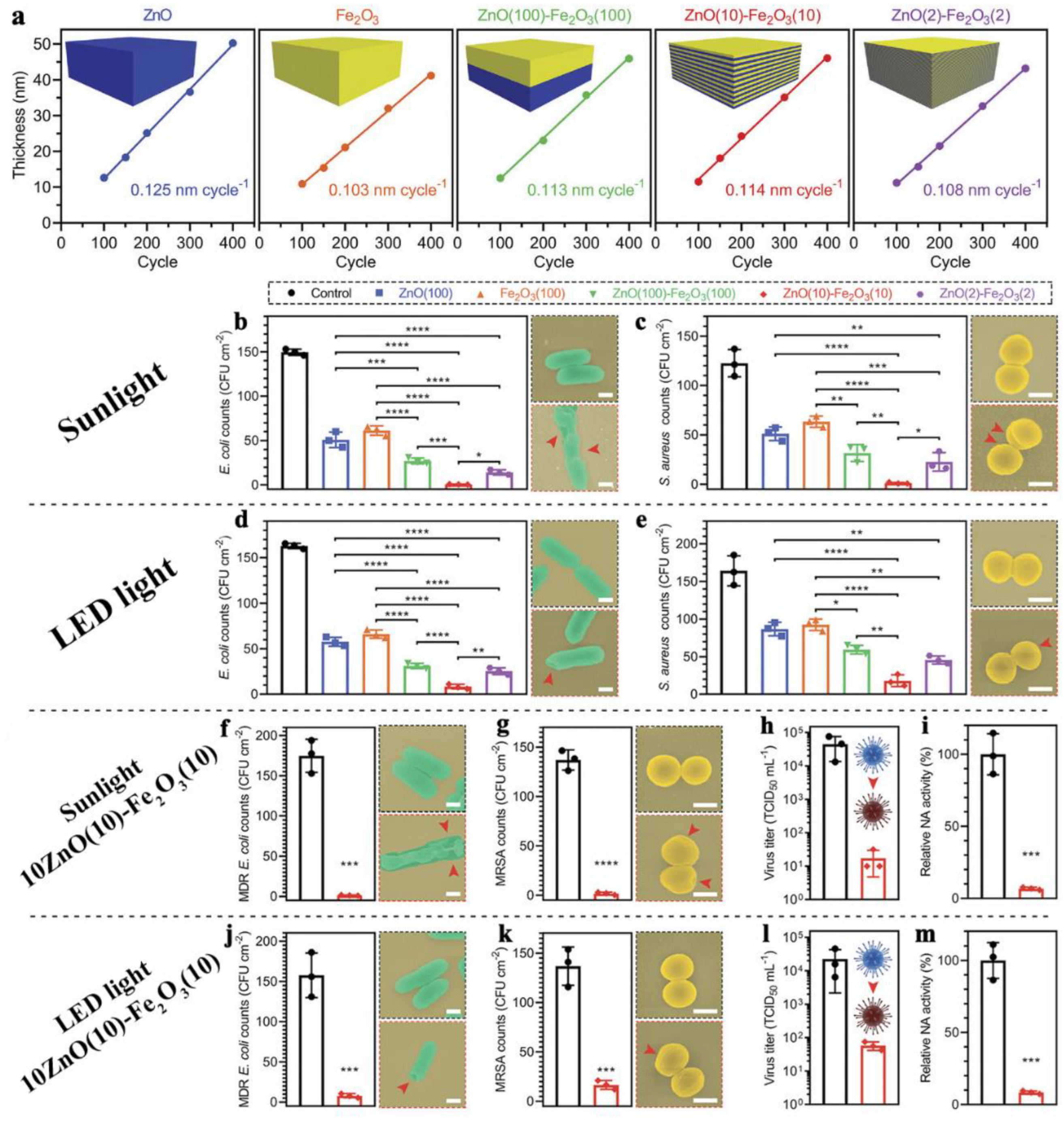
Recently, several mixed zinc oxide-based oxide systems have been successfully obtained. Li et al. synthesised ZnO/FeOx nanolaminates using 200 cycles of ALD (21-25 nm thickness) consisting of two, twenty and one hundred alternating layers (Figure 14a) [92]. The nanolaminates were activated using simulated solar light and LED light. The authors believe that such nanolaminates facilitate the transfer of numerous photoelectrons from ZnO to Fe2O3 under light irradiation, thus generating a significant amount of ROS. The best antibacterial activity against E. coli and S. aureus was observed for the twenty-layer sample in both light regimes (Figure 14b-e). At the same time, the nanolaminates showed high antiviral activity against H1N1 (Figure 14f-m) and no cytotoxicity against NIH-3T3 fibroblasts. The obtained heterostructures retain high transparency, have high abrasion resistance and can be used as effective antibacterial coatings for touchscreens. ALD coatings based on ZnO and Al2O3 (AZO), obtained in different ratios but with the same thickness of 50 nm, were investigated for antibacterial and antifungal properties [32]. AZO showed high efficacy against bacteria (E. coli and S. aureus) and 5 commonly found in real life moulds, both in the presence of natural light and in the absence of light. The Zn ion elution of AZO was significantly lower than that of pure zinc oxide, which is very important for the development of antibacterial moisture barrier films for next generation flexible touch display functional optical films.
Recently, we have investigated complex oxide systems based on ZnO and TiO2 (ZTO) with a thickness of about 40 nm [61]. ZnO and ZTO samples were very effective against S. aureus, A. baumannii, and P. aeruginosa. In this case, unlike pure zinc oxide, ZTO had no negative effect on the adhesion of fetal mesenchymal stem cells (FetMSCs).

3.5. Other Compounds

Many other oxide compounds can be produced through ALD, in addition to titanium oxide and zinc oxide. It is possible that some of these compounds also possess antibacterial properties. Godlewski et al. compared a series of rather thick films (about 200 nm) of wide bandgap oxides (ZnO, HfO2, TiO2, ZrO2, Al2O3) deposited on the surface of paper discs [55]. The authors tested 16 gram-positive and gram-negative strains using the agar disc diffusion method without illumination. No bactericidal effect was found for B. Cereus, S. agalactiae, Streptococcus alpha-haemolytic, E. Coli, Klebsiella spp., Salmonella BO, Yersinia spp, but against B. Subtilis, S. aureus ATCC and MRSA, S. Epidermidis, S. Pseudintermedius, Streptococcus beta-haemolytic, Proteus spp. and Salmonella DO most oxides were active. Compared to ZnO, ZrO2 and HfO2 nanocoatings showed similar or better antimicrobial activity [55]. ZrO2 is a biocompatible material that stimulates stromal and mesenchymal cell proliferation and differentiation in osteogenic direction [93,94,95], but 20 nm ZrO2 coatings reduce the adhesion of S. mutans and P. gingivalis [96]. Two recent studies have demonstrated the high efficiency of TiO2-ZrO2 complex oxide systems [63,64] against C. albicans and S. oralis on the PMMA surface. No significant antibacterial activity was observed for pure ZrO2.
It is noteworthy that according to Godlewski et al, Al2O3 performed even better than TiO2 in most of the cases [55]. As shown in [85], Al2O3 coatings of 13.5 and 27 nm thickness on cellulose-based fibres can also exert an antibacterial effect against S. aureus and E. coli, which is slightly lower than that of ZnO coatings. At the same time, Al2O3, unlike ZnO, was shown in the same study not to be cytotoxic against human HS-27 fibroblast and human keratinocyte standardized cell line (HaCaT). The antibacterial effect of 12.5 and 25 nm thick Al2O3 coatings against E. coli has also been demonstrated [82]. In general, all authors agree that Al2O3 is less active than ZnO [55,82,85].
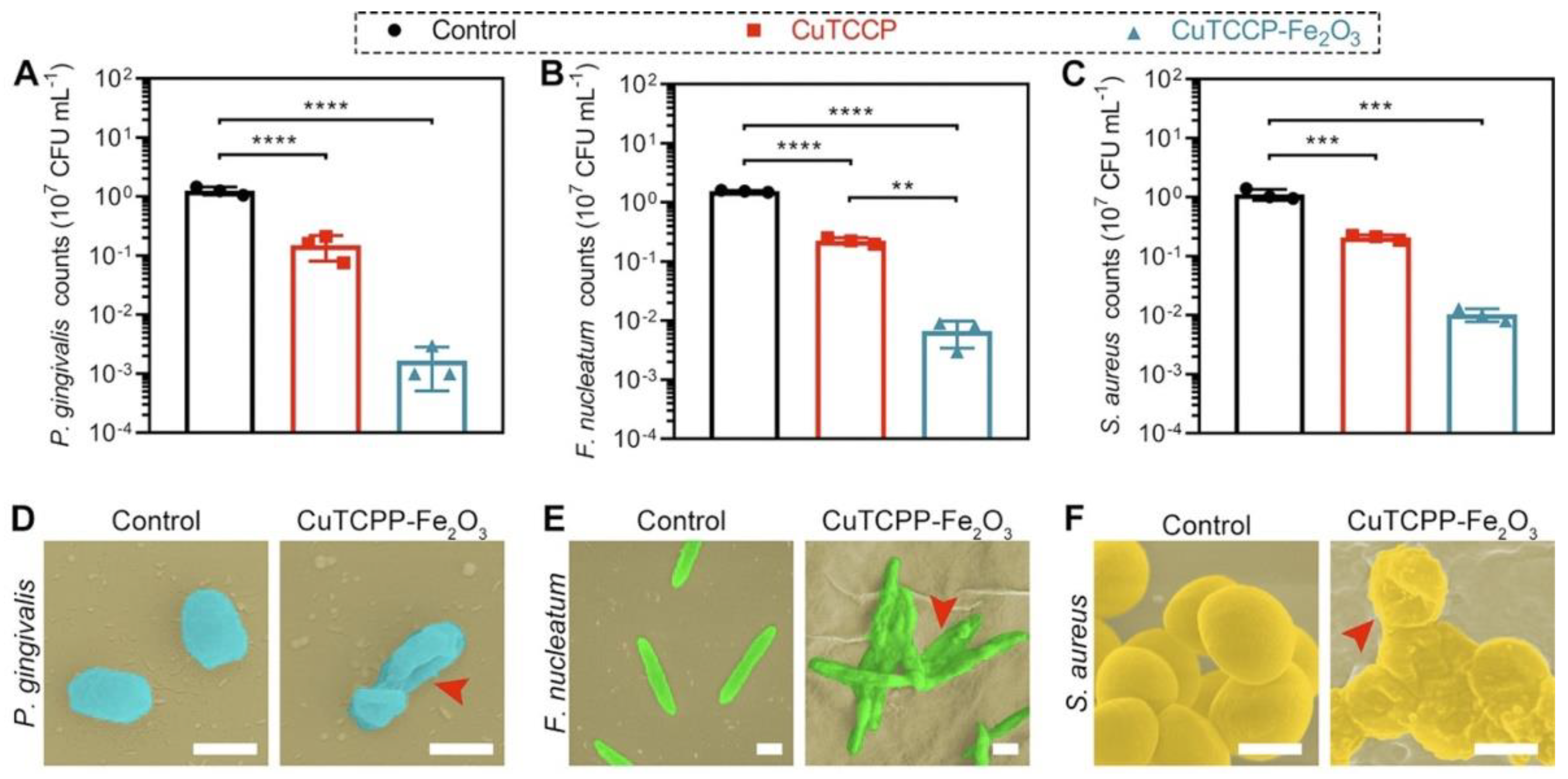
Of particular interest are pair of papers by Li et al. [86,97]. The authors prepared two-dimensional porphyrinic zinc and copper containig metal−organic framework (2D MOF) coated Fe2O3 using the ALD. Such materials have been shown to be effective in vitro and in vivo against various oral pathogens (P. gingivalis, Fusobacterium nucleatum, and S. aureus) due to both ROS formation and released ions (Figure 15). The enhanced photocatalytic activity of the ALD modified MOFs results from the lower adsorption energy and more charge transfer due to the synergistic effect of metal−linker bridging units, abundant active sites, and the excellent light-harvesting network. The in vivo results showed that this FeOx modified MOFs can be used in photodynamic ion therapy of periodontitis with a greater therapeutic effect than the reported clinical treatment with minocycline and vancomycin. In another study, it was shown that visible light transparent ALD ZnO–Fe2O3 superlattice nanocoatings are effective bactericidal and virucidal materials (H1N1) [92]. The action of such superlattices is based on the photocatalytic mechanism, where ZnO and Fe2O3 served as electronic donors and receptors, respectively. The heterointerface between ZnO and Fe2O3 facilitated the transfer of numerous photoelectrons from ZnO to Fe2O3 under light irradiation and promotes the ROS formation.
Furthermore, in addition to the aforementioned familiar compounds, obscure substances have exhibited antibacterial properties. For example, Radtke et al. demonstrated significant inhibition of staphylococcal colonisation and biofilm formation for titanium oxide nanotubes (TNT) with ALD-coated CaCO3 subsequently converted to hydroxyapatite. [98]. In previous work by this research group, TNT formed on the Ti6Al4V alloy were shown to have in vitro anti-biofilm properties themselves when tested on the S. aureus model. [99]. But the double modified biomaterials exhibited stronger antimicrobial activity than the surface with TNT, indicating a synergistic effect of both modifications [98]. A recent paper by Saha et al. demonstrated the antibacterial activity of conformal MgO nanocoatings deposited on collagen membranes against polymicrobial biofilm from human saliva [64].
TaN is widely used for the implant coating material due to its high mechanical strength, high chemical stability, and high biocompatibility but also TaN coating has a tremendous anti-bacterial effect [28]. TaN coating decreased E. coli and S. aureus colony formation, and due to the increased water contact angle and hydrophobic properties.

3.6. Silver and Other Metals

Metallic silver is one of the best known and most successful antimicrobial, antifungal, antiviral and anti-inflammatory materials [100]. Silver has been used as an antibacterial and medicinal agent for thousands of years [101], but its importance declined with the discovery of antibiotics. However, the problem of the emergence of antibiotic-resistant strains soon arose. This increase in antibiotic resistance has led to renewed interest in the use of silver as an antibacterial agent [102].
The antimicrobial activity of Ag-coated surfaces is commonly attributed to the strong oxidative effect of silver in the course of the gradual release of silver ions into the biological environment. However, the possible mechanisms of silver's antibacterial action are quite diverse. Ag ions induce the oxidative stress effect on cell membranes, damaging and penetrating them. In addition to its own chemical activity, silver ions can induce the formation of reactive oxygen species (ROS), which can also damage organelles and impair their function. Ag ions have many possibilities to disrupt the functioning of cell organelles and biochemical processes such as respiratory activity and intracellular ATP levels, inhibition of DNA replication. This is due to the fact that silver ions are Lewis acids and can easily interact with thiol and amino groups of proteins, nucleic acids and others [101]. Such diverse and potent effects make silver effective against gram-positive and gram-negative bacteria. However, this high activity can result in a negative cytotoxic response of body cells and cause silver accumulation in the internal organs.
However, despite these problems, a number of studies have shown that ultrathin nanosilver coatings do not exhibit this cytotoxicity. The effect of silver on different cells and tissues depends on both the size of the Ag structures and the cell type being studied in vitro. Several studies have shown that nanoparticles of 50 nm or less at low concentrations are not cytotoxic to mouse embryonic stem cells (mESC) [103], human umbilical vein endothelial cells (HUVEC) [104], normal human fibroblasts (FN1) [104], human mesenchymal stem cells hMSC [105,106,107], L929 fibroblasts [108,109] and human keratinocyte cell line HaCaT [110]. However, at high concentrations, such nanoparticles are toxic, as demonstrated in human embryonic kidney HEK293 [111,112], mouse embryonic fibroblast cells NIH3T3 [113] and human dermal fibroblasts HDFs [114].
The importance of dose and AgNPs size for their toxicity has also been demonstrated in a number of in vivo studies using different routes of administration (inhalation, intravenous and intraperitoneal) [115,116,117,118,119]. Thus, the issue of nanoparticle size and concentration, and in the case of nanofilms, their thickness and morphology, is the most important issue in studying the medical applications of silver.

3.6.1. ALD of Silver Nanoparticles and Nanocotings

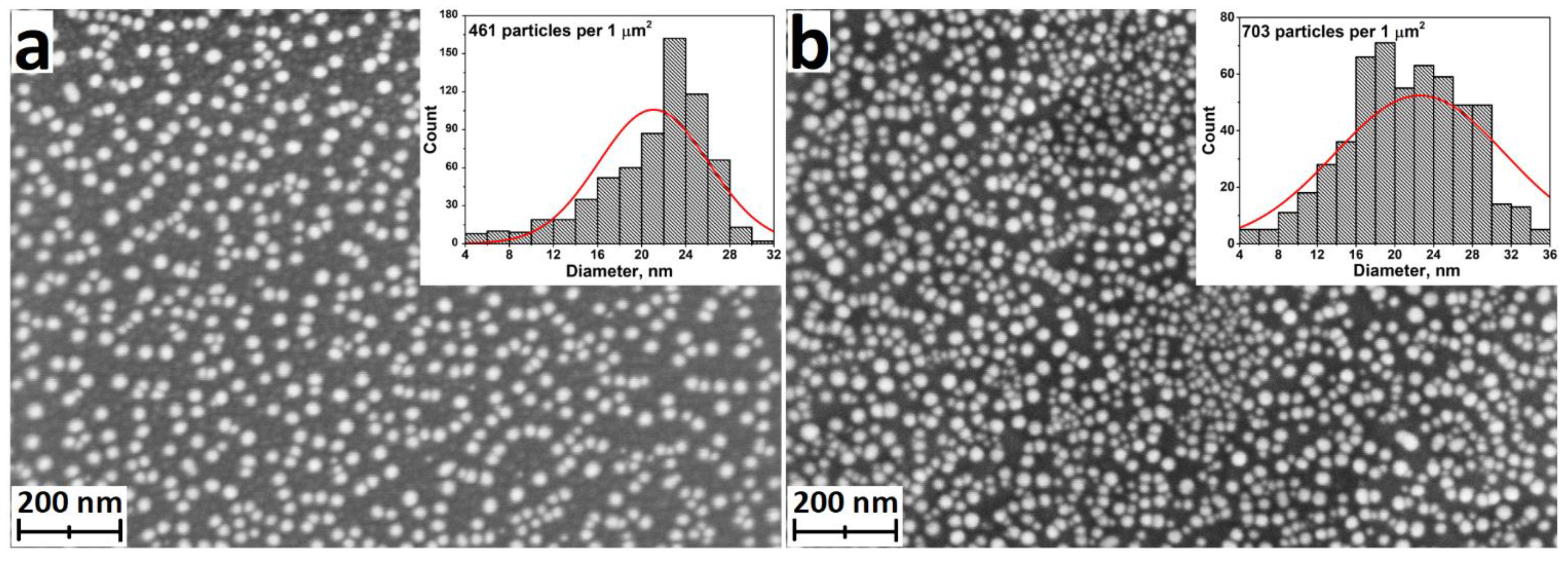
Although ALD is very good at producing oxides and a number of other binary compounds, there are approaches that allow the production of metallic films. However, continuous metallic nanofilms are not easily obtained by ALD. The problem is that growth of metals suffers from poor nucleation and initiates island growth [120,121]. This is particularly characteristic of silver ALD, where NPs grow in a relatively small number of cycles (Figure 16). Many ALD cycles are therefore required to obtain a continuous film. Nevertheless, this feature can be considered positive, as nanoparticles increase the specific surface area and hence the dissolution rate of silver ions compared to continuous films.
Although silver has been produced by ALD for a very long time, there are only a few papers investigating the antibacterial properties of ALD-Ag (Table 2). Radtke et al. investigated silver NPs with diameters of 7.8-9.2 nm deposited on titanium oxide nanotubes with diameters ranging from 10 to 84 nm [102]. An inhibitory effect on biofilm formation of S. aureus was found using the suspension method of study. The samples with the smallest diameter of nanotubes were the most effective as they have a large specific surface area.
Recently we synthesized larger diameter silver NPs (16-30 nm) on the surface of flat titanium discs. The results showed the inhibitory effect of silver on the growth of S. aureus. The ALD synthesis of an additional sublayer of crystalline TiO2 enhanced the inhibitory effect [57]. A similar synergistic effect on E. coli was observed in [68], where the antibacterial properties of an ALD layer of TiO2 and Ag NPs prepared by photodeposition were investigated. This effect has been discussed in more detail in Section 3.2.
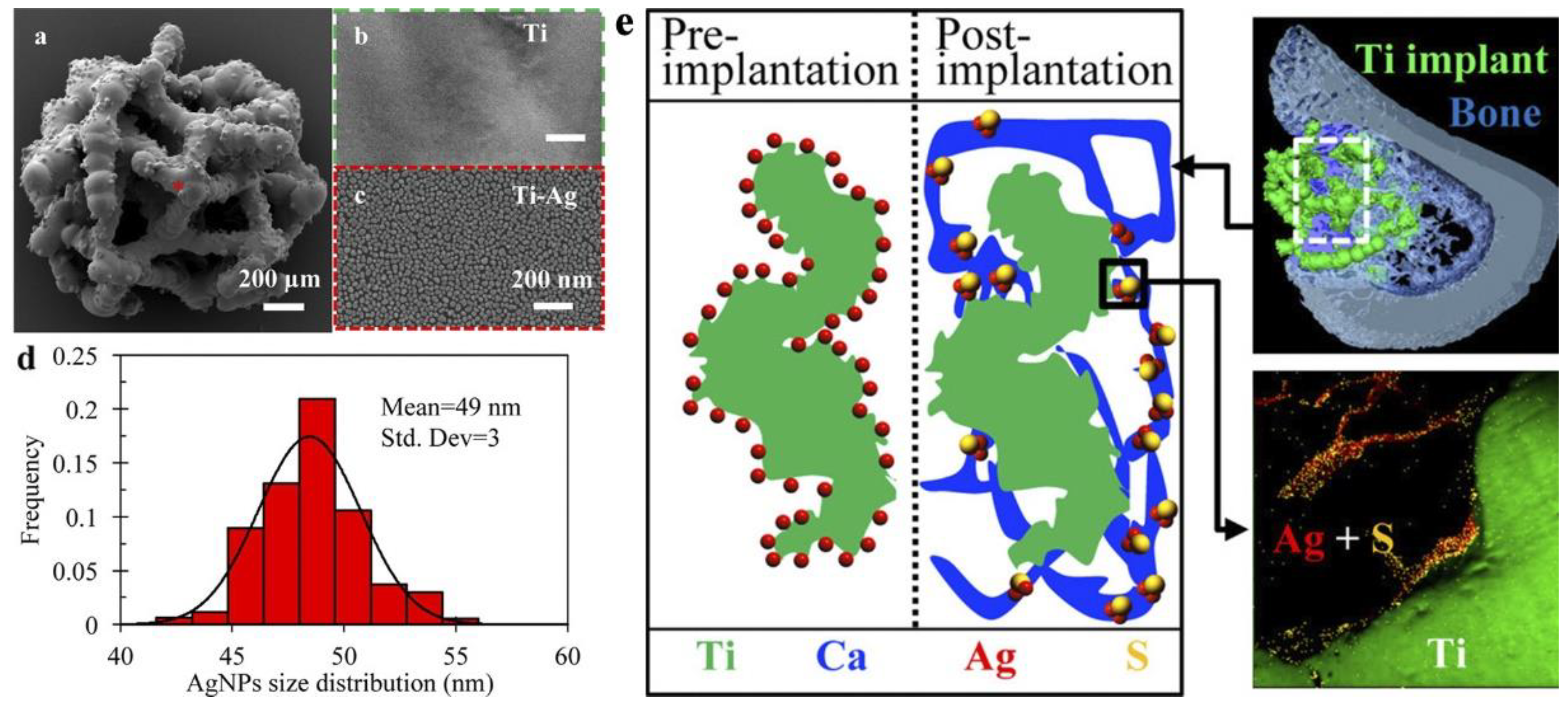
Several studies have shown that, in addition to their antibacterial effect, ALD silver NPs can have a positive effect on the adhesion and proliferation of fibroblast and osteoblast-like cells. [57,102,122]. In vivo studies of Ag-decorated porous titanium scaffolds in rat tibial defects rats showed that Ag NPs induced robust osteogenesis and angiogenesis with no evidence of systemic toxicity and silver accumulation in the liver. [27,122]. Further in vivo studies by the authors over 12 weeks showed that Ag accumulation occurs within the osseous tissue immediately adjacent to the implant surface. TEM and selected area electron diffraction (SAED) show that silver is part of the less toxic Ag2S within the newly formed bone (Figure 17) [27].
In terms of practical applications, the work [123], in which Ag nanoislands were obtained on the surface of N95 medical masks at relatively low temperatures (90 and 120 °C) by ALD is of great importance. Such coatings can be very effective in increasing the service life and reliability of personal protective equipment (PPE), as such particles prevent microbial contamination and accumulation in the membranes and fibres of PPE. In addition, the prepared Ag nanoparticles were stable to washing. According to the authors, ALD is ideal for coating PPE because it provides high adhesion of nanoparticles through chemisorption, does not require the use of capping agents, is performed in the gas phase, which can be important for liquid-sensitive materials, and avoids blocking the pores and negatively affecting the air permeability of the mask.
Although the results [123] show an improvement in the suppression of biofilm formation with smaller silver particles, the effect of particle size on its antibacterial activity and cytotoxicity/biocompatibility remains controversial. The duration of the silver effect and the onset of action also remain open, as the results showed that 2 h was not sufficient for effective bacteria/Ag contact, whereas after a longer time (24 h) effective contact occurred, resulting in a reduction in biofilm formation [123].
A major problem in silver ALD is the lack of a good precursor. A reproducible and controlled ALD process requires sufficient vapour pressure and thermal stability of the precursor. The most successful precursor is (2,2-dimethyl-6,6,7,7,8,8,8-heptafluorooctane-3,5-dionato) silver(I) triethylphosphine (Ag(fod)(PEt3)). However, to achieve the necessary vapour pressure, it must be heated to temperatures above 80-100 °C, which can lead to slow decomposition of Ag(fod)(PEt3). Furthermore, the optimum growth temperature for this compound varies within a fairly narrow range of 120-160 °C [24].
In addition, most synthesis techniques for Ag production by ALD use hydrogen plasma as a reducing agent. As has been shown in a number of papers, the use of plasma as a co-reagent causes problems in the treatment of complex shaped surfaces [124]. Plasma is very unstable and quickly "loses its activity" when treating deep channels. Therefore, it is necessary to carry out studies on the selection of an optimal reducing agent. In [123], borane dimethylamine complex ((CH3)2NH*BH3) was tested as a co-reagent. The results indeed showed silver growth in the inner layers of fibrous structure of the N95 filter media.

3.6.2. ALD of Other Metallic Nanocotings

Besides silver, several metals including copper, gold and iron have strong antibacterial properties [125,126]. These metal layers have been successfully fabricated by ALD, but only two studies have investigated their antibacterial properties. TiO2 inverse opal structures were functionalised with an ultrathin ALD Cu layer (1.1 nm Cu loading) [127]. A significant reduction in the CFU count of S. aureus was observed on the Cu containing samples pretreated at 550 °C to increase the stability of the Cu particles.
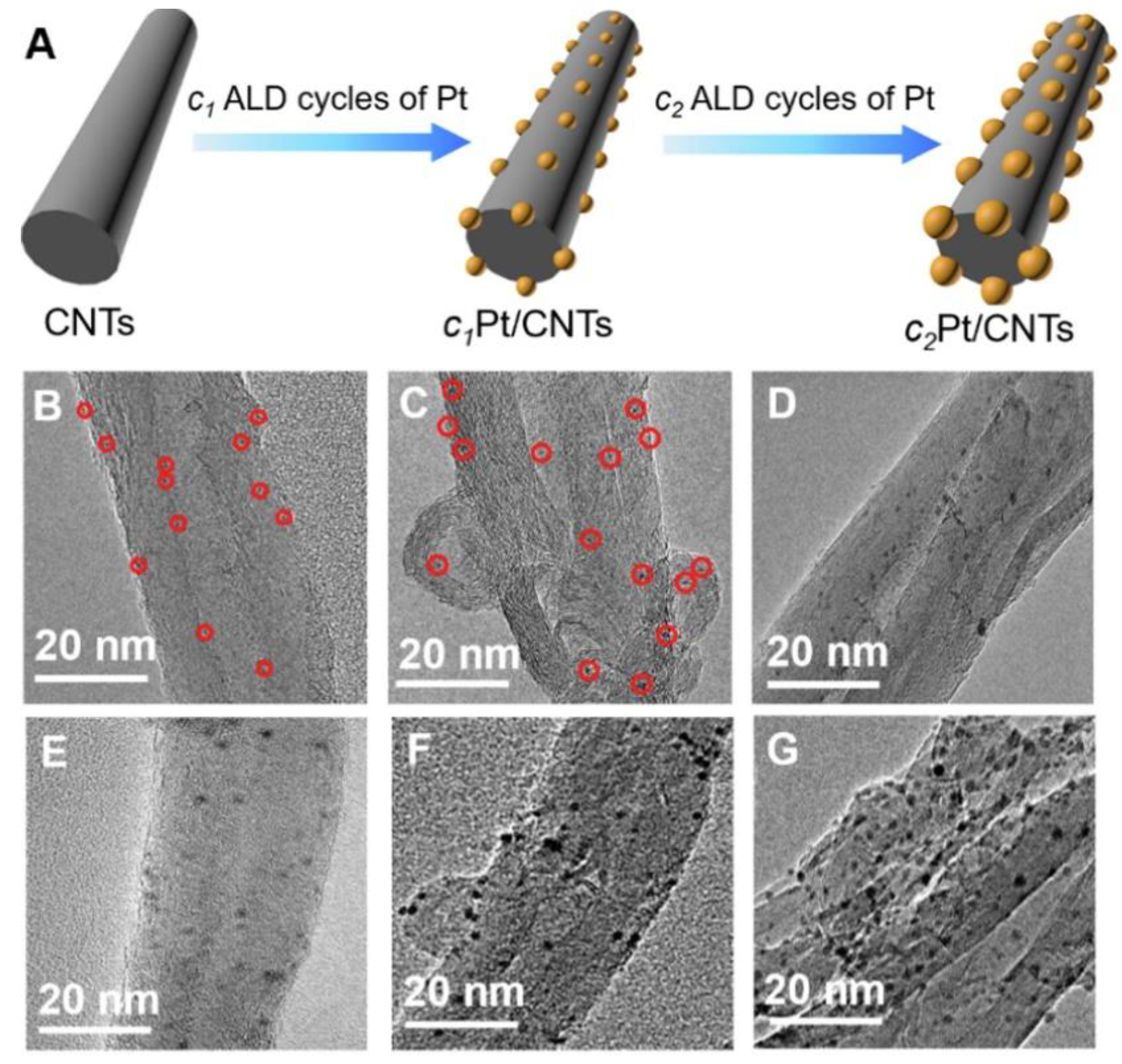
Yang et al. conducted an antimicrobial study of a whole series of nanozymes based on ALD-Pt ultra-small nanoparticles (0.55 - 2.81 nm) prepared on the surface of carbon nanotubes (CNTs) (Figure 18) [128]. The nanozymes with Pt NPs size of 1.69 nm exhibited much higher antibacterial activity than others in the presence of H2O2 due to effective formation of toxic free radicals. In the absence of H2O2, CNTs/Pt NPs showed negligible killing effect against both E. coli and S. aureus.

4. Applications for ALD Antibacterial Coatings

In most of the studies analysed in this review, the authors focused on specific applications of the antibacterial coatings produced. Therefore, the antibacterial properties were analysed using different specific bacterial strains and other functional properties of the materials and coatings were investigated. For implants, in addition to antibacterial properties, cytotoxicity was evaluated for different host-cells such as pre-osteoblasts, fibroblasts, epithelial cells, and an appropriate set of strains was selected depending on whether the implant was internal or in contact with the environment. For PPE and air purifying coatings, air permeability was studied and for water purifying coatings, stability in aqueous media was studied.

4.1. Medical Implants

The development of effective antibacterial coatings for orthopaedic implants is no less important than the development of coatings to accelerate their biointegration. The majority of orthopaedic implant rejections and failures are due to the formation of a biofilm of MRSA or S. epidermidis that prevents drug penetration to the infected site [122]. Such problems most often result in the need for costly and often painful reimplantation, and in some cases postinfection morbidity and mortality. Traditional clinical antimicrobial therapy methods, such as oral administration or intravenous injection, are therefore difficult to solve this problem [129,130]. Therefore, coatings that provide sustained release of antibacterial agents and inhibit biofilm formation are of great practical importance. Coatings that release metal ions (e.g. Zn2+, Ag+ and Cu2+) are very effective as they have broad-spectrum antibacterial properties and are effective even against antibiotic-resistant strains [131,132].
To date, there are about ten studies that have focused on antibacterial ALD coatings for potential applications in orthopaedic implants, and a slightly smaller number for dental implants. Substrates tested have included flat titanium [28,49,57,96], ZrO2 [80,87], stainless steel [26], as well as substrates with deposited carbon nanotubes [43], titanium nanotubes [44,98] or rough SLA surfaces [26,87]. In addition, porous substrates - 3D printed scaffolds made of titanium [27,122] and iron [86] as well as the denture material PMMA [53,63,64] have been used. Of particular note is the work [133] where osteopromotive MgO coatings were applied to the surface of collagen, which is an integral part of bone, cartilage, skin and tendon. ZnO [26,43,44,80,86,87], TiO2 [53,57,61], MgO [133], hydroxyapatite [98], ZrO2 [96], TaN [28], Ag [27,57,122] and TiO2-ZrO2 complex oxides [39,40] have been most commonly used as coatings [63,64].
In the majority of cases for orthopaedic implants, antibacterial properties were evaluated using S. aureus and E. coli, and less frequently MRSA [49,122] and S. epidermidis [122]. For dental implants, the set of strains included Porphyromonas gingivalis [80,87,96], S. Sanguinis , Bifidobacterium [80], S. oralis [63], C. albicans [53,63,64] and antibacterial - polymicrobial biofilm from human saliva [133]. In addition to the antibacterial properties, in vitro bicompatibility studies were performed using osteoblast-like MC3T3-E1 [43,44,84,86,87,96,134], MG-63 [26,57] snd SAOS-2 osteosarcoma [122], fibroblasts (L929) [26,98,134], gingival keratinocytes as well as epithelial cells [80,81,122].
In the absolute majority of studies, the coatings showed high antibacterial efficacy against both standard S. aureus and E. coli, as well as against specific strains, but it is difficult to compare them, as the studied samples differ greatly in thickness, morphology, used substrates and measurement techniques. Nevertheless, it can be stated that zinc oxide and silver coatings have the strongest antibacterial properties. At the same time, biocompatibility studies have been ambiguous for zinc oxide coatings. As demonstrated earlier in the text, most researchers have found that ALD ZnO does not pose a threat to eukaryotic cells. Nevertheless, our analysis revealed detrimental results of 40 nm thick zinc oxide on FetMSCs [61]. Basiaga et al. found a negative effect of thicker coatings on MG-63 proliferation [26], and Zhu et al. found a decrease in viability of MC3T3-E1 with increasing coating thickness. In contrast, silver nanoparticles obtained in three studies showed rather high biocompatibility and promotive effect on the differentiation of osteoblast-like MG-63 and FetMSCs in vitro [57] and no negative effect on new bone formation, robust osteogenesis and angiogenesis in in vivo [27,122]. In addition to the above, only a few studies have used the in vivo approach to evaluate the prospects of ALD coatings. The similar Wistar rat (Rattus norvegicus) in vivo model was used to confirm the biocompatibility of ALD MgO nanocoatings and to increase angiogenesis and bone formation [133]. The thicker coatings (500 vs. 200 cycles) were better candidates for guided bone regeneration due to higher release of magnesium ions.
Xue et al. investigated 3D customised earplugs coated with ZnO nanoaggregates produced by combined ALD and hydrothermal methods [134]. The in vitro study showed that the coatings were highly effective in preventing the growth of five major pathogens from patients with otitis media. A study of earplugs pre-impregnated with MRSA solution in an in vivo rat model showed that the subcutaneous tissue was more infected in the absence of ZnO coatings.
In addition to antibacterial effects and biocompatibility regulation, ALD coatings, due to their high quality and homogeneity (Figure 1), can fulfil the role of a reliable biocorrosion rate regulator for biodegradable implants and scaffolds, which are widely used for bone repair and regeneration, but also for vascular stents. For example, Li et al. showed that 76 nm thick ZnO coatings not only have strong antibacterial activity against both E. coli and S. aureus), do not significantly affect cytocompatibility with MC3T3-E1 cells, but also significantly slow down biocorrosion of 3D porous iron scaffolds [135].

4.2. Antibacterial Textile and Personal Protective Equipment

The COVID-19 pandemic has increased interest in the development of antibacterial textiles and effective PPE, and as a result the number of studies on ALD coatings for these applications has become very high in recent years. Both natural materials (cotton, silk) [33,34,36,76,85] and man-made polyamide [58,136], viscose [85], polypropylene [78], and combinations of natural materials and polymers [35,77] are used as substrates for coating. In the case of cotton and silk, the problem of bacterial contamination is particularly acute. These materials absorb large amounts of water and the aqueous environment is very conducive to the growth of bacteria [34].
In almost all the studies, zinc oxide is used as the coating material, which is significantly superior to ALD TiO2 deposited on polyamide 66 [58] and Al2O3 deposited on the surface of cotton and viscose [85]. Only in the study by Astaneh et al. the authors chose silver nanoparticles as antibacterial material deposited on the surface of N95 masks (the results more detailed described in Section 3.1.5.). Composites of ALD ZnO and photodeposited of Ag nanoparticles deposited on the cotton surface were very effective against S. aureus and E. coli under conditions of light irradiation and dark [33].
The thickness of the deposited coatings varied from fractions of a nanometer [15] to 116 nm [34]. Even relatively thick (> 100 nm) coatings had little effect on the air permeability of the material [34], which is particularly important for PPE and textile applications.
Almost all studies to date have shown some antibacterial activity of ZnO in both light and dark experiments using E. coli and S. aureus. For relatively thin films (up to 45 nm), an increase in thickness has a positive effect on the antibacterial activity [35]. In addition to the antibacterial properties, Kumar et al. investigated the antiviral activity [35]. ZnO 45 nm coatings deposited on electrospun nanofibrous silk showed 95% reduction in infectivity for OC43 and RV14 after 1 h of white light illumination and 99% reduction for RV14 after 2 h of illumination. It is worth noting that ZnO coatings with thicknesses of approximately 25 and 50 nm showed little cytotoxicity towards human fibroblasts and keratinocytes, in contrast to similar Al2O3 coatings [85], which is particularly important for textile and PPE applications.

4.3. Purification and Disinfection of Water and Air

Chemical disinfection of water is a very effective, simple and relatively inexpensive method. However, chemical reactions produce a number of toxic by-products that are not only health-threatening, but cause the water to taste bad. Therefore, photocatalytic ROS-based methods have attracted increasing interest. It is important to note that photocatalytic ROS generation not only kills bacteria, but also destroys many dangerous organic molecules and contaminants [37]. So far, the functional properties of both the pure oxide photocatalysts ZnO [38], TiO2 [39] and the composite materials ZnO-Ag [38], TiO2-Au [60], TiO2-Ag [68]. Polymeric porous membranes based on PVDF, PTFE and PMMA were commonly used as substrates. Pure oxide systems are less effective than metal oxide composites both as antibacterial coatings and as photocatalysts to degrade various organic contaminants such as MB, parecetomol, sodium lauryl sulphate and methyl orange [38,60]. Zhang et al. note that although TiO2 nanoarrays prepared by ALD and solvatothermal methods reduce water flux from 350 to 200 L/m2*hour with long-term use, the reduction is compensated by the absence of biofilm overgrowth in the pores of the PTFE ultrafiltration membrane [39]. In addition to solving membrane biofilm overgrowth, it should be noted that ALD antibacterial coatings have such an important property as high adhesion, which is very important for long-term use of ultrafiltration membranes.
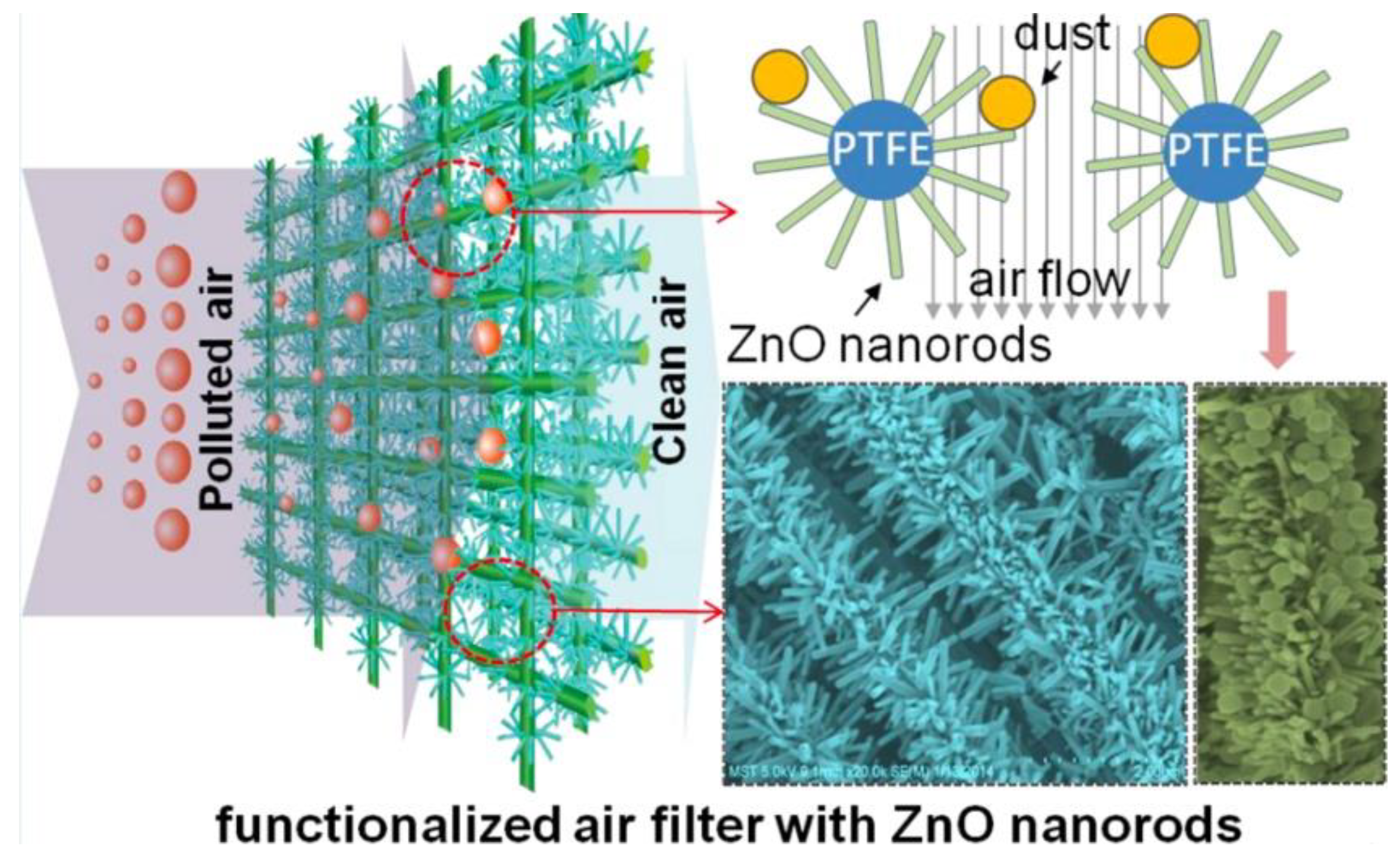
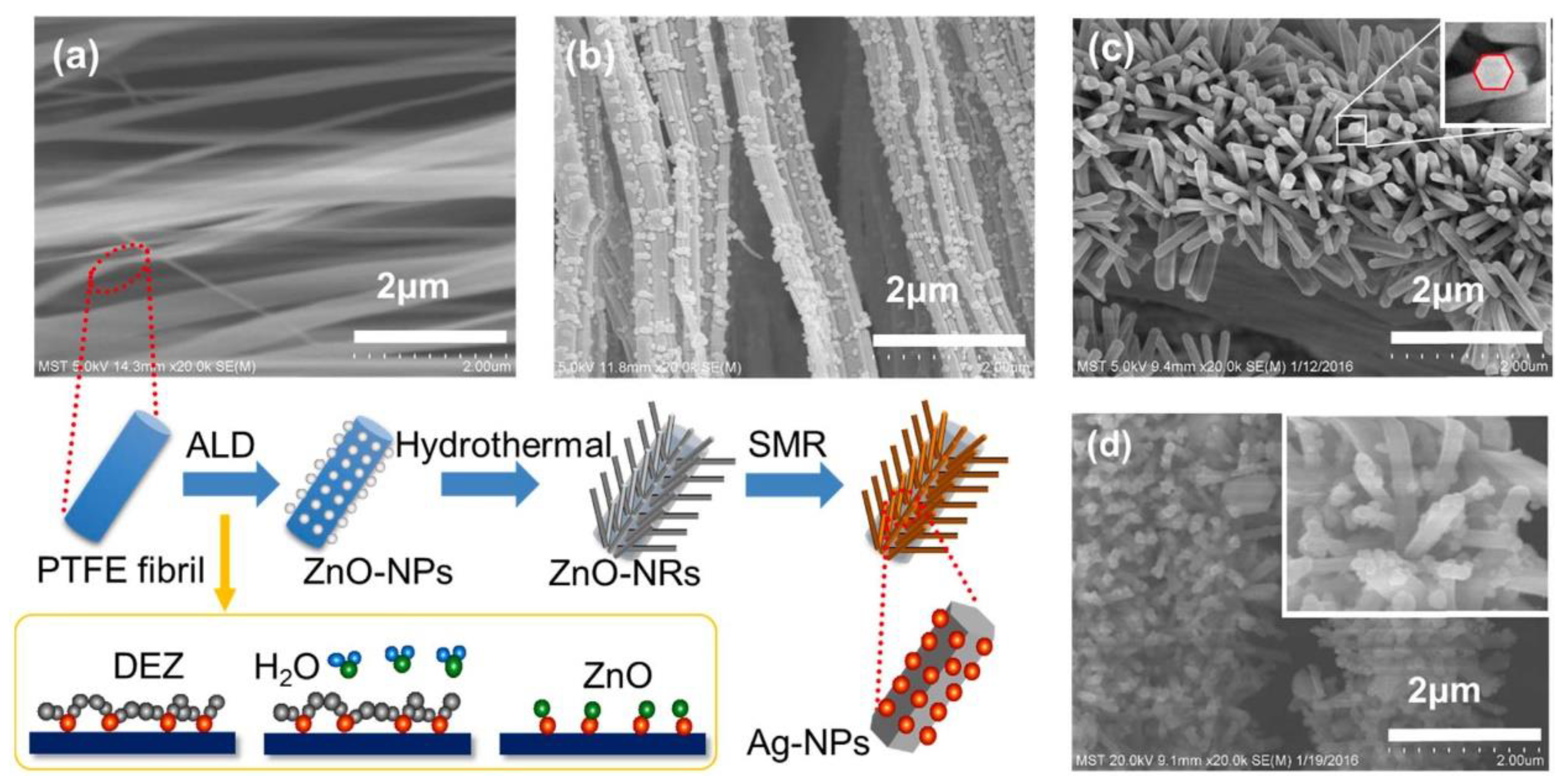
The use of ALD antimicrobial coatings also provides an effective solution to the problem of air purification. Zhong et al. obtained coatings of ALD ZnO seed layers and hydrothermally grown ZnO nanorods on the surface fibre matrix of ePTFE filters (Figure 19) [88]. These filters have a high antibacterial activity (> 99.0% sterilisation rates against S. aureus and E. coli) and are 40% more efficient in the capture of 0.30 and 1.2 μm simulated dust particles. Although the densely packed ZnO nanorods significantly altered the pore structure of the original ePTFE filter, the change in flow resistance was negligible. The application of silver nanoparticles to the ZnO nanorods resulted in even higher antibacterial efficacy (>99.9%) [91].

4.4.Оther Application

The problem of disinfecting various high-frequency touch surfaces can be effectively solved with stable antibacterial coatings. ALD is a very successful method for the synthesis of ultrafine coatings with a thickness of no more than a few tens of nanometres. Such coatings have high transparency and can be used for touch screens with permanent antibacterial properties. In recent years, work has been carried out on the synthesis of complex systems TiO2-Ag [30], Cu2O-ZnO [31], ZnO-Fe2O3 [92] and Al2O3-ZnO (AZO) [32] on transparent substrates such as glass, ITO, PET. The thicknesses of the coatings varied in the range of 10-50 nm and the coatings showed high transparency in the visible range. At the same time, regardless of the composition, they showed high activity not only against E. coli, S. aureus [30,31,32,92], MRD, MRSA [92], but also against several fungi such as Aspergillus brasiliensis, Talaromyces pinophilus, Chaetomium globosum, Trichoderma virens, and Aureobasidium pullulans [32]. All researchers agree that external illumination, at least sunlight, is required for the greatest efficiency of coatings, but Li et al. showed that LED illumination can be a substitute for sunlight and UV for photocatalytic disinfection using ZnO-Fe2O3 nanolaminates, which is relevant to modern personal electronic devices with LED backlit screens.
Transparent coatings may also be important for food packaging, although such application is only discussed in one publication [82], where ZnO and Al2O3 thin films were deposited on biaxially oriented polylactic acid (BOPLA) polymer films - 40 μm thick and 50 μm thick top side corona treated polypropylene (BOPP). Both types of coatings showed good oxygen and vapour barrier properties, but ZnO was more active against E. coli and some of the ZnO samples were effective against Bacillus subtilis and the fungus Aspergillus niger.
To date, there are several studies testing antimicrobial ALD coatings for potential use in treating various diseases and injuries. The idea of using alumina membranes conformally coated with 8 nm ZnO for the treatment of burns and other skin wounds was proposed by Skoog et al. in 2012 [89]. In vitro studies with specific strains showed that the materials were effective against B. subtilis, E. coli, S. aureus and S. epidermidis, but completely ineffective against P. aeruginosa, E. faecalis and C. albicans. Recently, Li et al. found high photocatalytic activity of metal-organic frameworks (MOFs) coated with ultrathin Fe2O3 layer (20 ALD cycles) [86]. S.aureus, E.coli and an in vivo cutaneous MRSA wound infection model were used to validate the prospective application of such composites. At 2 days post infection, the wound in the MOF-Fe2O3 group showed excellent MRSA killing (99.9995 ± 0.0003%), no obvious inflammatory response and complete escharosis in vivo. The group that didn't use MOF-Fe2O3 treatment showed severe suppuration due to heavy MRSA loads in the wounds [86]. Another study by this research group showed high antibacterial activity of MOF-Fe2O3 against oral pathogens (Porphyromonas gingivalis, Fusobacterium nucleatum and S. aureus). The authors believe that the obtained heterostructures can be effectively used for photodynamic ion therapy of periodontitis due to the rapid antibacterial activity, alleviated inflammation and improved angiogenesis [97].
In addition to the above-mentioned directions, the work of Yang et al. is of interest as they propose to use a composite of carbon nanotubes and ultra-small ALD platinum particles to obtain enzyme-like materials - nanozymes [128]. Also of great interest are the studies of TiO2-coated alumina membranes [51] and mesoporous TiO2/ZnO composites [84], which the authors suggest could potentially be used for efficient drug loading.

5. Analysis of the Results

5.1. Comparison of Different Antibacterial Coatings

To date, a variety of oxide and metallic ALD coatings with active antibacterial activity have been obtained and investigated. Comparison of different coatings in terms of effectiveness shows that among oxides, zinc oxide [54,55,58] is the most effective (Table 3). The antibacterial activity of ZnO is based on several independent mechanisms, (see Section 3.3.1). Therefore, ZnO is the most versatile material and the most commonly used for ALD antibacterial coatings. Furthermore, ALD of ZnO can be performed at low temperatures starting from 20 °C [81] and its growth rate reaches 0.19-0.20 nm per cycle [36,135], which is the highest value for all ALD coatings studied. Disadvantages of ZnO include low biocompatibility and low stability in aqueous medium, which limits its potential applications in water purification and disinfection. The effects of these drawbacks can be reduced or eliminated by doping or synthesising complex ZnO-based oxide systems.
In contrast, TiO2 has excellent biocompatibility and stability, and grows successfully at low temperatures, but has a slower growth rate (0.03-0.06 nm per cycle, depending on synthesis temperature and type of precursor and substrate). However, the main disadvantage of titania is its much lower antibacterial activity compared to ZnO [54,55,58]. Titanium oxide is bacteriocidal only in the presence of ambient light (mainly UV), but it can also be bacteriostatic in the dark. Bacteriostatic TiO2 coatings are biocompatible and non-toxic to the organism and may therefore be preferable for implant applications. The problem of low antibacterial activity of TiO2 can be solved by doping, synthesis of mixed oxide systems and noble metal sensitisation (see Section 3.2).
Iron oxide (Fe2O3), like titanium oxide, is not an active bactericidal oxide, but in the form of coatings for MOFs it shows high photocatalytic activity and can be considered as a promising antibacterial material. However, in our opinion, iron oxide is less promising than titanium oxide because of the difficulties in growing it by ALD. Other oxides such as ZrO2, Al2O3 and HfO2 are also very easily obtained by ALD, but they are expected to have weak antibacterial activity mainly due to bacteriostatic effect and, in our opinion, have limited prospects of application in areas where specific properties such as bioinertness (Al2O3) or high mechanical properties (ZrO2) are required.
Metallic silver, like zinc oxide, is a very potent bactericidal material with multiple mechanisms of action. Unlike oxides, silver is grown by ALD in the form of nanoparticles, resulting in high Ag+ release. In particular, ALD Ag has been reported to have high antibacterial activity against S. aureus and MRSA, while being non-toxic for eukaryotic cells in vitro and in vivo. However, synthesis difficulties (unstable precursors, its low vapour pressure, low reactivity, need to use non-standard co-reagents) limit the prospects of using this method to produce silver-based antibacterial coatings by ALD.
When comparing different antibacterial ALD coatings, it is very important to consider the influence of the substrate. The type and morphology of the substrate determines the morphology of the coatings, and for coatings with a thickness of a few tens of nanometres or less, wettability, surface charge and surface energy are influential. However, the influence of the substrate is not limited to this. Because ALD is a process based on reactions between chemical groups on the substrate surface and gaseous reagents, the chemical composition of the substrate surface determines which functional groups will react. For example, there are many hydroxyl groups on the surface of cotton and silk, and only amide groups on polyamide [58], which can greatly affect the growth rate and continuity of the coating. For example, the samples of ALD TiO2 coated polyamide-66 substrates contain the nitrogen peaks in the XPS spectra, indicating a non-cohesive film or the presence of a thinner film than expected. At the same time, for ZnO coatings, nitrogen is not observed in the XPS spectra, which means that the coating is continuous [58]. In the case of metals, even on inorganic substrates with a large number of hydroxyl groups on the surface, the density of growing NPs can vary greatly (Figure 16).

5.2. Advantages, Disadvantages and Perspectives of ALD

The main advantages and disadvantages of the ALD method have been described in numerous review articles [18,20,22,137,138] and these remarks are applicable when considering the application of ALD for the synthesis of antibacterial coatings. All oxide films deposited are characterised by high uniformity and conformality. These properties make ALD very promising for coating surfaces with complex topography, such as 3D implants and scaffolds. In this context, ALD coatings are very promising for regulating the biocorrosion rate of biodegradable implants as shown by Li et al. [135]. Drug release control and active antibacterial ion release control can also be achieved by applying high quality ALD coatings [139]. Membranes and filters for water and air purification have high specific surface areas and also require the use of coating technologies with high uniformity and conformality.
Another feature of ALD is the cyclic nature of the growth and the small growth per cycle, which allows the thickness of the films to be controlled with high precision. This advantage is particularly important when considering zinc oxide and silver coatings, as the antibacterial effect and biocompatibility are highly dependent on the thickness and particle size of the coating. In addition, for a number of applications, such as nanozymes, it is necessary to control the particle size/thickness as precisely as possible. For example, in the work of Yang et al, the optimal size of platinum nanoparticles for Pt/CNTs composites was sought for a range of particle sizes in the range 0.55 - 2.81 nm, which is difficult to achieve by methods other than ALD [128].
Due to the low growth per cycle, ALD is not suitable for producing films with thicknesses greater than a few hundred nanometres, and other methods should be used to produce relatively thick films and structures. However, even in this case, ALD can be effective for the synthesis of seed layers. Indeed, in a large number of studies ALD TiO2 [39] and ZnO [88,134,136] have been used as an effective method to create a homogeneous surface on which thicker layers of arrays of nanorods and other structures have been deposited (Figure 20), which due to their morphology and high specific surface area can be used as effective antibacterial ultrafiltration membranes, air filters or implants.
It should also be noted that in addition to their application value, uniform and conformal ALD coatings can play an important role in basic research to identify and separate the various factors that determine different antibacterial effects. For example, Jeong et al. used 15 and 30 nm Al2O3 layers to regulate and separate the effect of chemical action of ZnO rods and their morphology on antibacterial properties [83].
When considering ALD coatings, it is rarely mentioned that the high adhesion of such coatings compared to coatings produced by other methods is a very important feature for many applications. The high adhesion is due to the chemical bond between the coating materials and the substrate. In addition, the excellent mechanical properties of ALD coatings have been noted. In particular, Darwish et al. studied ALD TiO2 coating on PMMA using denture cleanser challenge test and showed high stability of titanium oxide, its mechanical strength and adhesion. According to Konopatsky et al. [69], low adhesion is one of the main problems of noble metal sensitised metal nanoparticle TiO2, as noble metals obtained by liquid phase deposition suffer from gradual removal from the surface and reduce the efficiency of photocatalysis. In our recent work [57], we have successfully obtained similar hybrid structures of TiO2 nanocoatings/Ag NPs using only ALD. Such structures should exhibit higher adhesion. Thus, we have shown that such complex structures can be obtained in a single ALD process, which simplifies the synthesis and reduces the risk of poor adhesion of Ag nanoparticles [69] and is likely to increase interest in such structures in the future.
The ALD of mixed oxide systems, as well as multilayer or complex oxide-metal structures, has great research potential and may yield new promising results. In this direction, encouraging results have already been obtained in recent years, showing the synergistic effect of combinations of different oxides [63,64] and noble metal sensitisation [30,60]. The ALD opens up great prospects for the synthesis of a wide variety of coatings due to the possibility to flexibly vary the reagents used at different stages of the coating growth.
Finally, it is necessary to briefly discuss the prospects for practical application of the technologies and approaches described in this review. Most of the work presented requires additional reproducibility testing as well as additional in vitro and in vivo studies, some of the work requires investigation of a number of other functional properties. Nevertheless, many of the coatings obtained can be used in practice and industry after careful evaluation of their cost effectiveness. Although ALD has already become an industrial method in microelectronics and is even successfully used in the medical industry for surface modification of titanium implants [140,141], ALD is still a very expensive and complex technology with a very low coating growth rate. However, technical advances in recent years suggest that the impact of these negative factors can be reduced. In particular, we should note the development of technically simpler technologies such as atmospheric pressure ALD [142,143] and faster roll-to-roll ALD, spatial ALD [19], as well as the emergence of techniques that avoid the use of plasma to produce silver [123,144].

6. Conclusions

This review details the results of using ALD for the synthesis of antibacterial coatings based on metals and oxides. The results of the analysis showed that the ALD method can be successfully applied to obtain a variety of different antibacterial nanocoatings on various inorganic and polymeric substrates. The influence of coating preparation conditions and their properties such as thickness, crystal structure, morphology, surface charge, wettability, etc. on the antibacterial properties was discussed in detail. It has been shown that for oxide coatings, increasing the thickness improves the antibacterial effect up to a certain limit, while for zinc oxide, increasing the thickness can induce a cytotoxic effect on eukaryotic cells. The surface topography, its charge and wettability are closely related and also have a significant influence on the antibacterial properties, mainly by achieving a bacteriostatic effect. It was also found that ALD coatings with a thickness typical for this method (not more than a few tens of nanometres) are characterised by a significant influence of the substrate. In addition, a significant influence of the ambient illumination factor on the bactericidal activity of photocatalytic coatings based on pure titanium and zinc oxides, as well as mixed and complex systems based on them, was shown. The analysis of the results showed that mixed oxide systems and composite materials based on oxides and noble metals are the most promising as antibacterial ALD coatings for a wide range of applications.
The results of the review showed that ALD coatings have wide application prospects due to ALD features such as high uniformity, conformality, precise composition and thickness control, and high adhesion. In particular, the most active and successful research is in the areas of antibacterial ALD nanocoatings for dental and orthopedic implants, water and air purification systems, and antibacterial textiles. Some progress is also being made in the field of ultra-thin transparent coatings for touch screens and the treatment of various diseases and injuries.

Author Contributions

Conceptualization, D.N., M.M. and L.K. (Lada Kozlova); methodology, D.N. and L.K. (Lada Kozlova), resources, D.N. and M.M.; formal analysis, D.N. and E.R.; investigation, D.N., E.R. and L.K. (Ludmila Kraeva); writing—original draft preparation, D.N. and L.K. (Lada Kozlova); writing—review and editing, M.M., E.R. and L.K. (Ludmila Kraeva); visualization, D.N. and L.K. (Lada Kozlova); project administration, D.N. and M.M.; funding acquisition, D.N. and M.M. All authors have read and agreed to the published version of the manuscript.

Funding

The research is partially funded by the Ministry of Science and Higher Education of the Russian Federation as part of the World-class Research Center program: Advanced Digital Technologies (contract No. 075-15-2022-311 dated 20.04.2022). The review of mixed and complex ALD oxides was conducted under the financial support of the Russian Science Foundation grant (project No. 22-73-00093), https://rscf.ru/project/22-73-00093/ (accessed on 17 October 2023).

Institutional Review Board Statement

Not applicable.

Informed Consent Statement

Not applicable.

Data Availability Statement

Not applicable.

Conflicts of Interest

The authors declare no conflict of interest.

References

  1. Elbourne, A.; Crawford, R.J.; Ivanova, E.P. Nano-structured antimicrobial surfaces: From nature to synthetic analogues. J Colloid Interface Sci 2017, 508, 603–616. [Google Scholar] [CrossRef] [PubMed]
  2. Modaresifar, K.; Azizian, S.; Ganjian, M.; Fratila-Apachitei, L.E.; Zadpoor, A.A. Bactericidal effects of nanopatterns: A systematic review. Acta Biomater 2019, 83, 29–36. [Google Scholar] [CrossRef] [PubMed]
  3. Li, X.; Huang, T.; Heath, D.E.; O'Brien-Simpson, N.M.; O'Connor, A.J. Antimicrobial nanoparticle coatings for medical implants: Design challenges and prospects. Biointerphases 2020, 15, 060801. [Google Scholar] [CrossRef] [PubMed]
  4. Imani, S.M.; Ladouceur, L.; Marshall, T.; Maclachlan, R.; Soleymani, L.; Didar, T.F. Antimicrobial Nanomaterials and Coatings: Current Mechanisms and Future Perspectives to Control the Spread of Viruses Including SARS-CoV-2. ACS Nano 2020, 14, 12341–12369. [Google Scholar] [CrossRef] [PubMed]
  5. Shirvanimoghaddam, K.; Akbari, M.K.; Yadav, R.; Al-Tamimi, A.K.; Naebe, M. Fight against COVID-19: The case of antiviral surfaces. APL Mater 2021, 9, 031112. [Google Scholar] [CrossRef]
  6. Akshaya, S.; Rowlo, P.K.; Dukle, A.; Nathanael, A.J. Antibacterial Coatings for Titanium Implants: Recent Trends and Future Perspectives. Antibiotics (Basel) 2022, 11. [Google Scholar] [CrossRef]
  7. Chouirfa, H.; Bouloussa, H.; Migonney, V.; Falentin-Daudre, C. Review of titanium surface modification techniques and coatings for antibacterial applications. Acta Biomater 2019, 83, 37–54. [Google Scholar] [CrossRef]
  8. Biyikli, N.; Haider, A. Atomic layer deposition: an enabling technology for the growth of functional nanoscale semiconductors. Semiconductor Science and Technology 2017, 32. [Google Scholar] [CrossRef]
  9. Ding, S.-J.; Wu, X. Superior Atomic Layer Deposition Technology for Amorphous Oxide Semiconductor Thin-Film Transistor Memory Devices. Chemistry of Materials 2020, 32, 1343–1357. [Google Scholar] [CrossRef]
  10. Koshtyal, Y.; Olkhovskii, D.; Rumyantsev, A.; Maximov, M. Applications and Advantages of Atomic Layer Deposition for Lithium-Ion Batteries Cathodes: Review. Batteries 2022, 8. [Google Scholar] [CrossRef]
  11. Ahmed, B.; Xia, C.; Alshareef, H.N. Electrode surface engineering by atomic layer deposition: A promising pathway toward better energy storage. Nano Today 2016, 11, 250–271. [Google Scholar] [CrossRef]
  12. Graniel, O.; Weber, M.; Balme, S.; Miele, P.; Bechelany, M. Atomic layer deposition for biosensing applications. Biosens Bioelectron 2018, 122, 147–159. [Google Scholar] [CrossRef] [PubMed]
  13. Xu, H.; Akbari, M.K.; Kumar, S.; Verpoort, F.; Zhuiykov, S. Atomic layer deposition – state-of-the-art approach to nanoscale hetero-interfacial engineering of chemical sensors electrodes: A review. Sensors and Actuators B: Chemical 2021, 331. [Google Scholar] [CrossRef]
  14. Zhang, B.; Qin, Y. Interface Tailoring of Heterogeneous Catalysts by Atomic Layer Deposition. ACS Catalysis 2018, 8, 10064–10081. [Google Scholar] [CrossRef]
  15. Eswar, N.K.R.; Singh, S.A.; Heo, J. Atomic layer deposited photocatalysts: comprehensive review on viable fabrication routes and reactor design approaches for photo-mediated redox reactions. Journal of Materials Chemistry A 2019, 7, 17703–17734. [Google Scholar] [CrossRef]
  16. Baker, M. Is there a reproducibility crisis in science? Nature 2016, 533, 452–454. [Google Scholar] [CrossRef]
  17. Hicks, D.J. Open science, the replication crisis, and environmental public health. Account Res 2023, 30, 34–62. [Google Scholar] [CrossRef] [PubMed]
  18. Sønsteby, H.H.; Yanguas-Gil, A.; Elam, J.W. Consistency and reproducibility in atomic layer deposition. Journal of Vacuum Science & Technology A: Vacuum, Surfaces, and Films 2020, 38. [Google Scholar] [CrossRef]
  19. Muñoz-Rojas, D.; Maindron, T.; Esteve, A.; Piallat, F.; Kools, J.C.S.; Decams, J.M. Speeding up the unique assets of atomic layer deposition. Materials Today Chemistry 2019, 12, 96–120. [Google Scholar] [CrossRef]
  20. George, S.M. Atomic Layer Deposition: An Overview. Chem. Rev. 2010, 110, 111–131. [Google Scholar] [CrossRef]
  21. Richey, N.E.; de Paula, C.; Bent, S.F. Understanding chemical and physical mechanisms in atomic layer deposition. J Chem Phys 2020, 152, 040902. [Google Scholar] [CrossRef] [PubMed]
  22. Cremers, V.; Puurunen, R.L.; Dendooven, J. Conformality in atomic layer deposition: Current status overview of analysis and modelling. Applied Physics Reviews 2019, 6. [Google Scholar] [CrossRef]
  23. Hämäläinen, J.; Ritala, M.; Leskelä, M. Atomic Layer Deposition of Noble Metals and Their Oxides. Chemistry of Materials 2013, 26, 786–801. [Google Scholar] [CrossRef]
  24. Wack, S.; Lunca Popa, P.; Adjeroud, N.; Guillot, J.; Pistillo, B.R.; Leturcq, R. Large-Scale Deposition and Growth Mechanism of Silver Nanoparticles by Plasma-Enhanced Atomic Layer Deposition. The Journal of Physical Chemistry C 2019, 123, 27196–27206. [Google Scholar] [CrossRef]
  25. Nazarov, D.V.; Smirnov, V.M.; Zemtsova, E.G.; Yudintceva, N.M.; Shevtsov, M.A.; Valiev, R.Z. Enhanced Osseointegrative Properties of Ultra-Fine-Grained Titanium Implants Modified by Chemical Etching and Atomic Layer Deposition. ACS Biomater Sci Eng 2018, 4, 3268–3281. [Google Scholar] [CrossRef] [PubMed]
  26. Basiaga, M.; Paszenda, Z.; Lisoń, J.; Taratuta, A.; Kazek-Kęsik, A.; Krok-Borkowicz, M.; Nuckowski, P.; Szindler, M.; Staszuk, M.; Major, Ł.; et al. Microstructure and antibacterial properties of a ZnO coating on a biomaterial surface. Archives of Civil and Mechanical Engineering 2022, 22. [Google Scholar] [CrossRef]
  27. Geng, H.; Poologasundarampillai, G.; Todd, N.; Devlin-Mullin, A.; Moore, K.L.; Golrokhi, Z.; Gilchrist, J.B.; Jones, E.; Potter, R.J.; Sutcliffe, C.; et al. Biotransformation of Silver Released from Nanoparticle Coated Titanium Implants Revealed in Regenerating Bone. ACS Appl Mater Interfaces 2017, 9, 21169–21180. [Google Scholar] [CrossRef] [PubMed]
  28. Gao, J.; Wang, M. Atomic layer deposition of TaN thin film on Ti–6Al–4V dental implant for enhanced anti-corrosion and anti-bacterial properties. Journal of Physics and Chemistry of Solids 2021, 150. [Google Scholar] [CrossRef]
  29. Pessoa, R.S.; dos Santos, V.P.; Cardoso, S.B.; Doria, A.C.O.C.; Figueira, F.R.; Rodrigues, B.V.M.; Testoni, G.E.; Fraga, M.A.; Marciano, F.R.; Lobo, A.O.; et al. TiO2 coatings via atomic layer deposition on polyurethane and polydimethylsiloxane substrates: Properties and effects on C. albicans growth and inactivation process. Applied Surface Science 2017, 422, 73–84. [Google Scholar] [CrossRef]
  30. Cai, P.F.; Li, J.; Wu, X.B.; Li, Z.Y.; Shen, J.; Nie, J.J.; Cui, Z.D.; Chen, D.F.; Liang, Y.Q.; Zhu, S.L.; et al. ALD-induced TiO(2)/Ag nanofilm for rapid surface photodynamic ion sterilization. Rare Metals 2022, 41, 4138–4148. [Google Scholar] [CrossRef]
  31. Liu, Z.; Liu, X.; Cui, Z.; Zheng, Y.; Li, Z.; Liang, Y.; Yuan, X.; Zhu, S.; Wu, S. Surface photodynamic ion sterilization of ITO-Cu2O/ZnO preventing touch infection. Journal of Materials Science & Technology 2022, 122, 10–19. [Google Scholar] [CrossRef]
  32. Eom, J.-H.; Cho, T.-Y.; Cho, S.-K. Performance of multifunctional antibacterial moisture barrier films with different Zn/Al ratios fabricated by plasma enhanced atomic layer deposition. Applied Surface Science 2023, 638. [Google Scholar] [CrossRef]
  33. Akyildiz, H.I.; Yilmaz, B.A.; Diler, S. Antibacterial Activity of Photodeposited Ag Nanoparticles on Cotton Fibers Enabled by Atomic Layer Deposition. Fibers and Polymers 2022, 23, 2769–2779. [Google Scholar] [CrossRef]
  34. Xiao, X.; Zhang, B.; Lyu, P.; Chen, F.; Zhang, Q.; Sun, J.; Jiang, Z.; Liu, X.; Xu, W. Controllable coating of zinc oxide on protein-based fibers/fabrics for superior antibacterial performance preserving wearable abilities. Applied Surface Science 2023, 610. [Google Scholar] [CrossRef]
  35. Kumar, U.; Fox, C.R.; Feit, C.; Kolanthai, E.; Sheiber, J.; Fu, Y.; Singh, S.; Banerjee, P.; Parks, G.D.; Seal, S. ALD based nanostructured zinc oxide coated antiviral silk fabric. RSC Adv 2022, 12, 19327–19339. [Google Scholar] [CrossRef] [PubMed]
  36. Puvvada, R.U.; Wooding, J.P.; Bellavia, M.C.; McGuinness, E.K.; Sulchek, T.A.; Losego, M.D. Bacterial Growth and Death on Cotton Fabrics Conformally Coated with ZnO Thin Films of Varying Thicknesses via Atomic Layer Deposition (ALD). Jom 2018, 71, 178–184. [Google Scholar] [CrossRef]
  37. Molina-Reyes, J.; Romero-Moran, A.; Uribe-Vargas, H.; Lopez-Ruiz, B.; Sanchez-Salas, J.L.; Ortega, E.; Ponce, A.; Morales-Sanchez, A.; Lopez-Huerta, F.; Zuñiga-Islas, C. Study on the photocatalytic activity of titanium dioxide nanostructures: Nanoparticles, nanotubes and ultra-thin films. Catalysis Today 2020, 341, 2–12. [Google Scholar] [CrossRef]
  38. Di Mauro, A.; Farrugia, C.; Abela, S.; Refalo, P.; Grech, M.; Falqui, L.; Nicotra, G.; Sfuncia, G.; Mio, A.; Buccheri, M.A.; et al. Ag/ZnO/PMMA Nanocomposites for Efficient Water Reuse. ACS Appl Bio Mater 2020, 3, 4417–4426. [Google Scholar] [CrossRef]
  39. Zhang, L.; Shi, X.; Sun, M.; Porter, C.J.; Zhou, X.; Elimelech, M. Precisely Engineered Photoreactive Titanium Nanoarray Coating to Mitigate Biofouling in Ultrafiltration. ACS Appl Mater Interfaces 2021, 13, 9975–9984. [Google Scholar] [CrossRef]
  40. Cabiscol, E.; Tamarit, J.; Ros, J. Oxidative stress in bacteria and protein damage by reactive oxygen species. International Microbiology 2000, 3, 3–8. [Google Scholar]
  41. Li, W.; Thian, E.S.; Wang, M.; Wang, Z.; Ren, L. Surface Design for Antibacterial Materials: From Fundamentals to Advanced Strategies. Adv Sci (Weinh) 2021, 8, e2100368. [Google Scholar] [CrossRef] [PubMed]
  42. Gristina, A.G.; Naylor, P.; Myrvik, Q. Infections from biomaterials and implants: a race for the surface. Medical progress through technology 1988, 14, 205–224. [Google Scholar] [PubMed]
  43. Zhu, Y.; Liu, X.; Yeung, K.W.K.; Chu, P.K.; Wu, S. Biofunctionalization of carbon nanotubes/chitosan hybrids on Ti implants by atom layer deposited ZnO nanostructures. Applied Surface Science 2017, 400, 14–23. [Google Scholar] [CrossRef]
  44. Zhang, K.; Zhu, Y.; Liu, X.; Cui, Z.; XianjinYang; Yeung, K. W.K.; Pan, H.; Wu, S. Sr/ZnO doped titania nanotube array: An effective surface system with excellent osteoinductivity and self-antibacterial activity. Materials & Design 2017, 130, 403–412. [Google Scholar] [CrossRef]
  45. Molina-Reyes, J.; Romero-Moran, A.; Sanchez-Salas, J.L. Enhanced photocatalytic bacterial inactivation of atomic-layer deposited anatase-TiO(2) thin films on rutile-TiO(2) nanotubes. Photochem Photobiol Sci 2020, 19, 399–405. [Google Scholar] [CrossRef]
  46. Page, M.J.; McKenzie, J.E.; Bossuyt, P.M.; Boutron, I.; Hoffmann, T.C.; Mulrow, C.D.; Shamseer, L.; Tetzlaff, J.M.; Akl, E.A.; Brennan, S.E.; et al. The PRISMA 2020 statement: an updated guideline for reporting systematic reviews. BMJ 2021, 372, n71. [Google Scholar] [CrossRef] [PubMed]
  47. Singh, J.; Hegde, P.B.; Avasthi, S.; Sen, P. Scalable Hybrid Antibacterial Surfaces: TiO(2) Nanoparticles with Black Silicon. ACS Omega 2022, 7, 7816–7824. [Google Scholar] [CrossRef] [PubMed]
  48. Scuderi, V.; Buccheri, M.A.; Impellizzeri, G.; Di Mauro, A.; Rappazzo, G.; Bergum, K.; Svensson, B.G.; Privitera, V. Photocatalytic and antibacterial properties of titanium dioxide flat film. Materials Science in Semiconductor Processing 2016, 42, 32–35. [Google Scholar] [CrossRef]
  49. Liu, L.; Bhatia, R.; Webster, T.J. Atomic layer deposition of nano-TiO2 thin films with enhanced biocompatibility and antimicrobial activity for orthopedic implants. Int J Nanomedicine 2017, 12, 8711–8723. [Google Scholar] [CrossRef]
  50. Singh, J.; Jadhav, S.; Avasthi, S.; Sen, P. Designing Photocatalytic Nanostructured Antibacterial Surfaces: Why Is Black Silica Better than Black Silicon? ACS Appl Mater Interfaces 2020, 12, 20202–20213. [Google Scholar] [CrossRef]
  51. Narayan, R.J.; Monteiro-Riviere, N.A.; Brigmon, R.L.; Pellin, M.J.; Elam, J.W. Atomic Layer Deposition of TiO2 Thin Films on Nanoporous Alumina Templates: Medical Applications. JOM 2009, 61, 12–16. [Google Scholar] [CrossRef]
  52. Goldfinger, Y.; Natan, M.; Sukenik, C.N.; Banin, E.; Kronenberg, J. Biofilm prevention on cochlear implants. Cochlear Implants Int 2014, 15, 173–178. [Google Scholar] [CrossRef] [PubMed]
  53. Darwish, G.; Huang, S.; Knoernschild, K.; Sukotjo, C.; Campbell, S.; Bishal, A.K.; Barao, V.A.; Wu, C.D.; Taukodis, C.G.; Yang, B. Improving Polymethyl Methacrylate Resin Using a Novel Titanium Dioxide Coating. J Prosthodont 2019, 28, 1011–1017. [Google Scholar] [CrossRef] [PubMed]
  54. Lee, S.-M.; Grass, G.; Kim, G.-M.; Dresbach, C.; Zhang, L.; Goselea, U.; Knez, M. Low-temperature ZnO atomic layer deposition on biotemplates: flexible photocatalytic ZnO structures from eggshell membranes. Phys Chem Chem Phys 2009, 11, 3608–3614. [Google Scholar] [CrossRef]
  55. Godlewski, M.; Gierałtowska, S.; Wachnicki, Ł.; Pietuszka, R.; Witkowski, B.S.; Słońska, A.; Gajewski, Z.; Godlewski, M.M. High-k oxides by atomic layer deposition—Applications in biology and medicine. Journal of Vacuum Science & Technology A: Vacuum, Surfaces, and Films 2017, 35. [Google Scholar] [CrossRef]
  56. Regonini, D.; Bowen, C.R.; Jaroenworaluck, A.; Stevens, R. A review of growth mechanism, structure and crystallinity of anodized TiO2 nanotubes. Materials Science and Engineering: R: Reports 2013, 74, 377–406. [Google Scholar] [CrossRef]
  57. Nazarov, D.; Ezhov, I.; Yudintceva, N.; Shevtsov, M.; Rudakova, A.; Kalganov, V.; Tolmachev, V.; Zharova, Y.; Lutakov, O.; Kraeva, L.; et al. Antibacterial and Osteogenic Properties of Ag Nanoparticles and Ag/TiO(2) Nanostructures Prepared by Atomic Layer Deposition. J Funct Biomater 2022, 13. [Google Scholar] [CrossRef] [PubMed]
  58. Akyildiz, H.I.; Diler, S.; Islam, S. Evaluation of TiO2 and ZnO atomic layer deposition coated polyamide 66 fabrics for photocatalytic activity and antibacterial applications. Journal of Vacuum Science & Technology A: Vacuum, Surfaces, and Films 2021, 39. [Google Scholar] [CrossRef]
  59. Grigal, I.P.; Markeev, A.M.; Gudkova, S.A.; Chernikova, A.G.; Mityaev, A.S.; Alekhin, A.P. Correlation between bioactivity and structural properties of titanium dioxide coatings grown by atomic layer deposition. Applied Surface Science 2012, 258, 3415–3419. [Google Scholar] [CrossRef]
  60. Zhang, L.; Shi, X.; Zhao, M.; Yin, Z.; Zhang, J.; Wang, S.; Du, W.; Xiang, J.; Cheng, P.; Tang, N. Construction of precisely controllable and stable interface bonding Au-TiO2/PVDF composited membrane for biofouling-resistant properties. Surfaces and Interfaces 2021, 24. [Google Scholar] [CrossRef]
  61. Nazarov, D.; Kozlova, L.; Rudakova, A.; Zemtsova, E.; Yudintceva, N.; Ovcharenko, E.; Koroleva, A.; Kasatkin, I.; Kraeva, L.; Rogacheva, E.; et al. Atomic Layer Deposition of Chlorine Containing Titanium–Zinc Oxide Nanofilms Using the Supercycle Approach. Coatings 2023, 13. [Google Scholar] [CrossRef]
  62. Amashaev, R.R.; Maksumova, A.M.; Rabadanov, M.K.; Abdullaeva, N.; Abdulagatov, I.M. STUDY OF ANTIBACTERIAL PROPERTIES OF MODIFIED TITANIUM OXIDE NANOFILMS SYNTHESIZED BY ATOMIC AND MOLECULAR LAYER DEPOSITION TECHNIQUES. New Materials, Compounds and Applications 2019, 3, 150–159. [Google Scholar]
  63. Shahmohammadi, M.; Nagay, B.E.; Barão, V.A.R.; Sukotjo, C.; Jursich, G.; Takoudis, C.G. Atomic layer deposition of TiO2, ZrO2 and TiO2/ZrO2 mixed oxide nanofilms on PMMA for enhanced biomaterial functionalization. Applied Surface Science 2022, 578. [Google Scholar] [CrossRef]
  64. Yang, B.; Ginsburg, S.; Li, W.; Vilela, M.M.; Shahmohammadi, M.; Takoudis, C.G.; Wu, C.D. Effect of nano-ceramic coating on surface property and microbial adhesion to poly(methyl methacrylate). J Biomed Mater Res B Appl Biomater 2023, 111, 1480–1487. [Google Scholar] [CrossRef] [PubMed]
  65. Niemelä, J.-P.; Marin, G.; Karppinen, M. Titanium dioxide thin films by atomic layer deposition: a review. Semiconductor Science and Technology 2017, 32. [Google Scholar] [CrossRef]
  66. Abdulagatov, I.M.; Ragimov, R.M.; Khamidov capital Em, C.A.C.; Maksumova, A.M.; Abdullaeva, N.M. ALD coated polypropylene hernia meshes for prevention of mesh-related post-surgery complications: an experimental study in animals. Biomed Mater 2021, 17. [Google Scholar] [CrossRef] [PubMed]
  67. Li, J.; Liu, C.-H.; Banis, M.N.; Vaccarello, D.; Ding, Z.-F.; Wang, S.-D.; Sham, T.-K. Revealing the Synergy of Mono/Bimetallic PdPt/TiO2 Heterostructure for Enhanced Photoresponse Performance. The Journal of Physical Chemistry C 2017, 121, 24861–24870. [Google Scholar] [CrossRef]
  68. Zhang, L.; He, D.; Jiang, Y.; Li, Y.; Shi, X.; Wang, S.; Xiang, J.; Zhang, J.; Cheng, P.; Tang, N. AgTi nanoparticle hybrid PVDF membrane with stable and high efficiency antibacterial performance by atomic layer deposition. Surfaces and Interfaces 2022, 30. [Google Scholar] [CrossRef]
  69. Konopatsky, A.; Teplyakova, T.; Sheremetyev, V.; Yakimova, T.; Boychenko, O.; Kozik, M.; Shtansky, D.; Prokoshkin, S. Surface Modification of Biomedical Ti-18Zr-15Nb Alloy by Atomic Layer Deposition and Ag Nanoparticles Decoration. J Funct Biomater 2023, 14. [Google Scholar] [CrossRef]
  70. Park, K.-H.; Han, G.D.; Neoh, K.C.; Kim, T.-S.; Shim, J.H.; Park, H.-D. Antibacterial activity of the thin ZnO film formed by atomic layer deposition under UV-A light. Chemical Engineering Journal 2017, 328, 988–996. [Google Scholar] [CrossRef]
  71. Kääriäinen, M.L.; Weiss, C.K.; Ritz, S.; Pütz, S.; Cameron, D.C.; Mailänder, V.; Landfester, K. Zinc release from atomic layer deposited zinc oxide thin films and its antibacterial effect on Escherichia coli. Applied Surface Science 2013, 287, 375–380. [Google Scholar] [CrossRef]
  72. Amna, T.; Hassan, M.S.; Barakat, N.A.; Pandeya, D.R.; Hong, S.T.; Khil, M.S.; Kim, H.Y. Antibacterial activity and interaction mechanism of electrospun zinc-doped titania nanofibers. Appl Microbiol Biotechnol 2012, 93, 743–751. [Google Scholar] [CrossRef] [PubMed]
  73. Zhou, G.; Li, Y.; Xiao, W.; Zhang, L.; Zuo, Y.; Xue, J.; Jansen, J.A. Synthesis, characterization, and antibacterial activities of a novel nanohydroxyapatite/zinc oxide complex. J Biomed Mater Res A 2008, 85, 929–937. [Google Scholar] [CrossRef] [PubMed]
  74. Tong, T.; Wilke, C.M.; Wu, J.; Binh, C.T.; Kelly, J.J.; Gaillard, J.F.; Gray, K.A. Combined Toxicity of Nano-ZnO and Nano-TiO2: From Single- to Multinanomaterial Systems. Environ Sci Technol 2015, 49, 8113–8123. [Google Scholar] [CrossRef] [PubMed]
  75. Guan, G.; Zhang, L.; Zhu, J.; Wu, H.; Li, W.; Sun, Q. Antibacterial properties and mechanism of biopolymer-based films functionalized by CuO/ZnO nanoparticles against Escherichia coli and Staphylococcus aureus. J Hazard Mater 2021, 402, 123542. [Google Scholar] [CrossRef]
  76. Mežnarić, S.; Jelovica Badovinac, I.; Šarić, I.; Peter, R.; Kolympadi Markovic, M.; Ambrožić, G.; Gobin, I. Superior UVA-photocatalytic antibacterial activity of a double-layer ZnO/Al2O3 thin film grown on cellulose by atomic layer deposition (ALD). Journal of Environmental Chemical Engineering 2022, 10. [Google Scholar] [CrossRef]
  77. Li, L.; Yu, P.; Li, Y.; Wu, X.; Li, W.; Cheng, X. A Facile Approach to Fabricating Antibacterial Textile with High Efficiency and Compact Process. Advanced Materials Interfaces 2021, 8. [Google Scholar] [CrossRef]
  78. Qian, X.; Xiong, S.; Rao, Y.; Low, Z.X.; Zhong, Z.; Wang, Y. Atomic layer deposition of ZnO on polypropylene nonwovens for photocatalytic antibacterial facemasks. Nanotechnology 2023, 34. [Google Scholar] [CrossRef]
  79. Yu, F.; Fang, X.; Jia, H.; Liu, M.; Shi, X.; Xue, C.; Chen, T.; Wei, Z.; Fang, F.; Zhu, H.; et al. Zn or O? An Atomic Level Comparison on Antibacterial Activities of Zinc Oxides. Chemistry 2016, 22, 8053–8058. [Google Scholar] [CrossRef]
  80. Wang, M.; Gao, J. Atomic layer deposition of ZnO thin film on ZrO2 dental implant surface for enhanced antibacterial and bioactive performance. Materials Letters 2021, 285. [Google Scholar] [CrossRef]
  81. Narayan, R.J.; Adiga, S.P.; Pellin, M.J.; Curtiss, L.A.; Stafslien, S.; Chisholm, B.; Monteiro-Rivierea, N.A.; Brigmon, R.L.; Elam, J.W. Atomic layer deposition of nanoporous biomaterials. Materials today 2010, 13, 60–64. [Google Scholar] [CrossRef]
  82. Vähä-Nissi, M.; Pitkänen, M.; Salo, E.; Kenttä, E.; Tanskanen, A.; Sajavaara, T.; Putkonen, M.; Sievänen, J.; Sneck, A.; Rättö, M.; et al. Antibacterial and barrier properties of oriented polymer films with ZnO thin films applied with atomic layer deposition at low temperatures. Thin Solid Films 2014, 562, 331–337. [Google Scholar] [CrossRef]
  83. Jeong, E.; Kim, C.U.; Byun, J.; Lee, J.; Kim, H.E.; Kim, E.J.; Choi, K.J.; Hong, S.W. Quantitative evaluation of the antibacterial factors of ZnO nanorod arrays under dark conditions: Physical and chemical effects on Escherichia coli inactivation. Sci Total Environ 2020, 712, 136574. [Google Scholar] [CrossRef] [PubMed]
  84. Wu, X.; Yao, L.; Al-Baadani, M.A.; Ping, L.; Wu, S.; Al-Bishari, A.M.; HiiRuYie, K.; Deng, Z.; Liu, J.; Shen, X. Preparation of multifunctional drug sustained-release system by atomic layer deposition of ZnO in mesoporous titania coating. Ceramics International 2020, 46, 9406–9414. [Google Scholar] [CrossRef]
  85. Popescu, M.C.; Ungureanu, C.; Buse, E.; Nastase, F.; Tucureanu, V.; Suchea, M.; Draga, S.; Popescu, M.A. Antibacterial efficiency of cellulose-based fibers covered with ZnO and Al2O3 by Atomic Layer Deposition. Applied Surface Science 2019, 481, 1287–1298. [Google Scholar] [CrossRef]
  86. Li, J.; Cui, Z.; Zheng, Y.; Liu, X.; Li, Z.; Jiang, H.; Zhu, S.; Zhang, Y.; Chu, P.K.; Wu, S. Atomic-layer Fe2O3-modified 2D porphyrinic metal-organic framework for enhanced photocatalytic disinfection through electron-withdrawing effect. Applied Catalysis B: Environmental 2022, 317. [Google Scholar] [CrossRef]
  87. Yao, L.; Wu, X.; Wu, S.; Pan, X.; Tu, J.; Chen, M.; Al-Bishari, A.M.; Al-Baadani, M.A.; Yao, L.; Shen, X.; et al. Atomic layer deposition of zinc oxide on microrough zirconia to enhance osteogenesis and antibiosis. Ceramics International 2019, 45, 24757–24767. [Google Scholar] [CrossRef]
  88. Zhong, Z.; Xu, Z.; Sheng, T.; Yao, J.; Xing, W.; Wang, Y. Unusual Air Filters with Ultrahigh Efficiency and Antibacterial Functionality Enabled by ZnO Nanorods. ACS Appl Mater Interfaces 2015, 7, 21538–21544. [Google Scholar] [CrossRef]
  89. Skoog, S.A.; Bayati, M.R.; Petrochenko, P.E.; Stafslien, S.; Daniels, J.; Cilz, N.; Comstock, D.J.; Elam, J.W.; Narayan, R.J. Antibacterial activity of zinc oxide-coated nanoporous alumina. Materials Science and Engineering: B 2012, 177, 992–998. [Google Scholar] [CrossRef]
  90. Dicastillo, C.L.; Vidal, C.P.; Falco, I.; Sanchez, G.; Marquez, P.; Escrig, J. Antimicrobial Bilayer Nanocomposites Based on the Incorporation of As-Synthetized Hollow Zinc Oxide Nanotubes. Nanomaterials (Basel) 2020, 10. [Google Scholar] [CrossRef]
  91. Feng, S.; Li, D.; Low, Z.-x.; Liu, Z.; Zhong, Z.; Hu, Y.; Wang, Y.; Xing, W. ALD-seeded hydrothermally-grown Ag/ZnO nanorod PTFE membrane as efficient indoor air filter. Journal of Membrane Science 2017, 531, 86–93. [Google Scholar] [CrossRef]
  92. Li, J.; Wang, C.; Wu, S.; Cui, Z.; Zheng, Y.; Li, Z.; Jiang, H.; Zhu, S.; Liu, X. Superlattice Nanofilm on a Touchscreen for Photoexcited Bacteria and Virus Killing by Tuning Electronic Defects in the Heterointerface. Adv Mater 2023, 35, e2300380. [Google Scholar] [CrossRef] [PubMed]
  93. Sa, M.W.; Nguyen, B.B.; Moriarty, R.A.; Kamalitdinov, T.; Fisher, J.P.; Kim, J.Y. Fabrication and evaluation of 3D printed BCP scaffolds reinforced with ZrO(2) for bone tissue applications. Biotechnol Bioeng 2018, 115, 989–999. [Google Scholar] [CrossRef] [PubMed]
  94. Jung, S.; Bohner, L.; Hanisch, M.; Kleinheinz, J.; Sielker, S. Influence of Implant Material and Surface on Differentiation and Proliferation of Human Adipose-Derived Stromal Cells. Int J Mol Sci 2018, 19. [Google Scholar] [CrossRef] [PubMed]
  95. Smieszek, A.; Donesz-Sikorska, A.; Grzesiak, J.; Krzak, J.; Marycz, K. Biological effects of sol-gel derived ZrO2 and SiO2/ZrO2 coatings on stainless steel surface--In vitro model using mesenchymal stem cells. J Biomater Appl 2014, 29, 699–714. [Google Scholar] [CrossRef]
  96. Jo, Y.; Kim, Y.T.; Cho, H.; Ji, M.K.; Heo, J.; Lim, H.P. Atomic Layer Deposition of ZrO(2) on Titanium Inhibits Bacterial Adhesion and Enhances Osteoblast Viability. Int J Nanomedicine 2021, 16, 1509–1523. [Google Scholar] [CrossRef] [PubMed]
  97. Li, J.; Song, S.; Meng, J.; Tan, L.; Liu, X.; Zheng, Y.; Li, Z.; Yeung, K.W.K.; Cui, Z.; Liang, Y.; et al. 2D MOF Periodontitis Photodynamic Ion Therapy. J Am Chem Soc 2021, 143, 15427–15439. [Google Scholar] [CrossRef]
  98. Radtke, A.; Ehlert, M.; Jedrzejewski, T.; Sadowska, B.; Wieckowska-Szakiel, M.; Holopainen, J.; Ritala, M.; Leskela, M.; Bartmanski, M.; Szkodo, M.; et al. Titania Nanotubes/Hydroxyapatite Nanocomposites Produced with the Use of the Atomic Layer Deposition Technique: Estimation of Bioactivity and Nanomechanical Properties. Nanomaterials (Basel) 2019, 9. [Google Scholar] [CrossRef]
  99. Radtke, A.; Topolski, A.; Jedrzejewski, T.; Kozak, W.; Sadowska, B.; Wieckowska-Szakiel, M.; Szubka, M.; Talik, E.; Pleth Nielsen, L.; Piszczek, P. The Bioactivity and Photocatalytic Properties of Titania Nanotube Coatings Produced with the Use of the Low-Potential Anodization of Ti6Al4V Alloy Surface. Nanomaterials (Basel) 2017, 7. [Google Scholar] [CrossRef]
  100. Nene, A.; Galluzzi, M.; Hongrong, L.; Somani, P.; Ramakrishna, S.; Yu, X.F. Synthetic preparations and atomic scale engineering of silver nanoparticles for biomedical applications. Nanoscale 2021, 13, 13923–13942. [Google Scholar] [CrossRef]
  101. Chernousova, S.; Epple, M. Silver as antibacterial agent: ion, nanoparticle, and metal. Angew Chem Int Ed Engl 2013, 52, 1636–1653. [Google Scholar] [CrossRef] [PubMed]
  102. Radtke, A.; Jedrzejewski, T.; Kozak, W.; Sadowska, B.; Wieckowska-Szakiel, M.; Talik, E.; Makela, M.; Leskela, M.; Piszczek, P. Optimization of the Silver Nanoparticles PEALD Process on the Surface of 1-D Titania Coatings. Nanomaterials (Basel) 2017, 7. [Google Scholar] [CrossRef] [PubMed]
  103. Yin, N.; Hu, B.; Yang, R.; Liang, S.; Liang, S.; Faiola, F. Assessment of the developmental neurotoxicity of silver nanoparticles and silver ions with mouse embryonic stem cells in vitro. Journal of Interdisciplinary Nanomedicine 2018, 3, 133–145. [Google Scholar] [CrossRef]
  104. Ottoni, C.A.; Maria, D.A.; Goncalves, P.; de Araujo, W.L.; de Souza, A.O. Biogenic Aspergillus tubingensis silver nanoparticles' in vitro effects on human umbilical vein endothelial cells, normal human fibroblasts, HEPG2, and Galleria mellonella. Toxicol Res (Camb) 2019, 8, 789–801. [Google Scholar] [CrossRef] [PubMed]
  105. Algazlan, A.S.; Almuraikhi, N.; Muthurangan, M.; Balto, H.; Alsalleeh, F. Silver Nanoparticles Alone or in Combination with Calcium Hydroxide Modulate the Viability, Attachment, Migration, and Osteogenic Differentiation of Human Mesenchymal Stem Cells. Int J Mol Sci 2022, 24. [Google Scholar] [CrossRef] [PubMed]
  106. Sitnikova, N.A.; Solovieva, A.O.; Permyakova, E.S.; Sheveyko, A.N.; Shtansky, D.V.; Manakhov, A.M. Silver Ions Incorporation into Nanofibers for Enhanced hMSC Viability. Chemistry 2022, 4, 931–939. [Google Scholar] [CrossRef]
  107. Sellami, H.; Khan, S.A.; Ahmad, I.; Alarfaj, A.A.; Hirad, A.H.; Al-Sabri, A.E. Green Synthesis of Silver Nanoparticles Using Olea europaea Leaf Extract for Their Enhanced Antibacterial, Antioxidant, Cytotoxic and Biocompatibility Applications. Int J Mol Sci 2021, 22. [Google Scholar] [CrossRef]
  108. Chavez-Andrade, G.M.; Tanomaru-Filho, M.; Rodrigues, E.M.; Gomes-Cornelio, A.L.; Faria, G.; Bernardi, M.I.B.; Guerreiro-Tanomaru, J.M. Cytotoxicity, genotoxicity and antibacterial activity of poly(vinyl alcohol)-coated silver nanoparticles and farnesol as irrigating solutions. Arch Oral Biol 2017, 84, 89–93. [Google Scholar] [CrossRef]
  109. Gecer, E.N. Synthesis and characterization of silver nanoparticles using Origanum onites leaves: Cytotoxic, apoptotic, and necrotic effects on Capan-1, L929, and Caco-2 cell lines. Green Processing and Synthesis 2023, 12. [Google Scholar] [CrossRef]
  110. Cataldi, S.; Ceccarini, M.R.; Patria, F.; Beccari, T.; Mandarano, M.; Ferri, I.; Lazzarini, A.; Curcio, F.; Albi, E. The Effect of Vitamin D3 and Silver Nanoparticles on HaCaT Cell Viability. Int J Mol Sci 2022, 23. [Google Scholar] [CrossRef]
  111. Jiang, X.; Lu, C.; Tang, M.; Yang, Z.; Jia, W.; Ma, Y.; Jia, P.; Pei, D.; Wang, H. Nanotoxicity of Silver Nanoparticles on HEK293T Cells: A Combined Study Using Biomechanical and Biological Techniques. ACS Omega 2018, 3, 6770–6778. [Google Scholar] [CrossRef] [PubMed]
  112. Liu, X.; Shan, K.; Shao, X.; Shi, X.; He, Y.; Liu, Z.; Jacob, J.A.; Deng, L. Nanotoxic Effects of Silver Nanoparticles on Normal HEK-293 Cells in Comparison to Cancerous HeLa Cell Line. Int J Nanomedicine 2021, 16, 753–761. [Google Scholar] [CrossRef] [PubMed]
  113. Gurunathan, S.; Qasim, M.; Park, C.; Yoo, H.; Choi, D.Y.; Song, H.; Park, C.; Kim, J.H.; Hong, K. Cytotoxicity and Transcriptomic Analysis of Silver Nanoparticles in Mouse Embryonic Fibroblast Cells. Int J Mol Sci 2018, 19. [Google Scholar] [CrossRef] [PubMed]
  114. Huang, Y.; Lu, X.; Chen, R.; Chen, Y. Comparative study of the effects of gold and silver nanoparticles on the metabolism of human dermal fibroblasts. Regen Biomater 2020, 7, 221–232. [Google Scholar] [CrossRef] [PubMed]
  115. Patlolla, A.K.; Hackett, D.; Tchounwou, P.B. Genotoxicity study of silver nanoparticles in bone marrow cells of Sprague-Dawley rats. Food Chem Toxicol 2015, 85, 52–60. [Google Scholar] [CrossRef] [PubMed]
  116. El Mahdy, M.M.; Eldin, T.A.; Aly, H.S.; Mohammed, F.F.; Shaalan, M.I. Evaluation of hepatotoxic and genotoxic potential of silver nanoparticles in albino rats. Exp Toxicol Pathol 2015, 67, 21–29. [Google Scholar] [CrossRef] [PubMed]
  117. Kim, J.S.; Sung, J.H.; Ji, J.H.; Song, K.S.; Lee, J.H.; Kang, C.S.; Yu, I.J. In vivo Genotoxicity of Silver Nanoparticles after 90-day Silver Nanoparticle Inhalation Exposure. Saf Health Work 2011, 2, 34–38. [Google Scholar] [CrossRef]
  118. Liu, Y.; Zheng, Z.; Zara, J.N.; Hsu, C.; Soofer, D.E.; Lee, K.S.; Siu, R.K.; Miller, L.S.; Zhang, X.; Carpenter, D.; et al. The antimicrobial and osteoinductive properties of silver nanoparticle/poly (DL-lactic-co-glycolic acid)-coated stainless steel. Biomaterials 2012, 33, 8745–8756. [Google Scholar] [CrossRef]
  119. Cheng, H.; Xiong, W.; Fang, Z.; Guan, H.; Wu, W.; Li, Y.; Zhang, Y.; Alvarez, M.M.; Gao, B.; Huo, K.; et al. Strontium (Sr) and silver (Ag) loaded nanotubular structures with combined osteoinductive and antimicrobial activities. Acta Biomater 2016, 31, 388–400. [Google Scholar] [CrossRef]
  120. Elam, J.W.; Zinovev, A.V.V.; Pellin, M.J.; Comstock, D.J.; Hersam, M.C. Nucleation and Growth of Noble Metals on Oxide Surfaces Using Atomic Layer Deposition. ECS Transactions 2007, 3, 271–278. [Google Scholar] [CrossRef]
  121. Kariniemi, M.; Niinistö, J.; Hatanpää, T.; Kemell, M.; Sajavaara, T.; Ritala, M.; Leskelä, M. Plasma-Enhanced Atomic Layer Deposition of Silver Thin Films. Chemistry of Materials 2011, 23, 2901–2907. [Google Scholar] [CrossRef]
  122. Devlin-Mullin, A.; Todd, N.M.; Golrokhi, Z.; Geng, H.; Konerding, M.A.; Ternan, N.G.; Hunt, J.A.; Potter, R.J.; Sutcliffe, C.; Jones, E.; et al. Atomic Layer Deposition of a Silver Nanolayer on Advanced Titanium Orthopedic Implants Inhibits Bacterial Colonization and Supports Vascularized de Novo Bone Ingrowth. Adv Healthc Mater 2017, 6. [Google Scholar] [CrossRef] [PubMed]
  123. Hashemi Astaneh, S.; Bhatia, H.; Nagay, B.E.; Barao, V.A.R.; Jursich, G.; Sukotjo, C.; Takoudis, C.G. Is atomic layer deposition of silver possible on N95 masks? Appl Surf Sci 2022, 591, 153195. [Google Scholar] [CrossRef]
  124. Knoops, H.C.M.; Faraz, T.; Arts, K.; Kessels, W.M.M. Status and prospects of plasma-assisted atomic layer deposition. Journal of Vacuum Science & Technology A: Vacuum, Surfaces, and Films 2019, 37. [Google Scholar] [CrossRef]
  125. Zhang, E.; Zhao, X.; Hu, J.; Wang, R.; Fu, S.; Qin, G. Antibacterial metals and alloys for potential biomedical implants. Bioact Mater 2021, 6, 2569–2612. [Google Scholar] [CrossRef] [PubMed]
  126. Altun, E.; Aydogdu, M.O.; Chung, E.; Ren, G.; Homer-Vanniasinkam, S.; Edirisinghe, M. Metal-based nanoparticles for combating antibiotic resistance. Applied Physics Reviews 2021, 8. [Google Scholar] [CrossRef]
  127. Pham, K.; Ali-Löytty, H.; Saari, J.; Zubair, M.; Valden, M.; Lahtonen, K.; Kinnunen, N.; Gunell, M.; Saarinen, J.J. Functionalization of TiO2 inverse opal structure with atomic layer deposition grown Cu for photocatalytic and antibacterial applications. Optical Materials 2022, 131. [Google Scholar] [CrossRef]
  128. Yang, J.; Ren, X.; Zhang, X.; Wang, X.; Zhang, R.; Bai, P.; Du, B.; Li, L.; Zhao, S.; Qin, Y.; et al. Mechanistic and kinetic insights into size-dependent activity in ultra-small Pt/CNTs nanozymes during antibacterial process. Arabian Journal of Chemistry 2022, 15. [Google Scholar] [CrossRef]
  129. Hoffman, L.R.; D'Argenio, D.A.; MacCoss, M.J.; Zhang, Z.; Jones, R.A.; Miller, S.I. Aminoglycoside antibiotics induce bacterial biofilm formation. Nature 2005, 436, 1171–1175. [Google Scholar] [CrossRef]
  130. Mangram, A.J.; Horan, T.C.; Pearson, M.L.; Silver, L.C.; Jarvis, W.R. Guideline for Prevention of Surgical Site Infection, 1999. Centers for Disease Control and Prevention (CDC) Hospital Infection Control Practices Advisory Committee. American journal of infection control 1999, 27, 97–132. [Google Scholar] [CrossRef]
  131. Liu, J.; Pathak, J.L.; Hu, X.; Jin, Y.; Wu, Z.; Al-Baadani, M.A.; Wu, S.; Zhang, H.; Farkasdi, S.; Liu, Y.; et al. Sustained Release of Zoledronic Acid from Mesoporous TiO(2)-Layered Implant Enhances Implant Osseointegration in Osteoporotic Condition. J Biomed Nanotechnol 2018, 14, 1965–1978. [Google Scholar] [CrossRef] [PubMed]
  132. He, X.; Zhang, X.; Wang, X.; Qin, L. Review of Antibacterial Activity of Titanium-Based Implants’ Surfaces Fabricated by Micro-Arc Oxidation. Coatings 2017, 7. [Google Scholar] [CrossRef]
  133. Saha, S.; Costa, R.C.; Silva, M.C.; Fonseca-Santos, J.M.; Chen, L.; Phakatkar, A.H.; Bhatia, H.; Faverani, L.P.; Barão, V.A.R.; Shokuhfar, T.; et al. Collagen membrane functionalized with magnesium oxide via room-temperature atomic layer deposition promotes osteopromotive and antimicrobial properties. Bioactive Materials 2023, 30, 46–61. [Google Scholar] [CrossRef] [PubMed]
  134. Xue, C.; Shi, X.; Fang, X.; Tao, H.; Zhu, H.; Yu, F.; Ding, X.; Liu, M.; Fang, F.; Yang, F.; et al. The "Pure Marriage" between 3D Printing and Well-Ordered Nanoarrays by Using PEALD Assisted Hydrothermal Surface Engineering. ACS Appl Mater Interfaces 2016, 8, 8393–8400. [Google Scholar] [CrossRef] [PubMed]
  135. Li, Y.-L.; He, J.; Ye, H.-X.; Zhao, C.-C.; Zhu, W.-W.; Lu, X.; Ren, F.-Z. Atomic layer deposition of zinc oxide onto 3D porous iron scaffolds for bone repair: in vitro degradation, antibacterial activity and cytocompatibility evaluation. Rare Metals 2021, 41, 546–558. [Google Scholar] [CrossRef]
  136. Wang, Z.; Zhang, L.; Liu, Z.; Sang, L.; Yang, L.; Chen, Q. The Antibacterial Polyamide 6-ZnO Hierarchical Nanofibers Fabricated by Atomic Layer Deposition and Hydrothermal Growth. Nanoscale Res Lett 2017, 12, 421. [Google Scholar] [CrossRef]
  137. Miikkulainen, V.; Leskelä, M.; Ritala, M.; Puurunen, R.L. Crystallinity of inorganic films grown by atomic layer deposition: Overview and general trends. Journal of Applied Physics 2013, 113. [Google Scholar] [CrossRef]
  138. Oviroh, P.O.; Akbarzadeh, R.; Pan, D.; Coetzee, R.A.M.; Jen, T.C. New development of atomic layer deposition: processes, methods and applications. Sci Technol Adv Mater 2019, 20, 465–496. [Google Scholar] [CrossRef]
  139. Brobbey, K.J.; Haapanen, J.; Gunell, M.; Toivakka, M.; Mäkelä, J.M.; Eerola, E.; Ali, R.; Saleem, M.R.; Honkanen, S.; Bobacka, J.; et al. Controlled time release and leaching of silver nanoparticles using a thin immobilizing layer of aluminum oxide. Thin Solid Films 2018, 645, 166–172. [Google Scholar] [CrossRef]
  140. Solovyev, A.A.; Markeev, A.M.; Tetyukhin, D.V.; Kozlov, E.N.; Molchanov, S.A. Applications of atomic layer deposition in implant dentistry. European Cells and Materials 2014, 27, 17. [Google Scholar]
  141. Picosun Customer Interview: CONMET LLC. 2016. Available online: https://press.picosun.com/picosun-customer-interview-conmet-llc.
  142. Yoo, K.S.; Kim, D.-G.; Lee, S.; Lee, W.-B.; Park, J.-S. Atmospheric pressure spatial ALD of Al2O3 thin films for flexible PEALD IGZO TFT application. Ceramics International 2022, 48, 18803–18810. [Google Scholar] [CrossRef]
  143. Chen, M.; Nijboer, M.P.; Kovalgin, A.Y.; Nijmeijer, A.; Roozeboom, F.; Luiten-Olieman, M.W.J. Atmospheric-pressure atomic layer deposition: recent applications and new emerging applications in high-porosity/3D materials. Dalton Trans 2023, 52, 10254–10277. [Google Scholar] [CrossRef]
  144. Mäkelä, M.; Hatanpää, T.; Mizohata, K.; Meinander, K.; Niinistö, J.; Räisänen, J.; Ritala, M.; Leskelä, M. Studies on Thermal Atomic Layer Deposition of Silver Thin Films. Chemistry of Materials 2017, 29, 2040–2045. [Google Scholar] [CrossRef]
Figure 1. Graph showing the main benefits of ALD.
Preprints 88173 g001
Figure 2. Flow diagram of the systematic literature search.
Preprints 88173 g002
Figure 3. (a) The mechanism of the antibacterial activity of titanium oxide. Adapted with permission from Ref. [47]. (b-e) Antibacterial activity of the pristine PTFE and TiO2 composite membranes. Representative SEM images of E. coli cells on (b) pristine PTFE membrane, (c) TiNP200-PTFE membrane, (d) TiNA200-PTFE membrane, and (e) TiNA200-PTFE membrane with sunlight irradiation. (f) Relative live cell numbers of E. coli cells after 3 h contact with different composite membranes, determined by plate-counting CFUs, normalized to the results of the PTFE membrane that had been presoaked in alcohol and rinsed for full membrane wetting. Values marked with an asterisk (*) are significantly different from the value of the random sample (n = 3; P < 0.05). Under a solar simulator, the illumination intensity was ∼8 × 10⁴ lux. (g-h) Analysis of antibacterial mechanisms for pristine PTFE and TiO2 composite membranes: (g) cumulative hydroxyl radical generation over time indicated by the for-mation of fluorescent hTPA from reaction with terephthalate; (h) H2O2 concentration measured by red-fluorescing Amplex Red Assay (10-acetyl-3,7-dihydroxyphenoxazine in the presence of horseradish peroxidase). Adapted with permission from Ref. [39]. Copyright 2021 American Chemical Society.
Preprints 88173 g003
Figure 4. (a) The two loss mechanisms in planar photocatalytic films: reflection of incident light and recombination of photogenerated charge carriers. Only excitons generated within a few diffusion lengths from the surface (active layer) reach the surface. (b) TiO2-coated nanopillars that generate ROS that must diffuse out of the nanostructured valleys before it can damage bacteria. The optical properties of nanopillars material (base pillar) affect light absorption in photocatalytic coating. Adapted with permission from Ref. [50]. Copyright 2020 American Chemical Society.
Preprints 88173 g004
Figure 5. General strategies to the development of antibacterial surfaces based on: (a) contact killing, (b) the area of the contact zones, (c) electrostatic and (d) wetting of the surface. (e-h) 45° view SEM images: (e) Black Si (B-Si) and (f) ALD-coated B-Si (AT_B-Si); mesoporous nanoparticles (g) spin-coated (Spin_MT_B-Si) and (h) spray-coated (Spray_MT_B-Si) on B-Si (Scale bar: 1 μm). (i-p) Scanning electron microscopy images of bacteria on different substrates: (i) undamaged bacteria on flat Si; (j−l) bacterial cell wall disruption on ALD-, spin-, and spray-coated TiO2 surfaces, respectively; (m) bacterial cell has been pierced by nanostructures of black Si; (n−p) bacteria have sunken inside the TiO2-coated pillars, indicating cell death (Scale bar: 1 μm). Blue arrows indicate the piercing of bacteria by pillars. Red arrows indicate cell wall damaged areas. Yellow arrows indicate sunken bacteria. Adapted with permission from Ref. [47].
Preprints 88173 g005
Figure 6. (a) Effect of film thickness on photocatalytic activity of TiO2 Reprinted with permission from Ref. [50]. Copyright 2020 American Chemical Society. (b) Percentage of reduction of the colonies of C. albicans on non-covered and TiO2-covered substrates after UV exposure. Adapted with permission from Ref. [29]. Copyright 2017 Elsevier.
Preprints 88173 g006
Figure 7. (a) The relative viability rate of bacteria after contact with the hybrid membrane surface for 3 hours. (b) SEM image of bacterial morphology after contact with the hybrid membrane surface for 3 hours. (c) SEM image of bacterial morphology on PVDF membrane surface. (d) The imagesand the relative viable bacteria rate of E. coli exposed to PVDF and AgTi/PVDF hybrid membranes for 1h, 2h, and 3h, respectively. Adapted with permission from Ref. [68]. Copyright 2022 Elsevier.
Preprints 88173 g007
Figure 8. Microscopic images of S. aureus cells under the three conditions: the glass substrate without ZnO film was exposed to UV light (Control Light), the ALD ZnO specimen was not exposed to UV light (ZnO Dark), and the ALD ZnO specimen was exposed to UV light (ZnO Light). (a), (b), and (c) are CLSM images. Cells with damaged membranes appear in red hue, while intact cells appear in green hue. (d), (e), and (f) are FESEM images. Arrows indicate cells with damaged membranes. Reprinted with permission from Ref. [70]. Copyright 2017 Elsevier.
Preprints 88173 g008
Figure 9. (a, b) Antibacterial activity of different samples against E. coli and S. aureus. (a) Antibacterial ratio of Ti, AHT, CNTs/CS, CNTs/CS-ZnO(30), CNTs/CS-ZnO(100) and CNTs/CS- ZnO(300) against E. coli and S. aureus. (b) SEM images of the attachment of E. coli and S. aureus cells to untreated Ti surface, CNTs/CS-ZnO(30), CNTs/CS-ZnO(100) and CNTs/CS-ZnO(300) after incubation at 37 ◦ C for 12 and 24 h, respectively. The scale bar is 1 μm. *P < 0.05 and **P < 0.01 versus the Ti group. Reprinted with permission from Ref. [43]. Copyright 2016 Elsevier.
Preprints 88173 g009
Figure 10. Top-view FE-SEM images of ZnO NR array surface after inactivation of E. coli under dark conditions. The length of the arrays was (a) 0.5 μm, (b) 1 μm, (c) 2 μm, and (d) 4 μm. Reprinted with permission from Ref. [83]. Copyright 2020 Elsevier.
Preprints 88173 g010
Figure 11. Schematic illustration of the differences in the antibacterial mechanisms between two atomic layers of ZnO (ZnO-O top, ZnO-Zn bottom). Adapted with permission from Ref. [79]. Copyright 2016 Wiley-VCH Verlag GmbH & Co. KGaA, Weinheim.
Preprints 88173 g011
Figure 12. (a-d) TEM images, in cross view, of Ag nanoparticles deposited on ZnO coatings and Si substrates using at 80 °C and varying cycles: (a) 600, (b) 300, (c) 150, and (d) 75. (e) Electron capture by Ag nanoparticles in contact with ZnO. (f) Normalized percentage of surviving E. coli bacteria in the dark and when exposed to UV irradiation in contact with PMMA, ZnO/PMMA, and Ag/ZnO/PMMA samples. Adapted with permission from Ref. [38].
Preprints 88173 g012
Figure 13. (a) Spread plates and (b) corresponding strain counts of ITO, ITO-Cu2O, ITO-ZnO, ITO-Cu2O/ZnO in the absence and presence of solar light. The error bar means ±standard deviations ( n = 3 independent samples): ∗∗p < 0.01, ∗∗∗p < 0.001, ∗∗∗∗p < 0.0 0 01. Adapted with permission from Ref. [31]. Copyright 2022 Published by Elsevier Ltd.
Preprints 88173 g013
Figure 14. (a) Thickness measurements of ZnO, Fe2O3, ZnO(100)– Fe2O3 (100), ZnO(10)– Fe2O3 (10), and ZnO(2)– Fe2O3 (2) with different number of cycles by spectroscopic ellipsometer. (b-m) Photocatalytic disinfection performance. (b) E. coli counts (all bacterial counts were achieved by using bacterial imprinting method) in Control, ZnO(100), Fe2O3(100), ZnO(100)–Fe2O3(100), ZnO(10)–Fe2O3(10), and ZnO(2)– Fe2O3 (2) groups under sunlight irradiation (1 sun) for 2 min, and corresponding SEM morphologies (scale bars = 500 nm for all SEM images) of E. coli in Control and ZnO(10)–Fe2O3(10) groups (they were marked by black color and red color, respectively). (c) S. aureus counts in Control, ZnO(100), Fe2O3(100), ZnO(100)– Fe2O3 (100), ZnO(10)– Fe2O3 (10), and ZnO(2)– Fe2O3 (2) groups under sunlight irradiation (1 sun) for 4 min, and corresponding SEM morphologies of S. aureus in Control and ZnO(10)– Fe2O3 (10) groups. (d) E. coli counts in Control, ZnO(100), Fe2O3(100), ZnO(100)– Fe2O3(100), ZnO(10)– Fe2O3(10), and ZnO(2)– Fe2O3(2) groups under LED light irradiation (2 W m−2) for 30 min, and corresponding SEM morphologies of E. coli in Control and ZnO(10)–Fe2O3(10) groups. (e) S. aureus counts in Control, ZnO(100), Fe2O3(100), ZnO(100)– Fe2O3(100), ZnO(10)– Fe2O3(10), and ZnO(2)– Fe2O3(2) groups under LED light irradiation (2 W m−2) for 60 min, and corresponding SEM morphologies of S. aureus in Control and ZnO(10)– Fe2O3(10) groups. (f) MDR E. coli counts and corresponding SEM morphologies in Control and ZnO(10)– Fe2O3(10) groups under sunlight irradiation (1 sun) for 2 min. (g) MRSA counts and corresponding SEM morphologies in Control and ZnO(10)– Fe2O3(10) groups under sunlight irradiation (1 sun) for 4 min. (h) H1N1 virus titer and (i) relative NA activity in Control and ZnO(10)– Fe2O3(10) groups under sunlight irradiation (1 sun) for 4 min.(j) MDR E. coli counts and corresponding SEM morphologies in Control and ZnO(10)– Fe2O3(10) groups under LED light irradiation (2 W m−2) for 30 min. (k) MRSA counts and corresponding SEM morphologies (scale bars = 500 nm for all SEM images) in Control and ZnO(10)– Fe2O3(10) groups under LED light irradiation (2 W m−2) for 60 min. (l) H1N1 virus titer and (m) relative NA activity in Control and ZnO(10)– Fe2O3(10) groups under LED light irradiation (2 W m−2) for 60 min. Adapted with permission from Ref. [92]. Copyright 2023 Wiley-VCH GmbH.
Preprints 88173 g014
Figure 15. Antibacterial mechanism of photodynamic ion therapy. Viabilities of (A) P. gingivalis, (B) F. nucleatum, and (C) S. aureus treated with CuTCPP and CuTCPP- Fe2O3 under 660 nm laser illumination for 20 min, followed by incubation in the dark for 2 h. SEM images (scale bars = 500 nm for all images) and corresponding EDS curves of (D) P. gingivalis, (E) F. nucleatum, and (F) S. aureus treated with CuTCPP- Fe2O3 under 660 nm light illumination for 20 min, followed by incubation in the dark for 2 h. Adapted with permission from Ref. [97]. Copyright 2021 American Chemical Society.
Preprints 88173 g015
Figure 16. SEM images of Ag nanoparticles deposited on the silicon surface (a) and titanium disks (b). Reprinted with permission from from [57].
Preprints 88173 g016
Figure 17. (a) Macroscopic SEM-SE image of the additively manufactured porous titanium scaffold. High-resolution SEM images of (b) titanium scaffolds and (c) silver coated titanium scaffolds. (d) Size distribution histogram of silver particles (125 °C for 500 ALD cycles). (e) Nanoscale elemental mapping of the bone−implant interface showed that silver was present primarily in the osseous tissue and colocalized with sulfur. TEM revealed silver sulfide nanoparticles in the newly regenerated bone, presenting strong evidence that the previously in vitro observed biotransformation of silver to silver sulfide occurs in vivo. Adapted with permission from Ref. [27].
Preprints 88173 g017
Figure 18. Synthesis and structural characterizations of Pt/CNTs nanozymes. (A) Illustration of the preparation process of Pt/CNTs nanozymes. (B-G) TEM images of 5Pt/CNTs, 10Pt/CNTs, 20Pt/CNTs, 30Pt/CNTs, 50Pt/CNTs and 70Pt/CNTs, respectively. Note: red circles refer to Pt nanoparticles. Adapted with permission from Ref. [128].
Preprints 88173 g018
Figure 19. Schematic of the functionalized air filter with ZnO nanorods (left). Schematic of the dust particle capture mechanism and SEM images of the functionalized air filter with ZnO nanorods (right). Reprinted with permission from Ref. [88]. Copyright 2015 American Chemical Society.
Preprints 88173 g019
Figure 20. Schematic of the preparation process of the Ag@ZnO-functionalized PTFE filters and the SEM images of the filters at different stages of production (a) Pristine PTFE, (b) ZnONPs@ PTFE, (c) ZnO-NRs@PTFE, (d) Ag/ZnO-NRs@PTFE; inset is a high magnification micrograph of Ag/ZnO-NRs@PTFE; (e) Schematic of the preparation of Ag/ZnO-NRs@PTFE. Reprinted with permission from Ref. [91]. Copyright 2017 Elsevier.
Preprints 88173 g020
Table 1. Results of the study antibacterial ALD TiO2 coatings.
Motivation Support/Precursors ALD Temperature/Thickness Bacteria / IIRADIATION Results Ref
Antibacterial coatings for purifying water Si / TiCl4/ H2O 100, 200, 300 °C / 50 nm E. coli /UV irradiation Increasing ALD temperature from 200°C to 300°C improves TiO2 photocatalytic activity but does not have an impact on antibacterial activity. E. coli survival: 200°C – 61%, 300°C – 66% [48]
Study the effect of coatings with different morphology and structure on osteoblast, fibroblast functions and bacterial activities Ti/ TDMAT/ H2O 120,160, 190 °C /100nm S. aureus, E. coli, MRSA TiO2 inhibit adhesion and growth bacteria and fibroblast but improve osteoblasts adhesion and proliferation. The 160 °C sample (amorphous) showed the best antibacterial activity. Increase in nanoscale roughness and greater hydrophilicity contribute to increased protein adsorption, which may affect the cellular/bacterial activities. [49]
The study of photocatalitic processess in antibacterial activity TiO2 Black Si and SiO2 nanopillars /(no data) No data / 50nm E. coli The increase in the light absorption does not lead to an increase in ROS production. TiO2-coated nanopillars arrays made of SiO2 have 73% higher bactericidal efficacies than those made of Si. [50]
Study of the synergic effect present in mixed anatase/rutile TiO2 on antibacterial properties Rutile-TiO2 nanotubes (RTNT) /TDMAT / H2O 250 °CAnnealing – 450C 2h / 10nm E. coli /UV light irradiation Considerable increase in photocatalytic activity and ROS generation using RTNT coated with anatase-TiO2 compared to only rutile or anatase. Considerable increase in bactericidal activity using RTNT coated with A-TiO2 compared to single R or A-TiO2 nanotubes [45]
Surface coating of nanoporous Al2O3 for controlled pore size reduction Porous Al2O3 membranes / TiCl4/H2O 300 °C/ 4.3 and 8.6 nm S. aureus, E. coli,UV irradiation 20 nm pore size TiO2-coated nanoporous alumina membrane inhibited microbial adhesion while the 100 nm pore size TiO2-coated membrane did not. [51]
Examine the efficiency of a bacteria-resistant coating for the PDMS cochlear implants PDMS1 / TDMAT/O2 plasma 100 °C / (10–40 nm) E. coli TiO2-coated surfaces save the integrity of polymeric materials and reduce E. coli colonization and biofilm formation with protein quantity on ALD- and LPD treated samples being reduced by 44 and 41% (BCA).CLSM - biofilm reduction of 91 and 89% for ALD- and LPD-coated surfaces [52]
Studies of antibacterial properties of TiO2 deposited on polymers used for catheters and contact lenses PU and PDMS/TiCl4/H2O 80 °C/ 50-200nm Candida albicansUV and without UV TiO2 suppressed the yeast hyphal transition of C. albicans onto PU+TiO2, however, a high adhesion of C. albicans was observed. For PDMS+TiO2 substrate, the yeast adhesion did not change, as observed in control. After UV treatment TiO2+PDMS had better reduction in CFU (up to 59.5%) compare to uncoated PDMS, while no difference was observed to TiO2-covered PU [29]
Improve the surface characteristics of denture PMMA PMMA/ TDMAT/O3 65 °C / 30 nm Candidaalbicans The Candida albicans reduction reached 63% to 77% for the attachment test and 56% for biofilm formation. [53]
1PDMS - polydimethylsiloxane 2PU – polyurethane 3PMMA - polymethyl methacrylate.
Table 2. Characteristics of Ag ALD antibacterial coatings.
Support/ALD Conditions Characteristics Bacteria Results Ref
TiO2 nanotubes 10-84 nm / 25 cycles of PEALD Ag(fod)(PEt3)1 +H2, 120 oC 7.8–9.2 nm dispersed Ag particles and dense Ag layer S. aureus Low level of Ag+ releasing The sample with the smallest nanotube diameter and a continuous layer of Ag showed the best bactericidal results. [102]
3d Ti scaffolds / direct liquid injection of 0.1M (hfac)Ag(1,5-COD)2 in toluene and propan-1-ol. 500 ALD cycles (125 °C) Effective layer - 13 nm (NPs 40-90nm diameter) S. aureus (MRSA) S. epidermidis. On bare titanium scaffolds, S. epidermidis growth was slow but on Ag-coated were significant further reductions (2 order of inhibition) in both bacterial recovery and biofilm formation. MRSA growth was similarly slow on bare titanium and not further affected by Ag coating. [122]
3d Ti scaffolds / direct liquid injection 0.1M (hfac)Ag(1,5-COD) in toluene and propan-1-ol. 500 ALD cycles (125 °C) Average NPs 49nm in diameter In vivo - implants in rat tibial defects No effect of Ag on bone formation and osseointegration after 2−12 weeks of implantation. Ag is a part of less toxic Ag2S within the newly formed bone tissue adjacent to the implant surface [27]
Ti / ALD TiO2 (TiCl4+H2O)+Ag (Ag(fod)(PEt3) +H2) 20–28 nm on Si16–30 nm on Ti S. aureus The ALD combination of TiO2+Ag is significantly more active against S. aureus than pure TiO2 and Ag [57]
N95 medical mask (PP, polyester) / Ag(fod)(PEt3) + (CH3)2NH*BH3 16 nm S. aureus 76% reduction in S. aureus CFU was observed after 24h. At early stage (2h) Ag had no bactericidal effect [123]
1 (2,2-dimethyl-6,6,7,7,8,8,8-heptafluorooctane-3,5-dionato) silver(I) triethyl-phosphine. 2 (hexafluoroacetylacetonato) silver(I)(1,5-cyclooctadiene).
Table 3. Growth and functional characteristics of antibacterial ALD coatings.
Characteristics TiO2 Doped TiO2 ZnO Ag ZrO2 Fe2O3 Al2O3
Antibacterial efficacy + ++ +++ +++ 0 + 0
Biocompatibility +++ ++ + + ++ + +
ALD temperature From 65°С From 65°С From RT 120 - 160°C ~200°С ~180°С From RT
Growth rate, uniformity and conformality ++ ++ +++ + ++ ++ +++
Morphology of the coating Dense coating Dense coating Dense nanocrystalline coating NPs Dense coating Dense coating Dense coating
Stability in aqueous environment +++ ++ + + +++ + +++
+++ - maximum performance ++ - high performance + - average performance 0 – minimal or no effectiveness. RT – room temperature.
Disclaimer/Publisher’s Note: The statements, opinions and data contained in all publications are solely those of the individual author(s) and contributor(s) and not of MDPI and/or the editor(s). MDPI and/or the editor(s) disclaim responsibility for any injury to people or property resulting from any ideas, methods, instructions or products referred to in the content.
Copyright: This open access article is published under a Creative Commons CC BY 4.0 license, which permit the free download, distribution, and reuse, provided that the author and preprint are cited in any reuse.
Prerpints.org logo

Preprints.org is a free preprint server supported by MDPI in Basel, Switzerland.

Subscribe

© 2024 MDPI (Basel, Switzerland) unless otherwise stated