Preprint
Article

Assessment of Microbiological Quality of Water Using Culture Methods, Flow Cytometry and Luminometry

Altmetrics

Downloads

85

Views

24

Comments

0

A peer-reviewed article of this preprint also exists.

Submitted:

25 October 2023

Posted:

25 October 2023

You are already at the latest version

Alerts
Abstract
A very important role in determining the quality of water is the assessment of its microbiological quality. Water quality control, which could pose a direct threat to human health and life, is performed in the case of water produced at the water treatment plants, tap water or water in swimming pools. However, these traditional methods used to assess its quality are laborious and time consuming. In emergency and incidental situations, in the era of terrorist threats, the need for quick, reliable and reproducible microbiological determinations seems to be essential. In this study, an attempt was made to compare various methods of assessing the microbiological quality of water. The assessment was carried out for waters with different microbiological characteristics: surface waters, rainwater, groundwater, and water supply. The evaluation was carried out using traditional culture methods and high-speed methods: flow cytometry and luminometry. The analysis of microbiological parameters were the basis for the statistical analysis. The conducted microbiological analysis of various types of water, along with their statistical evaluation, showed different dependencies for each of the analysed waters.
Keywords: 
Subject: Environmental and Earth Sciences  -   Environmental Science

1. Introduction

The aquatic environment contains a variety of microorganisms such as: viruses, bacteria, cyanobacteria, algae, fungi and protozoa. Therefore, it is necessary to monitor the microbiological quality of water. The problem mainly concerns the sources that are intended to supply us with drinking water, but also rainwater, which is increasingly being considered as an alternative water source. Accurate and fast detection of microbial cells is a constant challenge across a broad spectrum of research and application fields. This challenge covers issues as diverse as obtaining quantitative information on specific microbial populations in natural surface water [1,2], monitoring the quality of fluids used in the food and pharmaceutical industries [3] or the fast threat detection in drinking water [4]. The methods of assessing the microbiological quality of water can be divided into traditional (culture) methods and methods using modern technological achievements. Traditional methods have been used for many years, while the methods in which the flow cytometer and luminometer are used are rather new in the routine assessment of the microbiological quality of water.
This paper presents a microbiological analysis of surface, rain, ground and tap water. Surface waters have a very microbiologically diverse environment. The most numerous group of indigenous bacteria found in water bodies are chemoorganotrophic bacteria, which are classified as saprophytes. Typical representatives are ciliated gram-negative rods, which are represented by bacteria of the genus: Pseudomonas, Alcaligenes, Aeromonas, Achromobacter and Vibrio, as well as gram-positive cocci, which include bacteria of the genus Micrococcus and spiral bacteria of the genus Spirillum. On the submerged parts of higher plants and on underwater solid particles, stylic bacteria (eg Caulobacter), filamentous, sheath-like and bud-like bacteria (eg Hyphomicrobium) live in large numbers. The bottom sediments are inhabited mostly by anaerobic putrefactive bacteria, cellulolytic bacteria, and also anaerobic chemoorganotrophs. Surface waters rich in nutrients and heavily contaminated surface waters are an environment in which there are numerous autochthonous as well as allochthonous species: Escherichia coli, bacteria of the genus: Klebsiella, Proteus, Enterobacter, Pseudomonas aeruginosa, as well as bacteria belonging to the genus Corynebacterium and Arthrobacter [5]. Allochthonous bacteria also include bacteria of the genera Clostridium and Bacillus, which enter the water from the soil as a result of surface runoff, often during heavy rainfall. Fungi thrive in aquatic environments with pH values below 6.0. In the aquatic environment, the most abundant moulds are those the class of Clostridia (Mucor sp. and Rhizopus sp.). Fungi belonging to the phylum Ascomycota, yeasts and moulds (e. g. moulds belonging to the genera Aspergillus and Penicillium) as well as mitospore fungi (Deuteromycota) are frequently found in surface waters. The occurrence of potentially pathogenic fungi in water reservoirs which are sources of supply for water supply stations has been confirmed by many authors [6,7,8]. Candidia albicans, Rhodotorula glutinis and Trichosporon beigelli, among others, were predominant and their abundance correlated with the presence of E. coli [9]. The kingdom Procaryota includes cyanobacteria, which are often found in surface waters. The forms in which they occur range from unicellular to colonial to those thread-like. Cyanobacteria are widely distributed, this is due to their resistance to extreme environmental conditions. Some of them secrete toxic metabolites [10].
Rainwater is characterized by a highly diverse quality, and its composition is shaped by many factors. The source of rainwater contamination are to a large extent substances leached from the atmosphere, however the greatest pollution is caused by the runoff on the ground surface, roof, gutter or pipeline network. It is estimated that during rainfall, about 20-25% of pollutants are produced, the sources of which are: dust, furnace fumes, industrial fumes and dust, as well as volatile seeds or plant protection products sprayed into the air. Nevertheless, the greatest contamination is caused by the entry of microbial pathogens such as bacteria, viruses and protozoa [11]. In addition, other constituents such as inert solids and dust, and faecal deposits from rodents and birds collected on roofs during the dry season can also affect the collected rainwater quality. Large quantities of pathogenic microorganisms including bacteria Escherichia coli, Salmonella spp., and protozoa Giardia lamblia have been detected in rainwater. Therefore, the first stream of roof runoff water, i.e. occurring when the rainfall starts, may contain contaminants in the form of these microorganisms with relatively increased concentrations [12,13]. The quality of rainwater and the amount of pathogens present in it will be determined by the water collection system, because both microbiological and chemical pollutants get into rainwater during runoff from roofs, washing off its surface: organic substances, dust, bird and animal faeces. All of them are responsible for the most dangerous contamination in terms of microbiology and pollution resulting from human activity [14,15].
Groundwater is inhabited by microflora that is not very diverse in terms of species composition. The autochthonous microflora in groundwater includes mainly microorganisms that have low nutritional requirements. The representatives of the indigenous body of such waters are bacteria, mostly psychrophiles. The main representatives of bacteria are species of the genera: Flavobacterium and Achromobacter, few cocci develop. These waters also contains actinomycetes that grow on dead organic matter, and yeasts. The most common are actinomycetes of the genera; Actinomycetes, Nocardia, Streptomycetes, Micromonospora and yeasts belonging to the genera Candida, Pichia, Cryptococcus, Debaryomyces, Torulopsis, Saccharomyces and Hansenula. Sulfur, ferrous and manganese bacteria are important for groundwater [16].
In the case of tap waters, biological growth in pipelines is a serious microbiological threat. The biodiversity of the biofilm depends on the environmental conditions in the water distribution system. Among the organisms form the biofilm, and are also present in the corrosion products (chemical sediment), there are mixotrophic, heterotrophic and autotrophic bacteria, algae, protozoa, fungi and viruses [17,18,19]. Taking into account the aspect of microbiological contamination, the most significant are opportunistic pathogenic microorganisms whose occurrence (despite the fact that disinfection was used) is found in biological sediment present in the water supply network. These include Mycobacterium avium, Escherichia coli, Aeromonas hydrophila, Klebsiella oxytoca, and bacteria of the genera Pseudomonas, Legionella, Enterobacter, Salmonella, Shigella, Campylobacter, Nocardia, Flavobacterium, Micrococcus, Corynebacterium, Xanthomonas, and Serratia. Also, fungi, and above all the metabolic products of microscopic fungi present in the water distribution network - mycotoxins, are the cause of many human diseases [20]. The release of microorganisms and biofilm fragments into water occurs when the velocity increases and direction of water flow in water pipes changes [21]. In model tests it was shown that during water flow in a ductile iron water supply system at a velocity of 0.1 m/s, ferric, psychrophilic, ammonifying bacteria and proteolytic were released from the biofilm. About the thinning of biological growths and transport to the water released from them, excluding hydraulic conditions, is determined by many factors. About blurring biofilm and transferring them to the water released from them, excluding hydraulic conditions, is co-dictated by many factors. These include the type and the number of microorganisms inhabiting the biofilm, its age, cohesiveness, structure, thickness, type and content of metabolites, how long was the water present in the water supply system and its temperature, the type of installation materials together with their sanitary and technical condition, and the geometry of water supply pipes with other elements of the distribution system. Along with the density of the biofilm, which is the highest in summer, the number of microorganisms increases. Water pipes made of corrosive non-toxic materials and inadequate control of biofilm growth throughout the water supply system are also causing an increased amount of microorganisms. The highest bacteriological contamination of water was found at the ends of the network, in the water after a longer period of stagnation in the pipelines and derived after a break in its supply [22,23].
In the last three decades, significant progress has been made in the study of microflora in various environments. Significant technological advances, including the appearance of molecular microbiology, have revealed the complex and abundant presence of microorganisms in almost any aquatic environment. This new approach highlighted the huge underestimation of bacteria detected by conventional methods compared to bacteria detected by culture-independent methods - a phenomenon often referred to as the "great plate count anomaly" [24]. Flow cytometry (FCM) is defined as the counting of cells, and the different cytometric techniques used, are currently being compared to each other [25]. Luminometry, in comparison to culture methods, is also characterized by high measurement accuracy and sensitivity as well as relatively low apparatus cost. It allows accurate and fast measurement of the amount of biomass and metabolic activity of microorganisms.
The aim of this study was to compare methods for assessing the microbiological quality of water. This evaluation was conducted for waters with different microbiological characteristics: surface water, rainwater, groundwater, tap water. In the presented studies, determinations were made in terms of the number of microorganisms, and correlation of results obtained using different methods. Each water sample was tested using the culture method (using reference agar - agar A and R2A agar), and by flow cytometry and luminometry. The creation of such a database allowed for the statistical evaluation of the obtained results of the quality of different types of waters.

2. Materials and Methods

2.1. Research material

Surface water
Samples were taken from the surface water reservoir (dam reservoir in Rzeszów) for a period of two years, in four seasons:
Spring – water temperature: 12oC - 18oC – 50 samples,
Summer – water temperature: 19oC - 24oC – 82 samples,
Autumn – water temperature: 9oC - 18oC – 60 samples,
Winter – water temperature: 3oC - 8oC – 42 samples.
The samples were taken at a depth of about 20-30 cm, below the water table, with a sterile bottle. When collecting the sample, care was taken to ensure that no solid particles floating under the water surface entered the bottle. After the bottle was filled with water, it was taken out and ¼ has been poured out.
Rainwater
Samples of rainwater collected in Strażów (Podkarpackie Voivodeship), within a single-family housing estate, were used for microbiological research. Water was collected in spring, summer, and fall. Water was taken directly from the air. Water was collected in:
Spring: 115 samples
Summer: 90 samples
Autumn: 75 samples
Groundwater and tap water
Samples of groundwater were taken at 4 water treatment stations in the Rzeszów district using groundwater intakes (Trzebownisko, Huta Komorowska, Głogów Młp. and Cmolas). Non-disinfected water was collected for the tests. Properly disinfected water taps were used for this purpose. The samples for testing tap water were taken in several points within the city of Rzeszów. Sterile bottles containing sodium thiosulfate were used to draw water from the water supply with chlorine-containing disinfectants. A total of 220 tap water and 240 groundwater samples were collected for the analysis.
All collected water samples were immediately transported to the laboratory in a portable refrigerator cooled with ice containers (temp. 3-5oC). The samples were analysed within two hours from the collection.

2.2. Methodology of bacteriological determinations

The scope of the study included (Table 1).
The total number of bacteria was determined by the Koch method using the submersible inoculation method on the agar medium – agar A. Due to its high contamination, surface water was diluted with Ringer's fluid in the range from 10-1 to 10-6, prior to testing. In the case of culture on R2A agar (to allow the culturing of many other bacteria that will not readily grow on fuller, complex organic media), the incubation temperatures were as above. The culture time was extended to 7 days. All bacterial count results are presented as cfu [1 ml].
The total number of microorganisms present in the tested waters was determined by flow cytometry. Cells were counted using a Partec Cube 6 flow cytometer (Sysmex-Partec) equipped with a 488 nm blue laser, forward scattering detector (FSC), side scattering detector (SSC) and three fluorescence detectors (FL1- 536 ± 20 nm, FL2-590 ± 25 nm, FL3N 615 nm). The fluid flow rate was 4 μl/s and the absolute number (TVAC) of particles was obtained as the result. All data were processed using Flowmax software (Partec). Fluorescent dyes, which are perfect for the excitation by the blue argon laser line at 488 nm, were used for the determinations: SYBRGreen I nucleic acid stain (10,000 x diluted in DMSO). SYBRGreen I (the term SYBRGreen was used in the further part of the study) has excitation / emission maxima respectively at 497/520 nm [26]. Diluted dye in the amount of 20 μl, was introduced into 2 ml of test water placed in a sterile test tube, where necessary (surface water, rainwater), samples were diluted just prior to measurement in filtered (0.22 μm, Millex®-GP, Millipore) bottled mineral water (EVIAN, France) so that the concentration measured with FCM was always less than 2 x 10⁵ cell ml. The solution was shaken for about 5 seconds using a Vortex device, the prepared sample was placed in the incubator for 10 min. at 37oC. After removing the sample from the incubator, it was placed in the flow cytometer and the determinations were made. Determinations were performed for the sample of 100 μl. The results obtained, i.e. the number of particles in μl was converted to the number of particles present in 1 ml of the tested water, taking into account the dilutions used earlier. The final result was the number of particles - bacteria with a high content of nucleic acids (HNA). Autofluorescence in surface water samples was tested according to the same methodology, without the use of a fluorochrome. FCM analysis with fluorescence dye was performed according to the method described in [25].
A PhotonMaster luminometer was used to determine ATP concentration in microbial cells. In the first stage, the reagent for the determination was prepared. Two solutions, BacTiter-GloTM Buffer and BacTiter-GloTM Substrate, reached the room temperature and then the appropriate volume of BacTiter-GloTM Buffer was added to the bottle containing BacTiter-GloTM Substrate. The resulting reagent was gently mixed to obtain a homogeneous solution. The prepared reagent was stored at – 18o C. Prior to the determination, the reagent was placed in a sand bath at 37o C. The measurement stage begins with determination of luminase activity contained in the luciferin-luciferase complex used, which directly affects the result obtained. For the determination of total ATP (intracellular and extracellular), 100 μl of tested water was taken into a tube. The collected water was then placed in a sand bath for 30 seconds. After heating, 100 μl of reagent was added to the tube. The sample was mixed with a vortex motion, placed in a PhotonMaster luminometer and the RLU (Relative Light Unit) was interpreted. A standard curve was made for converting RLU values into ATP concentrations. However, in the case of the luminometer used in waters with a low number of bacteria (groundwater, tap water), the RLU values were often at the detection limit of this apparatus. The ATP values read from the standard curve were very low and it was considered that the RLU value would be more reliable for comparison purposes.

2.3. Statistical analysis

The statistical analysis was performed using the STATISTICA 12 program and MS Excel 2013. The Pearson's linear correlation coefficient was calculated - a coefficient determining the level of linear dependence between the variables.
The strength of the correlation coefficient was:
< 0.2 – no linear relationship.
0.2-0.4 – low dependence.
0.4-0.7 – moderate dependence.
0.7-0.9 – strong enough dependence.
> 0.9 – very strong dependence.

3. Results and discussion

3.1. Surface water

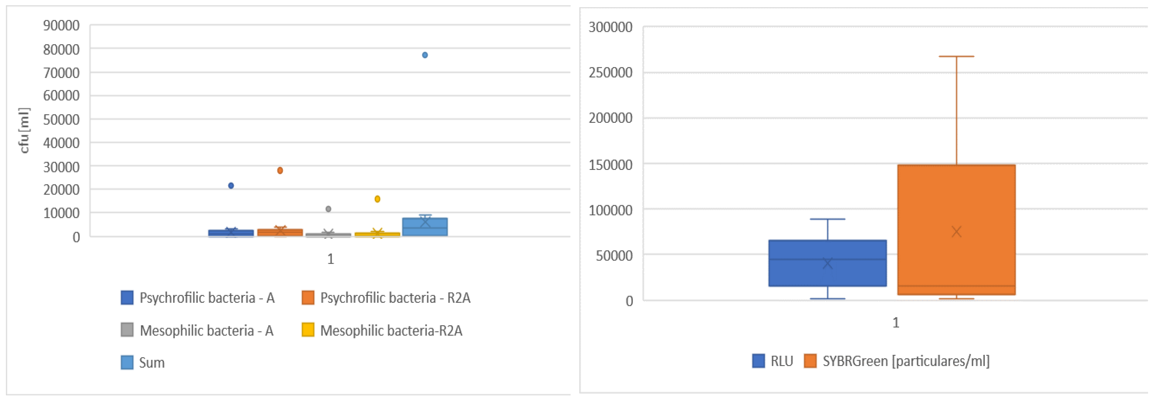
The values of the number of bacteria in the tested surface water samples are very diverse and depend mainly on the season. The lowest values were observed in the winter season, while the highest numbers in the summer season. The amounts of particles measured with a flow cytometer using SYBRGreen dye are many times higher than the amounts of bacteria determined by the culture method. In all the performed determinations, the numbers of bacteria on R2A agar were significantly higher than the numbers on agar A (Figure 1).
The statistical parameters for the determined parameters are presented in tab. 2. When analysing the values of correlation coefficients for the assessed parameters of surface water, very strong relationships can be noticed. They concern the number of psychrophilic bacteria determined on A agar and R2A agar (Table 2).
In contrast, fairly strong relationships were observed for:
the number of mesophilic bacteria on both tested agars,
RLU values, and the number of psychrophilic bacteria on Agar A and R2A,
total number of bacteria determined and the RLU value.
This would suggest the possibility of using a luminometric ATP concentration determination for the rapid assessment of microbiological quality of surface water. Unfortunately, no linear relationships were observed for cytometric determinations with the other tested microbiological parameters of water. This corresponds to previous results presented by Velten et al. [27] and Magic-Knezev and van der Kooij [28] for environmental aquatic bacteria. Correlation coefficients after adjusting for autofluorescence increased slightly. The significance of the correlation cannot be observed in any case.
The microbiological quality of water is the main factor determining health and the lives of its users. This problem applies to all types of water presented and analyzed in the research. Surface waters are a source of drinking water for approximately 50% of the Polish population and 66% of the European population [European Environment Agency (EEA)]. Currently, surface waters are assessed according to the classification of ecological status, ecological potential and chemical status. The microbiological quality, which is often very bad, is not assessed.
The literature gives examples of quantitative assessment in surface waters using different methods. Such studies were conducted on the Grand River, located in southern Ontario, Canada. A modified flow cytometry method was used to monitor water quality in the river. The results were compared to total cell counts (divided into HNA and LNA groups) for the same river water sample using flow cytometry and counts for other water quality parameters including phosphorus and nitrogen concentrations, temperature and turbidity. The flow cytometry method provided reproducible results with a standard error of ≤ 12% [29]. Quantities of microorganisms were also measured in seawater. The total bacterial count determined by the culture method was 1.10·105 cfu/ml, while by flow cytometry the value was much higher and amounted to 8.00·105 particles/ml [30]. Similar differences in bacterial abundance were shown by the results obtained in this research.

3.2. Rainwater

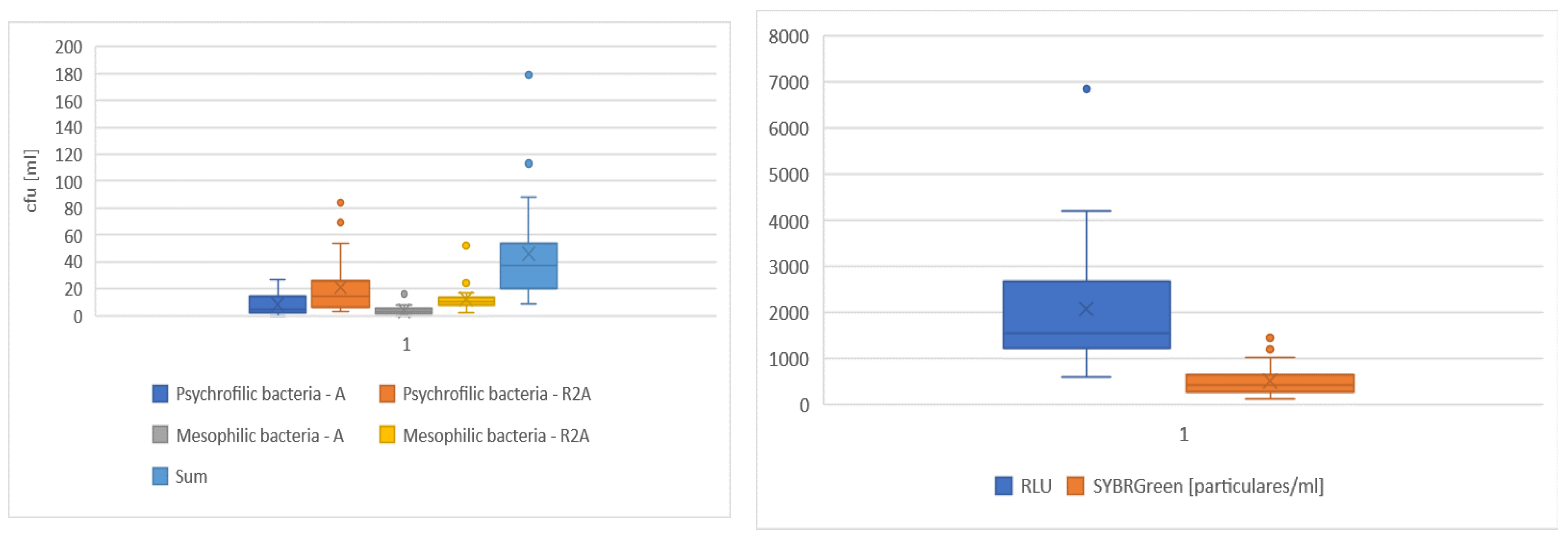
The microbiological quality of rainwater, similar to surface water, was dependent on the season. The lowest values were observed in the spring season, while the highest numbers in the summer season. The amounts of particles measured with a flow cytometer using SYBRGreen dye are many times higher than the amounts of bacteria determined by the culture method. In all the performed determinations, the numbers of bacteria on agar R2A were significantly higher than the numbers on agar A (Figure 2).
In case of rainwater, very strong linear Pearson correlations between the examined parameters were found. A very strong relationship was found between:
sum of all bacteria, and psychrophilic bacteria on R2A agar,
sum of all bacteria, and mesophilic bacteria on R2A agar,
number of psychrophilic bacteria on agar A and agar R2A,
the number of psychrophilic bacteria on Agar A, with the number of mesophilic bacteria on agar R2A,
number of mesophilic bacteria on agar A and R2A.
There is a rather strong relationship between RLU value and the number of particles measured cytometrically (Table 3).
Water resources mainly come from precipitation, which is characterised by significant diversity in time and space. The use of rainwater, even for drinking purposes, is increasingly being addressed. Rainwater is characterized by a highly varied quality, and its composition is shaped by many factors. The results of the microbiological quality of rainwater presented in the work prove their great difference that depend on the season. From an ecological point of view, the solution to use rainwater is attractive. It is therefore necessary to know its quality. Especially microbiological contamination, which pose risk to human health, is very important.
Recently, many countries including Thailand, USA, Nigeria, New Zealand, India, Zambia, Brazil, Canada, Australia, Jordan, New Guinea and South Korea have studied the quality of rainwater [31,32,33,34,35]. In Europe, rainwater quality assessment has been studied by Polkowska et al.[36], Fewtrell and Kay [37], Melidis et al. [38], Sazakli et al. [39], Tsakovski et al. [40]. Many studies have found that the rainwater has unacceptable levels of microbiological contamination and poor physicochemical properties. A clear position on the quality and health risks associated with water collected from roof coverings has not been reached [41].

3.3. Grundwater

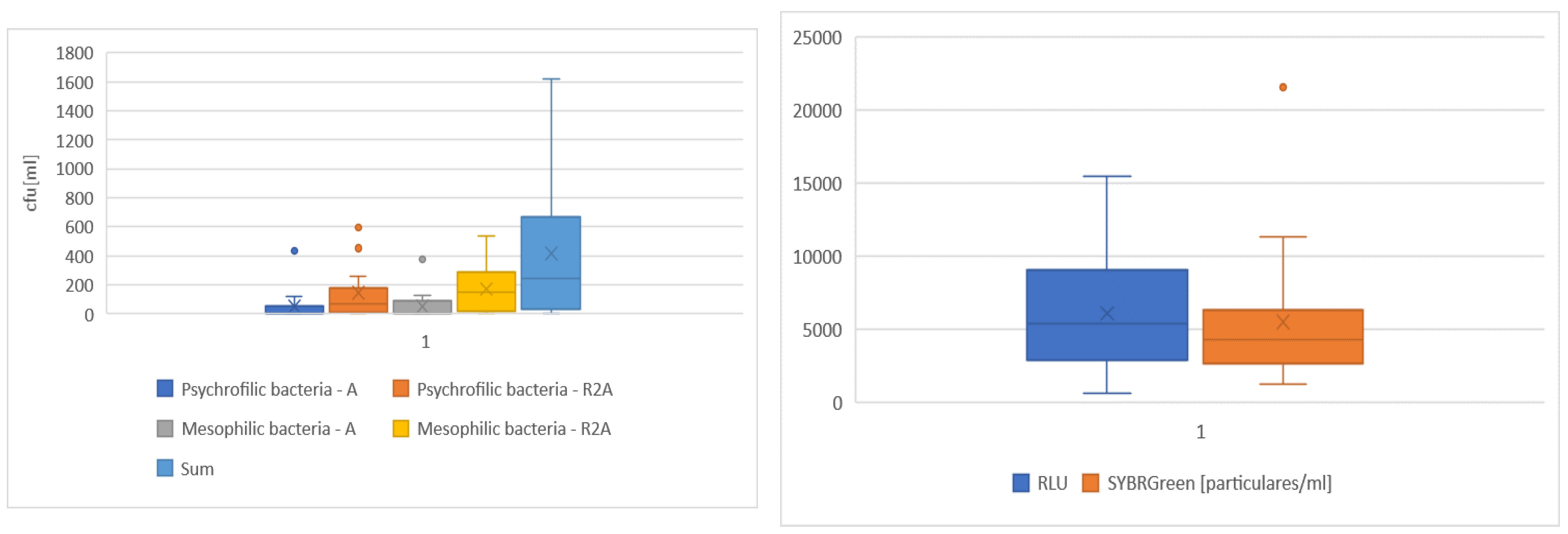
The maximum numbers of psychrophilic and mesophilic bacteria in this water determined by culture methods on agar A constituted 27 and 16 cfu/ml, respectively. In some samples, no bacteria could be detected on agar A, but were always detected on agar R2A (minimum values were 4 and 2 cfu/ml for psychrophiles and mesophiles, respectively). Similarly to the total bacterial count values measured by the culture method, the RLU values in the tested water samples were also very low and ranged from 132 to 461 RLUs, with an average of 267. The number of particles in the groundwater samples measured using SYBRGreen ranged from 860 to 6,860 [1 ml] (Figure 3).
When analysing the Pearson correlation coefficients a strong correlation between the number of psychrophilic bacteria on R2A agar and the sum of all the bacteria obtained by the culture method can by observed (Table 4).
A fairly strong linear correlation is observed between:
number of psychrophilic and mesophilic bacteria on both agars,
the number of mesophilic bacteria and the sum of all bacteria,
RLU value and the number of mesophilic bacteria on agar A,
RLU value and the number of mesophilic bacteria on R2A agar,
RLU value and the sum of all bacteria.
In case of groundwater, the results obtained indicate luminometric determination of ATP as a rapid and reliable method for assessing its quality.
In accordance with the principle that the best quality water resources should be used as a source of drinking water, the amount of groundwater used for collective water supply is increasing in Poland. Often the quality of these waters is so good that they do not require disinfection. In Poland nearly 70% of tap water comes from underground resources, but storing it in reservoirs and then transferring it through extensive systems of pipelines and water installations often adversely affects the quality of that water. This makes it necessary to use disinfection processes also for water from underground intakes. There is also local contamination of underground water intakes, as well as secondary pollutants, often caused by poor technical and sanitary condition of installations inside buildings, supplying water directly to the point of collection by the consumer. However, in emergency situations, a microbiological hazard may arise and then rapid determination methods can inform us about it.
Concentration of ATP is often used as a microbiological parameter along with other microbiological methods to characterize drinking water quality [42,43,44] and for assessing biological stability [45,46]. Some studies confirm a significant correlation between ATP and HPC in drinking water [41,47], as also indicated by the obtained results of the statistical analysis for the data presented. Sometimes no correlation has been observed between ATP and HPC [26,43,46]. It is important to understand that these two microbiological parameters are very different; ATP is a total measurement, bearing in mind that culture methods often reveal only less than 1% of the bacteria present in a sample [48]. Therefore, these two microbiological parameters will not necessarily correlate with each other, and in fact the correlation values obtained may indicate quite the opposite.
Other methods of total counts such as microscopy and flow cytometry are also frequently used and have been found to correlate significantly with ATP concentrations[49,50]. Correlations between total cell counts obtained by different methodologies can be expected, as these are measurements of total biomass. However, Liu et al. [46] observed no correlation between the values obtained by flow cytometry methods and ATP. This situation is similar to the results presented. Overall, it is important to recognize that different methods provide us with different information about the microbiological status of drinking water.

3.4. Tap water

Microbiological quality was assessed in 220 samples of tap water. Statistical parameters indicate large differences in the values of individual indices on both agars used. In all samples tested, a significantly higher number of bacteria was observed in cultures on agar R2A (Figure 4).
Analysing Pearson linear correlations (Table 5), a strong linear correlation is found between:
the number of psychrophilic bacteria on R2A agar and the sum of all bacteria,
the number of mesophilic bacteria on R2A agar and the sum of all bacteria.
A fairly strong correlation has been found between the number of particles measured cytometrically. Unfortunately, the correlation coefficient values indicate a lack of linear correlation between the bacterial count results obtained by culture methods and the RLU and particle count values measured by flow cytometry. This applies to both the abundance values of individual bacterial groups and their sum.
Total microbial counts in drinking water are usually monitored using heterotrophic plate indicators (HPC). This method has been used for over 100 years and is recommended in drinking water guidelines. However, the HPC method has its drawbacks. This method is time consuming and limited to cultureable bacteria. Microbiological water safety is a very important issue. Recently, rapid and accurate detection methods have developed, such as adenosine triphosphate (ATP) measurement to assess microbial activity in drinking water and flow cytometry (FCM) to determine total cell concentration (TCC). When it comes to drinking water quality control, it is necessary to understand the relationship between conventional and new methods. All three methods were used to assess the quality of 200 drinking water samples obtained from two local buildings connected to the same distribution system. Samples were taken on both normal working days and weekends and correlations between different microbiological parameters were determined. TCC in the samples ranged from 0. 37 to 5. 61 x 105 cells [1 ml], and two clusters, the so-called high nucleic acid (HNA) and low nucleic acid (LNA) bacterial groups, were clearly distinguished. The results showed that the rapid determination methods (i. e. , FCM and ATP) has correlated well (R2 = 0. 69), but only a low correlation (R2 = 0.31) has been observed between the rapid methods and the conventional HPC data [51]. Such results are also confirmed by the values obtained in the presented studies. With respect to drinking water monitoring, both FCM and ATP measurements have been recognized as useful parameters for rapid assessment of microbiological quality of drinking water [51,52].
The quality of tap water depends on the source of its intake, the method of collecting and treating it, the sanitary conditions of water intakes and tanks, water supply network, connections and internal water supply installation. There are many factors that determine the quality of water, which ultimately reaches the recipient. Often the good quality of produced water that enter into the water supply system is not equivalent to the same good quality of water that reaches the recipient. Interruptions in water supply and the resulting storage of water by the population favor bacteriological contamination. Pressure fluctuations in the network may cause its contamination with other foreign waters sucked into the water supply network. Water intended for human consumption should meet certain standards and it is regularly examined. Physico-chemical parameters are more and more often being assessed by rapid tests. On the other hand, the problem of assessing microbial contamination is still being solved by tradition, labor-intensive and long-lasting culture methods. It takes a minimum of 24 hours from the time the determination is performed to the first result. Therefore, it is necessary to the search for fast, reliable, and delivering reproducible results microbiological determinations nowadays. It should be noted that these rapid methods – luminometric ATP determination as well as flow cytometry will not replace culture method for the time being. Traditional methods, as shown in the presented work, are not reliable. The presented research clearly show how much difference the is in the results obtained with different media. The A-reference nutrient agar, depending on the type of water tested, detects only a small number of bacteria. And is this result satisfactory in the case of the waters we use on a daily basis? Luminometric ATP determination is a very quick and relatively cheap method which allows to assess the microbiological quality of water. This is a general indicator. Continuous ATP determination would allow for hazard identification and could be used for various types of water [52]. Surface waters are used as intakes for drinking water. Changing microbiological quality, detected very quickly, would allow e.g. quick correction of unit processes (e.g. correction of disinfectant doses), detection of incidental contamination, and thus a quick response from the services.

4. Conclusions

  • The number of bacteria assessed on Agar R2A for all types of analyzed waters were higher compared to the reference Agar A. The lowest values of standard deviation for 4 groups of bacteria (psychrophilic and mesophilic bacteria on both agars) in waters with low bacteriological contamination were obtained for groundwater.
  • The total number of bacteria quite strongly correlates with the RLU values for surface and groundwater. This suggest the possibility of using the luminometric ATP determination for the quick assessment of the microbiological quality of this type of water.
  • Quite strong correlations between the RLU values and cytometrically determined particle number were observed for rainwater
  • Strong and relatively strong linear relationships for the tested microbiological parameters differ in each of the assessed waters.
The research conducted and statistical analysis also allow additional conclusions to be drawn for each type of the water.
In the case of surface water, the microbiological quality was highly dependent on the season during which the research was conducted. Low Pearson’s correlation coefficients factors indicate the lack of linear relationships for cytometric determinations with other microbiological water parameters examined. In case of cytometric determinations, the obtained result should be correlated with autofluorescence.
The microbiological quality of rainwater is highly variable. Very strong correlations were found for the determination of the bacteria number performed with the use of culture methods. On the other hand, low values of linear correlation were obtained for the number of bacteria, and the values obtained by cytometry and luminometry methods.
In the case of non-disinfected groundwater of very good microbiological quality, it is suggested to quickly measure and evaluate the microbiological quality using the luminometric method of ATP determination.
For tap water, no linear correlation was found between the number of bacteria obtained with the culture methods and the values of RLU and the number particles measured with the flow cytometer. Such a result indicates the presence of uniform microflora of the examined waters. On the other hand, the luminometric measurements may have been influenced by extracellular ATP, which was released from microbial cells during ozonation. This indicates the need to assess the concentration of extracellular ATP for disinfected tap water.

Author Contributions

Conceptualization, J.Z.; methodology, J.Z.; software, J.Z.; validation, J.Z.; formal analysis, J.Z.; investigation, J.Z.; resources, J.Z; data curation, J.Z.,; writing—original draft preparation ; writing—review and editing, J.Z., E.K. and W.P; visualization J.Z.; supervision, J.Z.; project administration, J.Z.; funding acquisition, J.Z. All authors have read and agreed to the published version of the manuscript.

Funding

This research received no external funding.

Acknowledgments

Not applicable.

Conflicts of Interest

The authors declare no conflict of interest.

References

  1. Maier R.M.; Pepper I.L; Gerba Ch.P. Environmental Microbiology, Secend Edidtion , Academic Press, 2009.
  2. Błaszczyk M. Environmental microbiology, PWN, Warszawa 2010.
  3. Kalsoom M.; Rehman F.; Shafique T.; Junaid S.; Khalid N.; Adnan M.; Zafar I.; Tariq M.A.; Raza M.A.; Zahra A.; Ali H. Biological importance of microbes in agriculture, food and pharmaceutical industry: a review, Innovare Journal of Life Sciences, 2020,Vol 8, Issue 6, 1-4. [CrossRef]
  4. Berry D., Xi CH., Raskin L. Microbial ecology of drinking water distribution systems, Curr. Opin. Biotechnnol, 2006, 17930, 297-302. [CrossRef]
  5. Mara D.; Horan N. Handbook of Water and Wastewater Microbiology, Academic Press, 2003.
  6. Caggiano G.; Diella G.; Triggiano F.; Bartolomeo N.; Apollonio F.; Carmen Campanale C.; Lopuzzo M.; Maria Montagna M.T. Occurrence of Fungi in the Potable Water of Hospitals, A Public Health Threat: Pathogens, 2020, 9, 783. [CrossRef]
  7. Gonçalves A.B.; Paterson, R.R.M.; Lima, N. Survey and significance of filamentous fungi from tap water. International Journal of Hygiene and Environmental Health, 2006, 209, 257-264. [CrossRef]
  8. Hageskal G.; Lima, N.; Skaar I. The study of fungi in drinking water. Mycological Research, 2009, 113: 165-172. [CrossRef]
  9. Frimmel F.H.; Saravia F.; Gorenflo F. NOM removal from different raw waters by membrane filtration, Water Sci.Technol. 2004, 4, 16-174. [CrossRef]
  10. Jones M.R.; Pinto E.; Torres, M A.; Dörr F.; Mazur-Marzec H.; Szubert K.; Tartaglione L.; Dell'Aversano C.; Miles, Ch. O.; Beach D. G.; McCarron P.; Sivonen K.; Fewer D.P.; Jokela J.; Janssen E. M.-L. CyanoMetDB, a comprehensive public database of secondary metabolites from cyanobacteria, Water Research, 2021, Vol. 196, 15. [CrossRef]
  11. Helmreich B.; Horn H. Opportunities in rainwater harvesting. Science Direct, Desalination, 2009, 248, 118- 124. [CrossRef]
  12. Georgios D.; Gikas Vassilios A.; Tsihrintzis A. Assessment of water quality of first-flush roof runoff and harvested rainwater, Journal of Hydrology, 2012, 466-467, 115-126. [CrossRef]
  13. Proctor C.R.; Hammes F. Drinking water microbiology – from measurement to management. ScienceDirect, Current Opinion in Biotechnology, 2015, 33, 87-94. [CrossRef]
  14. Jo W.K.; Kang J.H. Workplace exposure to bioaerosols in pet shop, pet clinics and flower garden, Chemosphere, 2006, 65, 1755-1761. [CrossRef]
  15. Amin M.T.; Han M.Y. Improvement of solar based rainwater disinfection by using leman and vinegar as catalysts. Desalination, 2011, 276, 416-424. [CrossRef]
  16. Cullimore D.R. Practical manual of groundwater microbiology, CRC Press, 2007, ISBN-13: 978-0849385315.
  17. Bienz K.; Eckert J.; Kayser F.; Zinkernagel R. Medical microbiology, Wydawnictwo Lekarskie PZWL, Warszawa 2007.
  18. Liu S., Grunawan C.; Barraud N.; Rice S.A.; Harry E.J.; Amal R. Understanding, Monitoring and Controling Biofilm Growth in Drinking Water Distribution Systems, Environ. Sci. Technol., 2016, 50, 17, 8954–8976 . [CrossRef]
  19. Świderska - Bróż M.The effects of the presence of biofilm in water distribution systems intended for human consumption, Ochrona Środowiska, 2012, 1, 9-14.
  20. Pierścieniak M.; Trzcińska N.; Słomczyński T.; Wąsowski J. The problem of secondary contamination of tap water, Ochrona Środowiska i Zasobów Naturalnych, 2009, 39, 29-32.
  21. Łebkowska M.; Pajor E.; Rutkowska-Narożniak A.; Kwietniewski M.; Wąsowski J.; Kowalski D. Research on the development of microorganisms in water supply pipes made of ductile iron with cement lining, Ochrona Środowiska, 2011, Vol. 33, 3, 9-16.
  22. Liu G., Zhang Y.; Knibbe W.; Feng C.; Liu W.; Medema G.; Meer W. Potential impacts of changing supply-water quality on drinking water distribution: A review, Water Research, 2017, 116, 136-137. [CrossRef]
  23. Grabińska – Łoniewska A.; Siński E. Pathogenic and potentially pathogenic microorganisms in aquatic ecosystems and water supply networks, Wydawnictwo „Seidel-Przywecki” Sp. z o.o., Warszawa, 2010.
  24. Cabral J. Water Microbiology. Bacterial Pathogens and Water, International Journal of Environmental Research and Public Health, 2010, 7, 3657-3703. [CrossRef]
  25. Hammes F.; Berney M.; Wang Y.; Vital M.; Köster O.; Egli T.. Flow-cytometric total bacterial cell counts as a descriptive microbiological parameter for drinking water treatment processes, Water Res., 2008, 42 (1-2), 269-77. [CrossRef]
  26. Hammes F.; Egli T. New Method for Assimilable Organic Carbon Determination Using Flow-Cytometric Enumeration and a Natural Microbial Consortium as Inoculum, Environ. Sci. Technol., 2005, 39 (9), 3289-3294. [CrossRef]
  27. Velten S.; Hammes F.; Boller M.; Egli T. Rapid and direct estimation of active biomass on granular activated carbon through adenosine tri-phosphate (ATP) determination, Water Res., 2007, 41(9),1973-83. [CrossRef]
  28. Magic-Knezev A.; Van Der Kooij D.. Optimisation and significance of ATP analysis for measuring active biomass in granular activated carbon filters used in water treatment, Water Res., 2004, 38(18), 3971-9. [CrossRef]
  29. Elhadidy A.M.; Van Dyke M.I.; Peldszus S.; Huck P.M. Application of flow cytometry to monitor assimilable organic carbon (AOC) and microbial community changes in water, Journal of Microbiological Methods, 2016, 130, 154-163. [CrossRef]
  30. Joachimsthal E.L.; Ivanov V.; Tay J-H.; Stephen T. Flow cytometry and conventional enumeration of microorganisms in ships’ ballast water and marine samples, Marine Pollution Bulletin, 2003, 46 (3), 308-313. [CrossRef]
  31. Pinfold J.; Nigel V.; Horan J. Measuring the effect of a hygiene behaviour intervention by indicators of behaviour and diarrhoeal disease, Transactions of The Royal Society of Tropical Medicine and Hygiene, 1993, Vol. 90, Issue 4, 366-371. [CrossRef]
  32. Uba B.N.; Aghogho O. Rainwater quality from different roof catchments in the Port Harcourt district, Rivers State, Nigeria, Journal of Water Supply, Research and Technology-Aqua, 2000, 49 (5), 281-288. [CrossRef]
  33. Handia L.; Madalitso J.; Tembo J.; Mwiindwa C. Potential of rainwater harvesting in urban Zambia, Physics and Chemistry of the Earth, Parts A/B/C, 2003, 28 (20-27), 893-896. [CrossRef]
  34. Despins CH.; Farahbakhsh K.; Leidl CH. Assessment of rainwater quality from rainwater harvesting systems in Ontario, Canada, Journal of Water Supply: Research and Technology AQUA, 2009, 117-135. [CrossRef]
  35. Lee J.Y.; Yang J-S.; Han M.; Choi J. Comparison of the microbiological and chemical characterization of harvested rainwater and reservoir water as alternative water resources, Science of the Total Environment, 2010, 408, 896-905. [CrossRef]
  36. Polkowska Ż.; Astel A.; Walna B.; Małek S.; Mędrzycka K.; Górecki T.; Siepak J.; Namieśnik J. Chemometric analysis of rainwater and throughfall at several sites in Poland, Atmospheric Environment, 2005, 39 (5), 837-855. [CrossRef]
  37. Fewtrell L.; Kay D. Microbial quality of rainwater supplies in developed countries: a review, Urban Water Journal, 2007, 4, 253-260. [CrossRef]
  38. Melidis P.; Akratos Ch.S.; Tsihrintzis V.A.; Trikilidou E.. Characterization of Rain and Roof Drainage Water Quality in Xanthi, Greece, Environmental Monitoring and Assessment, 2007, 127 (1-3), 15-27. [CrossRef]
  39. Sazakli E.; Alexopoulos A.; Leotsinidis M. Rainwater harvesting, quality assessment and utilization in Kefalonia Island, Greece, Water Research, 2007, 41 (9), 2039-2047. [CrossRef]
  40. Tsakovski S.; Tobiszewski M.; Simeonov V.; Polkowska Ż.; Namieśnik J. Chemical composition of water from roofs in Gdansk, Poland, Environmental Pollution, 2010, 158 (1), 84-91. [CrossRef]
  41. Evans C.A.; Coombes P.J.; Dunstan R.H. Wind, rain and bacteria: The effect of weather on the microbial composition of roof-harvested rainwater, Water Research, 2006, 40 (1), 37-44. [CrossRef]
  42. Deininger R.A.; Lee J.Y.. Rapid determination of bacteria in drinking water using an ATP assay, 2001. [CrossRef]
  43. Siebel E.; Wang Y.; Egli T.; Hammes F. Correlations between total cell concentration, total adenosine tri-phosphate concentration and heterotrophic plate counts during microbial monitoring of drinking water, Drink. Water Eng. Sci., 2008, 1, 1-6. [CrossRef]
  44. Vital M.; Hammes F.; Egli T. Competition of Escherichia coli O157 with a drinking water bacterial community at low nutrient concentrations, Water Research, 2012, 46 (19), 6279-6290. [CrossRef]
  45. Lautenschlager K.; Hwang CH.; Liu W.T.; Boon N.; Köster O.; Vrouwenvelder H.; Egli T.; Hammes F. A microbiology-based multi-parametric approach towards assessing biological stability in drinking water distribution networks, Water Research, 2013, 47 (9), 3015-3025. [CrossRef]
  46. Liu G.; Van Der Mark E.J; Verberk J.J.C.; Van Dijk J.C. Flow Cytometry Total Cell Counts: A Field Study Assessing Microbiological Water Quality and Growth in Unchlorinated Drinking Water Distribution Systems, BioMed Research International, 2013, . [CrossRef]
  47. Delahaye E.; Welté B.; Levi Y.; Leblon G.; Montiel A. An ATP-based method for monitoring the microbiological drinking water quality in a distribution network, Water Research, 2003, 37 (15), 3689-3696. [CrossRef]
  48. Maki J.S.; Lacroix S.J.; Hopkins B.S.; Staley J.T. Recovery and diversity of heterotrophic bacteria from chlorinated drinking waters, Applied and Environmental Microbiology, 1986, 51 (5), 1047-1055 . [CrossRef]
  49. Eydal H.S.C.; Pedersen K. Use of an ATP assay to determine viable microbial biomass in Fennoscandian Shield groundwater from depths of 3–1000 m, Journal of Microbiological Methods, 2007, 69A (4), 273-280. [CrossRef]
  50. Hammes F.; Egli T. Cytometric methods for measuring bacteria in water: ad-vanteges pitfalls and application, Anal. Bioanal. Chem., 2010, 397 (3), 1083—1095. [CrossRef]
  51. Stevens M.; Köhler D.; Göhde R. Flow Cytometric enumeration of total bacterial cell count (TCC) in drinking water with the Partec Cyflow® Cube 6 flow cytometer, Görlitz 72, 2013, 12.
  52. Vang Ó.K.; Corfitzen C.B.; Smith C; Albrechtsen H.J. Evaluation of ATP measurements to detect microbial ingress by wastewater and surface water in drinking water. Water Res., 2014, 64, 309-320. [CrossRef]
Figure 1. Box and whisker plot showing median and variation in samples taken from surface water.
Preprints 88704 g001
Figure 2. Box and whisker plot showing median and variation in samples taken from rainwater.
Preprints 88704 g002
Figure 3. Box and whisker plot showing median and variation in samples taken from grundwater.
Preprints 88704 g003
Figure 4. Box and whisker plot showing median and variation in samples taken from tap water.
Preprints 88704 g004
Table 1. Scope and methodology for determining microbiological properties of water.
Tested parameter Method / Standard
Total bacteria count at 22 ° (72h) Culture method on standard nutrient agar; PN-EN ISO 6222:2004
Total bacteria count at 37 °C (48h)
Enumeration of microorganisms Partec Cube 6 flow cytometer
ATP concentration Luminometric determination;
www.promega.com/protocols
Table 2. Statistical parameters - correlation coefficients for surface water.
Average Psychrophilic bacteria AGAR A Psychrophilic bacteria AGAR R2A Mesophilic bacteria AgarA Mesophilic bacteria AgarR2A Sum RLU The number of particles SYBRGreen
Psychrophilic bacteria AGAR A 43724 1,000 0,985 0,753 0,710 0,988 0,780 0,111
Psychrophilic bacteria AGAR R2A 62397 - 1,000 0,764 0,732 0,993 0,832 0,116
Mesophilic bacteria AgarR2A 85326 - - 1,000 0,951 0,820 0,647 0,126
Mesophilic bacteria AgarR2A 13590 - - - 1,000 0,789 0,611 0,217
Sum 128235 - - - - 1,000 0,813 0,128
RLU 459769 - - - - - 1,000 0,093
The number of particles SYBRGreen 236571 - - - - - - 1,000
Table 3. Statistical parameters - correlation coefficients for rainwater.
Average Psychrophilic bacteria AGAR A Psychrophilic bacteria AGAR R2A Mesophilic bacteria AgarA Mesophilic bacteria AgarR2A Sum RLU The number of particles SYBR Green
Psychrophilic bacteria AGAR A 1834 1,000 0,994 0,981 0,982 0,997 0,532 0,389
Psychrophilic bacteria AGAR R2A 2307 - 1,000 0,984 0,974 0,996 0,519 0,411
Mesophilic bacteria AgarR2A 823 - - 1,000 0,982 0,991 0,491 0,419
Mesophilic bacteria AgarR2A 1361 - - - 1,000 0,988 0,458 0,375
Sum 632 - - - - 1,000 0,509 0,401
RLU 41687 - - - - - 1,000 0,725
The number of particles SYBR Green 93781 - - - - - - 1,000
Table 4. Statistical parameters - correlation coefficients for grundwater.
Average Psychrophilic bacteria AGAR A Psychrophilic bacteria AGAR R2A Mesophilic bacteria AgarA Mesophilic bacteria AgarR2A Sum RLU The number of particles SYBR Green
Psychrophilic bacteria AGAR A 8 1,000 0,852 0,461 0,476 0,851 0,609 0,497
Psychrophilic bacteria AGAR R2A 21 - 1,000 0,621 0,670 0,968 0,697 0,264
Mesophilic bacteria AgarR2A 3 - - 1,000 0,766 0,738 0,842 0,270
Mesophilic bacteria AgarR2A 12 - - - 1,000 0,809 0,768 0,316
Sum 44 - - - - 1,000 0,798 0,361
RLU 267 - - - - - 1,000 0,287
The number of particlesSYBR Green 2069 - - - - - - 1,000
Table 5. Statistical parameters - correlation coefficients for tap water.
Average Psychrophilic bacteria AGAR A Psychrophilic bacteria AGAR R2A Mesophilic bacteria AgarA Mesophilic bacteria AgarR2A Sum RLU The number of particles SYBR Green
Psychrophilic bacteria AGAR A 52 1,000 0,531 0,308 0,500 0,650 0,016 0,013
Psychrophilic bacteria AGAR R2A 144 - 1,000 0,784 0,857 0,953 0,153 -0,014
Mesophilic bacteria AgarR2A 54 - - 1,000 0,721 0,802 0,110 -0,025
Mesophilic bacteria AgarR2A 166 - - - 1,000 0,932 0,266 -0,044
Sum 416 - - - - 1,000 0,184 0,003
RLU 9291 - - - - - 1,000 0,053
The number of particles SYBR Green 5308 - - - - - - 1,000
Disclaimer/Publisher’s Note: The statements, opinions and data contained in all publications are solely those of the individual author(s) and contributor(s) and not of MDPI and/or the editor(s). MDPI and/or the editor(s) disclaim responsibility for any injury to people or property resulting from any ideas, methods, instructions or products referred to in the content.
Copyright: This open access article is published under a Creative Commons CC BY 4.0 license, which permit the free download, distribution, and reuse, provided that the author and preprint are cited in any reuse.
Prerpints.org logo

Preprints.org is a free preprint server supported by MDPI in Basel, Switzerland.

Subscribe

© 2024 MDPI (Basel, Switzerland) unless otherwise stated