Submitted:
02 November 2023
Posted:
02 November 2023
You are already at the latest version
Abstract
Keywords:
1. Introduction
2. Morphological characteristics of pericytes
3. Markers of pericytes
4. A role of pericytes in angiogenesis
5. Pericytes in osteogenesis
6. Recent pre-clinical and clinical application of pericytes
7. Conclusions
Author Contributions
Funding
Institutional Review Board Statement
Informed Consent Statement
Data Availability Statement
Acknowledgments
Conflicts of Interest
References
- Roddy, E.; De Baun, M.R.; Daoud-Gray, A.; Yang, Y.P.; Gardner, M.J. Treatment of critical-sized bone defects: clinical and tissue engineering perspectives. Eur. J. Orthop. Surg. Traumatol. 2018, 28, 351–362. [Google Scholar] [CrossRef] [PubMed]
- Nishida, J.; Shimamura, T. Methods of reconstruction for bone defect after tumor excision: a review of alternatives. Med. Sci. Monit. 2008, 14, 107–113. [Google Scholar]
- Goulet, J.A.; Senunas, L.E.; De Silva, G.L.; Greenfield, M.L.V.H. Autogenous iliac crest bone graft: complications and functional assessment. Clin. Orthop. Relat. Res. 1997, 339, 76–81. [Google Scholar] [CrossRef]
- Rabitsch, K.; Maurer-Ertl, W.; Pirker-Frühauf, U.; Wibmer, C.; Leithner, A. Intercalary reconstructions with vascularised fibula and allograft after tumour resection in the lower limb. Sarcoma 2013, 2013, 160295. [Google Scholar] [CrossRef]
- Tamai, N.; Myoui, A.; Tomita, T.; Nakase, T.; Tanaka, J.; Ochi, T.; Yoshikawa, H. Novel hydroxyapatite ceramics with an interconnective porous structure exhibit superior osteoconduction in vivo. J. Biomed. Mater. Res. 2002, 59, 110–117. [Google Scholar] [CrossRef] [PubMed]
- James, A.W.; Zara, J.N.; Corselli, M.; Askarinam, A.; Zhou, A.M.; Hourfar, A.; Nguyen, A.; Megerdichian, S.; Asatrian, G.; Pang, S.; et al. An abundant perivascular source of stem cells for bone tissue engineering. Stem Cells Transl. Med. 2012, 1, 673–684. [Google Scholar] [CrossRef]
- Zhao, H.; Chappell, J.C. Microvascular bioengineering: a focus on pericytes. J. Biol. Eng. 2019, 13, 1–12. [Google Scholar] [CrossRef] [PubMed]
- Meijer, E.M.; van Dijk, C.G.M.; Kramann, R.; Verhaar, M.C.; Cheng, C. Implementation of pericytes in vascular regeneration strategies. Tissue Eng. - Part B Rev. 2022, 28, 1–21. [Google Scholar] [CrossRef] [PubMed]
- Zhang, Z.S.; Zhou, H.N.; He, S.S.; Xue, M.Y.; Li, T.; Liu, L.M. Research advances in pericyte function and their roles in diseases. Chinese J. Traumatol. - English Ed. 2020, 23, 89–95. [Google Scholar] [CrossRef]
- Herndon, J.M. Chapter 9 – Development and maintenance of the blood-brain barrier. In Primer on cerebrovascular diseases.Second edition. Ed. Caplan, L.R. Publisher: Academic Press, 2017, 51-56. [CrossRef]
- Attwell, D.; Mishra, A.; Hall, C.N.; O'Farrell, F.M.; Dalkara, T. What is a pericyte? J. Cereb. Blood Flow Metab. 2016, 36, 451–455. [Google Scholar] [CrossRef]
- Brown, L.S.; Foster, C.G.; Courtney, J.-M.; King, N.E.; Howells, D.W.; Surtherland, B.A. Pericytes and neurovascular function in the healthy and diseased brain. Front. Cell.Neurosci. 2019, 13, 282. [Google Scholar] [CrossRef] [PubMed]
- Sun, Z.; Gao, C.; Gao, D.; Sun, R.; Li, W.; Wang, F.; Wang, Y.; Cao, H.; Zhou, G.; Zhang, J.; Shang, J. Reduction in pericyte coverage leads to blood-brain barrier dysfunction via endothelial transcytosis following chronic cerebral hypoperfusion. Fluids Barriers CNS. 2021, 18, 21. [Google Scholar] [CrossRef]
- Heymans, M.; Figueiredo, R.; Dehouck, L.; Francisco, D.; Sano, Y.; Shimizu, F.; Kanda, T.; Bruggmann, R.; Engelhardt, B.; Winter, P.; Gosselet, F.; Culot, M. Contribution of brain pericytes in blood–brain barrier formation and maintenance: a transcriptomic study of cocultured human endothelial cells derived from hematopoietic stem cells. Fluids Barriers CNS. 2020, 17, 48. [Google Scholar] [CrossRef] [PubMed]
- Ferland-McCollough, D.; Slater, S.; Richard, J.; Reni, C.; Mangialardi, G. Pericytes, an overlooked player in vascular pathobiology. Pharmacol.Ther. 2017, 171, 30–42. [Google Scholar] [CrossRef] [PubMed]
- He, S.; Zhang, Z.; Peng, X.; Wu, Y.; Zhu, Y.; Wang, L.; Zhou, H.; Li, T.; Liu, L. The protective effect of pericytes on vascular permeability after hemorrhagic shock and their relationship with Cx43. Front. Physiol. 2022, 13, 1–16. [Google Scholar] [CrossRef]
- Welberg, L. Pericytes set the tone. Nature Rev. Neorosci. 2014, 15, 283. [Google Scholar] [CrossRef]
- Randall Harrell, C.; SimovicMarkovic, B.; Fellabaum, C.; Arsenijevic, A.; Djonov, V.; Volarevic, V. Molecular mechanisms underlying therapeutic potential of pericytes. J. Biomed. Sci. 2018, 25, 21. [Google Scholar] [CrossRef]
- Wu, Y.; Fu, J.; Huang, Y.; Duan, R.; Zhang, W.; Wang, C.; Wang, S.; Hu, X.; Zhao, H.; Wang, L.; Liu, J.; Gao, G.; Yuan, P. Biology and function of pericytes in the vascular microcirculation. Animal Model Exp. Med. 2023, 6, 337–345. [Google Scholar] [CrossRef] [PubMed]
- Nakagomi, T.; Kubo, S.; Nakano-Doi, A.; Sakuma, R.; Lu, S.; Narita, A.; Kawahara, M.; Taguchi, A.; Matsuyama, T. Brain vascular pericytes following ischemia have multipotential stem cell activity to differentiate into neural and vascular lineage cells. Stem Cells. 2015, 33, 1962–1974. [Google Scholar] [CrossRef]
- Ahmed, T.A.; El-Badri, N. Pericytes: the role of multipotent stem cells in vascular maintenance and regenerative medicine. Adv. Exp. Med. Biol. 2018, 1079, 69–86. [Google Scholar] [CrossRef]
- Cheng, J.; Korte, N.; Nortley, R.; Sethi, H.; Tang, Y.; Attwell, D. Targeting pericytes for therapeutic approaches to neurological disorders. ActaNeuropathologica. 2018, 136, 507–523. [Google Scholar] [CrossRef]
- Courtney, J.-M.; Sutherland, B.A. Harnessing the stem cell properties of pericytes to repair the brain. Neural.Regen. Res. 2020, 15, 1021–1022. [Google Scholar] [CrossRef]
- Garrison, A.T.; Bignold, R.E.; Wu, X.; Johnson, J.R. Pericytes: the lung-forgotten cell type. Front. Physiol. 2023, 14, 1–16. [Google Scholar] [CrossRef]
- Sagare, A.P.; Bell, R.D.; Zhao, Z.; Ma, Q.; Winkler, E.A.; Ramanathan, A.; Zlokovic, B.V. Pericyte loss influences Alzheimer-like neurodegeneration in mice. Nature Communications. 2013, 4, 2932. [Google Scholar] [CrossRef]
- HosseinGeranmayeh, M.; Rahbarghazi, R.; Farhoudi, M. Targeting pericytes for neurovascular regeneration. Cell Commun.Signalling. 2019, 17, 26. [Google Scholar] [CrossRef]
- Jiang, Z.; Zhou, J.; Li, L.; Liao, S.; He, J.; Zhou, S.; Zhou, Y. Pericytes in the tumor microenvironment. Cancer Lett. 2023, 556, 216074. [Google Scholar] [CrossRef]
- Sun, R.; Kong, X.; Qiu, X.; Huang, C.; Wong, P.-P. The emerging roles of pericytes in modulating tumor microenvironment. Front. Cell Dev. Biol. 2021, 9, 1–10. [Google Scholar] [CrossRef]
- Picoli, C.C.; Gonçalves, B.Ô.P.; Santos, G.S.P.; Rocha, B.G.S.; Costa, A.C.; Resende, R.R.; Birbrair, A. Pericytes cross-talks within the tumor microenvironment. BiochemicaetBiophysicaActa (BBA) – Reviews on Cancer. 2021, 1876, 188608. [Google Scholar] [CrossRef]
- Warmke, N.; Griffin, K.J.; Cubbon, R.M. Pericytes in diabetes-associated vascular disease. J. Diabetes Complications. 2016, 30, 1643–1650. [Google Scholar] [CrossRef]
- Harrell, C.R.; Simovic Markovic, B.; Fellabaum, C.; Arsenijevic, A.; Djonov, V.; Volarevic, V. Molecular mechanisms underlying therapeutic potential of pericytes. J. Biomed. Sci. 2018, 25, 1–12. [Google Scholar] [CrossRef]
- Mills, S.J.; Cowin, A.J.; Kaur, P. Pericytes, mesenchymal stem cells and the wound healing process. Cells 2013, 2, 621–634. [Google Scholar] [CrossRef]
- Cathery, W.; Faulkner, A.; Maselli, D.; Madeddu, P. Concise review: the regenerative journey of pericytes toward clinical translation. Stem Cells 2018, 36, 1295–1310. [Google Scholar] [CrossRef]
- Armulik, A.; Genové, G.; Betsholtz, C. Pericytes: developmental, physiological, and pathological perspectives, problems, and promises. Dev. Cell 2011, 21, 193–215. [Google Scholar] [CrossRef]
- Chen WC, Saparov A, Corselli M, Crisan M, Zheng B, Péault B, Huard J. Isolation of blood-vessel-derived multipotent precursors from human skeletal muscle. J Vis Exp. 2014 Aug 21;(90):e51195. PMID: 25177794; PMCID: PMC4762055. [CrossRef]
- Meyers, C.A.; Casamitjana, J.; Chang, L.; Zhang, L.; James, A.W.; Péault, B. Pericytes for therapeutic bone repair. In Pericyte Biology – Novel Concepts. Advances in Experimental Medicine and Biology; Birbrair, A., Ed.; Publisher: Springer, Cham, 2018; Volume 1109, pp. 21–32. [Google Scholar] [CrossRef]
- Blocki, A.; Beyer, S.; Jung, F.; Raghunath, M. The controversial origin of pericytes during angiogenesis - Implications for cell-based therapeutic angiogenesis and cell-based therapies. Clin. Hemorheol. Microcirc. 2018, 69, 215–232. [Google Scholar] [CrossRef]
- Gökçinar-Yagci, B.; Uçkan-Çetinkaya, D.; Çelebi-Saltik, B. Pericytes: properties, functions and applications in tissue engineering. Stem Cell Rev. Rep. 2015, 11, 549–559. [Google Scholar] [CrossRef]
- Wong, S.P.; Rowley, J.E.; Redpath, A.N.; Tilman, J.D.; Fellous, T.G.; Johnson, J.R. Pericytes, mesenchymal stem cells and their contributions to tissue repair. Pharmacol. Ther. 2015, 151, 107–120. [Google Scholar] [CrossRef]
- Caporarello, N.; D'Angeli, F.; Cambria, M.T.; Candido, S.; Giallongo, C.; Salmeri, M.; Lombardo, C.; Longo, A.; Giurdanella, G.; Anfuso, C.D.; et al. Pericytes in microvessels: from ‘mural’ function to brain and retina regeneration. Int. J. Mol. Sci. 2019, 20, 6351. [Google Scholar] [CrossRef]
- Yamazaki, T.; Mukouyama, Y.S. Tissue specific origin, development, and pathological perspectives of pericytes. Front. Cardiovasc. Med. 2018, 5, 1–6. [Google Scholar] [CrossRef]
- Yianni, V.; Sharpe, P.T. Perivascular-derived mesenchymal stem cells. J. Dent. Res. 2019, 98, 1066–1072. [Google Scholar] [CrossRef]
- Esteves, C.L.; Donadeu, F.X. Pericytes and their potential in regenerative medicine across species. Cytom. Part A 2018, 93, 50–59. [Google Scholar] [CrossRef]
- Grant, R.I.; Hartmann, D.A.; Underly, R.G.; Berthiaume, A.A.; Bhat, N.R.; Shih, A.Y. Organizational hierarchy and structural diversity of microvascular pericytes in adult mouse cortex. J. Cereb. Blood Flow Metab. 2019, 39, 411–425. [Google Scholar] [CrossRef]
- James, A.W.; Hindle, P.; Murray, I.R.; West, C.C.; Tawonsawatruk, T.; Shen, J.; Asatrian, G.; Zhang, X.; Nguyen, V.; Simpson, H.; et al. Pericytes for the treatment of orthopedic conditions. Pharmacol. Ther. 2017, 171, 93–103. [Google Scholar] [CrossRef]
- Lee, L.L.; Khakoo, A.Y.; Chintalgattu, V. Cardiac pericytes function as key vasoactive cells to regulate homeostasis and disease. FEBS Open Bio 2021, 11, 207–225. [Google Scholar] [CrossRef]
- Chiaverina, G.; di Blasio, L.; Monica, V.; Accardo, M.; Palmeiro, M.; Peracino, B.; Vara-Messler, M.; Puliafito, A.; Primo, L. Dynamic interplay between pericytes and endothelial cells during sprouting angiogenesis. Cells 2019, 8, 1–13. [Google Scholar] [CrossRef]
- Laredo, F.; Plebanski, J.; Tedeschi, A. Pericytes: problems and promises for CNS repair. Front. Cell. Neurosci. 2019, 13, 1–15. [Google Scholar] [CrossRef]
- James, A.W.; Péault, B. Perivascular mesenchymal progenitors for bone regeneration. J. Orthop. Res. 2019, 37, 1221–1228. [Google Scholar] [CrossRef]
- Avolio, E.; Alvino, V.V.; Ghorbel, M.T.; Campagnolo, P. Perivascular cells and tissue engineering: current applications and untapped potential. Pharmacol. Ther. 2017, 171, 83–92. [Google Scholar] [CrossRef]
- James, A.W.; Zara, J.N.; Zhang, X.; Askarinam, A.; Goyal, R.; Chiang, M.; Yuan, W.; Chang, L.; Corselli, M.; Shen, J.; et al. Perivascular stem cells: a prospectively purified mesenchymal stem cell population for bone tissue engineering. Stem Cells Transl. Med. 2012, 1, 510–519. [Google Scholar] [CrossRef]
- Askarinam, A.; James, A.W.; Zara, J.N.; Goyal, R.; Corselli, M.; Pan, A.; Liang, P.; Chang, L.; Rackohn, T.; Stoker, D.; et al. Human perivascular stem cells show enhanced osteogenesis and vasculogenesis with Nel-like molecule I protein. Tissue Eng. - Part A 2013, 19, 1386–1397. [Google Scholar] [CrossRef]
- Meyers, C.A.; Xu, J.; Zhang, L.; Asatrian, G.; Ding, C.; Yan, N.; Broderick, K.; Sacks, J.; Goyal, R.; Zhang, X.; et al. Early immunomodulatory effects of implanted human perivascular stromal cells during bone formation. Tissue Eng. - Part A 2018, 24, 448–457. [Google Scholar] [CrossRef]
- Tawonsawatruk, T. , West, C., Murray, I.; et al. Adipose derived pericytes rescue fractures from a failure of healing – non-union. Sci Rep 2016, 6, 22779 (2016). [Google Scholar] [CrossRef] [PubMed]
- Chung, C.G.; James, A.W.; Asatrian, G.; Chang, L.; Nguyen, A.; Le, K.; Bayani, G.; Lee, R.; Stoker, D.; Pang, S.; et al. Human perivascular stem cell-based bone graft substitute induces rat spinal fusion. Stem Cells Transl. Med. 2015, 4, 538. [Google Scholar] [CrossRef]
- Lee, S.; Zhang, X.; Shen, J.; James, A.W.; Chung, C.G.; Hardy, R.; Li, C.; Girgius, C.; Zhang, Y.; Stoker, D.; et al. Brief report: Human perivascular stem cells and Nel-Like Protein-1 synergistically enhance spinal fusion in osteoporotic rats. Stem Cells. 2015, 33, 3158–3163. [Google Scholar] [CrossRef]
- Gruskin, E.; Doll, B.A.; Futrell, F.W.; Schmitz, J.P.; Hollinger, J.O. Demineralized bone matrix in bone repair: history and use. Adv. Drug Deliv. Rev. 2012, 64, 1063–1077. [Google Scholar] [CrossRef]
- Lin, S.S.; Yeranosian, M.G. The role of orthobiologics in fracture healing and arthrodesis. Foot Ankle Clin. 2016, 21, 727–737. [Google Scholar] [CrossRef]
- Dodwad, S.N.M.; Mroz, T.E.; Hsu, W.K. Biologics in spine fusion surgery. Benzel’s Spine Surg. Tech. Complicat. Avoid. Manag. Vol. 1-2, Fourth Ed. 2017, 1–2, 280–284. [CrossRef]
- Zhu, S.; Chen, M.; Ying, Y.; Wu, Q.; Huang, Z.; Ni, W.; Wang, X.; Xu, H.; Bennett, S.; Xiao, J.; et al. Versatile subtypes of pericytes and their roles in spinal cord injury repair, bone development and repair. Bone Res. 2022, 10, 30. [Google Scholar] [CrossRef]
- Crisan, M.; Yap, S.; Casteilla, L.; Chen, C.W.; Corselli, M.; Park, T.S.; Andriolo, G.; Sun, B.; Zheng, B.; Zhang, L.; et al. A perivascular origin for mesenchymal stem cells in multiple human organs. Cell Stem Cell 2008, 3, 301–313. [Google Scholar] [CrossRef]
- Asatrian, G.; Pham, D.; Hardy, W.R.; James, A.W.; Peault, B. Stem cell technology for bone regeneration: current status and potential applications. Stem Cells Cloning Adv. Appl. 2015, 8, 39–48. [Google Scholar] [CrossRef]
- Sacchetti, B.; Funari, A.; Michienzi, S.; Di Cesare, S.; Piersanti, S.; Saggio, I.; Tagliafico, E.; Ferrari, S.; Robey, P.G.; Riminucci, M.; et al. Self-renewing osteoprogenitors in bone marrow sinusoids can organize a hematopoietic microenvironment. Cell 2007, 131, 324–336. [Google Scholar] [CrossRef]
- Xu, J.; Li, D.; Hsu, C.-Y.; Tian, Y.; Zhang, L.; Wang, Y.; Tower, R.J.; Chang, L.; Meyers, C.A.; Gao, Y.; et al. Comparison of skeletal and soft tissue pericytes identifies CXCR4+ bone forming mural cells in human tissues. Bone Res. 2020, 8, 22. [Google Scholar] [CrossRef] [PubMed]
- Birbrair, A. Pericyte biology in disease. In Adv. Exp, Med. Biol. Publisher: Springer, Chem., Switzerland, 2019; Volume 1147. [CrossRef]
- Supakul, S.; Yao, K.; Ochi, H.; Shimada, T.; Hashimoto, K.; Sunamura, S.; Mabuchi, Y.; Tanaka, M.; Akazawa, C.; Nakamura, T.; et al. Pericytes as a source of osteogenic cells in bone fracture healing. Int. J. Mol. Sci. 2019, 20, 1079. [Google Scholar] [CrossRef] [PubMed]
- Çelebi-Saltik, B. Pericytes in tissue engineering. In Pericyte Biology - Novel Concepts Adv. Exp. Med. Biol.; Birbrair, A.; Publisher: Springer, Chem., 2018; Volume 1109, 125–137. [CrossRef]
- Annabi, N.; Tamayol, A.; Uquillas, J.A.; Akbari, M.; Bertassoni, L.E.; Cha, C.; Camci-Unal, G.; Dokmeci, M.R.; Peppas, N.A.; et al. Rational design and applications of hydrogels in regenerative medicine. Adv. Mater. 2014, 26, 85–124. [Google Scholar] [CrossRef] [PubMed]
- Asatrian, G.C.C.; James, A.W.; Liang, P.; et al. Human perivascular mesenchymal stem cells promote lumbar spinal fusion via induction of osteogenesis and vasculogenesis. Seattle, WA: International Association for Dental Research; 2013.
- Chung, C.G.K.J.; Velasco, O.; Asatrian, G.; et al. Perivascular stem cells with NELL-1 protein induce robust spinal fusion. Charlotte, NC: American Association of Dental Research; 2014.
- James, A.W.; Zara, J.N.; Corselli, M.; Chiang, M.; Yuan, W.; Nguyen, V.; Askarinam, A.; Goyal, R.; Siu, R.K.; Scott, V.; et al. Use of human perivascular stem cells for bone regeneration. J. Vis. Exp. 2012, 63, e2952. [Google Scholar] [CrossRef]
- Alakpa, E.V.; Jayawarna, V.; Burgess, K.E.V.; West, C.C.; Péault, B.; Ulijn, R.V.; Dalby, M.J. Improving cartilage phenotype from differentiated pericytes in tunable peptide hydrogels. Sci. Rep. 2017, 7, 6895. [Google Scholar] [CrossRef] [PubMed]
- Alakpa, E.V.; Jayawarna, V.; Lampel, A.; Burgess, K.V.; West, C.C.; Bakker, S.C.J.; Roy, S.; Javid, N.; Fleming, S.; Lamprou, D.A.; et al. Tunable supramolecular hydrogels for selection of lineage-guiding metabolites in stem cell cultures. Chem 2016, 1, 298–319. [Google Scholar] [CrossRef]
- Sacchetti, B.; Funari, A.; Remoli, C.; Giannicola, G.; Kogler, G.; Liedtke, S.; Cossu, G.; Serafini, M.; Sampaolesi, M.; Tagliafico, E.; et al. No identical ‘mesenchymal stem cells’ at different times and sites: Human committed progenitors of distinct origin and differentiation potential are incorporated as adventitial cells in microvessels. Stem Cell Reports 2016, 6, 897–913. [Google Scholar] [CrossRef]
- Chang, L.; Nguyen, V.; Nguyen, A.; Scott, M.A.; James, A.W. Pericytes in sarcomas of bone. Med. Oncol. 2015, 32, 202. [Google Scholar] [CrossRef]
- Zhang, C.; Hu, K.; Liu, X.; Reynolds, M.A.; Bao, C.; Wang, P.; Zhao, L.; Xu, H.H.K. Novel hiPSC-based tri-culture for pre-vascularization of calcium phosphate scaffold to enhance bone and vessel formation. Mater. Sci. Eng. C 2017, 79, 296–304. [Google Scholar] [CrossRef] [PubMed]
- Lin, Y.; Huang, S.; Zou, R.; Gao, X.; Ruan, J.; Weir, M.D.; Reynolds, M.A.; Qin, W.; Chang, X.; Fu, H.; et al. Calcium phosphate cement scaffold with stem cell co-culture and prevascularization for dental and craniofacial bone tissue engineering. Dent. Mater. 2019, 35, 1031–1041. [Google Scholar] [CrossRef]
- Schroeder, G.D.; Hsu, W.K.; Kepler, C.K.; Kurd, M.F.; Vaccaro, A.R.; Patel, A.A.; Savage, J.W. Use of recombinant human bone morphogenetic protein-2 in the treatment of degenerative spondylolisthesis. Spine (Phila. Pa. 1976) 2016, 41, 445–449. [Google Scholar] [CrossRef]
- Dhote, R.; Charde, P.; Bhongade, M.; Rao, J. Stem cells cultured on beta tricalcium phosphate (β-TCP) in combination with recombinant human platelet-derived growth factor - BB (RH-PDGF-BB) for the treatment of human infrabony defects. J. Stem Cells 2015, 10, 243–254. [Google Scholar] [PubMed]
- Zhang, X.; Péault, P.; Chen, W.; Li, W.; Corselli, M.; James, A.W.; Lee, M.; Siu, R.K.; Shen, P.; Zheng, Z.; et al. The nell-1 growth factor stimulates bone formation by purified human perivascular cells. Tissue Eng. - Part A 2011, 17, 2497–2509. [Google Scholar] [CrossRef] [PubMed]
- Pang, S.; Shen, J.; Liu, Y.; Chen, F.; Zheng, Z.; James, A.W.; Hsu, C.-Y.; Zhang, H.; Lee, K.S.; Wang, C.; et al. Proliferation and osteogenic differentiation of mesenchymal stem cells induced by a short isoform of NELL-1. Stem Cells 2015, 33, 904–915. [Google Scholar] [CrossRef] [PubMed]
- Shen, J.; Chen, X.; Jia, H.; Meyers, C.A.; Shrestha, S.; Asatrian, G.; Ding, C.; Tsuei, R.; Zhang, X.; Peault, B.; et al. Effects of WNT3A and WNT16 on the osteogenic and adipogenic differentiation of perivascular stem/stromal cells. Tissue Eng. - Part A 2018, 24, 68–80. [Google Scholar] [CrossRef] [PubMed]
- Granjeiro, J.M.; Oliveira, R.C.; Bustos-Valenzuela, J.C.; Sogayar, M.C.; Taga, R. Bone morphogenetic proteins: from structure to clinical use. Brazilian J. Med. Biol. Res. 2005, 38, 1463–1473. [Google Scholar] [CrossRef] [PubMed]
- Mumcuoglu, D.; Fahmy-Garcia, S.; Ridwan, Y.; Nicke, J.; Farrell, E.; Kluijtmans, S.G.; van Osch, G.J. Injectable BMP-2 delivery system based on collagen-derived microspheres and alginate induced bone formation in a time-and dose-dependent manner. Eur. Cells Mater. 2018, 35, 242–254. [Google Scholar] [CrossRef]
- Engstrand, T.; Veltheim, R.; Arnander, C.; Docherty-Skogh, A.-C.; Westermark, A.; Ohlsson, C.; Adolfsson, L.; Larm, O. A novel biodegradable delivery system for bone morphogenetic protein-2. Plast. Reconstr. Surg. 2008, 121, 1920–1928. [Google Scholar] [CrossRef] [PubMed]
- Yang, H.S.; La, W.-G.; Bhang, S.H.; Jeon, J.-Y.; Lee, J.H.; Kim, B.-S. Heparin-conjugated fibrin as an injectable system for sustained delivery of bone morphogenic protein-2. Tissue Eng. Part A 2010, 16, 1225–1233. [Google Scholar] [CrossRef]
- Bai, J.; Khajavi, M.; Sui, L.; Fu, H.; Krishnaji, S.T.; Birsner, A.E.; Bazinet, L.; Kamm, R.D.; D'Amato, R.J. Angiogenic responses in a 3D micro-engineered environment of primary endothelial cells and pericytes. Angiogenesis 2021, 24, 111–127. [Google Scholar] [CrossRef]
- Morrison, K.A.; Weinreb, R.H.; Dong, X.; Toyoda, Y.; Jin, J.L.; Bender, R.; Mukherjee, S.; Spector, J.A. Facilitated self-assembly of a prevascularized dermal/epidermal collagen scaffold. Regen. Med. 2020, 15, 2273–2283. [Google Scholar] [CrossRef]
- van Dijk, C.G.M.; Brandt, M.M.; Poulis, N.; Anten, J.; van der Moolen, M.; Kramer, L.; Homburg, E.F.G.A.; Louzao-Martinez, L.; Pei, J.; Krebber, M.M.; et al. A new microfluidic model that allows monitoring of complex vascular structures and cell interactions in a 3D biological matrix. Lab Chip 2020, 20, 1827–1844. [Google Scholar] [CrossRef]
- Lu, L.; Dai, C.; Du, H.; Li, S.; Ye, P.; Zhang, L.; Wang, X.; Song, Y.; Togashi, R.; Vangsness, T.; et al. Intra-articular injections of allogeneic human adipose-derived mesenchymal progenitor cells in patients with symptomatic bilateral knee osteoarthritis: a phase I pilot study. Regen. Med. 2020, 15, 1625–1636. [Google Scholar] [CrossRef] [PubMed]
- Lu, L.; Dai, C.; Zhang, Z.; Du, H.; Li, S.; Ye, P.; Fu, Q.; Zhang, L.; Wu, X.; Dong, Y.; et al. Treatment of knee osteoarthritis with intra-articular injection of autologous adipose-derived mesenchymal progenitor cells: a prospective, randomized, double-blind, active-controlled, phase IIb clinical trial. Stem Cell Res. Ther. 2019, 10, 143. [Google Scholar] [CrossRef]
- Lee, W.-S.; Kim, H.J.; Kim, K.I.; Kim, G.B.; Jin, W. Intra-articular injection of autologous adipose tissue-derived mesenchymal stem cells for the treatment of knee osteoarthritis: a phase IIb, randomized, placebo-controlled clinical trial. Stem Cells Transl. Med. 2019, 8, 504–511. [Google Scholar] [CrossRef]
- Freitag, J.; Bates, D.; Wickham, J.; Shah, K.; Huguenin, L.; Tenen, A.; Paterson, K.; Boyd, R. Adipose-derived mesenchymal stem cell therapy in the treatment of knee osteoarthritis: a randomized controlled trial. Regen. Med. 2019, 14, 213–230. [Google Scholar] [CrossRef]
- Kuah, D.; Sivell, S.; Longworth, T.; James, K.; Guermazi, A.; Cicuttini, F.; Wang, Y.; Craig, S.; Comin, G.; Robinson, D.; et al. Safety, tolerability and efficacy of intra-articular Progenza in knee osteoarthritis: a randomized double-blind placebo-controlled single ascending dose study. J. Transl. Med. 2018, 16, 49. [Google Scholar] [CrossRef] [PubMed]
- Jones, I.A., Wilson, M.; Togashi, R.; Han, B.; Mircheff, A.K.; Vangsness, T.J.R. A randomized, controlled study to evaluate the efficacy of intra-articular, autologous adipose tissue injections for the treatment of mild-to-moderate knee osteoarthritis compared to hyaluronic acid: a study protocol. BMC Mus. Dis. 2018, 19, 383. [CrossRef]
- Mikkelsen, R.K.; Blønd, L.; Hölmich, L.R.; Mølgaard, C.; Troelsen, A.; Hölmich, P.; Barfod, K.W. Treatment of osteoarthritis with autologous, micro-fragmented adipose tissue: a study protocol for a randomized controlled trial. Trials 2021, 22, 748. [Google Scholar] [CrossRef] [PubMed]
- Polancec, D.; Zenic, L.; Hudetz, D.; Boric, I.; Jelec, Z.; Rod, E.; Vrdoljak, T.; Skelin, A.; Plecko, M.; Turkalj, M.; et al. Immunophenotyping of a stromal vascular fraction from microfragmented lipoaspirate used in osteoarthritis cartilage treatment and its lipoaspirate counterpart. Genes 2019, 10, 474. [Google Scholar] [CrossRef]
- Borić, I.; Hudetz, D.; Rod, E.; Jeleč, Ž.; Vrdoljak, T.; Skelin, A.; Polašek, O.; Plečko, M.; Trbojević-Akmačić, I.; Lauc, G.; et al. A 24-month follow-up study of the effect of intra-articular injection of autologous microfragmented fat tissue on proteoglycan synthesis in patients with knee osteoarthritis. Genes 2019, 10, 1051. [Google Scholar] [CrossRef]
- Hudetz, D.; Borić, I.; Rod, E.; Jeleč, Ž.; Kunovac, B.; Polašek, O.; Vrdoljak, T.; Plečko, M.; Skelin, A.; Polančec, D.; et al. Early reasults of intra-articular micro-fragmented lipoaspirate treatment in patients with late stages knee osteoarthritis: a prospective study. Croat. Med. J. 2019, 60, 227–236. [Google Scholar] [CrossRef]
- Panchal, J.; Malanga, G.; Sheinkop, M. Safety and efficacy of percutaneous injection of lipogems micro-fractured adipose tissue for osteoarthritic knees. Am. J. Orthop. 2018, 47. [Google Scholar]
- Russo, A.; Condello, V.; Madonna, V.; Guerriero, V.; Zorzi, C. Autologous and micro-fragmented adipose tissue for the treatment of diffuse degenerative knee osteoarthritis. J. Exp. Orthop. 2017, 4, 33–10. [Google Scholar] [CrossRef] [PubMed]
- Yamamoto, M.; Takahashi, Y.; Tabata, Y. Controlled release by biodegradable hydrogels enhances the ectopic bone formation of bone morphogenetic protein. Biomaterials 2003, 24, 4375–4383. [Google Scholar] [CrossRef]


| Function | Comments/reference |
|---|---|
| Physiological | |
| Blood brain barrier (BBB) | BBB is a specialized vascular structure that restricts the passage of most molecules from the systemic circulation into the central nervous system (CNS). It is crucial for proper neuronal function and is maintained by various cell types, collectively known as the neurovascular unit [10]. Pericytes, found along capillary walls, play a vital role in BBB maintenance, immune cell regulation, and brain blood flow control within the CNS ([11]. They are part of the neurovascular unit, which manages interactions between neurons and cerebral blood vessels to meet the brain's energy needs [12]. Loss of pericyte coverage can lead to BBB dysfunction and the accumulation of neurotoxic molecules, impacting white matter lesions [13]. The interaction between endothelial cells and brain pericytes can induce BBB characteristics during embryogenesis and is used in in vitro BBB models [14]. |
| Vascular permeability |
Increased pericyte coverage on tumor vasculature reduces vessel permeability, limiting the entry of pro-inflammatory and pro-tumorigenic cells [15]. In the context of hemorrhagic shock, pericytes act as protective cells situated on the basolateral side of endothelium, playing a crucial role in maintaining vascular barrier function in pulmonary and peripheral vessels [15]. |
| Vaso- constriction |
Pericytes play a key role in regulating blood flow in microvessels. They exhibit contractility through proteins like α-SMA, desmin, vimentin, and C-GMP, which affect actin filament bundles near endothelial cells. Pericytes respond to vasoconstrictors (e.g., angiotensin-II, serotonin) and vasodilators (e.g., nitric oxide, cholinergic agonists, adenosine) by changing the collagen lattice's surface area in vitro [9]. In rat cortex slices, ischemia causes vasoconstriction near pericytes, followed by pericyte death, suggesting their involvement in blood flow regulation and a potential role in reperfusion after ischemia [17]. |
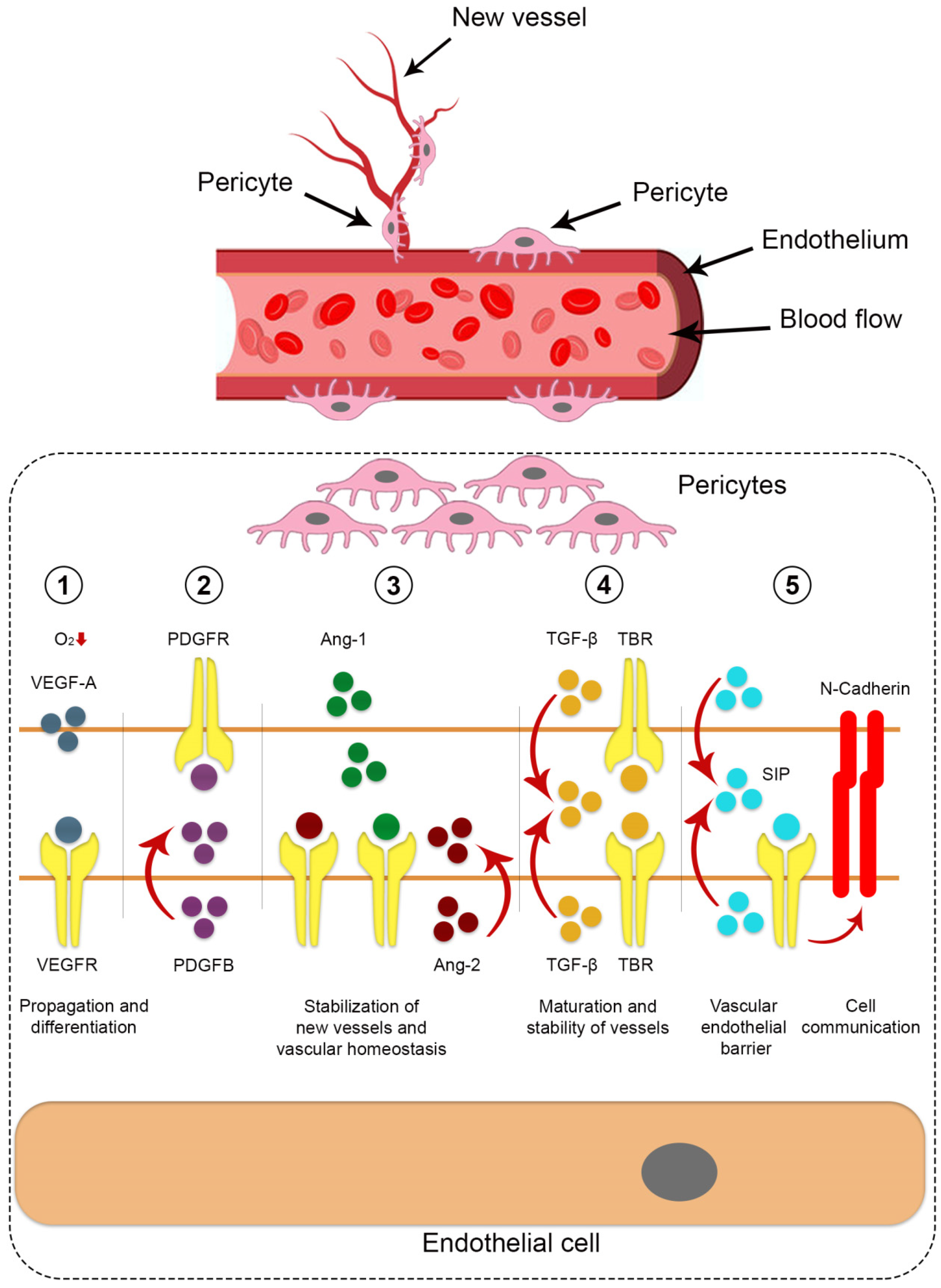
| Angiogenesis | Pericytes, found in microvessels like capillaries and venules, play a vital role in maintaining structural integrity, regulating blood flow, promoting angiogenesis, stabilizing vasculature, and controlling permeability. In the central nervous system and retina, they form barriers protecting cells from harmful blood factors [18]. Pericytes are crucial for angiogenesis, influencing vessel stability by interacting with sprouting endothelial cells. They can also adjust capillary diameters, similar to smooth muscle cells, regulating microvessel blood flow [19]. |
| Impact on immune function |
Pericytes facilitate immune cell migration by releasing molecules, recruiting various immune cells, and promoting M2-like macrophages. They regulate the immune system in the central nervous system. Reduced CD4+ T-cells lead to decreased pericyte coverage, and retinal pericytes inhibit CD4+ T-cell activation [19]. |
| Stem cell | Pericytes maintain blood vessel integrity, prevent issues like vessel dilation and hemorrhaging, and exhibit stem cell-like qualities [20]. In the brain, vascular pericytes are crucial for the blood-brain barrier and demonstrate stem cell capabilities, particularly after brain damage from conditions like ischemia and hypoxia [21,22]. After ischemia, they can transform into various cell types, including neurons, microglia, and vascular cells, and assist in clearing damaged areas by becoming glial cells [23]. |
| Pathological | |
| Fibrosis |
Pericytes contribute to lung diseases like pulmonary arterial hypertension (PAH) and allergic asthma by transforming into scar-forming myofibroblasts, which leads to tissue fibrosis through collagen deposition and matrix remodeling [24]. |
| Neuro- degeneration |
Pericytes in the blood-brain barrier degenerate in Alzheimer's disease (AD), associated with neurovascular dysfunction, Aβ elevation, tau pathology, and neuronal loss [25]. Pericyte loss in neurological disorders increases blood-brain barrier permeability and may lead to vascular dementia [26]. |
| Cancer | Pericytes (PCs) in the tumor microenvironment have diverse roles, including forming the pre-metastatic niche, promoting cancer cell growth and drug resistance, and influencing M2 macrophage polarization [27]. In carcinogenesis, disrupted interaction between PCs and endothelial cells leads to dysfunctional tumor vasculature [28]. Recent studies employing advanced technologies confirm pericytes' communication with cancer cells [29]. |
| Diabetic retinopathy |
Pericyte loss is an early hallmark of diabetes-related microvascular diseases, including retinopathy and nephropathy. Pericytes actively contribute to vascular dysfunction by secreting pro-angiogenic factors, initiating neovascularization, thickening the basement membrane, and causing vasoconstriction [30]. |
| Model | Type of cells and tissue origin | Animal type | Dosage | Method of investigation | Therapeutic outcome | Reference l |
|---|---|---|---|---|---|---|
| Intramuscular ectopic bone model | Adipose human perivascular stem cells (hPSCs) and adventitial cells derived from the same patient | severe combined immunodeficient (SCID) mice | 2.5 × 10⁵ cells, sponge size 2.0 × 1.0 × 0.5 cm | Micro-CT imaging (bone mineral density and bone volume) | Both perivascular populations had a similar baseline osteogenic potential | [45] |
| Intramuscular ectopic bone model | Adipose hPSCs, SVF from the same patient | SCID mice | 2.5 × 10⁵ cells, sponge size 2.0 × 1.0 × 0.5 cm | High resolution radiographic/3-Dimensional Micro-CT imaging | Larger particles of bone formation, increase in vascularity of the implant site, osteocalcin and bone sialoprotein expression | [51] |
| 3 mm non-healing calvarial defect centered in the parietal bone | Adipose hPSCs or SVF from the same patient | SCID mice | Radiographic imaging and histological examination | Significant bone defect healing overtime | [6] | |
| Intramuscular ectopic bone model | Adipose hPSCs | SCID mice | The demineralized bone matrix (DBX), with NELL-1 (3 μg/μL), hPSC (2.5×10⁵ cells), or hPSC+NELL-1 | Micro-CT imaging, histological examination, immunohistochemical staining over 4 weeks | The additive effect of hPSC+NELL-1 on bone formation and vasculogenesis | [52] |
| Intramuscular ectopic bone model in the hindlimb | Adipose hPSCs or SVF from the same patient | SCID mice | DBX with SVF or hPSC | Histological examination, immunohistochemical staining | Significantly greater neutrophilic and macrophage infiltrates within and around SVF in comparison to PSC-laden implants, robust immunomodulatory effect | [53] |
| Tibial model | Adipose hPSCs, bone marrow MSC | Wistar rats | 5 × 10⁶ cells were percutaneously injected into the fracture gap | Radiographic, micro-CT imaging and immunohistochemical staining | At eight weeks, 80% of animals in the cell treatment groups showed evidence of bone healing compared to only 14% of those in the control group | [54] |
| Posterolateral lumbar spinal fusion model | Adipose hPSCs | Athymic rats | DBX, 0,15Х106 hPSCs, 0,50x10⁶ hPSCs, 1.5x10⁶ hPSCs | Micro-CT imaging, immunohistochemical staining | Regulate bone formation via direct and paracrine mechanisms | [55] |
| Osteoporotic spinal fusion mode | Adipose hPSCs or NELL-1, BMP-2 | Athymic rats | 0.25 × 10⁶ cells per milliliter of hPSCs or 33.3 μg/ml of NELL-1, 0.75 × 10⁶ cells per milliliter of hPSCs or 66.6 μg/ml of NELL-1 | Micro-CT imaging, histological examination, immunohistochemical staining | The hPSC combined with NELL-1 synergistically enhances spinal fusion in osteoporotic rats | [56] |
| Systemic | Local |
|---|---|
| Bisphosphonates | INFUSE (rhBMP-2) |
| Recombinant parathyroid hormone | Regranex (rhPDGF-BB) |
| RANKL inhibitors | rhBMP-7* |
| SOST inhibitors (pending) | Healos (GDF-5) (pending) |
| Demineralized bone matrix | |
| Fibula allograft | |
| Iliac crest autograft |
| Dose (cell) (×106), donor, type of study | Age, Kellgren-Lawrence Grade, sample (M:F), follow up | Outcome, measures, effect | Study (year) |
|---|---|---|---|
| 1 × 10, 2 × 10, 5 × 10 x × 106, Autologous adipose tissue-MSC (Ad-MSC) In this single-site, randomized, double-blind, dose-ranging, phase I study |
|
WOMAC, VAS, WORMS, MRI, others MRI assessments showed slight improvements in the low-dose group |
Lu, L.; et al. (2020) [90] |
| 50 × 2 x × 106, Autologous Ad-MSC Randomized double-blind phase IIb clinical trial |
|
WOMAC, VAS, MRI, others 50% improvement of WOMAC 70% improvement rate in Re-Join® group after 12 months |
Lu, L.; et al. (2019) [91] |
| 100 × 106, autologous Ad-MSC Double-blinded, randomized controlled phase IIb clinical trial |
|
KOOS, WOMAC, VAS, MRI, others Improvement of WOMAC score at 6 months No significant change of cartilage defect in MSC group |
Lee, W.S.; et al. (2019) [92] |
| 100, 100 × 2 x × 106 at baseline and 6 months, autologous Ad-MSC Randomized controlled trial (RCT) |
|
KOOS, NPRS, WOMAC, others Significant pain and functional improvement in both treatment groups |
Freitag, J.; et al. (2019) [93] |
| 3.9 (Progenza 3.9M, n = 8) or placebo (n = 2) and 6.9 (Progenza 6.7M, n = 8) or placebo (n = 2), allogeneic Ad-MSC Double-blinded RCT |
|
|
Kuah, D.; et al. (2018) [94] |
| 6 ml, intra-articular injection, autologous adipose tissue (AT) or hyaluronic acid Prospective, single-center, parallel-group RCT |
|
WOMAC, WOMAC-A, PROMIS, force plate analysis, others |
Jones, I.; et al. (2018) [95] |
| 10 ml intra-articular injection, autologous, micro-fragmented AT or isotonic saline (placebo) Blinded RCT |
|
KOOS4, Tegner activity score, work status, others |
Mikkelsen, R.; et al. (2021) [96] |
| Intra-articular knee injection of autologous microfragmented lipoaspirate (MLA) |
|
Stromal vascular fraction isolation, flow cytometry | Polanec, D.; et al. (2019) [97] |
| 4-15 ml intra- articular injection, autologous microfragmented AT containing Ad-MSCs Prospective, non-randomized, interventional, single-center, open label clinical trial |
|
dGEMRIC, VAS A single intra-articular injection of autologous microfragmented AT improves glycosaminoglycans (GAG) content on a significant scale |
Borić, I.; et al. (2019) [98] |
| Intra-articular injection, autologous MLA Prospective, non-randomized study |
|
VAS, WOMAC, KOOS KOOS score improved from 46 to 176% when compared with baseline WOMAC decreased from 40 to 45% VAS rating decreased from 54% to 82% |
Hudetz, D.; et al. (2019) [99] |
| Intra-articular injection, autologous microfragmented AT |
|
NPRS, 100-point KSS with FXN and LEAS KSS score improved from 74 to 82 FXN score improved from 65 to 76 LEAS score improved from 36 to 47 |
Panchal, J.; et al. 2018 [100] |
| Intra-articular injection, autologous and microfragmented AT Retrospective study |
|
KOOS, IKDC, Tegner Lysholm knee, VAS IKDC-subjective and total KOOS improved by 20 points VAS-point and Tegner Lysholm knee improved by 24 and 31 points, respectively |
Russo, A.; et al. (2017) [101] |
Disclaimer/Publisher’s Note: The statements, opinions and data contained in all publications are solely those of the individual author(s) and contributor(s) and not of MDPI and/or the editor(s). MDPI and/or the editor(s) disclaim responsibility for any injury to people or property resulting from any ideas, methods, instructions or products referred to in the content. |
© 2023 by the authors. Licensee MDPI, Basel, Switzerland. This article is an open access article distributed under the terms and conditions of the Creative Commons Attribution (CC BY) license (http://creativecommons.org/licenses/by/4.0/).


