Submitted:
02 November 2023
Posted:
02 November 2023
You are already at the latest version
Abstract
Keywords:
1. Background
2. Material Method
2.1. Experimental Reagent and Dose Selection
2.2. Animals
2.3. Tissue Sampling
2.4. Neurobehavioural Tests
2.4.1. Open Field Experiment
2.4.2. New Object Recognition Experiment
2.4.3. High Plus Maze Experiment
2.4.4. Morris Water Maze Test
2.5. HE/Nissl Staining
2.6. Immunohistochemistry
2.7. Transmission Electron Microscopy
2.8. WB
2.9. Q-PCR
2.10. ATP/Glu Test
2.11. Statistical Analysis
3. Results
3.1. Learning and Memory Ability and Hippocampal Neuron Damage Induced by Cy Exposure in Rats
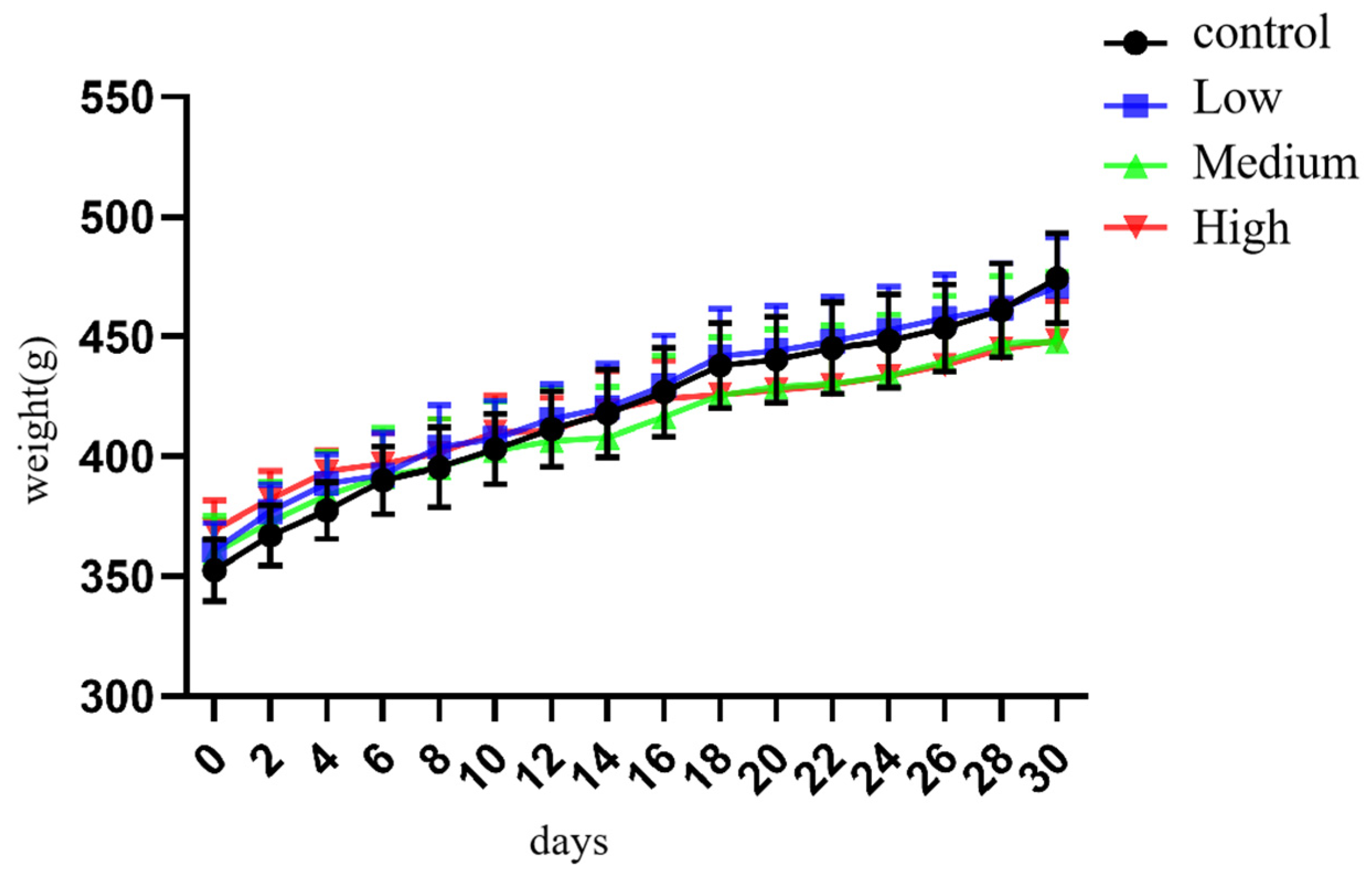
3.1.1. General Changes in Rats Exposed to Cy
3.2. Changes in Learning and Memory Ability in Cy-Exposed Rats
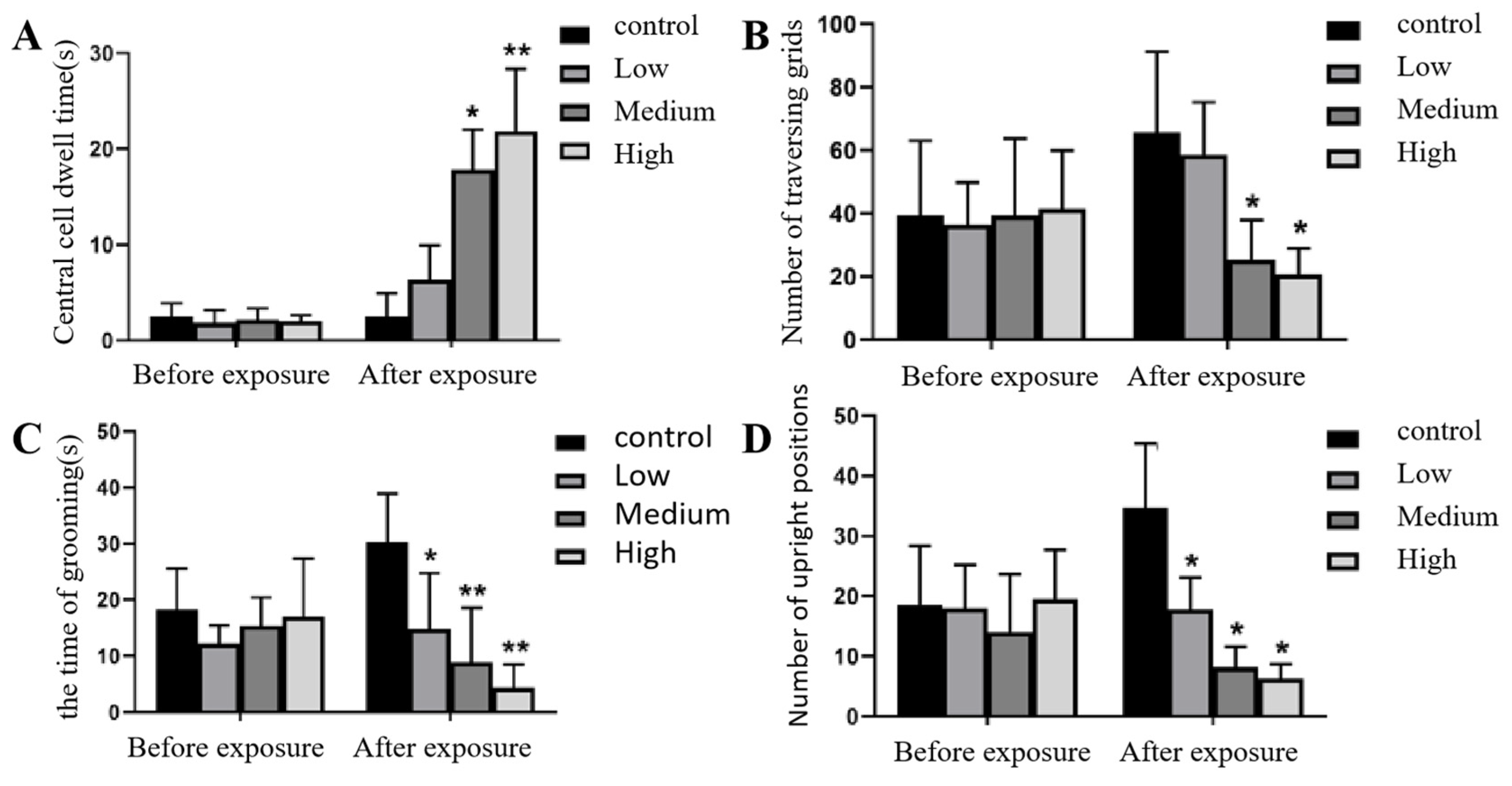
3.2.1. Open Field Experiment
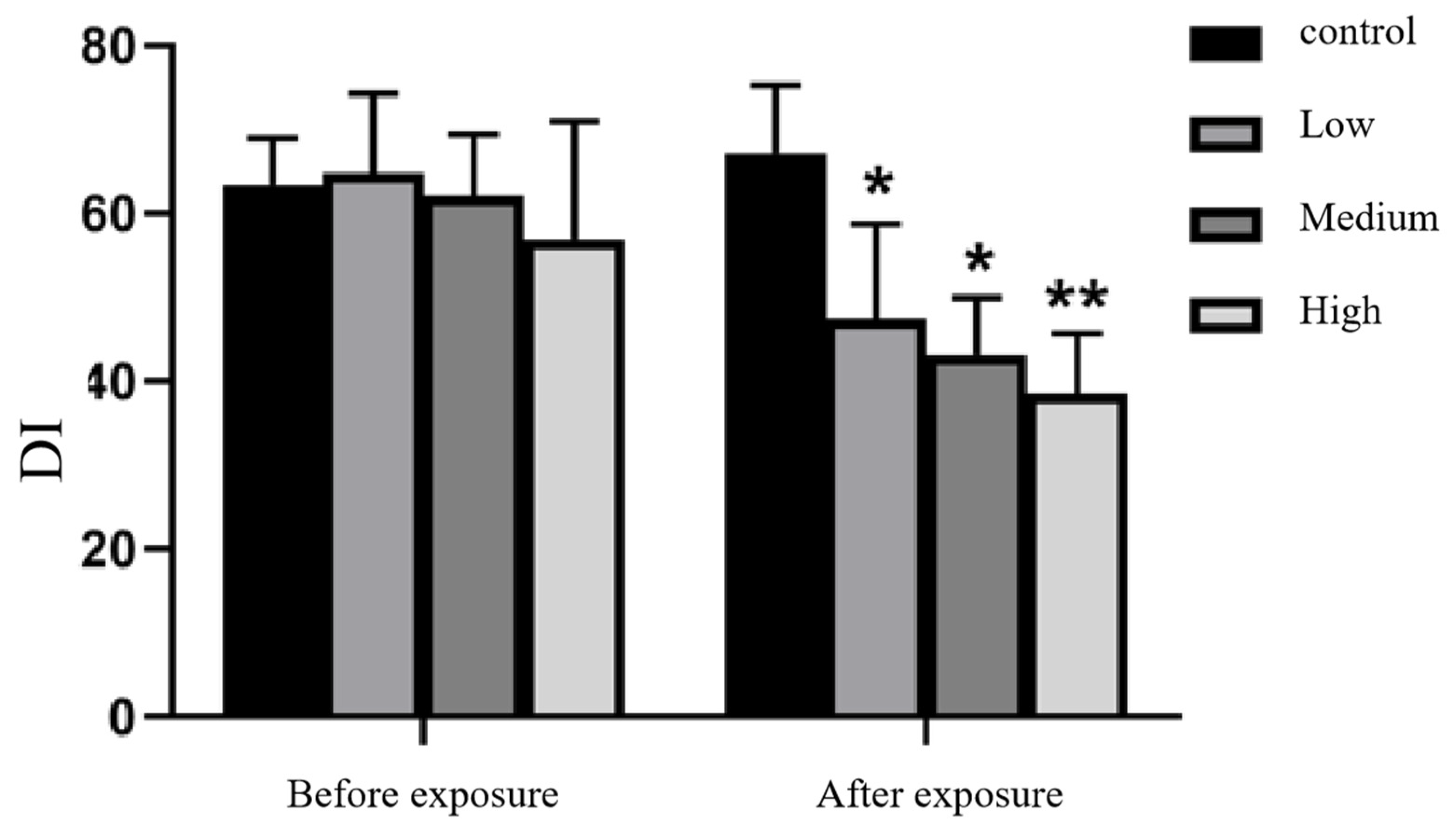
3.2.2. New Object Recognition Experiment
3.2.3. Elevated Plus Maze Test
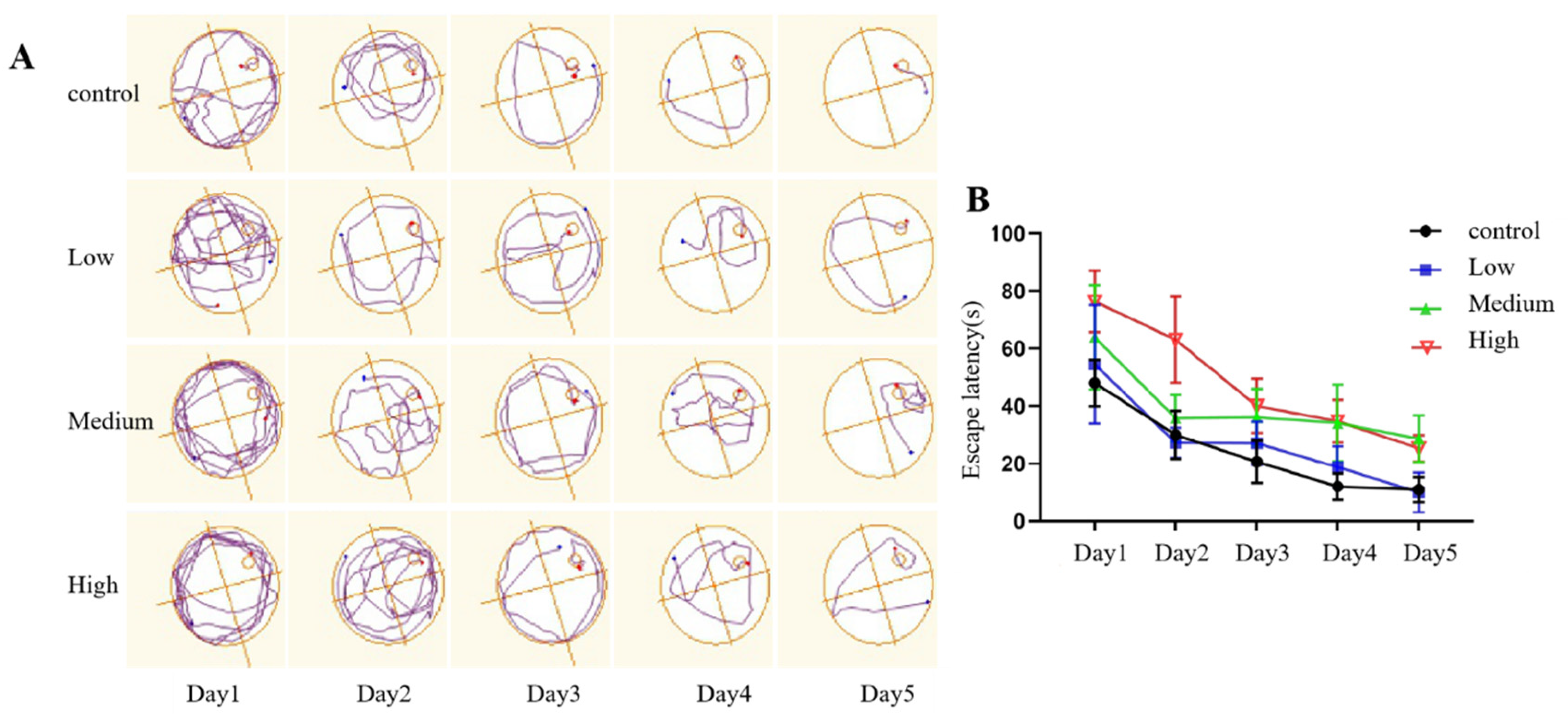
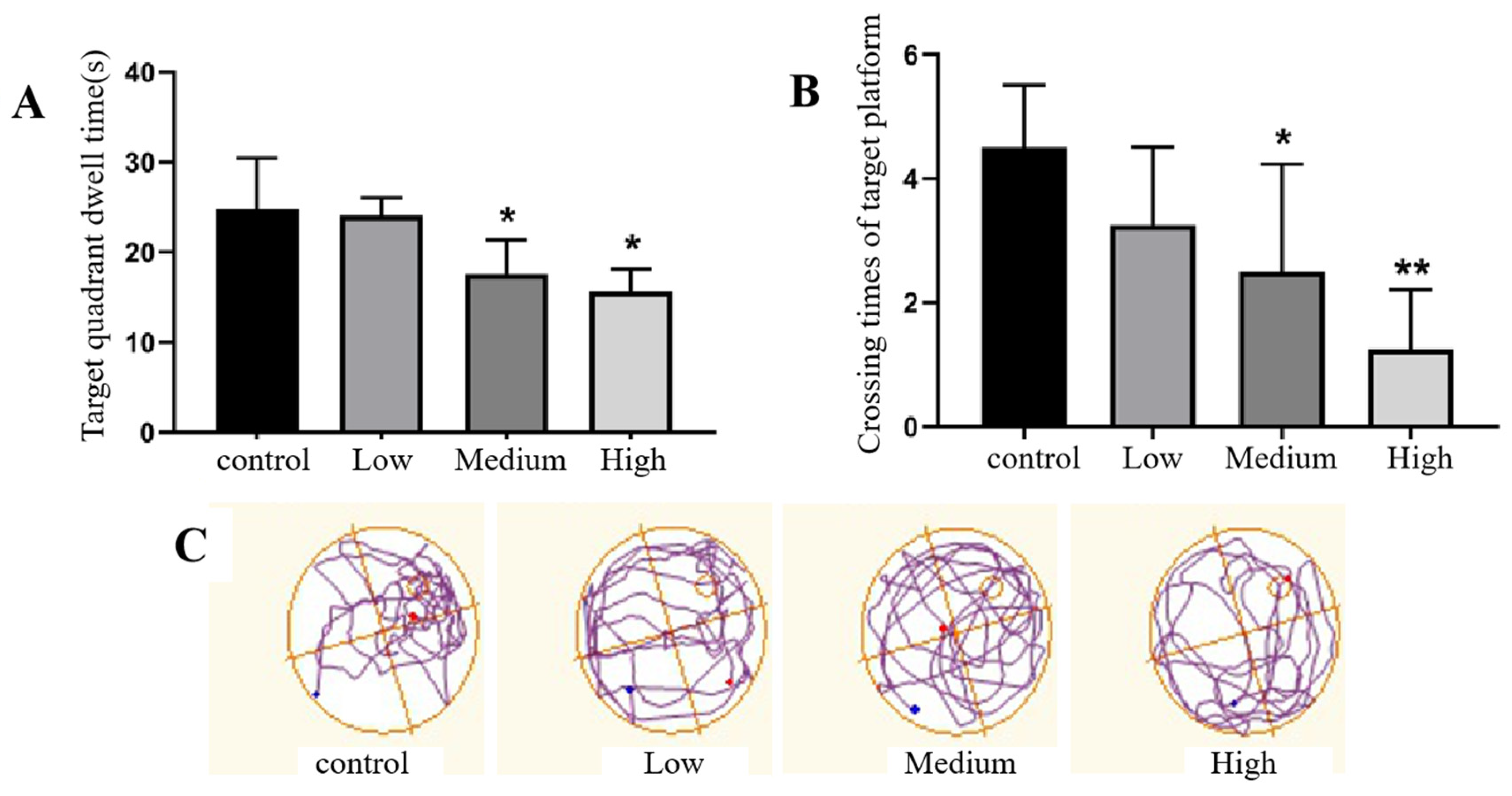
3.2.4. Morris Water Maze Test
- (1)
- Directional navigation experiment
- (2)
- Space exploration experiment.
3.3. Injury of Hippocampal Neurons in Rats Exposed to Cy
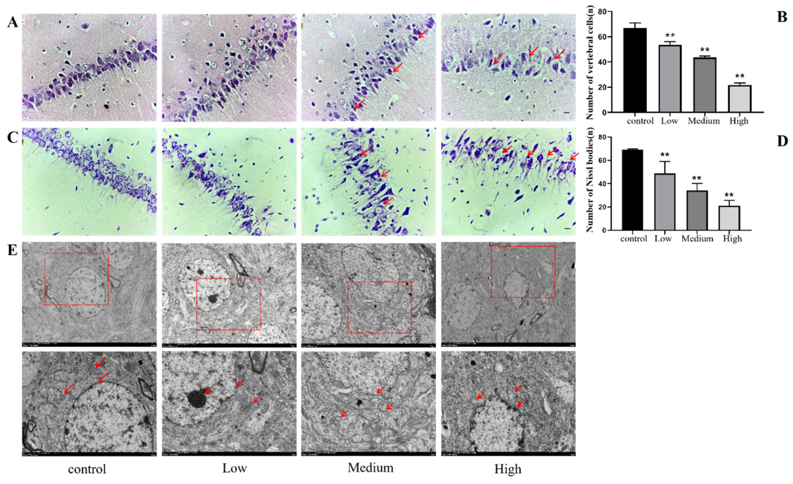
3.3.1. HE Staining
3.3.2. Nissl Staining
3.3.3. Electron Microscopy
3.4. Damage to the Synaptic Plasticity of Hippocampal Neurons Induced by Cy Exposure in Rats
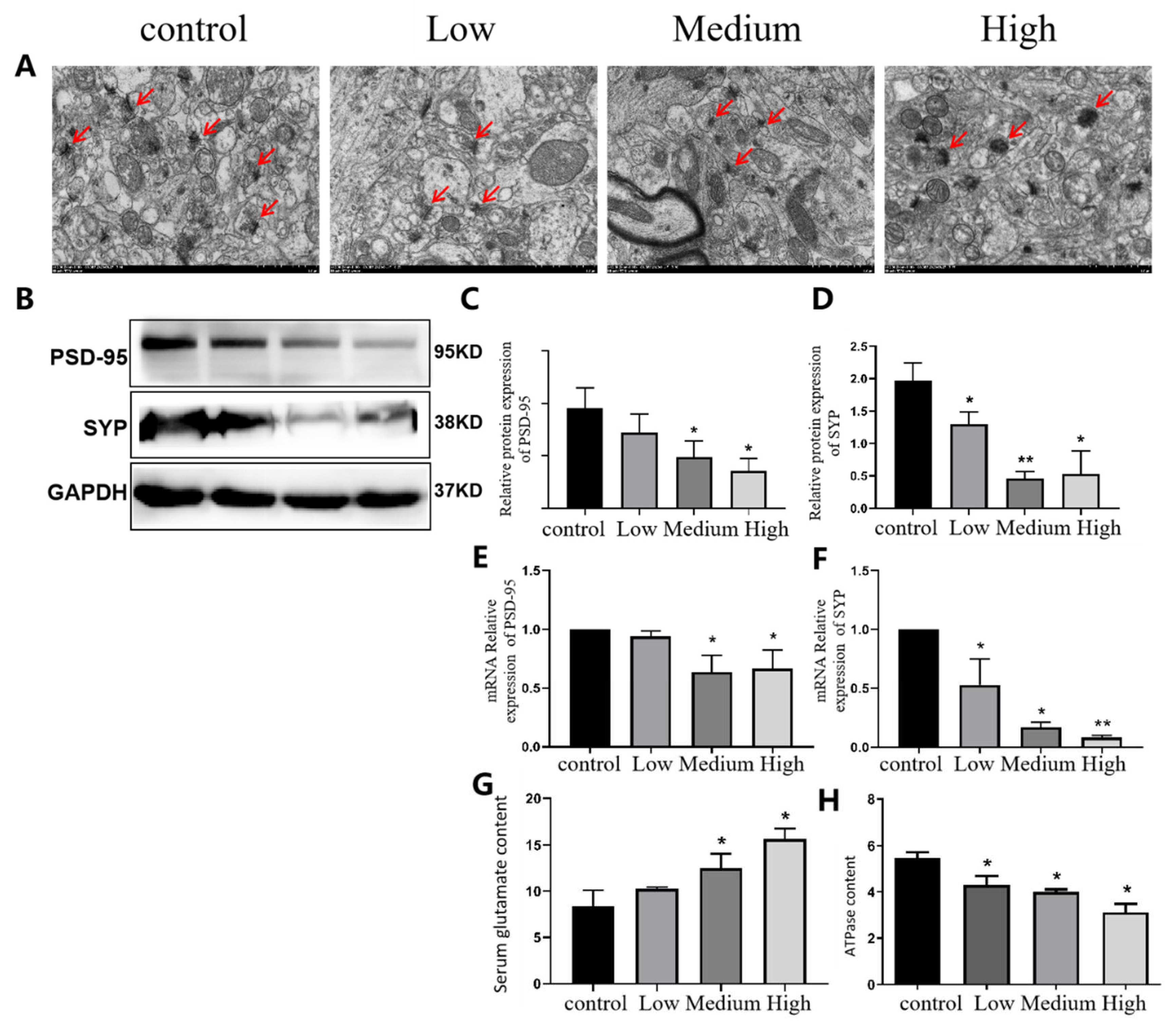
3.4.1. Ultrastructural Changes in the Synapses of Hippocampal Cells in Cy-Exposed Rats
3.5. Changes in Inflammatory Factors and A2AR Factors in the Neurons of Rats Exposed to Cy
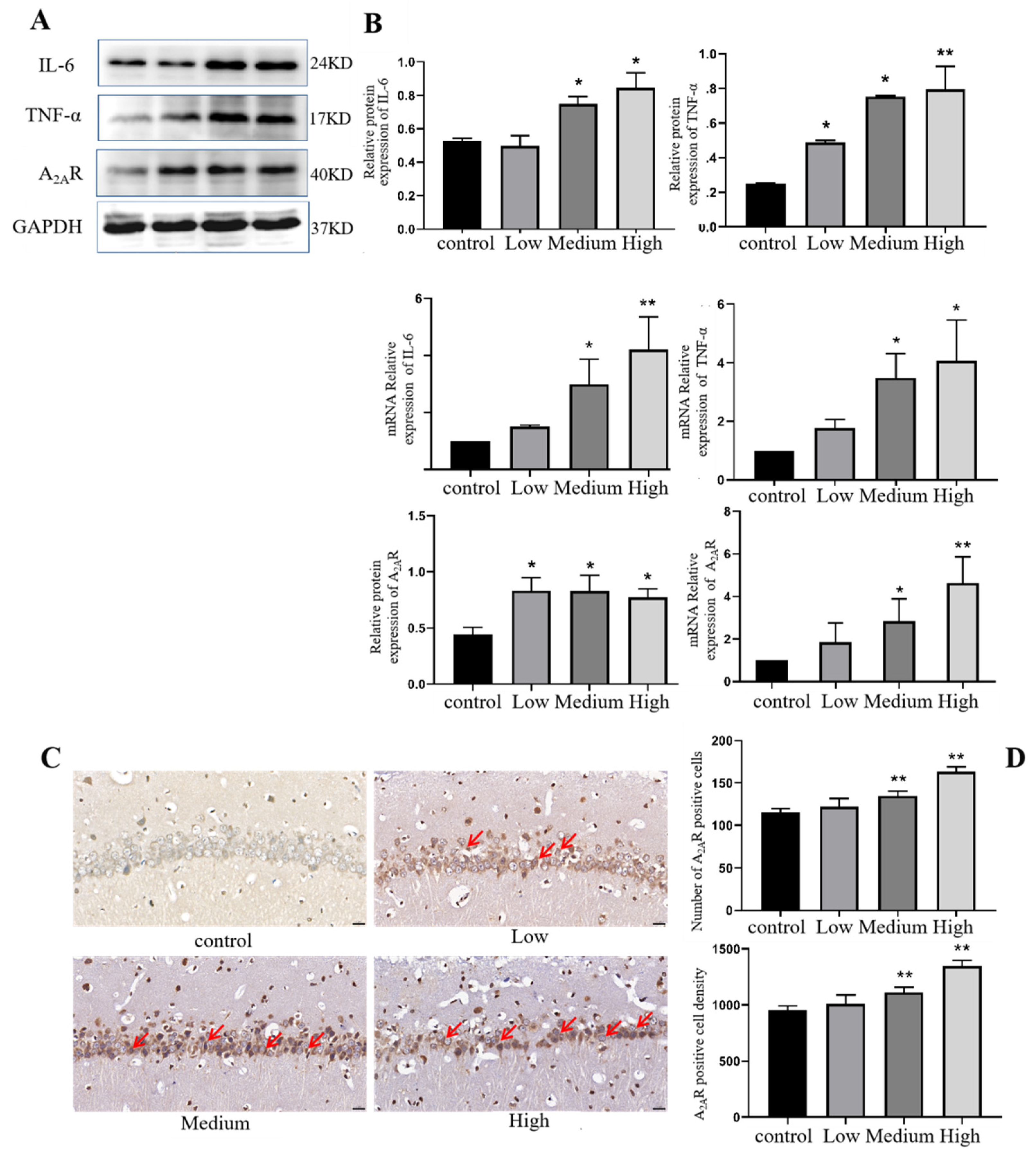
3.5.1. Expression of Inflammatory Factors in the Hippocampus of Cy-Exposed Rats
3.5.2. Expression of the A2AR Factor in the Hippocampus of Cy-Exposed Rats
3.6. Correlation Analysis of A2AR with Neurobehavioural Indices, Inflammatory Factors and Synaptic Plasticity Factors
| Experimental category | Indicators | Pearson correlation coefficient r | P |
|---|---|---|---|
| Open field test | |||
| Central grid dwell time | 0.7154 | <0.001 | |
| The time of grooming | -0.6354 | 0.001 | |
| Number of standing | -0.4381 | 0.032 | |
| Number of grid crossings | -0.5682 | 0.004 | |
| New object recognition experiment | |||
| Discrimination index | -0.6896 | <0.001 | |
| Plus maze experiment | |||
| Open arm entry times | -0.375 | 0.071 | |
| Open arm entry time | -0.4633 | 0.023 | |
| Water maze test | |||
| Average latency | 0.7000 | <0.001 | |
| Target Quadrant Dwell Time | -0.6044 | 0.002 | |
| Target platform traversal times | -0.6364 | 0.001 | |
| Inflammation related factors | |||
| IL-6 | 0.8612 | <0.001 | |
| TNF | 0.8433 | 0.001 | |
| Synaptic plasticity related factors | |||
| PSD-95 | -0.6377 | 0.026 | |
| SYP | -0.7935 | 0.002 | |
4. Discussion
4.1. Cy Exposure Can Cause Neurobehavioural Changes in Rats
4.2. Cy Exposure Can Cause Morphological Changes in Rat Hippocampus
4.3. Cy Exposure Can Cause Abnormal Changes in the Inflammatory Response and Adenosine A2AR Expression in the Rat Hippocampus
5. Conclusions
Author Contributions
Funding
Institutional Review Board Statement
Informed Consent Statement
Data Availability Statement
Conflicts of Interest
References
- YC, L.; WZ, L.; CC, K.; LJ, H.; CT, C.; CR, J. Action of the insecticide cyfluthrin on Ca signal transduction and cytotoxicity in human osteosarcoma cells. Hum Exp Toxicol 2020, 39, 1268-1276. [CrossRef]
- FAO; Specifications, R.I.P.O.; Standards, R.A.; AGP. Manual on the Development and Use of FAO Specifications for Plant Protection Products. 1999.
- F, H.; T, K. Pesticide residues and health risk appraisal of tomato cultivated in greenhouse from the Mediterranean region of Turkey. Environmental science and pollution research international 2021, 28, 22551-22562. [CrossRef]
- MK, M. Dietary Pyrethroid Exposures and Intake Doses for 188 Duplicate-Single Solid Food Items Consumed by North Carolina Adults. Toxics 2020, 8. [CrossRef]
- W, N.; H, G.; B, W.; J, Z.; J, S.; Y, S.; Y, S.; H, Y. Gestational Exposure to Cyfluthrin through Endoplasmic Reticulum (ER) Stress-Mediated PERK Signaling Pathway Impairs Placental Development. Toxics 2022, 10, 733. [CrossRef]
- J, Z.; LY, M.; YX, X.; LQ, Z.; WS, N.; R, W.; YN, S.; XY, L.; HF, Y. The role of stimulator of interferon genes-mediated AMPK/mTOR/P70S6K autophagy pathway in cyfluthrin-induced testicular injury. Environ Toxicol 2022, 38, 727-742. [CrossRef]
- L, M.; E, Z.; M, I. β-Cyfluthrin-Mediated Cytotoxicity of Cultured Rat Primary Hepatocytes Ameliorated by Cotreatment with Luteolin. Evidence-based complementary and alternative medicine: eCAM 2022, 2022, 3647988. [CrossRef]
- Xiaoyu, Z.; Bing, W.; Lijun, D.; Ji, Z.; Huifang, Y. Analysis on the current status and influencing factors of neurological subhealth among vegetable greenhouse growers in the suburbs of Yinchuan City. Journal of Ningxia Medical University 2017, 039, 293-298. [CrossRef]
- D, D.; S, K.; N, B.; R, E.; M, C.; RP, S.; M, S.; JO, G.; J, B.; V, K. Chlorpyrifos, permethrin and cyfluthrin effect on cell survival, permeability, and tight junction in an in-vitro model of the human blood-brain barrier (BBB). Neurotoxicology 2022, 93, 152-162. [CrossRef]
- M, V.; P, Q.; K, A.; C, M.; P, G.; C, B.; A, C. Aggregate and cumulative chronic risk assessment for pyrethroids in the French adult population. Food and chemical toxicology: an international journal published for the British Industrial Biological Research Association 2020, 143, 111519. [CrossRef]
- W, Z.; R, F.; S, L.; Y, L.; Y, J.; Y, L.; B, L.; Y, C.; L, J.; X, Y. Combined effects of chlorpyrifos and cyfluthrin on neurobehavior and neurotransmitter levels in larval zebrafish. Journal of applied toxicology: JAT 2022, 42, 1662-1670. [CrossRef]
- X, Q.; L, L.; X, N.; Q, N. Effects of Chronic Aluminum Lactate Exposure on Neuronal Apoptosis and Hippocampal Synaptic Plasticity in Rats. Biol Trace Elem Res 2020, 197, 571-579. [CrossRef]
- Ennaceur, A.; Delacour, J.A. A new one-trial test for neurobiological studies of memory in rats. I. Behavioral data. Behav Brain Res 1988, 31, 47-59. [CrossRef]
- P, K.; R, C.; R, W.; A, B.; S, P.; AW, B. Sex differences in the elevated plus-maze test and large open field test in adult Wistar rats. Pharmacology, biochemistry, and behavior 2021, 204, 173168. [CrossRef]
- N, G.; S, M.V.; A, N.; M, M.; M, M.; S, S.; M, G.; S, V.; M, H. Pharmacological Evidences for Curcumin Neuroprotective Effects against Lead-Induced Neurodegeneration: Possible Role of Akt/GSK3 Signaling Pathway. Iranian journal of pharmaceutical research: IJPR 2020, 19, 494-508. [CrossRef]
- M, J.; K, A.; S, A.; J, M.; W, W.; M, K.; U, I.; T, M.; I, I. Changes in cholinergic function in the frontal cortex and hippocampus of rat exposed to ethanol and acetaldehyde. Neuroscience 2007, 144, 232-238. [CrossRef]
- A, H.Q.C.; B, J.Z.A.; C, Y.L.A.; D, L.X.H.; D, Y.J.H.; D, W.Q.S.; A, J.C.; A, W.B.L.; A, J.Y.L. Gene expression network regulated by DNA methylation and microRNA during microcystin-leucine arginine induced malignant transformation in human hepatocyte L02 cells. Toxicol Lett 2018, 289, 42-53. [CrossRef]
- IA, A.; SE, O.; AK, O.; EO, F. Neuroprotective role of gallic acid in aflatoxin B -induced behavioral abnormalities in rats. J Biochem Mol Toxic 2021, 35, e22684. [CrossRef]
- ED, L.; E, U.; RE, B. Neurobehavioral toxicology of halothane in rats. Neurotoxicol Teratol 1991, 13, 461-470. [CrossRef]
- Z, L.; Z, H.; J, L.; H, L.; L, M.; Y, L.; J, Z.; Y, X.; Y, L. Regulation of CRMP2 by Cdk5 and GSK-3β participates in sevoflurane-induced dendritic development abnormalities and cognitive dysfunction in developing rats. Toxicol Lett 2021, 341, 68-79. [CrossRef]
- FDS, C.; de Souza Oliveira Tavares C; BHS, A.; F, M.; RÁB, L.; S, G.D.S. Improved Spatial Memory And Neuroinflammatory Profile Changes in Aged Rats Submitted to Photobiomodulation Therapy. Cell Mol Neurobiol 2022, 42, 1875-1886. [CrossRef]
- Hughes, M.F.; Ross, D.G.; Starr, J.M.; Scollon, E.J.; Wolansky, M.J.; Crofton, K.M.; Devito, M.J. Environmentally relevant pyrethroid mixtures: A study on the correlation of blood and brain concentrations of a mixture of pyrethroid insecticides to motor activity in the rat. Toxicology 2016, 359, 19-28. [CrossRef]
- ES, C. Dopamine Release in the Midbrain Promotes Anxiety. Biol Psychiat 2020, 88, 815-817. [CrossRef]
- S, C.; J, D.; K, C.; D, L.; K, C.; M, C.; L, K.; D, F.; C, T. NavWell: A simplified virtual-reality platform for spatial navigation and memory experiments. Behav Res Methods 2020, 52, 1189-1207. [CrossRef]
- K, B.; Y, D.; W, S. Morris water maze test for learning and memory deficits in Alzheimer’s disease model mice. Journal of visualized experiments: JoVE 2011, 20, 2920. [CrossRef]
- F, S.; LP, C.; VK, K.; I, S. Beta-cyfluthrin induced neurobehavioral impairments in adult rats. Chem-Biol Interact 2016, 243, 19-28. [CrossRef]
- D, L.; L, Y.; M, L.; Q, Z.; F, T.; W, C. Behavioral disorders caused by nonylphenol and strategies for protection. Chemosphere 2021, 275, 129973. [CrossRef]
- J, W.; B, L.; Y, X.; M, Y.; C, W.; M, S.; J, L.; W, W.; J, Y.; F, S., et al. Activation of CREB-mediated autophagy by thioperamide ameliorates β-amyloid pathology and cognition in Alzheimer’s disease. Aging Cell 2021, 20, e13333. [CrossRef]
- M, G.; DM, H. Targeting pre-synaptic tau accumulation: a new strategy to counteract tau-mediated synaptic loss and memory deficits. Neuron 2021, 109, 741-743. [CrossRef]
- G, P.; V, S.; A, V.; N, K.; N, N.; M, G.; T, B.; A, Y.; I, M.; S, K. A combined use of silver pretreatment and impregnation with consequent Nissl staining for cortex and striatum architectonics study. Front Neuroanat 2022, 16, 940993. [CrossRef]
- Y, Y.; H, G.; W, L.; X, L.; X, J.; X, L.; Q, W.; Z, X.; Q, Z. Arctium lappa L. roots ameliorates cerebral ischemia through inhibiting neuronal apoptosis and suppressing AMPK/mTOR-mediated autophagy. Phytomedicine: international journal of phytotherapy and phytopharmacology 2021, 85, 153526. [CrossRef]
- Y, Y.; SW, Z.; SQ, W.; QP, Z.; L, W.; H, Z.; JH, G.; XZ, L.; JC, B.; ZP, L. Alpha-Lipoic Acid Attenuates Cadmium- and Lead-Induced Neurotoxicity by Inhibiting Both Endoplasmic-Reticulum Stress and Activation of Fas/FasL and Mitochondrial Apoptotic Pathways in Rat Cerebral Cortex. Neurotox Res 2021, 39, 1103-1115. [CrossRef]
- Z, C.; Z, Y.; S, Y.; Y, Z.; M, X.; J, Z.; L, L. Brain Energy Metabolism: Astrocytes in Neurodegenerative Diseases. Cns Neurosci Ther 2023, 29, 24-36. [CrossRef]
- I, G.; T, T.; AL, S.; F, Y.; J, L.; D, V.; MS, H.; J, N.; AU, B. Systematic expression analysis of plasticity-related genes in mouse brain development brings PRG4 into play. Developmental dynamics: an official publication of the American Association of Anatomists 2022, 251, 714-728. [CrossRef]
- De la Torre-Iturbe S; RA, V.; De la Cruz-López F; G, F.; L, G. Dendritic and behavioral changes in rats neonatally treated with homocysteine; A proposal as an animal model to study the attention deficit hyperactivity disorder. J Chem Neuroanat 2022, 119, 102057. [CrossRef]
- QQ, L.; J, C.; P, H.; M, J.; JH, S.; HY, F.; FC, Q.; YY, Z.; YY, S.; G, C., et al. Enhancing GluN2A-type NMDA receptors impairs long-term synaptic plasticity and learning and memory. Mol Psychiatr 2022, 27, 3468-3478. [CrossRef]
- HL, T.; SL, C.; Q, Z.; RL, H. GRIP1 regulates synaptic plasticity and learning and memory. P Natl Acad Sci Usa 2020, 117, 25085-25091. [CrossRef]
- SD, S.; L, F.; M, Z.; P, G. Synaptic Plasticity, Dementia and Alzheimer Disease. CNS & neurological disorders drug targets 2017, 16, 220-233. [CrossRef]
- A, T.; MI, Á.; A, T.; JJ, M.; JJ, R. Brain local and regional neuroglial alterations in Alzheimer’s Disease: cell types, responses and implications. Curr Alzheimer Res 2016, 13, 321-342. [CrossRef]
- T, X.; J, L.; XR, L.; Y, Y.; X, L.; X, Z.; Y, C.; PQ, Y.; Y, L. The mTOR/NF-κB Pathway Mediates Neuroinflammation and Synaptic Plasticity in Diabetic Encephalopathy. Mol Neurobiol 2021, 58, 3848-3862. [CrossRef]
- D, D.; Y, C.; S, G.; Z, X.; S, C.; K, C.; S, W.; G, S.; L, Y.; S, B., et al. Sinisan alleviates depression-like behaviors by regulating mitochondrial function and synaptic plasticity in maternal separation rats. Phytomedicine: international journal of phytotherapy and phytopharmacology 2022, 106, 154395. [CrossRef]
- MA, C. Synaptophysin-dependent synaptobrevin-2 trafficking at the presynapse-Mechanism and function. J Neurochem 2021, 159, 78-89. [CrossRef]
- J, Y.; J, G.; KQ, S.; XD, F. Electroacupuncture improves learning and memory impairment and enhances hippocampal synaptic plasticity through BDNF/TRKB/CREB signaling pathway in cerebral ischemia-reperfusion injury rats. Zhen ci yan jiu = Acupuncture research 2023, 48, 843-851. [CrossRef]
- JL, R.; I, A.; V, C.; M, M.; MR, M.; A, A.; MA, M. Effects of exposure to pyrethroid cyfluthrin on serotonin and dopamine levels in brain regions of male rats. Environ Res 2016, 146, 388-394. [CrossRef]
- Y, G.; N, L.; J, C.; P, Z.; J, N.; S, T.; X, P.; J, W.; J, Y.; L, M. Neuropharmacological insight into preventive intervention in posttraumatic epilepsy based on regulating glutamate homeostasis. Cns Neurosci Ther 2023, 29, 2430-2444. [CrossRef]
- M, W.; H, Z.; J, L.; J, H.; N, C. Exercise suppresses neuroinflammation for alleviating Alzheimer’s disease. J Neuroinflamm 2023, 20, 76. [CrossRef]
- KM, L.; Y, H.; PP, W.; YH, L.; N, Z.; JJ, L.; CY, Y.; JJ, C.; L, Z.; C, Z., et al. Ursolic Acid Protects Neurons in Temporal Lobe Epilepsy and Cognitive Impairment by Repressing Inflammation and Oxidation. Front Pharmacol 2022, 13, 877898. [CrossRef]
- S, S.; A, Z.; P, B.; F, Y.; N, A.; E, N. The methanolic extract of Cinnamomum zeylanicum bark improves formaldehyde-induced neurotoxicity through reduction of phospho-tau (Thr231), inflammation, and apoptosis. Excli J 2020, 19, 671-686.
- Y, Z.; Q, F.; J, C.; Y, W.; X, L.; X, Z.; Y, S.; H, Z.; X, Z.; M, Y., et al. Melatonin Improves Mitochondrial Dysfunction and Attenuates Neuropathic Pain by Regulating SIRT1 in Dorsal Root Ganglions. Neuroscience 2023, S0306-4522, 454-455. [CrossRef]
- CA, M.; CN, F.; CMG, L.; VG, F.; AC, C.; ACR, O.; de Souza LC; EPF, R.; KC, C.; HC, G., et al. Leptin, hsCRP, TNF-α and IL-6 levels from normal aging to dementia: Relationship with cognitive and functional status. Journal of clinical neuroscience: official journal of the Neurosurgical Society of Australasia 2018, 56, 150-155. [CrossRef]
- Jia-Qi, S.; Qi, W.; Chao, T.; Ru-Nan, Z.; Shuai, Z.; Xiao-Rong, Z. Study on effect of beta-cypermethrin on expression of TH and TNF-α in rat brain tissue. Occupation and Health 2019, 35, 2187-2190.
- M, T.; DG, F.; VL, B.; I, M.; JE, C.; P, P.; R, G.; A, P.; S, C.; PM, C., et al. Age-related shift in LTD is dependent on neuronal adenosine A receptors interplay with mGluR5 and NMDA receptors. Mol Psychiatr 2020, 25, 1876-1900. [CrossRef]
- Silvia, V.D.S.; Haberl, M.G.; Zhang, P.; Bethge, P.; Lemos, C.; Gon Alves, N.; Gorlewicz, A.; Malezieux, M.; Gon Alves, F.Q.; Grosjean, N.L. Early synaptic deficits in the APP/PS1 mouse model of Alzheimer’s disease involve neuronal adenosine A2A receptors. Nat Commun 2016, 7, 11915. [CrossRef]
- Chen, J.F.; Pedata, F. Modulation of ischemic brain injury and neuroinflammation by adenosine A2A receptors. Curr Pharm Design 2008, 14, -. [CrossRef]
- S, H.; H, D.; H, Z.; S, W.; L, H.; S, C.; J, Z.; L, X. Noninvasive limb remote ischemic preconditioning contributes neuroprotective effects via activation of adenosine A1 receptor and redox status after transient focal cerebral ischemia in rats. Brain Res 2012, 1459, 81-90. [CrossRef]


| Antibody name | Company | Country |
|---|---|---|
| Rabbit Anti-A2AR (ab3461) | Abcam | U.S.A |
| Rabbit Anti-PSD-95 (ab76115) | Abcam | U.S.A |
| Rabbit Anti-SYP (ab32127) | Abcam | U.S.A |
| Rabbit Anti-IL-6 | Affinity | China |
| Rabbit Anti-TNF-α | Affinity | China |
| Rabbit Anti-GAPDH | Bioss | China |
| Forward primer | Reverse primer | Species | |
|---|---|---|---|
| A2AR | GAAAGACGGGAACTCCACGAAGAC | GGCAGTAACACGAACGCAAAGAAG | rat |
| PSD-95 | TCCAGTCTGTGCGAGAGGTAGC | GGACGGATGAAGATGGCGATGG | rat |
| SYP | GCTGTGTTTGCCTTCCTCTACTC | TGATAATGTTCTCTGGGTCCGTG | rat |
| IL-6 | ACTTCCAGCCAGTTGCCTTCTTG | TGGTCTGTTGTGGGTGGTATCCTC | rat |
| TNF-α | AAAGGACACCATGAGCACGGAAAG | CGCCACGAGCAGGAATGAGAAG | rat |
Disclaimer/Publisher’s Note: The statements, opinions and data contained in all publications are solely those of the individual author(s) and contributor(s) and not of MDPI and/or the editor(s). MDPI and/or the editor(s) disclaim responsibility for any injury to people or property resulting from any ideas, methods, instructions or products referred to in the content. |
© 2023 by the authors. Licensee MDPI, Basel, Switzerland. This article is an open access article distributed under the terms and conditions of the Creative Commons Attribution (CC BY) license (http://creativecommons.org/licenses/by/4.0/).


