Preprint
Article

Antioxidant and Antifungal Activities and Characterization of Phenolic Compounds Using HPLC- MS in Empetrum rubrum Vahl ex Willd.

Altmetrics

Downloads

98

Views

57

Comments

0

A peer-reviewed article of this preprint also exists.

Submitted:

20 November 2023

Posted:

22 November 2023

You are already at the latest version

Alerts
Abstract
In searching for compounds with antioxidant and antifungal activity, our study focused on the subshrub species Empetrum rubrum Vahl ex Willd. (Ericaceae), commonly known as Brecillo or Murtilla de Magallanes. We measured the antioxidant activity of its methanolic extract (MEE) obtained from the aerial parts (leaves and stems). The antioxidant activity of MEE was evaluated in vitro by 2,2-Diphenyl-1-picrylhydrazyl (DPPH) free radical and 2,2′-Azino-bis(3-ethylbenzothiazoline-6-sulfonic acid) diammonium salt (ABTS) cationic radical. Results were expressed in gallic acid and Trolox equivalents for DPPH and ABTS assays, respectively. The antioxidant activities, for the DPPH and ABTS assays, were also calculated as an inhibitory concentration of extract needed to inhibit 50% of the absorbance (IC50 value). The results of IC50 values measured by the DPPH and ABTS methods were 0.4145 mg mL-1 and 0.1081 mg mL-1, respectively. In relation to the antioxidant activity, the total phenolic content (TPC) in MEE was determined by the Folin-Ciocalteu method, and the presence of secondary metabolites groups, including phenolic compounds, was determined by using different standardized test procedures. Phenolic compounds contained in leaves, stems, and fruits of E. rubrum were determined qualitatively by means of high-performance liquid chromatography coupled with mass spectrometry (HPLC-MS) analysis. The antifungal activity of MEE obtained from aerial parts of E. rubrum was tested against Rhizoctonia solani. In vitro, mycelial growth of this fungus was reduced from 90% to 100% in the presence of MEE.
Keywords: 
Subject: Biology and Life Sciences  -   Plant Sciences

1. Introduction

Plants, due to their biodiversity and the broad presence of secondary metabolites [1] in plant tissues, provide a source of antioxidants [2]. A nonenzymatic antioxidant [3] is defined as a molecule capable of stabilizing another that has a missing electron, by giving one [4]. This way prevents reactions with other molecules [5] and prevents oxidative stress [6]. Phenols and polyphenols are intrinsically antioxidants [7] and this group of secondary plant metabolites is present in all parts of plants [8] and possesses nonenzymatic antioxidant properties [9]. Furthermore, plant extracts with phenolic compounds exert different biological functions [10], such as antifungal [11,12], antibacterial [13,14], antiviral [15], and anti-inflammatory activities [16]. Another aspect to be considered is the benefit for human health, because phenolic compounds prevent coronary heart diseases [17], have cytotoxic effects [18], have been used in traditional medicine for the treatment of diabetes [19], possess inhibitory activities of enzymes related to Alzheimer`s disease [20] and anti-obesity activities [21]. Exposure of plants to abiotic stress affects plant secondary metabolism and influences the polyphenolic composition and therefore antioxidant properties [22]. Abiotic stresses are mainly extreme temperature, drought, salinity, and flooding [23]. Species of the genus Empetrum are keystone species, for maintaining mammals and birds, and dominate many tundra and heathland ecosystems excluding other plants through allelopathic toxins [24]. A wide range of biologically active compounds with pharmacological effects are present in the genus Empetrum, and Empetrum species from the northern hemisphere are used in traditional medicine [25]. Following a common practice and in order to avoid confusion, all Southern Hemisphere natural populations are assignable as E. rubrum Vahl ex Willd and all northern hemisphere natural populations as Empetrum nigrum L. [24]. Other species of the genus Empetrum present in the Northern Hemisphere are Empetrum atropurpureum and Empetrum eamesii [26]. There are no scientific reports about biological activities and chemical compounds such as polyphenols in aerial parts of this species E. rubrum Vahl ex Willd. This vascular plant lives in high-Andean zones [27] in the presence of living conditions that involve strong winds, high exposition to the sun during the summer months, and low temperatures down to -20 ºC. E. rubrum is a straight subshrub, that develops on sand of volcanic origin and survives after being covered by snow during the winter months [28]. The environmental conditions of E. rubrum play a role in the accumulation of polyphenolic compounds that have antioxidant properties. Empetrum nigrum L. var. andinum DC. and Empetrum maclovianum Gand. are synonymous with E. rubrum, it is native to Chile and is distributed across the Valparaiso Region to the Magallanes Region in Chile, and in adjacent areas of Argentina [29]. The distribution of Empetrum nigrum L. is predominantly circum-arctic-boreal [30] and there are scientific studies about antioxidant, anti-inflammatory, and α-glucosidase inhibitory effects of aerial parts extract [31], about the antioxidant effect of fruit extracts [32], about antifungal and antibacterial effects [33] and about cytotoxic activity against human cancer cells of a compound isolated from the leaves [34], of this species. The extraction, isolation, and structural elucidation of chemical compounds contained in Empetrum nigrum, have underpinned the uses in traditional and folk medicine for the treatment of tuberculosis [35], diarrhea [36], coronary heart disease, and alcoholic liver disease [37]. Considering that the antioxidant and antifungal activities of the southern hemisphere species of the genus Empetrum have not been investigated, the present study considers the evaluation of the above-mentioned biological activities in the methanolic extract of E. rubrum. With the aim of contributing to a preliminary knowledge of secondary metabolites contained in fruits, stems, and leaves of E. rubrum, we also carried out high-performance liquid chromatography (HPLC) and Mass Spectroscopic (MS) characterization of this species.

2. Materials and Methods

2.1. Collection of Wild Plant Material

The leaves, stems, and fruits of E. rubrum were collected during the summer from the locality of Callaqui, in the Region of Bio Bio, in Chile, particularly at 1550 meters above sea level on the west side of the Volcano Callaqui (37º54’57.2’’S 71º28’44.8’’O), on the Andes mountain range. The plant material was transported to the Laboratorio de Extractos Vegetales of the Universidad de Concepción, to be washed with water and dried at room temperature (25 °C) and stored for its later use. The plant material was authenticated at the Departamento de Botánica of the Universidad de Concepción.

2.2. Preparation of the Extracts

To obtain the MEE for the assays mentioned below in points 2.3 to 2.5 and in point 2.7, the dried and ground leaves and stems were treated in a Soxhlet apparatus (Glassco, 304918), carrying out 6 cycles at 65 ± 5 °C, and an overnight maceration of 14 hours between the cycles. Methanol was used as an extraction solvent for the obtention of the extract. The extract was then concentrated in a rotary evaporator (Heidolph, Laborota 4001 efficient) under reduced pressure, at 65 ± 5 °C. Subsequently, the extract was dried in a glass vessel at 35 °C in an oven, and stored at 4 °C until use. The percentage yield (Y(%)), expressed on the dry weight basis of plant material was calculated from the following equation (1):
Y(%) = W1 x 100 / W2
Where W1 is the weight of the extract obtained after solvent removal and W2 is the weight of plant material before the extraction procedure.
Three kinds of extracts from E. rubrum were prepared for the assay mentioned in point 2.7, about HPLC and MS analyses. The first (F1) was obtained by employing distilled water as an extraction solvent, from the leaves and stems. The extraction process was performed in a Soxhlet apparatus (Glassco, 304918) at 65 ± 5 °C, for two days, until the extract surrounding the leaves and stems was colorless. The drying process for calculating Y (%), and calculated by the equation (1), was done in the same way as the method for the obtention of MEE, as described above. The second extract (F2) was obtained with methanol, from the leaves and stems, which corresponds to the same method for obtaining MEE. For the obtention of the third kind of extract (F3) from the fruits, the lyophilized fruits were dissolved in methanol, and then the mixture was treated with ultrasound (Merck) for 30 seconds, followed by agitation for 16 hours. For separating the solvent from the remained fruits, the mixture was centrifuged (Merck Centrifuge) and was repeated until the supernatant methanol was colorless, adding pure methanol to the remained fruits after each centrifugation, carrying out three centrifugation cycles. The supernatant methanol was treated in a rotary evaporator (Heidolph, Laborota 4001 efficient) and finally was dried in a glass vessel at 35 °C in an oven, and stored at 4 °C until use.

2.3. Determination of Antioxidant Activity

2.3.1. Scavenging Activity against DPPH Radical

The method with modifications, proposed by Gaviria et al. [38] was used for the measurement of DPPH inhibition in the presence of MEE. The DPPH solution was prepared by dissolving 1.8 mg of DPPH (Sigma-Aldrich) in methanol, in a 50 mL volumetric flask, for obtaining the DPPH concentration of 36 µg mL-1 and the DPPH solution was kept at 4 °C protected from light. Before the measurements, the DPPH solution was adjusted with methanol to obtain an absorbance of 0.90 ± 0.10 at a wavelength of 517nm. The homogenized mixtures composed of 2.0 mL of DPPH solution and 0.175 mL of different concentrations of MEE, were incubated for 1 minute (min), prior to the registration of absorbance in a spectrophotometer (Merck, Spectroquant Pharo 300). The MEE solutions employed in this assay were prepared in methanol, in a range of concentrations between 0.04 and 0.80 mg mL-1. The assays were performed in triplicate, using 2.0 mL of DPPH solution and 0.175 mL of methanol as negative control. The inhibition percentage of absorbance (IA (%)) was calculated according to the equation (2) as indicated below:
IA(%) = (A0 – A1 / A0 ) x 100
Where A0 is the absorbance of the negative control and A1 is the absorbance of the samples. The IC50 value was calculated from the equation of the curve from the graph of MEE concentrations versus inhibition (%), and using the IC50 value a kinetic assay was carried out. The kinetic assay consisted of absorbance measurements at 517 nm every 5 min for 3 hours, and it was carried out in triplicate. The antioxidant effect of MEE was also expressed as the gallic acid equivalents (GAE) in the unit of mg of gallic acid per 1000 mg (1 g) of MEE. The results of GAE were obtained from a calibration curve performed in duplicate, and it consisted of DPPH absorbance versus gallic acid concentrations, of 0.01, 0.02, 0.03, 0.04, and 0.05 mg mL-1, and the regression equation of the calibration curve to express the results was y = 0.6883 – 0.0262 x + 0.0003 x2 with R2 = 0.9875. The GAE was calculated, interpolating the absorbance in the calibration curve, of a mixture composed of 2.0 mL of DPPH solution and 0.175 mL of MEE, incubated for one minute before the absorbance measurement at 517 nm. The concentration in 0.175 mL of MEE was 0.5 mg/mL, and this absorbance measurement was performed in triplicate.

2.3.2. Scavenging Activity against ABTS Radical

The method developed by Kuskoski et al. [39] was carried out with some modifications. The ABTS radical cation was obtained through the reaction of ABTS (7 mM) (Sigma-Aldrich) with potassium peroxodisulfate (2.45 mM) (Merck), in a 50 mL volumetric flask and methanol as the solvent The reaction was performed at room temperature, during 16 hours in darkness. Once the ABTS radical cation is formed, it acquired a dark blue color, and the solution was diluted with methanol to obtain an absorbance of 0.70 ± 0.02 at a wavelength of 734 nm (Merck, Spectroquant Pharo 300). After adjusting the ABTS radical cation solution, a 2.0 mL aliquot of this solution was mixed with 0.175 mL of different concentrations of MEE. The mixture was incubated for 1 min for the determination, performed in triplicate, of the absorbance at a wavelength of 734 nm. The negative control was a mixture of 2.0 mL of ABTS radical cation solution with 0.175 mL of methanol. The inhibition percentage of the absorbance with concentrations of MEE between 0.01 and 0.20 mg mL-1, was calculated using the equation (2), and with the value of IC50, obtained from the graph of MEE concentrations versus inhibition (%), a kinetic assay was performed in triplicate. The kinetic assay was developed with the values obtained for absorbance at 734 nm every 5 min for 3 hours. The antioxidant effect was also expressed as Trolox equivalents (TEAC), by interpolating the absorbance of MEE in a calibration curve, according to the method described by dos Santos et al. [40] with some modifications, and the result was expressed as mg of Trolox per 1000 mg (1 g) of MEE. The absorbance of MEE was determined in triplicate at 734 nm, with a mixture composed of 2.0 mL of ABTS radical cation solution and 0,175 mL of MEE, after one minute of incubation. The concentration in 0.175 mL of MEE was 0.125 mg/mL. The calibration curve was performed in duplicate, with ABTS absorbance versus Trolox concentrations of 0.005, 0.010, 0.015, 0.020, 0.025, 0.030, 0.035, 0.040, 0.045, 0.050, and 0.055 mg mL-1 and the regression equation of the calibration curve to express the results was y = 0.5799 – 0.0143 x + 7.2967 x 10-5 x2 with R2 = 0.9966.

2.4. Assay for Determination of TPC

The determination of TPC in MEE, was carried out using the Folin-Ciocalteu method [41], with modifications, and gallic acid was used as a reference substance, for expressing the TPC as gallic acid equivalents in the unit of mg of gallic per 1000 mg (1 g) of MEE. In a 25 mL graduated flask, 180 µL of MEE at a concentration of 1.0 mg mL-1, 12.5 mL of distilled water, 1.25 mL of Folin-Ciocalteu reagent, 5 mL of sodium carbonate solution at 20 %, were added, and the volume of 25 mL was completed with distilled water. According to the dilution used, the concentration of MEE in the 25 mL graduated flask was 0.0072 mg mL-1. The absorbance of the homogenized mixture described above was measured at a wavelength of 765 nm (Merck, Spectroquant Pharo 300), after thirty min of incubation at room temperature in darkness, and the assay was performed in triplicate. The phenol used as a reference for the calibration curve was gallic acid, at concentrations of 1, 2, 3, 5, and 6 µg mL-1 in two series of five 25 mL graduated flasks because the calibration curve was performed in duplicate. The blank solutions were prepared with all the components, except gallic acid and MEE, that were not added to the 25 mL graduated flasks. The regression equation of the calibration curve to calculate the TPC was y = 0.041 + 0.1061 x + 0.0033 x2 with R2 = 0.9969.

2.5. Qualitative Phytochemical Screening of Secondary Metabolites

With the aim to detect secondary metabolites, leaves and stems of E. rubrum , and also MEE, were subjected to a preliminary phytochemical screening, by utilizing standard procedures for detecting flavonoids [42], tannins [42], coumarins [43] , saponins [42], alkaloids [43]. The results of the qualitative assays were expressed as a marked presence of the metabolites (+++), a normal presence (++), a weak presence of the metabolites (+), and the absence of secondary metabolites was also informed (–).

2.6. HPLC and MS conditions for the analyses of phenolic compounds

A comparative study of the presence of phenolic compounds in aqueous and methanolic extracts of leaves and stems (F1 and F2 respectively) and fruits (F3).
Chromatographic analyses of phenolic compounds were conducted with a Shimadzu HPLC NEXERA system (Kyoto, Japan), equipped with a quaternary LC-30AD pump, a DGU-20A5R degasser unit, CTO-20AC column oven, a SIL-30AC autosampler, a CBM-20A controller system, and a UV–Vis diode array (DAD). An SPD-M20A detector coupled in tandem with a QTrap LC/MS/MS 3200 Applied Biosystems MDS Sciex detector (Foster City, CA, USA). Instrument control and data collection were done by using a CLASS-VP DAD Shimadzu Chromatography Data System and Analyst Software (version 1.5.2).
The anthocyanin analysis in F3 extract was carried out using a C18 column (Kromasil C18 250×4.6 mm, 5 μm) with a C-18 pre-column (Nova-Pak Waters, 22×3.9 mm, 4 μm) at 40 °C, using a mobile phase gradient constituted by water/acetonitrile/formic acid (87:3:10% v/v/v) (solvent A) and water/acetonitrile/formic acid (40:50:10% v/v/v) (solvent B). The flow rate was 0.8 mL/min, and the gradient program was from 6 to 30% of solvent B in 15 min, from 3 to 50% in 15 min, from 50 to 60% in 5 min, and from 60 to 6% in 6 min, followed by 9 min of stabilization at 94% [44].
The chromatographic analyses of flavonols and hydroxycinnamic acid derivatives (HCAD) to F1, F2, and F3 extracts were performed according to a method previously described by Ruiz et al. 2013b with some modifications. HPLC analyses were carried out on a Kinetex C18 column (core-shell 150 × 4.6 mm, 2.6 μm) with a pre-column (Phenomenex, Torrance, CA, USA) and a binary mobile phase of 0.1 % formic acid in water and acetonitrile at a flow rate of 0.5 mL/min with an injection volume of 10 μL. For flavonols, the mobile-phase gradient ranged from 15 to 25 % acetonitrile for 14 min, from 25 to 35 % for 11 min, from 35 to 100 % for 1 min, from 100 to 15 % for 1 min, and finally a stabilization period of 10 min. The column temperature was set at 40 °C. An additional clean-up step was performed on F3 fruit extract using solid-phase extraction previously described by Ruiz et al. [44], which was used to remove anthocyanins to improve flavonols and HCAD identification. Five mL of the fruit extract was diluted with 5 mL of hydrochloric acid 0.1 N. This solution was loaded on 500 mg Oasis MCX (Waters, USA) cartridges previously conditioned with 5.0 mL of methanol and 5.0 mL of water, followed by a rinsing step using 5.0 mL of hydrochloric acid 0.1 N and 5.0 mL of water. The fraction of interest that contains flavonols and hydroxycinnamic acid derivatives was eluted with three 5 mL volumes of methanol. Finally, the pooled solvents were evaporated and resuspended in 5 mL of the mobile phase.
Identity assignment was carried out considering the retention times and by analysis of DAD and ESI-MS/MS spectra. In positive ionization mode for anthocyanins and negative ionization mode for flavonols and HCAD, under the following parameters: 5 V collision energy, 4000 V ionization voltage, capillary temperature at 450 °C, nebulizer gas 40 psi and auxiliary gas 50 psi.

2.7. Evaluation of Antifungal Activity of MEE

The antifungal activity of MEE was assayed against the pathogenic fungus Rhizoctonia solani Kühn (LBH-Rs-12), obtained from the Laboratorio de Biotecnología de Hongos of the Universidad de Concepción, Campus Los Angeles, and the fungal strain was conserved in potato dextrose agar at 4 degrees Celsius.

2.7.1. Evaluation of the Inhibitory Activity in Relation to the Fungal Growth

The antifungal activity of MEE, based on the growth inhibition of the mycelium was carried out according to Elgorban et al. [45] with some modifications. In sterile Petri dishes 19 mL of potato dextrose agar (PDA) and 1 mL of MEE concentrations of 100, 200, 300, and 400 mg mL-1, were mixed to obtain the final MEE concentrations of 5, 10, 15, and 20 mg mL-1. Plates with 19 mL of PDA and 1 mL of sterile water were used as the negative control. Mycelial disks of 0.91 cm2 obtained from 8-day-old cultures of R. solani using a sterile cork borer, were inoculated at the center of the plates and the incubation temperature was 24 degrees Celsius. The growing area of R. solani was measured every 12 hours for 3.5 days, using the ImageJ2x software program, and all assays were performed in triplicate. The percentage of inhibitory activity in relation to the area of mycelial growth (IMG (%)) was calculated according to the following equation (3):
IMG (%) = (AC – AT / AC ) x 100
Where AC is the area of fungal colony without MEE (negative control) and AT is the area of fungal colony treated with different MEE concentrations.

2.7.2. Evaluation of the Inhibitory Activity in Relation to the Mycelial Weight

Additionally, in order to verify the inhibitory activity of MEE in relation to the fungal growth, the mycelial biomass was determined, by measuring the dry weight. After the 3.5 days of incubation as described above, the mycelial dry weight was determined according to Pereira et al. [46] with some modifications. After 4 days of incubation, the plates used as the negative control and the plates with different MEE concentrations were autoclaved at 121 degrees Celsius and 1 atmosphere for 15 min. After autoclaving, plates containing mycelium were filtered through filter paper, and the filter paper containing the mycelium was dried at 60 degrees Celsius to a constant weight. Finally, the filter paper containing the dry biomass was weighed, and the dry weight of mycelium was determined by means of the difference in weight. The assays conducted for determining the percentage inhibition of the dry mycelial weight (IMW (%)) were performed in triplicate, and the following equation was used (4):
IMW (%) = (MN – MM / MN) x 100
Where MN is the dry mycelial weight without MEE (negative control) and MM is the dry mycelial weight treated with different MEE concentrations.

2.8. Statistical Analysis

The results from the absorbance measurements for the DPPH, ABTS, and TPC assays, as well as for the antifungal effect were obtained using an analysis of variance (ANOVA) and multiple comparisons using the test of Tukey (p<0.05) with the statistical software InfoStat.

3. Results and Discussion

3.1. Extraction Yield

The total mass of dry leaves and stems was 97.989 g and the total mass of MEE was 27.739 g. The yield of extraction with methanol was 28.308 %, making three extractions in all, in the Soxhlet apparatus. In each extraction cycle, the extraction was carried out until the solvent remained transparent and colorless. The total mass for F1 was 18.254 g, extracted with water from 92.334 g of leaves and stems, obtaining a 19.769 % yield of extraction, after five extractions. According to Dias et al. [47] the Soxhlet apparatus was chosen to carry out a conventional technique to determine the total phenolic content and antioxidant capacity, making this technique an effective method for obtaining bioactive compounds.

3.2. Antioxidant Activity of MEE

The results of scavenging activity against DPPH and ABTS radicals reflect the antioxidant activity in all concentrations of MEE, observing a decrease of IA (%), which is directly proportional to MEE concentration. The assay performed with DPPH resulted in a decrease of 88,83 ± 0.62% and 4.98 ± 0.82% with a MEE concentration of 0.80 mg mL-1 and 0.04 mg mL-1, respectively. The decreases observed in the ABTS assay consisted of 92.63 ± 0.83% and 4.52 ± 0.29% with a MEE concentration of 0.20 mg mL-1 and 0.01 mg mL-1, respectively. The antioxidant activity also reflected in the IC50 values of DPPH and ABTS, and in the GAE and TEAC values, is shown in Table 1. In the DPPH assay, gallic acid is commonly used to compare the antioxidant activity of a given substance [48]. On the other hand, in the ABTS assay as an antioxidant measuring method, Trolox is commonly used as a reference in measurements of antioxidant activity [49].
The antioxidant effect of aerial parts extracted from Korean crowberry (Empetrum nigrum var. japonicum) has been studied and a lower value of IC50 measured by the DPPH method was reported [50]. However, the method employed to obtain this lower value of 77.99 µg mL-1, was different, using a longer reaction time of 30 min between DPPH and the aqueous fraction. The aqueous fraction was obtained from a methanolic extract, and all stages of the extraction process were carried out at room temperature. The method employed by Gao et al. [51] , using ultrasound-assisted enzymatic extraction, yielded IC50 values of 212.919 µg mL-1 and 182.242 µg mL-1, measured by the DPPH method and the ABTS method, respectively. The authors noted above performed the extraction from Empetrum nigrum aerial parts. Gallic acid was used as a reference substance, and as a DPPH scavenger, owing to its effects on health, such as the strong antioxidant and free radical scavenging activities. Oxidation processes are of significant scientific interest, due to their involvement in the progressive genesis of diseases such as cancer, myocardial infarction, Alzheimer’s dementia, diabetes mellitus, and obesity [52]. On the other hand, Trolox has been used for ABTS assay, as a reference substance for measuring the antioxidant capacities of plant extracts [53]. TEAC of MEE could be compared with a lower TEAC value of 25.42 ± 1.98 for methanolic extract obtained from Nassauvia dentata, and it was collected at the same time as E. rubrum, at 1550 meters above sea level on the west side of the Volcano Callaqui [54]. When assessing the antioxidant effect of MEE at IC50 concentration (0.414 mg mL-1) during a period of 3 hours, it was observed a diminution of the percentage of absorbance for DPPH. Just 5 min after that the antioxidant effect began, a decrease of 73.13 % in DPPH absorbance was observed, and subsequently a recuperation of the free radical was observed (Figure 1). The recuperation of DPPH was reflected in a gradual decline of the antioxidant effect, reaching 44.62 % after 180 min. For the assay with ABTS using IC50 concentration (0.108 mg mL-1) of MEE, to determine the antioxidant effect during 3 hours, it was observed a decline of 68.17 %, of absorbance for ABTS, in the first 5 min. The decrease of absorbance for ABTS showed an increase until min 100, reaching a decrease of 91.11 %. Subsequently, the antioxidant effect of MEE remained practically constant, with slight variations of 1-2% (Figure 1).

3.3. Determination of TPC

According to the measurements carried out in triplicate, and based on the regression equation of the calibration curve, 0.007 mg mL-1 of MEE is equivalent to 1.683 ± 0.024 µg mL-1 of gallic acid. Consequently, the TPC expressed as gallic acid equivalents are 240.428 ± 3.428 mg of gallic per 1000 mg (1 g) of MEE. This result for MEE, in comparison with the total polyphenols from Empetrum nigrum aerial parts, extracted with ethanol at concentrations of 50 to 70 % and made under different extraction conditions [55], revealed a higher content of phenols in MEE.

3.4. General Qualitative Analysis of Secondary Metabolites

According to the phytochemical screening (Table 2), leaves, stems and MEE contain a marked presence of flavonoids and condensed tannins, and these classes of secondary metabolites are directly related to the observed antioxidant effects of MEE [56].

3.5. HPLC and MS analyses of phenolic compounds

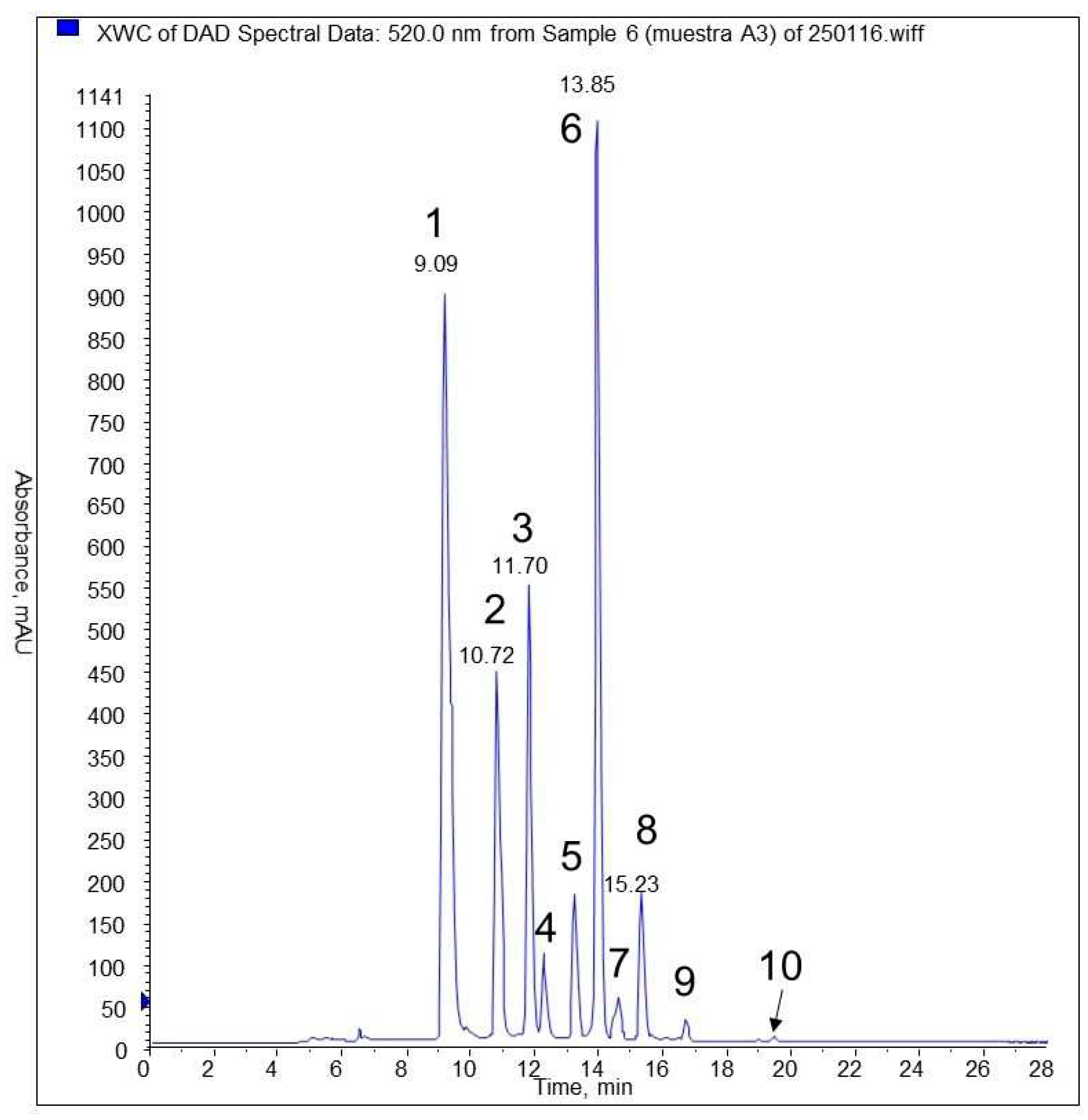
The anthocyanin chromatographic profile in E. rubrum fruit extract F3 shows (Figure 2, Table 3) that the main anthocyanins identified are delphinidin-3-glucoside and malvidin-3-glucoside, followed by cyanidin and petunidin-3-glucosides. In a lesser proportion, some pentoside conjugates can be observed, but their identity was not confirmed with commercial standards, as the glucoside derivatives were. Other authors [57] identified the anthocyanin profile in other Empetrum berries (Black Crowberry) with the main identified anthocyanins were delphinidin, cyanidin, peonidin, and malvidin, but conjugated with galactose instead of glucose.
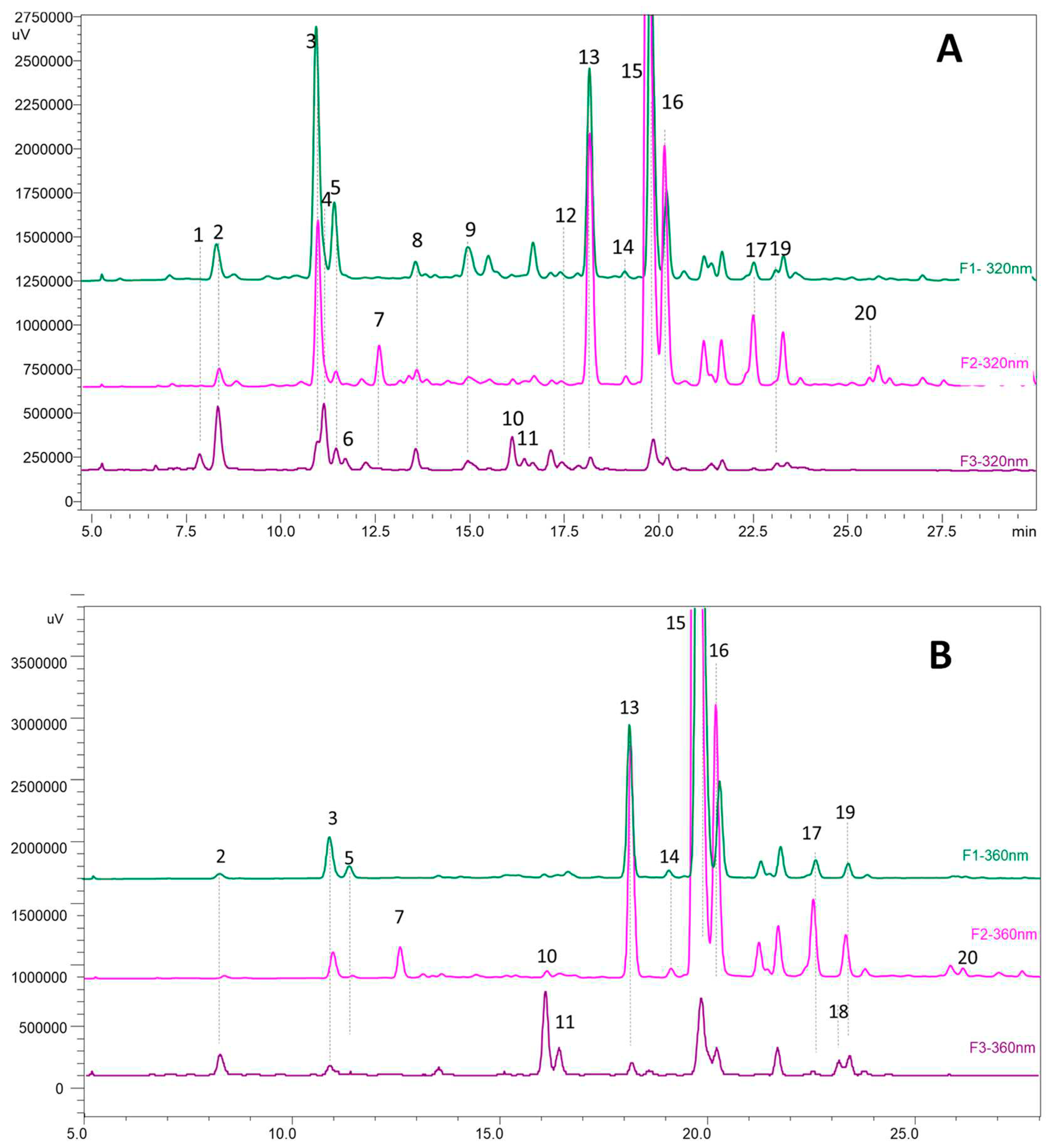
The phenolic compounds chromatographic analysis of the F1 (leaves and steams aqueous extract), F2 (leaves and steams methanolic extract), and the cleaned F3 fruit extract showed that the main compounds were HCAD and conjugated flavonols (Figure 3). There was no significant presence of flavanol or larger molecular weight procyanidins. The principal flavonols found in the samples were quercetin-3-glucoside and a laricitin hexoside, which can be either galactoside or glucoside in the steam and leaves samples. The other significant signals were tentatively identified as a caffeoylquinic acid isomer, and a coumaric acid and caffeic acid derivate (Table 4), also more prevalent in steam and leaves samples. In the F3 sample, which was previously treated to remove the anthocyanins, maintained the same proportion of flavonols and HCAD, but in much lesser quantities. Laaksonen et al. [58] analyzed flavonol and HCAD in Empetrum nigrum berries and found that the main flavonols identified were similar in profile with galactosides of laricitin and quercetin, and coumaric acid derivates.

3.6. Antifungal Activity of MEE

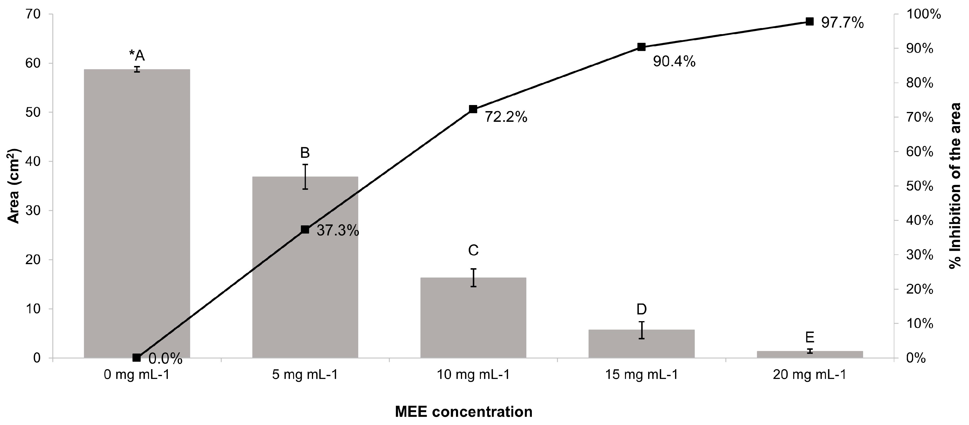
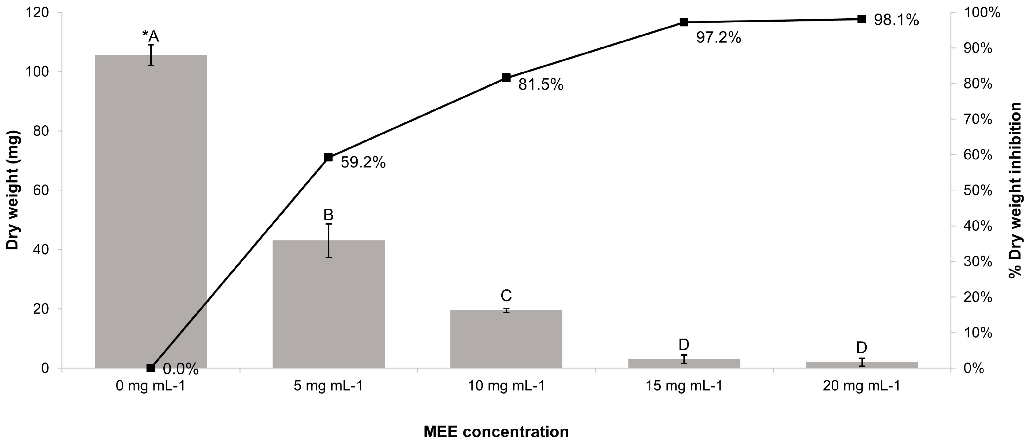
The observed antifungal activity of MEE against R. solani, consisted of a growth inhibition of mycelium and a diminution of the mass of fungus. The reduction of both parameters mentioned above was directly related to the MEE concentration. In fact, as shown in Figure 4, the maximum concentration of 20 mg mL-1 inhibited the mycelial growth in a percentage of 97.70 ± 0.72 %, after 84 hours. With respect to the inhibition of the mass of fungus, and as seen in Figure 5 the same concentration, of 20 mg mL-1, mentioned above leads to a decrease of 98.10 ± 1.34 % after 84 hours.
Similarly, the research of an algal methanolic extract performed by Al-Nazwani et al. [59] demonstrated that there is a correlation between the increase of the extract concentration tested, and the inhibition of growth and the biomass decline of R. solani. The authors mentioned above also concluded that phenol in comparison with other tested compounds, exerted the greatest antifungal effect, reflected in the mycelial growth of R. solani. Other research demonstrated the association of antifungal and antioxidant activities of methanolic plant extracts, with their phenols, tannins, and flavonoids contents [60]. According to the observed inhibition of mycelial growth of R. solani, Sclerotium rolfsii, and another fungus, a higher content of total phenols and flavonoids in a 70% ethanolic extract in comparison with an aqueous extract, is directly related to the growth inhibition of these microorganisms [61]. The above-mentioned research is in agreement with the higher levels of TPC detected in MEE, in comparison with TPC detected in the methanolic extract obtained from Nassauvia dentata [54], because MEE has a higher antifungal effect than the methanolic extract from Nassauvia dentata, against R. solani. Another modality of antifungal activity consists of biocontrol agents against the fungus Botrytis cinerea, using microorganisms inhabiting the leaf surface of E. rubrum [62].

4. Conclusions

The results of our research revealed a considerable content of phenols, extracted from the aerial parts of E. rubrum, using a polar solvent such as methanol, and these phenol compounds exhibited antioxidant activity. Furthermore, and based on our results, the phenol compounds in E. rubrum could have an antifungal effect against R. solani. Therefore, the methanol extract obtained from E. rubrum may be a natural antifungal agent in the future.

Author Contributions

Conceptualization, C.S. and C.V.; methodology, C.S., C.V., M.G.-R and P.Z.; formal analysis, C.S., C.V., M.G.-R and P.Z.; investigation, C.S., C.V., M.G.-R and P.Z; resources, C.S. and C.V.; data curation, C.S., C.V., M.G.-R and P.Z.; writing—original draft preparation, C.S. and C.V.; writing—review and editing, C.S. and C.V.; supervision, C.S.; project administration, C.S.; funding acquisition, C.S. and C.V. All authors have read and agreed to the published version of the manuscript.

Funding

This work has been supported by the Vicerrectoría de Investigación y Desarrollo (Project Nr. 214.418.007-1.0) of the Universidad de Concepción.

Data Availability Statement

The data presented in this manuscript are available from the authors upon reasonable request.

Acknowledgments

The authors wish to thank Mrs. Claudia Flores for her technical support.

Conflicts of Interest

The authors declare that they have no conflicts of interest.

References

  1. Chassagne, F.; Cabanac, G.; Hubert; G., David, B.; Marti, G. The landscape of natural product diversity and their pharmacological relevance from a focus on the Dictionary of Natural Products. Phytochem. Rev. 2019, 18, 601-622. [CrossRef]
  2. Wu, G.; Chang, C.; Hong, C.; Zhang, H.; Huang, J.; Jin, Q.; Wang, X. Phenolic compounds as stabilizers of oils and antioxidative mechanisms under frying conditions: A comprehensive review. Trends Food Sci. Tech. 2019, 92, 33-45. [CrossRef]
  3. Su, L.J.; Zhang, J.H.; Gomez, H.; Murugan, R.; Hong, X.; Xu, D.; Jiang, F.; Peng, Z.Y., 2019. Reactive Oxygen Species-Induced Lipid Peroxidation in Apoptosis, Autophagy, and Ferroptosis. Oxid. Med. Cell. Longev. 2019, 2019,1-13. [CrossRef]
  4. Rai, V.; Kumar, A.; Das, V.; Ghos, S. Evaluation of chemical constituents and in vitro antimicrobial, antioxidant and cytotoxicity potential of rhizome of Astilbe rivularis (Bodho-okhati), an indigenous medicinal plant from Eastern Himalaya region of India. BMC Complem. Altern. M. 2019, 19 , 1-10. [CrossRef]
  5. Zafar, S.; Ahmed, R.; Khan, R. Biotransformation: a green and efficient way of antioxidant synthesis. Free Radical Res. 2016, 50, 939-948. [CrossRef]
  6. Galano, A.; Castañeda-Arriaga, R.; Pérez-González, A.; Tan, D.X.; Reiter, R.J. Phenolic Melatonin-Related Compounds: Their Role as Chemical Protectors against Oxidative Stress. Molecules 2016, 21, 1-42. [CrossRef]
  7. Piccolella, S.; Crescente, G.; Candela, L.; Pacifico, S. Nutraceutical polyphenols: New analytical challenges and opportunities. J. Pharmaceut. Biomed. 2019, 175, 112774. [CrossRef]
  8. Kafkas, N.E.; Kossar, M.; Öz, A.T.; Mitchell, A.E. Advanced Analytical Methods for Phenolics in Fruits. J. Food Quality 2018, 2018, 1-6. [CrossRef]
  9. Haida, Z.; Hakiman, M. A comprehensive review on the determination of enzymatic and nonenzymatic activities. Food Sci. Nutr. 2019, 7, 1555-1563. [CrossRef]
  10. Laganà, P.; Anastasi, G.; Marano, F.; Piccione, S.; Singla, R.K.; Dubey, A.K.; Delia, S.; Coniglio, M.A.; Facciolà, A.; Di Pietro, A.; Haddad, M.A.; Al-Hiary, M.; Caruso, G. Phenolic Substances in Foods: Health Effects as Anti-Inflammatory and Antimicrobial Agents. J. AOAC Int. 2019,102, 1378-1387. [CrossRef]
  11. Ramamoorthy, R.; Muthalagu, M.; Andra, S.; Ravichandran, B.; Narayanasamy, M. Investigation on antimicrobial, antioxidant and cytotoxicity properties of triple bark extract formulated using traditional medicinal plants. SN Appl. Sci. 2019, 1, 1-7. [CrossRef]
  12. Ribeiro Alves, D.; de Morais, S.M.; Tomiotto-Pellissier, F.; Menegazzo Miranda-Sapla, M.; Vasconcelos, F.R.; da Silva, I.N.G.; de Sousa, H.A.; Assolini, J.P.; Conchon-Costa, I.; Rogéiro Pavanelli, W.; das Chagas Oliveira Freire, F. Flavonoid Composition and Biological Activities of Ethanol Extracts of Caryocar coriaceum Wittm., a Native Plant from Caatinga Biome. Evid.-based Complement. Altern. Med. 2017, 2017, 1-7. [CrossRef]
  13. Sharifi-Rad, M.; Varoni, E.M.; Iriti, M.; Martorell, M.; Setzer, W.N.; Contreras, M,M.; Salehi, B.; Soltani-Nejad, A.; Rajabi, S.; Tajbakhsh, M.; Sharifi-Rad, J. Carvacrol and human health: A comprehensive review. Phytother. Res. 2018, 32, 1675-1687. [CrossRef]
  14. Ynalvez, R.A.; Compean, K.L.; Addo-Mensah, A. Qualitative Determination of the Secondary Metabolites and Evaluation of the Antimicrobial Activity of Leaf Extracts from Different Plant Families (Boraginaceae, Fabaceae, Lamiaceae and Lauraceae) against Microorganisms of Clinical Importance. J. Pharm. Res. Int. 2018, 23, 1-12. [CrossRef]
  15. Liang, Q.P.; Wu, C.; Xu, T.Q.; Jiang, X.Y.; Tong, G.D.; Wei, C.S.; Zhou, G.X. Phenolic Constituents with Antioxidant and Antiviral Activities from Phyllanthus urinaria Linnea. Indian J. Pharm. Sci. 2019, 81, 424-430. [CrossRef]
  16. Aourahoun, K.A.K.; Fazouane, F.; Benayache, S.; Bettache, Z.; Benayad, T.; Denni, N. Antioxidant and anti-inflammatory activity of phenolic extracts of Genista ferox (Fabaceae). Pakistan J. Pharm. Sci. 2019, 32, 2643-2649.
  17. Topi, D.; Guclu, G.; Kelebek, H.; Selli, S., 2020. Comparative elucidation of phenolic compounds in Albanian olive oils using LC-DAD-ESI-MS/MS. J. Liq. Chromatogr. R. T. 2020, 43, 203-212.
  18. Kandil, A.S.; Abou-Elella, F.; El Shemy, H.A. Cytotoxic profile activities of ethanolic and methanolic extracts of chicory plant (Cichorium intybus L.). J. Radiat. Res. Appl. Sci. 2019, 12, 106-111. [CrossRef]
  19. Talba, M.A.; Miaffo, D.; Kamani, S.L.P.; Kamanyi, A.; La Wansi, S. Antioxidant Properties and Digestive Enzyme Inhibitory Activity of the Aqueous Extract from Leafy Stems of Cissus polyantha. Evid.-based Complement. Altern. Med. 2019, 2019, 1-8. [CrossRef]
  20. Ergul, M.; Ergul, M.; Eruygur, N.; Atas, M.; Ucar, E. In Vitro Evaluation of the Chemical Composition and Various Biological Activities of Ficus carica Leaf Extracts. Turk. J. Pharm. Sci. 2019, 16, 401-409. [CrossRef]
  21. Utami, S.; Endrini, S.; Nafik, S.; Lestari, I.M.T.; Anindya, D.; Abu Bakar, E.; Rozy, F.; Said, F.F.; Afifah, E.; Arumwardana, S.; Nufus, H.; Rihibiha, D.D.; Kusuma, H.S.W.; Wibowo, S.H.B.; Widowati, W. In vitro Antioxidant and Anti-obesity Activities of Freeze-dried Canarium sp., Averrhoa bilimbi L. and Malus domestica. Indones. Biomed. J. 2019, 11, 320-326. [CrossRef]
  22. Piccolella, S.; Crescente, G.; Pacifico, F.; Pacifico, S. Wild aromatic plants bioactivity: a function of their (poly)phenol seasonality? A case study from Mediterranean area. Phytochem. Rev. 2018, 17, 785-799. [CrossRef]
  23. Feng, Z.; Ding, C.Q.; Li, W.H.; Wang, D.C.; Cui, D. Applications of metabolomics in the research of soybean plant under abiotic stress. Food Chem. 2020, 310, 1-8. [CrossRef]
  24. Lorion, J.; Small, E. Crowberry (Empetrum): A Chief Arctic Tradtional Indigenous Fruit in Need of Economic and Ecological Management. Bot. Rev. 2021, 87, 259-310. [CrossRef]
  25. Bezverkhniaia, E.A.; Emilova, E.V.; Kadyrova, T.V.; Krasnov, E.A.; Brazovskii, K.S.; Ponkratova, A.O.; Luzhanin, V.G., Belousov; M.V. Phytochemistry, ethnopharmacology and pharmacology of the genus Empetrum: a review. Adv.Trad. Med. 2021, 10, 1-14. [CrossRef]
  26. Popp, M.; Mirré, V.; Brochmann, C. A single Mid-Pleistocene long-distance dispersal by a bird can explain the extreme bipolar disjunction in crowberries (Empetrum). P. Natl. Acad. Sci. USA 2011, 108, 6520-6525. [CrossRef]
  27. Hoffmann, A.; Liberona, F.; Muñoz, M.; Watson, J. Plantas Altoandinas en la Flora Silvestre de Chile, 1st ed.; Ediciones Fundación Claudio Gay, Santiago de Chile, Chile, 1998; pp. 82-83.
  28. San Martín, C.; Montenegro, D.; Pérez, Y.; Solís, J.L. Vegetation and woody flora of Tortel commune (Aisén Region, Chile): a key to identify the species. Agro Sur 2014, 42, 15-29.
  29. Rodriguez, R.; Marticorena, C.; Alarcón, D.; Baeza, C.; Cavieres, L.; Finot, V.L.; Fuentes, N.; Kiessling, A.; Mihoc, M.; Pauchard, A.; Ruiz, E.; Sanchez, P.; Marticorena, A. 2018. Catalogue of the vascular plants of Chile. Gayana Bot. 2018, 75, 1-430.
  30. Hein, N.; Merkelbach, J.; Zech, K.; Weijers S. Drought sensitivity of Empetrum nigrum shrub growth at the species’ southern lowland distribution distribution range margin. Plant Ecol. 2021, 222, 305-321. [CrossRef]
  31. Hyun, T.K.; Kim, H.C.; Ko, Y.J.; Kim, J.S. Antioxidant, alpha-glucosidase inhibitory and anti-inflammatory effects of aerial parts extract from Korean crowberry (Empetrum nigrum var. japonicum). Saudi J. Biol. Sci. 2016, 23, 181-188. [CrossRef]
  32. Jurikova, T.; Durisova L.; Elias, P.; Mlcek, J.; Sochor, J.; Ondrasova, M. Evaluation of Fruit Anatomy, Accumulation and Detection of Polyphenols in Black Crowberry (Empetrum nigrum) from NW Slovakia. Acta Biol. Cracov. Bot. 2019, 61, 25-33. [CrossRef]
  33. Jurikova, T.; Mlcek, J.; Skrovankova, S.; Balla, S.; Sochor, J.; Baron, M.; Sumczynski, D. Black Crowberry (Empetrum nigrum L.) Flavonoids and Their Health Promoting Activity. Molecules 2016, 21, 1-11. [CrossRef]
  34. Oka, S.; Kuniba, R.; Tsuboi, N.; Tsuchida, S.; Ushida, K.; Tomoshige, S.; Kuramochi, K. Isolation, synthesis, and biological activities of a bibenzyl from Empetrum nigrum var. japonicum. Biosci. Biotech. Bioch. 2020, 84, 31-36. [CrossRef]
  35. Li, H.X.; Jean, S.; Webster, D.; Robichaud, G.A.; Calhoun, L.A.; Johnson, J.A.; Gray, C.A. Dibenz[b,f]oxepin and Antimycobacterial Chalcone Constituents of Empetrum nigrum. J. Nat. Prod. 2015, 78, 2837-2840. [CrossRef]
  36. Moerman, D.E. Native American medicinal plants: an ethnobotanical dictionary, abridged ed.; Timber Press, Portland (OR), USA, 2009.
  37. Wang, Q.B.; Zheng, C.L.; Jin, S.Y.; Guo, H.; Wang, Z.Y. Chemical constituents of ethyl acetate extract of Empetrum nigrum var. japonicum and their effect on alcoholic fatty liver of rats. Chin. Tradit. Herb. Drugs 2016, 47, 3164-3168. [CrossRef]
  38. Gaviria, C.; Ochoa, C.; Sánchez, N.; Medina C.; Lobo M.; Galeano P.; Mosquera, A.; Tamayo, A.; Lopera, Y.; Rojano, B. Actividad antioxidante e inhibición de la peroxidación lipídica de extractos de frutos de mortiño (Vaccinium meridionale SW). Bol. Latinoam. Caribe 2009, 8, 519-528.
  39. Kuskoski, E.M.; Asuero, A.G.; García-Parrilla, M.C.; Troncoso, A.M.; Fett, R. Actividad Antioxidante de Pigmentos Antociánicos. Food Sci. Technol. (Campinas) 2004, 24, 691-693. [CrossRef]
  40. Dos Santos, M.N.G.; da Silva, E.P.; Godoy, H.T.; da Silva, F.A.; Celestino, S.M.C.; Pineli, L.D.D.; Damiani, C. Effect of freezing and atomization on bioactive compounds in cagaita (Eugenya dysenterica DC) fruit. Food Sci. Technol. 2018, 38, 600-605. [CrossRef]
  41. Grzegorczyk-Karolak, I.; Kuzma, Ł.; Wysokinska., H. Study on the chemical composition and antioxidant activity of extracts from shoot culture and regenerated plants of (Scutellaria altissima L.). Acta Physiol. Plant. 2015, 37, 1-9. [CrossRef]
  42. Hussain, I.; Khattak, M.U.R.; Ullah, R.; Muhammad, Z.; Khan, N.; Khan, F.A.; Ullah, Z.; Haider, S. Phytochemicals screening and antimicrobial activities of selected medicinal plants of Khyberpakhtunkhwa Pakistan. Afr. J. Pharm. Pharmaco. 2011, 5, 746-750. [CrossRef]
  43. Sethy, R.; Kullu, B. Comparative phytochemical screening and antimicrobial activity of Calotropis sp. of ethnomedicinal significance. Int. J. Pharm. Sci. Res. 2020, 11, 3242-3251. [CrossRef]
  44. Ruiz, A.; Mardones, C.; Vergara, C.; Hermosín-Gutiérrez, I.; von Baer; D.; Hinrichsen, P.; Rodríguez, R.; Arribillaga, D.; Dominguez, E. Analysis of hydroxycinnamic acids derivatives in calafate (Berberis microphylla G. Forst) berries by liquid chromatography with photodiode array and mass spectrometry detection. J. Chromatogr. A 2013, 1281, 38-45. [CrossRef]
  45. Elgorban, A.M.; Bahkali, A.H.; El-Metwally, M.A.; Elsheshtawi, M.; Abdel-Wahab, M.A. In vitro Antifungal Activity of Some Plant Essential Oils. Int. J. Pharmacol. 2015, 11, 56-61. [CrossRef]
  46. Pereira, F.D.; Wanderley, P.A.; Viana, F.A.C.; de Lima, R.B.; de Sousa, F.B.; Lima, E.D. Growth Inhibition and Morphological Alterations of Trichophyton rubrum induced by Essential Oil from Cymbopogon winterianus Jowitt ex Bor. Braz. J. Microbiol. 2011, 42, 233-242. [CrossRef]
  47. Días, A.L.B.; Sergio, C.S.A.; Santos, P.; Barbero, G.F.; Rezende, C.A.; Martinez, J. Ultrasound-Assisted Extraction of Bioactive Compounds from Dedo de Moca Pepper (Capsicum baccatum L.): Effects on the Vegetable Matrix and Mathematical Modeling. J. Food Eng. 2017, 198, 36-44. [CrossRef]
  48. Pyrzyńska, K.; Pękal, A. Application of free radical diphenylpicrylhydrazyl (DPPH) to estimate the antioxidant capacity of food samples. Anal. Methods 2013, 5, 4288-4295. [CrossRef]
  49. Arts, M.J.T.J.; Haenen, G.R.M.M.; Vost, H.P.; Bast, A. Antioxidant capacity of reaction products limits the applicability of the Trolox equivalent antioxidant capacity (TEAC) assay. Food Chem. Toxicol. 2004, 42, 45-49. [CrossRef]
  50. Hyun, T.K.; Kim, H.C.; Ko, Y.J.; Kim, J.S. Antioxidant, α-Glucosidase Inhibitory and Anti-Inflammatory Effects of Aerial Parts Extract from Korean Crowberry (Empetrum nigrum var. japonicum). Saudi J. Biol. Sci. 2016, 23, 181-188. [CrossRef]
  51. Gao, Y.; Shi, Y.T.; Miao, N.; Xing, W.X., Yun, C.; Wang, S.F.; Wang, W.J., Wang, H.M. A green ultrasound-assisted enzymatic extraction method for efficient extraction of total polyphenols from Empetrum nigrum and determination of its bioactivities. J. Ind. Eng. Chem. 2022, 109, 559-567. [CrossRef]
  52. Gao, J.Y.; Hu, J.X.; Hu, D.; Yang, X. A Role of Gallic Acid in Oxidative Damage Diseases: A Comprehensive Review. Nat. Prod. Commun. 2019, 14, 1-9. [CrossRef]
  53. Qamar, F.; Naveed, S.; Faizi, S.; Sana, A. Formulation and Evaluation of Natural Antioxidant Cream of Ocinum basilicum. Lat. Am. J. Pharm. 2021, 40, 2293-2300.
  54. Schneider, C.; Zapata, P., Gonzalez-Reyes, M. Evaluation of the Antioxidant Properties and Antifungal Activity of Methanolic Extract obtained from Nassauvia dentata Griseb. (Asteraceae). Pak. J. Bot. 2023, 55, 1041-1049. [CrossRef]
  55. Gao, Y.; Wang, S.F.; Dang, S.K.; Han, S.L.; Yun,C.L.; Wang, W.J.; Wang, H.M. Optimized ultrasound-assisted extraction of total polyphenols from Empetrum nigrum and its bioactivities. J. Chromatogr. B 2021, 1173, 122699. [CrossRef]
  56. Yin, P.P.; Yang, L.G.; Li, K.; Fan, H.; Xue, Q.; Li, X.; Sun, L.W.; Liu,Y.J. Bioactive components and antioxidant activities of oak cup crude extract and its four partially purified fractions by HPD-100 macroporous resin chromatography. Arab. J. Chem. 2019, 12, 249-261. [CrossRef]
  57. Ogawa, K.; Sakakibara, H.; Iwata, R.; Ishii, T.; Sato, T.; Goda, T.; Shimoi, K.; Kumazawa, S. Anthocyanin Composition and Antioxidant Activity of the Crowberry (Empetrum nigrum) and Other Berries. J. Agric. Food Chem. 2008, 56, 4457–4462. [CrossRef]
  58. Laaksonen, O.; Sandell, M.; Järvinen, R.; Kallio, H. Orosensory contributing compounds in crowberry (Empetrum nigrum) press-byproducts. Food Chem. 2011, 124, 514-1524. [CrossRef]
  59. Al-Nazwani, M.S.; Aboshousha, S.S.; El-Saedy, M.A.M.; Gareeb, R.Y.; Komeil, D.A. Antifungal activities of Chlorella vulgaris extract on black scurf disesase, growth performance and quality of potato. Arch. Phytopathol. Plant Prot. 2021, 54, 2171-2190. [CrossRef]
  60. Kengne, I.C.; Fankam, A.G., Yamako, E.K.; Tamokou, J.D. Phytochemical Analysis, Antifungal, and Antioxidant Properties of Two Herbs (Tristemma mauritianum and Crassocephalum bougheyanum) and One Tree (Lavigeria macrocarpa) Species. Adv. Pharmacol. Pharm. Sci. 2023, 2023, 1-13.
  61. Lopez-Palestina, C.U.; Joaquin-Ramos, A.D.; Santiago-Saenz, Y.O.; Velazquez-Jimenez, R.; Altamirano-Romo, S.E.; Gutierrez-Tlahque, J. Antifungal and Antioxidant Potential of Ethanolic and Aqueous Extracts of the Wild Plant Consuela (Tinantia erecta). Acta Biol. Colomb. 2023, 28, 143-153.
  62. Sanhueza, T.; Herrera, H.; Arriagada, C. Contribution of Leaf-Associated Microorganisms from Native Andean Ericaceae against Botrytis cinerea in Vaccinium corymbosus Cultivars. J. Soil Sci. Plant Nut. 2023, 23, 2637-2650.
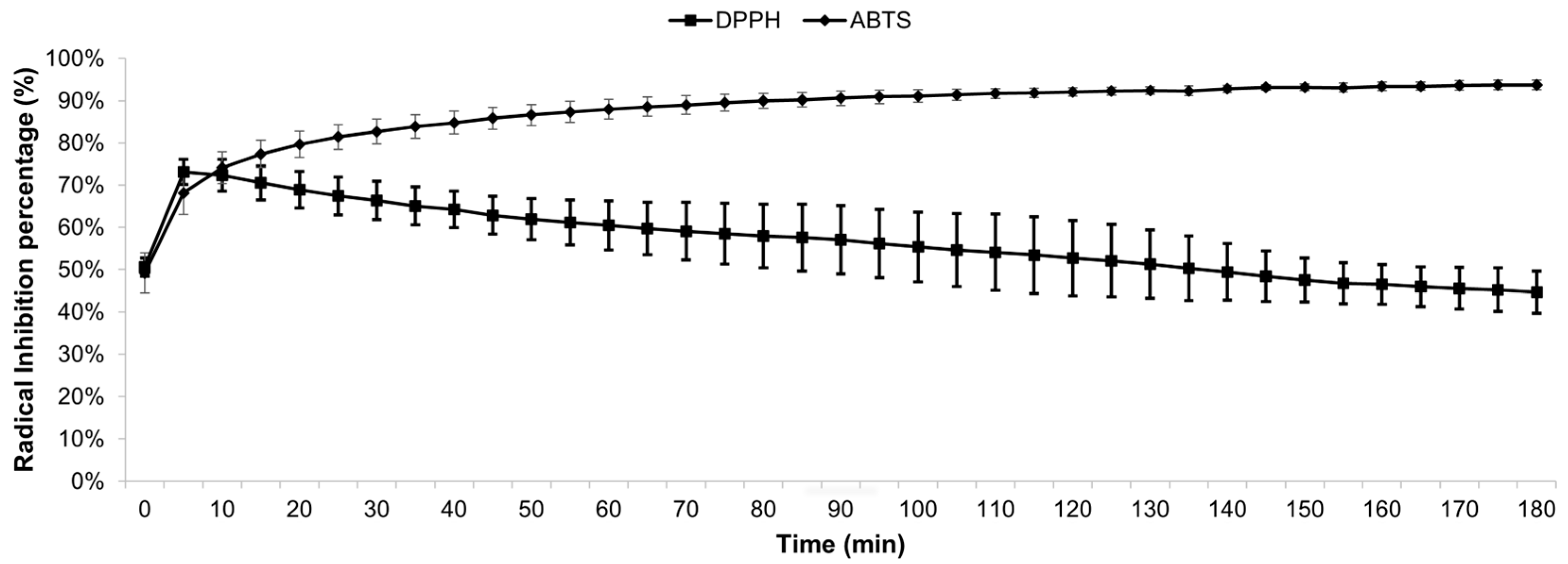
Figure 1. Antioxidant effect of MEE during 3 hours, using IC50 concentrations.
Preprints 91001 g001
Figure 2. Chromatogram E. rubrum fruit extract F3. Identification numbers in table 3.
Preprints 91001 g002
Figure 3. Chromatograms of F1, F2 and F3 extracts at 320 nm for hydroxycinnamic acid derivates (A) and 360 nm for flavonols determination (B). Identification in Table 4.
Preprints 91001 g003
Figure 4. Influence on the mycelial growth according to increasing concentrations of MEE. The different letters indicate statistically significant differences between the MEE concentrations, in accordance with the Tukey test results (p≤ 0.05).
Preprints 91001 g004
Figure 5. Relation between MEE concentrations and the accumulated biomass of R. solani. There are no statistically significant differences between the same letters, based on the results of the Tukey test (p≤ 0.05).
Preprints 91001 g005
Table 1. Antioxidant Activity of MEE expressed in IC50, GAE and TEAC.
Radical IC50 GAE* TEAC**
DPPH 0.414 mg mL-1 26.31 ± 1.51 mg -
ABTS 0.108 mg mL-1 - 200.24 ± 2.61 mg
*The expression of the result is mg of gallic per 1000 mg (1 g) of MEE. **The expression of the result is mg of Trolox per 1000 mg (1 g) of MEE.
Table 2. Results of preliminary qualitative assays for groups of secondary metabolites.
Secondary metabolites Leaves and Stems MEE
Flavonoids +++ +++
Hydrolizable Tannins
Condensed Tannins +++ +++
Coumarins
Saponins
Alkaloids
Marked presence of metabolites (+++), normal presence (++), weak presence of metabolites (+) and (–) indicates absence of secondary metabolites.
Table 3. Identification of main anthocyanins present in E. rubrum fruit methanolic extract by HPLC-DAD-ESI-MS/MS.
Identification N° peak tr
(min)
λ
(nm)
[M+H]+ fragments
delphinidin-3-glucoside (*) 1 9,09 523 465 303,9
cyanidin-3- glucoside (*) 2 10,7 520 449 287
petunidin-3- glucoside (*) 3 11,7 525 479 317
cyanidin pentoside 4 12,14 519 419 287
petunidin pentoside 5 13,14 522 449 317; 302; 274
peonidina-3-glucoside (*) 5 13,14 522 463 301; 286
malvidin-3- glucoside (*) 6 13,85 527 493 331; 315; 287
peonidin pentoside 7 14,49 522 433 301; 286; 158
malvidin pentoside 8 15,24 528 463 331; 315; 287
malvidin derivate 9 16,6 527 521 331
malvidin 10 19,38 535 331
Note: (*): identification confirmed with commercial standards.
Table 4. Identification of the main phenolic compound present in E. rubrum extracts by HPLC-DAD-ESI-MS/MS.

peak
Identifications tR
(min)
DAD (nm) [M-H]- fragments
1 coumaric acid derivate 7,84 320 361 163, 119
2 caffeoylquinic acid isomer 8,33 324 535 191
3 caffeoylquinic acid isomer 10,9 324 553 191
4 caffeoylquinic acid isomer 11,11 324 553 191
5 coumaroylquinic acid 11,43 306 337 191, 163, 119, 155
6 coumaric acid derivate 11,65 284 325 163, 119
7 myricetin-3-galactoside (*) 12,5 340 479 317, 287, 271
8 feruloylquinic acid 13,52 320 367 161, 133
9 coumaroylquinic acid 14,9 320 337 191, 173
10 myricetin-3-glucoside(*) 16,05 360 479 317, 287, 271
11 myricetin rutinoside 16,1 360 625;479 317, 287, 271
12 quercetin-3-rutinoside(*) 17,93 360 609 301
13 caffeic acid derivate 18,1 320 367 179, 191, 161, 135
14 quercetin galactoside 19,33 360 463 301
15 laricitin hexoside 19,75 360 493 331, 315, 287
16 quercetin-3-glucoside(*) 20,12 360 463 301, 271, 179, 163
17 quercetin pentoside 22,35 355 433 301, 271, 255, 151
18 kaempferol-3-glucoside(*) 23,1 348 447 285, 255, 227, 151
19 isorhamnetin hexoside 23,67 351 477 315, 285, 299, 271
20 unknown quercetin hexoside 26,8 354 583 463, 301
Note: (*): identification confirmed with commercial standards.
Disclaimer/Publisher’s Note: The statements, opinions and data contained in all publications are solely those of the individual author(s) and contributor(s) and not of MDPI and/or the editor(s). MDPI and/or the editor(s) disclaim responsibility for any injury to people or property resulting from any ideas, methods, instructions or products referred to in the content.
Copyright: This open access article is published under a Creative Commons CC BY 4.0 license, which permit the free download, distribution, and reuse, provided that the author and preprint are cited in any reuse.
Prerpints.org logo

Preprints.org is a free preprint server supported by MDPI in Basel, Switzerland.

Subscribe

© 2024 MDPI (Basel, Switzerland) unless otherwise stated