Preprint
Article

Assessment of Biocidal Efficacy of Zinc Oxide-Zeolite Nanocompoites as a Novel Water Disinfectant against Commercial Disinfectants Used in Water Purification

Altmetrics

Downloads

155

Views

43

Comments

0

Submitted:

24 November 2023

Posted:

27 November 2023

You are already at the latest version

Alerts
Abstract
Water microbial contamination is a serious issue that poses a risk to both animal and human health. 120 water samples collected from main water source and drinkers from a poultry farm. Different bacterial pathogens were isolated from water sources. Escherichia (E.) coli, Pseudomonas (P.) aureoginosa, (Salmonella) S. Typhimurim, Aeromonas (A.) hydrophila at different percentages. Variable degree of bacterial resistance to some commercial disinfectants commonly used to disinfect water system (iodine, terminator and H2O2). Nanoparticles were used to control bacteria in water. About the safety investigation for the prepared nanomaterials, the work results demonstrated that zinc oxide (ZnO) nanoparticles (NPs) exhibit the highest safety profile among the manufactured materials. The median fatal dose (LD50) for ZnO NPs was determined to be 3709 mg/kg body weight. In comparison, the LD50 values for zeolites and nanocomposites were 3251 mg/kg and 2658 mg/kg, respectively. Therapeutic dosages were estimated based on the LD50. Zeolite NPs, ZnO NPs and ZnO/zeolite NPs showing promising results in control of those bacteria. It was concluded that the escalating resistance of bacteria to disinfectants have led to a need to find alternative such as nanoparticles that proved promising results in control of pathogens, particularly it showed a safe effect on laboratory animals.
Keywords: 
Subject: Public Health and Healthcare  -   Public, Environmental and Occupational Health

1. Introduction

The quality of drinking water is a matter of ultimate importance for livestock especially poultry affecting both health and performance of the birds [1] (Umar et al., 2014). Unfortunately, most of farmers are unaware of the impact of the water quality on their animals [1]. Besides, many rural areas in developing countries lack the hygienic quality of drinking water offered to livestock [2]. Water is an essential nutrient, it is required for all vital processes done in the animal body, transport nutrient to/out the cells, remove waste materials, and maintain proper fluid and ion balance [3].
In Egypt, the River Nile is the main source of drinking water for both humans and animals, also it supplies about 97.0% of water reserves across the country [4,5]. Drinking water sources (tab water or ground water) receive many contaminants through many ways such industry, agriculture and domestic wastes [6]. There is a wide range of pathogens could be found in water especially this offered to animals. The major type of these pathogens is coliform bacteria; the most important bacteria in this group include Escherichia coli, Klebsiella spp. and Enterobcater spp. Besides, Psuedomonas spp., Salmonella, Aeromonas spp. and Protus which are highly pathogenic and of zoonotic importance [7,8].
Drinking water purification plants are designed specifically to eliminate chemical and microbiological pollution in raw water sources through many treatment stages; noteworthy in the last step of disinfection, bacteria and other pathogens which cause water-borne diseases are eliminated [9]. Many disinfectants have been used in poultry farms such: chlorine and chlorine containing substances, iodine, hydrogen peroxide, and quaternary ammonium compounds (QAC) that exhibited high efficacy in controlling water borne pathogens [10], but due to their abuse and /or overuse and other factors that have led to development of microbial resistance toward disinfectants [11].
To overcome microbial resistance to disinfectants and environmental pollution including water pollution, researchers have resorted to nanoparticles [12,13]. NPs can be made of metals, metal oxides, carbon nanotubes, zeolites, and other materials. The actual mechanisms of action of different NPs against microorganism vary depending on the type of NP and the target microorganism [14]. One of the promising materials that could be used in water purification is zinc-zeolite (ZnO-Z) nanocomposite where zeolites are used as an adsorbing platform for zinc nanoparticles which act as a source of zinc ions that are released slowly from zeolite matrix and attack bacterial cells inhibiting their growth [15].
The aim of this study was to investigate the bacterial profile of commercial poultry farms that impair the quality of drinking water of the farm under the study causing water-borne diseases for both birds and farm workers and evaluating the effectiveness of some disinfectants commonly used in water treatment and then comparing their antibacterial effect to ZnO,Z and ZnO-Z nanocomposites in controlling virulent pathogenic bacteria and In vivo evaluating the pathological effect of the tested nanomaterial on lab animal.

2. Materials and methods

2.1. Study area and period

A field study was conducted in Beni-Suef district, Beni-Suef province (coordinates 29°04 N-31° 05'E) during the period from March till November 2022. Representative water samples were collected from main water supplies tap, underground [hand pump] and drinkers from two commercial poultry farms. Many birds and some of the farm workers were suffering from nausea, diarrhea, vomiting and weight loss which mainly associated with water-borne infections after exclusion of other causes, despite continuous water treatment using different disinfectants. The ethical approval for the in-vivo assessments of the tested nanoparticles was authorized by of Institutional Animal Care and Use Committee (IACUC), Ref. No: IORG 022-412), Beni-Suef university.

2.2. Water sample collection

A total of 120 water samples were collected from the main water sources (n=10) and drinkers (n=30) intended for poultry and the farm workers' drinking. The water samples represented three different sources (tap and underground [hand pump]) from the investigated two farms in different areas. Water samples were gathered from tap and hand pump wells in 250 ml sterilized Schott Duran bottles for bacteriological examination. The tap's outlets were thoroughly disinfected with 70.0% ethyl alcohol, water is then allowed to flow, and the samples were taken [16]. Sampling bottles were tightly sealed, properly labeled and then immediately sent to laboratory for bacteriological analysis as described by APHA [17].

2.3. Isolation of bacterial pathogens in the screened water samples

For isolation of Pseudomonas spp., initially 10 ml of the water samples were enriched in 90 ml of tryptic soya broth (TSB, Oxoide, CM0129T), incubated at 37 °C for 24 h, in the second day the enriched broth was subsequently inoculated at surface of Pseudomonas cetrimide agar (Oxoide, CM0579), incubated at 37°C for 24h. Blue, blue-green pigmented or non-pigmented colonies with specific sweaty grape odor on the surface of cetrimide agar were picked up and purified on tryptic soya agar (TSA) media (Oxoide, PO0163) [18,19]. For recovery of Aeromonas organism, another 10 ml of the water samples were poured into 90 ml of buffered peptone water (BPW, Oxoide, CM509), incubated at 37°C for 24 h, followed by plating on Aeromonas specific media (Oxoide, CM0833), supplemented with ampicillin antibiotic (Oxoide, SR0136) and incubated at 37°C for 24h, Green and yellow colonies were taken on nutrient agar (Oxoide, CM0003) plates and incubated at 37°C for 24 h for purification [20].
While, the isolation of Salmonella spp., the third portion of water samples (10 ml) was firstly enriched into BPW, incubated at 37°C for 24h, followed by post-enrichment on Rappaport-Vassiliadis (RV, Oxoide, CM0669), incubated at 42°C for 24 h and finally selectively plated on the surface of Salmonella-Shigella agar (S-S, Oxoide, CM0099), incubated at 37°C for 24 h, colonies of white color with black centers were subsequently purified on the surface of nutrient agar plates [21]. For cultivation of E. coli O157:H7, the fourth part of water samples (10 ml) was added to 90 ml of tryptic soya broth supplemented with novobiocin (20 mg/L, Oxoid, SR0181). After incubation for 24 h at 37°C, a volume of 100-µL of the broth media was spread onto Sorbitol MacConkey (SMAC, Oxoid, CM0813) agar supplemented with cefixime (0.25 mg/ml) and tellurite potassium (2.5 mg/ml) (Oxoid, SR0172) [22]. Plates were incubated for 24 h at 37°C and examined for typical E. coli O157 colonies (non-sorbitol fermenters). The suspected typical colonies were streaked on TSA plates. The suspected non-fermenting colonies were assessed for agglutination using specific E. coli O157:H7 agglutination latex kit (Oxoid, DR0620M) according to manufacturer instruction. Bacterial colonies showing an agglutination reaction were considered as E. coli O157:H7 positive. After purification of the all previous isolates they were preserved on TSA slopes at 4°C where they were subjected for confirmation by biochemical and molecular analysis.

2.4. Identification of bacterial pathogens in screened water samples

2.4.1. Biochemical and serological identification of bacterial pathogens

All recovered Pseudomonas colonies were biochemically identified according to Palleroni [18] and Holtz et al. [19], while the suspected Aeromonas isolates were identified using morphological and biochemical characters [23]. On the other hand, the obtained E. coli and Salmonella isolates were serologically identified according to Kauffman-White Scheme by slide agglutination test using polyvalent and monovalent O and H antigen (Difco Laboratories, Detroit, Michigan, USA) [24,25]. The serological analyses were applied in Serology Unit, Animal Health Research Institute, Dokki, Giza, Egypt.

2.4.2. Molecular identification of the bacterial pathogens

All biochemically or serologically identified isolates of A. hydrophila, P. aeruginosa, E. coli and Salmonella spp. were submitted to the molecular characterization in the biotechnology center at the animal health research institute, Egypt, as well determining the virulence genes as mentioned in Table 1.

2.5. Synthesis and characterization of ZnO NPs, Z NPs and ZnOnanocomposite

Commercial ZnO powder (size 0.6-1 m, purity 99.9%, Loba, Chemi, Pvt. Ltd., India) was ground using hardened steel balls (diameter) in steel cells (250 mL). 15 mm, 32 gm) in ambient conditions for various times between 2 and 10 hours. The automated milling was carried out in a mill with a horizontal oscillator (Retsch, PM 400) with a 25 Hz frequency. Steel ball ratio in the combination and ZnO powders was approximately 15.0% by weight. The use of already-milled materials without further milling media. Each compartment contained 10 g and five balls of the powdered sample. There were two parallel cells utilized in the total powder sample weight for this experiment is 20 g).
Commercial zeolite (Clinoptilolite ore and Nano Zeolite with particle sizes ranging from 1 to 10 m were utilized in the mailing process for commercial zeolite. The Nano-zeolite was produced by combining commercial zeolite with processed natural clay zeolite that had been dried at 100°C for 48 hours, and then passed through a photon ball mill for 12 hours at a constant mechanical speed of 300 rpm. Then, 200 ml of DMF (di-methyleformamide) solution were combined with 0.5 g of ZnO to synethsis ZnO-Z. The liquid was then filtered to get rid of any settled solids after spending 24 hours in an ultrasonic bath. Then, 1 g of zeolite was added to the mixture, and the ultrasonication process was repeated for 24 hours. The mixture was then centrifuged, dried at 100 °C for 24 hours, and filtered.
The crystal structure and crystallinity of the prepared materials were detected using X-ray diffraction (XRD), while the vibration and chemical bonds of the materials were screened by fourier-transform infrared spectrum (FT-IR). The average size and morophologic shape of zinc oxide, zeolite nanocomposite was characterized by Scanning Electron microscope (SEM) at National Research Center (NRC), Egypt.

2.6. Evaluation of biocidal activity of tested disinfectants

The efficacy of different disinfectants commercially used for sanitation and disinfection of drinking water at animal and poultry facilities and listed by Food and Drug Administration also proven to be effective under field condition [10], (Aksoy et al.,2019), and ZnO-Z nanocomposite at different concentrations using broth macro dilution method against different bacterial pathogens isolates from water samples from the farm under the study as following: Iodine at concentrations of (0.5 and 1.0%) (India), commercial preparation of hydrogen peroxide (H2O2) 50% stock solution (Sigma) at concentration of (3.0 and 5.0%), Terminator (glutraladehyde and cocobenzyl dimethyl ammonium chloride) (Bomac laboratories, Ltd, Newzealand) at concentration of (0.3 and 0.5%), and ZnO-Z nanocomposite at concentrations of (0.1 and 0.5 mg/ml), against 40 isolates of different pathogens that were isolated from different water sources E. coli, Salmonella sp., Pseudomonas sp. and Aeromonas sp. (10 isolates each) using broth macro dilution method as described by Li et al. [14] at contact time (30 min, 2 h and 24 h).

2.7. Experimental animals

The Department of Physiology at Beni-Suef University, Faculty of Veterinary Medicine acquired rats for use as laboratory animals. The rats were kept in a standard laboratory environment with a 12-hour light/dark cycle, a humidity level of 60.0%, and a temperature of 23°C. The Faculty of Veterinary Medicine at Beni-Suef University evaluated and approved the animal handling guidelines, which included weighing and gavage techniques. Protocol of Animal Rights for Laboratory Experiments, IACUC was approved at Beni-Suef University for ethical handling of laboratory animals. The experiment was conducted under a standard 12-hour cycle of light and darkness. To perform the LD50 values, adult rats weighing 160 to 180 g b.wt. were used in toxicological experiments. All of the animals included in the acute investigation had a constant diet and had access to water every single day.

2.7.1. Experimental groups and medications for 10 days’ acute study

In this investigation, 54 adult male albino rats were used, with an average body weight of 100 to 150 gm and an average age of 3 to 4 months. Rats were divided into equal groups of nine for each dose increase during each experiment, beginning with 200 mg/kg b.wt. in the first group, the concentration of all investigated compounds was raised in subsequent groups, reaching 10000 mg/kg b.wt. in the final group.

2.7.2. The assessment of LD50, LD90 as a measure of toxicity (Probit analysis)

The independent variable X can collect experimental data by converting the value evaluation of a mathematical model that best fits the dependent variable Y (percent) into probit values. Results of the investigation (X-different concentrations) Regression and probit analyses are much inferior to traditional LD50 calculation methods, which involve (percent) at specified doses of the investigated components and Y-experimental animal death (Regression analysis) [37].
In order to calculate the number, data and interpolation are needed. Since the Miller and Tainter method similarly causes a 50.0% rise in the experimental data variable Y [38], it is very noteworthy. If the mortality of the lowest and/or highest doses is 0.0% and/or 100.0%, respectively, Miller-Tainter method also converts mortality results (in percent) into probit values; however, the percentage values are first corrected against the number of experimental animals; these corrected values are then transformed into probit values for additional processing. The doses were expressed in mg/kg rather than percentages, allowing the LD50 and LD90 to be calculated.

2.7.3. Estimation of the maximum LD100 and median lethal dose LD50 for the tested nanomaterials

Rats were used in the tests, which were divided into six groups of nine each. The various test materials received oral doses ranging from 200 to 10,000 mg/kg b.wt. Following administration, the animals were observed for two hours, then again after twenty-four, then again after 10 days. The death rate (%) was estimated after 24 hours up until 10 days [39]. The outcomes were processed using Miller-interactive Tainter's LD50 calculation techniques. The trials employed rats with LD0, LD20, LD50, LD90, and LD100. The SPSS program's probit analysis was used to determine the linear correlation coefficient and analyses mortality trends as a function of the measured drug concentrations.

2.7.4. Mortality and Toxic Signs

Ocular observations of mortality, distinct changes in physical appearance, behavior (sleepy, salivation, lethargy), and any damage or illness were made at 2, 6, and up to 24 hours after dosing, and throughout the course of the next 10 days [40].

2.8. Histopathological observation

Standard histological procedures were used to fix a tiny piece of the liver, kidney, and brain before embedding it in paraffin, sectioning it for 5–6 mm thick, and mounting it on glass microscope slides. Hematoxylin and eosin was used to stain the sections, and light microscopy was used to examine the prepared slides [41].

3. Statistical analysis

Data obtained were recorded the prevalence of identified bacterial traits in the collected water samples as well as the germicidal efficacy of tested disinfectants and zinc oxide-zeolite nanocomposite using non-parametric test (Chi-square test) using SPSS (Inc. version 22.0, Chicago, IL, USA).

4. Results and discussion

E. coli was the most prevalent bacteria isolated from different water sources 34/75 (45.3%) followed by Salmonella spp. 21/75 (28.0%). Besides E. coli was mainly recovered from drinkers filled from surface water followed by drinkers filled from tap water (53.0 and 41.7%, respectively). Meanwhile Salmonella spp. were mainly recovered from surface water (40.0%), also P. aeruginosa and A. hydrophila were mainly obtained from tap water and drinkers filled out from tap water (100.0 and 20.8%, respectively) (Table 2). High detection rates of the isolated bacteria were found in the drinkers in comparing to the main water rates.
E. coli isolation in the screened water samples was not in consistent to those found by Selim et al. [42], (8.0%) and Barbosa et al. [43], (16.5%) also, Momtaz et al. [44] , who found that only 4 out of 448 water samples (0.89%) were positive for E. coli. While, our findings for Salmonellae isolates were not matched to those isolated by Yam et al. [45], (18.0%), Haley et al. [46] (79.2%), Adingra et al. [47] (15.4%), Momtaz et al. ( [44] (7.58%), Tracogna et al. [48] (8.8%), Yhils and Bassey [49] (12.9%) and Abd El-Tawab et al.[50] (25.0%). Furthermore, P. aeruginosa was found in 38.9% and A. hydrophila wasn’t detected in any of the examined water samples as reported by Mohammed [51].
Although E. coli is a normal inhabitant of the intestinal tract of bird, man and animals.It must be pointed out that the water that is supplied to the birds should be free from this pathogen which is a requirement for water intended for the birds. Drinkers are considered important foci for the microbiological quality of the water provided to the birds. Open water supplies, such as troughs and bell drinkers, may present high contamination levels of 107 and 104 per ml for mesophiles and fecal coliforms [52]. In the closed system (nipple), the quality of the water offered to the birds is better protected and there are no deleterious effects on bird performance compared to the open systems [53].
The risk of contamination with Salmonellas was 6 to 7 times higher when the water given to birds was exposed to the environment [54]. Besides, more water samples were positive to Salmonellas in a broiler facility when water was provided in troughs and therefore water was considered an important means of re-infection in birds [55]. Salmonellas were isolated from 21.6% of the broiler farms and from 12.3% of the water samples examined in Canada by Poppe et al. [56]. The use of open drinkers in the majority of the farms acts as favorable media to contamination and the presence of Salmonellas in the litter was considered an important contamination route of the water provided to the birds.
Water systems could provide as an important environmental vehicle (reservoir) for pathogenic organisms and serve as a potential source of water contamination, resulting in a possible health risk for man, accompanied with severe gastrointestinal, food-borne infections in addition to, high mortality rate in the immuno-compromised individuals [57,58]. The existence of the pathogenic enteric bacteria represents an alarming circumstance for water- and food-borne epidemics in the screened settings. The isolation of those pathogenic bacteria from water with high percentage in poultry farms necessitated the strict application of biosecurity measures inside the farms and using safe and efficient disinfectants to control those pathogens.
Concerning the serological identification of the isolated Salmonella and E. coli as investigated in Table 3, it showed that the most predominant serotype recovered traits of E. coli was O157 16/34 (47.1%), followed by O144 8/34 (23.5%). Meanwhile the percentage of serogroups S. Kentucky was higher than other recovered serogroups 10/21 (47.6%), followed by S. Typhimurium. S. Infants 5 and 4/21 (23.8 and 19.0 %, respectively). The obtained results for determining E. coli O157 was the parallel to those obtained by Momba et al. [59], Mersha et al. [60], El-Leithy et al. [61] and Goma et al. [62] (25.56, 4.2, 32.0 33.3, respectively). S. Kentucky was the prominent serotype detected in poultry water sources; this was in accordance to those found by Hassan et al. [63], and Djeffal et al. [64]. E. coli O157 is belonged to the Enterohemorrhagic E. coli (EHEC), that causes hemorrhagic colitis and are often associated with devastating or life-threatening systemic manifestations, the hemolytic uremic syndrome (HUS), results from Shiga toxins (Stxs) produced by the bacteria in the intestine of the diseased man. While the attention devoted to EHEC O157:H7 is justified by the pathogenicity, low infectious dose, and ability of the bacteria to survive in extra-intestinal environments, a number of non-O157:H7 EHEC cause severe human disease and are often implicated in HUS as O26. Also, S. Kentucky is a common causative agent of gastroenteritis in humans, poultry act as the main reservoir of S. Kentucky, and also domestic poultry has played an important role in its global spread of this species. S. Kentucky has been identified as one of the most prominent Salmonella serovars isolated from broilers causing diarrhea and high mortalities resulting in severe economic losses [65,66].Upon the molecular characterizations (Figure 1) for the screened traits, it was denoted that all the examined isolates revealed their specific genus identification as well as virulence related genus, indicated their severity at the farm or consumer levels.
Biocidal efficacy of tested disinfectants (Table 4) showed that all of the isolated bacteria exhibited resistance against both concentrations of iodine (0.5 and 1.0%) and only P. aeruginosa isolates showed moderate sensitivity by 50.0% after 24h of exposure to 1.0% concentration. A similar pattern was exhibited by most of the recovered isolates to H2O2 3.0% conc. where all of them showed resistance at variable degree except for P. aeruginosa was sensitive by 40.0% after 24h contact time, meanwhile both E. coli and P. aeruginosa were sensitive to 5.0% concentration of H2O2 after 24h of exposure (55.0 and 40.0%, respectively). Concerning Terminator disinfectant Salmonella spp. was resistant to both of its concentration (0.3 and 0.5%) by 80.0 and 70.0%, respectively. Whilst E. coli, P. aeruginosa and A. hydrophila were sensitive to both concentrations (0.3 and 0.5%) variably where E. coli was sensitive (45.0 and 50.0%, respectively) after 24h, P. aeruginosa was sensitive by (45.0 and 60.0%, respectively) after 24h and A. hydrophila showed sensitivity by (50.0 and 60.0%, respectively) a for the same contact time (Table 4). Our findings were Similar to those obtained by Amini Tapouk et al. [67] who reported that E. fecalis showed high sensitivity to 2.0% glutaraldehyde.
Concerning in-vitro sensitivity of zeolite nanoparticles, zinc oxide nanoparticles and ZnO-Z nanocomposite at different contact time against tested pathogens as shown in Table 5, it revealed that all of the tested pathogens (E. coli, Salmonella spp., P. aeroginosa and A. hudrophila were significantly resistant to zeolite nanoparticles at p = 0.000 at all contact times, on the other hand they showed less resistant pattern to ZnO nanoparticles particularly with increasing the contact time mainly after 24hr of exposure where A. hydrophila, Salmonella spp., P. aeroginosa and E. coli were sensitive to it as following 48, 43, 42 and 40%, respectively after 24 hr of exposure (at p= 0.000). Meanwhile ZnO nanocomposite showed a significantly promising results in control of those pathogens where their sensitivity to composite increase with the increase of contact time where Salmonella spp. was the most affected pathogen (76.0%), followed by E. coli (73.0%) then P. aeroginosa (69.0%) and finally A. hydrophila (63.0%) at p= 0.000 after 24hr of exposure.
In contrast to the finding in this study de Souza et al. [68] found that P. aeruginosa was highly resistant to ZnO-Nps, meanwhile S. aureus and S. Typhimurium were sensitive. Stankovic et al. [69] and Talebian et al. [70] reported that antimicrobial activity of ZnO-NPs is mainly affected by the morphology of particles. On the other hand Wang et al. [71] had to some extent similar results to our study were ZnO-coated zeolite was significantly effect in controlling S. aureus compared to ZnO NPs that showed less effect in their control. Also Wakweya and Jifar [72] reported that ZnO NPs had lesser antibacterial effect on both E. coli and S. aureus thanZnO-Z. Incorporation of the matrix of zeolites with metal oxides such ZnO NPs increases the antibacterial properties of composite [73] which increase the capacity of it to penetrate the bacterial layer and subsequently increasing its biocidel effect.
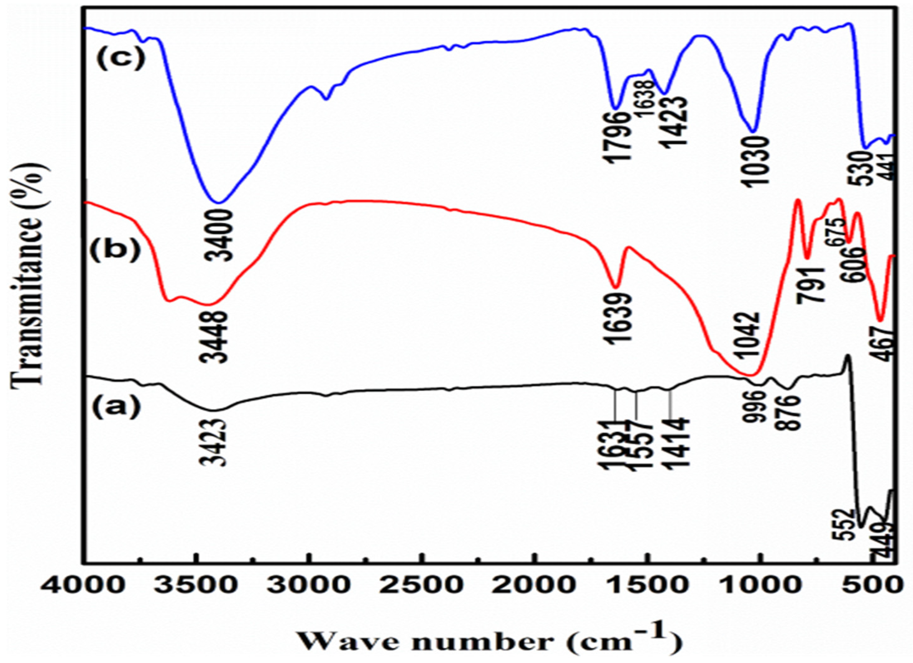
FTIR of the synthesized materials as showed in Figure 2, (a) FTIR of ZnO nanoparticles showed a broad absorption peak at 3423 cm-1 may be attributed to the characteristic peak of hydroxyl group (O-H) [74,75]. Peak appeared at 1631 cm-1 may be due to the bending of water molecules and the absorption peaks located in the range from 450-600 cm-1 is due to the presence of Zn-O bond [76,77,78]. In Figure 2 (b) FTIR of zeolite nanoparticles show, the -OH, -Si-O, and Al-O bonds in the prepared zeolite are presumably responsible for the characteristic absorbance at 3448 cm-1, 1639 cm-1, and below 1042 cm-1 to 467cm-1due to symmetric and asymmetric stretching vibration of zeolite [79]. In Figure 2 (c) FTIR of Zeolite/ ZnO nanoparticles show peaks of zeolite ZnO nanoparticles are somewhat sharper and stronger than those of pure zeolite or ZnO indicating weaker interactions and ordered arrangements of ZnO molecules in the zeolite. All stretching vibrations associated with the hydroxide fictional group at frequencies over 3000 cm-1 in the FTIR spectra of zeolite change towards lower frequency, possibly as a result of the chemical bonding activity between Zn+2 and O atoms [79].
Additionally, the IR spectra of the ZnO-Z nanoparticles showed little variation from the reference material (zeolite) at frequencies below 1640 cm-1, which should be caused by the disordered alignment and irregular conformation of ZnO molecules in the zeolite network [80]. (Hara et al., 2000). The range between 460 and 530 cm1 is where zinc oxide concentrations peak [80]. (Hara et al., 2000). We can see that the IR peak of zinc oxide nanoparticles is clearly defined in the FT-IR spectra of the samples containing ZnO nanoparticles and appeared at 441-530 cm-1, also the presence of ZnO nanoparticles in zeolite was confirmed by the interaction between zeolite and ZnO nanoparticles leading to the shift in zeolite peaks like from 1639 cm-1 to 1638 cm-1 and also from 1042 cm-1 to 1030 cm-1.
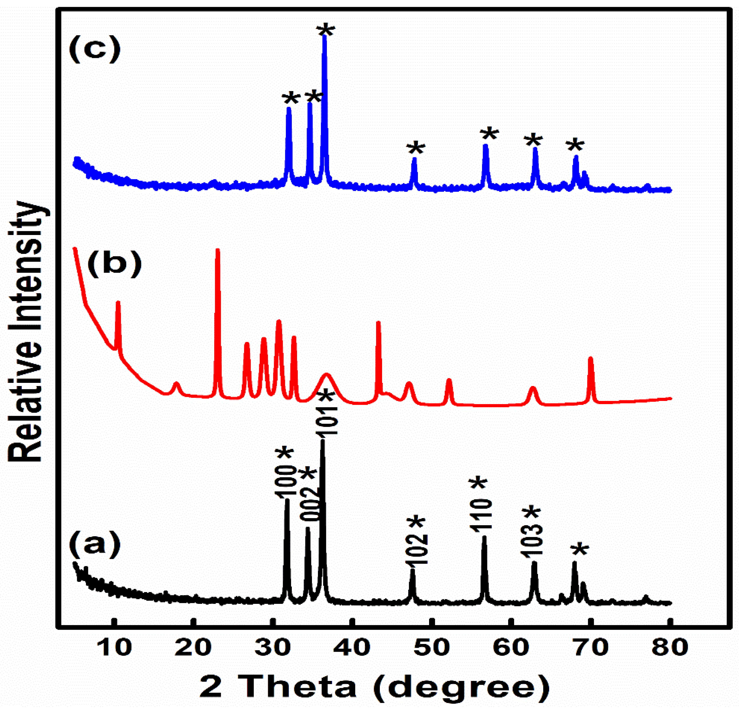
The current findings of XRD spectrum for the synthesized ZnO NPs, zeolite NPs and ZnO-Z NPs was showed in Figure. 3. The crystalline structure of ZnO NPs Figure 3(a) was confirmed by the observation of distinct diffraction peaks at 31.7°, 34.4°,36.15°, 47.47°, 56.56°and 62.73° in the spectra which corresponds to the indices of (100), (002), (101), (102), (110) and (103), respectively [81,82,83]. Figure 3 (b) showed XRD of zeolite nanoparticles, the patterns of zeolite peaks were in agreement with (Ref Cod 01-087- 1619) [84]. (Alswat et al., 2017). While, Figure 3 (c) showed XRD of ZnO-Z NPs, from which we observed the similar peaks of ZnO NPs were appeared in that composite that showed successful incorporation and preparation of zeolite NPs and ZnO NPs.
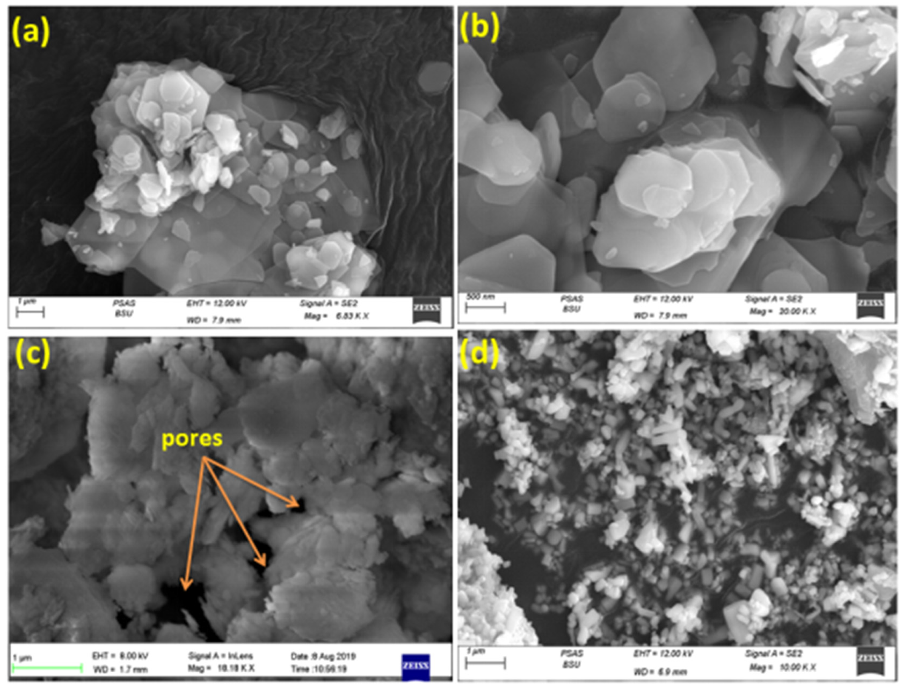
Regarding Figure 5 (a, b), SEM of ZnO nanoparticles showed that the particles were discovered to be less than 100 nm. The particles were discovered to have a large surface area and surface energy, whereby larger-sized particles will aggregate [85]. The homogenous, smooth, and devoid of any fractures surfaces of the nanoparticles demonstrated proper material production [86]. While, in the figure 5c, SEM of zeolite nanoparticles have hollow cores and mesoporous shells which offer excellent room for interactions with ZnO nanoparticles. Also, Figure 5d displayed the SEM pictures that reveal the morphological makeup of the zeolite/ZnO nanoparticles. The morphology of the Zeolite/ZnO nanoparticles showed many pores and voids indicating a larger surface area and porosity. Also small spherical granules of the ZnO nanoparticles injected into the surface of the zeolite are plainly seen.
Shifting to the acute toxicity of zeolite, zinc nanoparticles, and their nanocomposites (Table 6) in rats was investigated for 10 days following oral administration. Tremors, rapid breathing, an arched back, convulsions, and unconsciousness were toxicity signs that were followed by death. The mortality probability began to rise around 1247, 1805 and 1046 mg/kg b.wt. After oral administration of zeolite, Zinc NPs and their nanocomposites, respectively. The LD50 was discovered to be 3251, 3709 and 2658 mg/kg respectively, and (LD100) was reached 8467, 7620 and 6636 mg/kg b.wt., as shown in the Table 6. These findings showed that zeolites, Zinc NPs and their nanocomposites can be used safely in pharmacological research. For any biological applications or as a therapeutic dose, we used LD50 values of 1/20 for the zeolites, ZnO NPs and their nanocomposites at doses of 162.5, 185.4 and 129.3 mg/kg respectively. Toxicity increased when the medicine dose was increased in the trial, as seen in Table 7 and Table 8. For acute oral testing, the maximum dosage (2,000 mg/kg body weight) indicated in OECD Guideline 423 was applied. By oral gavage, a 25.0% aqueous solution of the dosage was given. Prior to dosing, after two hours, on day 1, at least once each day for a total of one week, the animals were monitored for treatment-related effects. All rats underwent gross pathology 10 days following oral treatment. No animals perished while the study was being conducted. One rat showed reductions in body weight and fecal excretion on day 3 of observation, but these findings vanished four days following medication. Two weeks following oral delivery, there were no unusual findings during necropsy. The approximate acute oral toxicity (LD50) was >2,000 mg/kg b.wt. for male Sprague-Dawley rats.
From these study and through the probit analysis LD50 had been estimated and measured; depends upon the LD50 results; the tested therapeutic doses were determined and calculated for its use in the biomedical applications at this research as 1/20 from the calculated LD50 had been tested as follow;
Zeolite === LD50 = 3251 x 1/20 = 162.50 mg/kg
ZnO NPs==== LD50 = 3709 x 1/20 = 185.45 mg/kg
Nanocomposite ==== LD50 = 2658 x 1/20 = 129.3 mg/kg
That’s indicate the highly significant safety of the tested nanomaterials
From the obtained data its illustrates that ZnO NPs are the most safe prepared materials at this study with median lethal dose equal to 3709 mg/kg b.wt., while 3251 in zeolites whereas 2658 in the nanocomposites. Depends upon the LD50, therapeutic doses were estimated.
According to the delivery routes, the liver, kidney, lung, and brain may be the target organs for ZnO NPs, according to the histopathological examination (Figure 6). The current research will also provide a deeper knowledge of the toxicity and in vivo behaviors of ZnO NPs in rats based on the different routes of administration. After a 10-day acute treatment, no abnormalities were discovered in any of the many organs that were taken for histological investigation. The liver's hepatocytes were positioned properly, and its cords and major vein were both largely patent. The kidney's glomeruli were seen to be organized normally, without any congestion or cyanosis. Normal dermal and epidermal blood vessels on the skin with no damage or congestion. The intestinal or cecal epithelium was unaffected by zeolites, zinc nanoparticles, or their nanocomposites, and no harm was seen. Additionally, there was no stenosis or damage, and the intestinal villi were orientated correctly. As demonstrated in Figure 6, the brain's hippocampus area in particular did not exhibit any abnormal or degenerative changes. Therefore, unlike earlier studies concerning intravenous dosing of Zno NPs for a number of days, the acute oral administration of Zno NPs had resulted in that there was no inflammation or pathological lesions in the body organs [87].
In order to increase therapeutic effectiveness, active or passive targeting, controlled or prolonged release, and decrease systemic drug adverse effects, medication delivery systems are becoming more and more common [79]. To our knowledge, no prior research has been done on the interaction between zeolites, zinc nanoparticles, and their nanocomposites with the aim of identifying novel uses [90]. By enabling the regulated and continuous delivery of medications, nanotechnology has shown to be helpful in the treatment of a number of biological disorders. The creation of novel materials for use in cutting-edge medical technologies as well as a rise in the targeting effectiveness of multifunctional. Nano carriers were made possible by the nanometer size. Nanoparticles or layers can include small molecules that change the efficacy, bioavailability, and safety of drugs [91]. Drug pharmacokinetics and pharmaco-dynamics are significantly impacted by the nano carrier size and incorporation into layers [92]. Nanoparticles enhanced the effects of carrier molecules like drugs due to their high surface-to-volume ratio. Reactive oxygen species are also produced at a higher rate under oxidative stress, which speeds up cellular activity (ROS). Zn helps achieve a high degree of activity in a short amount of time as a consequence [93,94]. Nanoparticles containing drugs have been shown to be effective in the treatment of brain diseases and infections due to their small size particles adhering effectively and crossing the blood-brain barrier as well as their sustained or controlled release, which reduces dosing treatment and drug side effects on organ function.

5. Conclusion

Over use/abuse commercial disinfectants to control bacterial pathogens in water system in poultry farms have attributed in the incidence of bacterial resistance as well spreading of resistance genes among different genera of bacterial pathogens through the environment that displayed a significant role in this process. Therefore finding an alternative became a necessity to overcome this residence using nanoparticles such zeolite NPs, ZnONPs and particularly, ZnO-Z NPs that showed promising results in controlling zoonotic pathogens that contaminate poultry water systems and poses a risk to human population. Remarkable results were presented by this study proving the in-vitro safety of using ZnO-Z nanocomposite in lab animals that could be used in the future under field condition to compromise effective control to different bacterial pathogens in livestock water system.

Supplementary Materials

The following supporting information can be downloaded at the website of this paper posted on Preprints.org.

Author Contributions

Manar Bahaa El Din Mohamed1 and Sahar Abdel Aleem Abdel Aziz1: Conceptualization, Methodology, Writing, Reviewing and editing the original draft. Fatma I. Abo El-Ela2: Conceptualization, Validation, Investigation the toxicity analysis for the tested nanomaterials, Rehab K. Mahmoud3 and Ahmed A. Farghali4; Supervision, Writing Analysis and characterization of tested nanomaterials. Sarah I. Othman5, Ahmed A. Allam6: Conceptualization, writing draft and funding the publication.

Acknowledgments

The authors acknowledge Princess Nourah Bint Abdulrahman University researchers supporting project number (PNURSP2012R5) Princess Nourah Bint Abdulrahman University, Riyadh, Saudi Arabi.

Conflicts of Interest

The authors declare no conflict of interest.

Abbreviation:

E. Escherichia
P. Pseudomonas
S. Salmonella
A. Aeromonas
H2O2 hydrogen peroxide
NPs Nanoparticles
ZnO Zinc oxide
Z Zeolite
LD Lethal Dose
QAC quaternary ammonium compounds
TSB tryptic soya broth
XRD X-ray diffraction
FT-IR Fourier-transform infrared spectrum
SEM Scanning Electron microscope

References

  1. Umar, S. , Munir, M.T., Azeem, T., Ali, M., Shah, A. Effects of Water Quality on Productivity and Performance of Livestock: A Mini Review. Veterinaria 2014, 2, 11–15. [Google Scholar]
  2. Guppy, L. , Shantz, H. Groundwater Quality in Rural Cambodia: Measures and Perceptions. Geograph. Res. 2011, 49, 384–394. [Google Scholar] [CrossRef]
  3. Amna, M.M. , Abdelga, A. Bacteriological evaluation of the drinking water quality in dairy farms in Khartoum state, Sudan. J of Vet Med. and Anim. Hlth. 2004, 6, 95–100. [Google Scholar] [CrossRef]
  4. Abdel-Shafey, H.I. , Aly, F.O. Water issue in Egypt: resources, pollution and protection endeavours. Cent.Eurp. J. of Environ. Med. 2002, 8, 3–21. [Google Scholar]
  5. El-Sadek, A. Upscaling field scale hydrology and water quality modeling to catchment scale. Water Res. Managem. 2007, 21, 149–169. [Google Scholar] [CrossRef]
  6. Annual Drinking Report, UNEP GEMS/Water Programme, 2005. Workshop report: Development and use of global water quality indicators and indices. Vienna, Austria 4-6th May 2005. (http://www.gemswater.org/publications/pdfs/indicators_workshop_report.pdf).
  7. LeJeune, J.T. , Thomas, E., Hancock, D.D. Cattle water troughs as reservoirs of E. coli 0157. Appl. Envir. Microbiol. 2001, 67, 3053–3057. [Google Scholar] [CrossRef]
  8. Murinda, L. , Miliwebsky, E., Gioffre, A., Chinen, I., Basckier, A., Chillemi, G., Guth, B.E.C., Masana, M.O., Cataldi, A., Rodriguez, H.R. Novel single-tube agar-based test system for motility enhancement and immunocapture of E.coli O157:H7 by H7 flagellar antigen-specific antibodies. J ClinMicrobiol. 2002, 40, 4685–4690. [Google Scholar] [CrossRef]
  9. Ezzat, S.M. , Mohammed, T., Moustafa, Fouda, A., S. El-Gamal, M., and Ibrahim, A. M. Assessment of some drinking water purification plants efficiency at Great Cairo in Egypt. Curr.Scie. Inter. 2017, 06, 761–776. [Google Scholar]
  10. Aksoy A, El Kahlout KEM, Yardimci H. Comparative Evaluation of the Effects of Binzalkonium Chloride, Iodine, Gluteraldehyde and Hydrogen Peroxide Disinfectants against Avian Salmonellae Focusing on Genotypic Resistance Pattern of the Salmonellae Serotypes toward Benzalkonium Chloride. Brazilian Journal of Poultry Science. [CrossRef]
  11. Davies, R. , Wales, A.. Antimicrobial resistance on farms: A review including biosecurity and the potential role of disinfectants in resistance selection. Comp. Rev. Food Sci. Food Saf 2019, 18, 753–774. [Google Scholar] [CrossRef] [PubMed]
  12. Jegadeesan, G. , Mondal,K., Lalvani S.B. Arsenate remediation using nanosized modified zerovalent iron particles. Environ. Progress 2005, 24, 289–296. [Google Scholar] [CrossRef]
  13. Hardiljeet, K.B. , Meera, J., Dennis, M.O. Kinetics and thermodynamics of Cadmium ion removal by adsorption onto nanozervalent iron particles. J. Harzard. Mater 2010, 186, 458–65. [Google Scholar] [CrossRef]
  14. Li, W.R. , Xie, X.B., Shi, Q.S., Zeng, H.Y., Ou-Yang, Y.S., Chen, Y.B. Antibacterial activity and mechanism of silver nanoparticles on E. coli. ApplMicrobiol Biotechnol. 2010, 85, 1115–1122. [Google Scholar] [CrossRef]
  15. Petrik, L. , Missengue, R., Fatoba, M., Tuffin, M., Sachs, J. 2014. Silver/zeolite nano composite-based clay filters for water disinfection. Report to the Water Research Commission. No KV 297/12. Available online: http://www.ircwash.org/resources/silver-zeolite-nano-composite-based-clay-filters-water-disinfection.
  16. Azam, A. , Ahmed, A.S., Oves, M., Khan, M.S., Habib, S.S., Memic, A. Antimicrobial activity of metal oxide nanoparticles against Gram-positive and Gram-negative bacteria: a comparative study. Int. J. Nanomedicine 2012, 7, 6003–6009. [Google Scholar] [CrossRef] [PubMed]
  17. APHA, A.W.W. A, W.E.F. 2012.Standard methods for examination of water and wastewater, 22nd. American Public Health Association, Washington. ISBN: 978-087553-013-0. https://www.scirp.org/(S(vtj3fa45qm1ean45vvffcz55))/ reference/ References Papers.aspx?.
  18. Palleroni, N.J. Genus I.Pseudomonas. In Bergey’s Manual of Systematic Bacteriology; Krieg, N. R. and Holt, J. C., Ed.; Williams and Wilkins: Baltimore; Volume 1, pp. 141–199.
  19. Holtz, J.G. , Krieg, N.R., Sneath, P.H.A., Staley, J.T., Williams, S.T. Bergey’s manual of determinative bacteriology, 9th ed.; Lippincott Williams & Wilkins: Philadelphia, PA, USA, 2000. [Google Scholar]
  20. Ashiru, A. , Uaboi-Egbeni, P., Oguntowo, J., Idika, C. 2011. Isolation and antibiotic profile of Aeromonas species from tilapia fish (Tilapia nilotica) and catfish (Clariasbetrachus). Pakistan J. Nutr. 2011, 10, 982–986. [Google Scholar] [CrossRef]
  21. Ewing, W.H. The genus Salmonella. In Edwards and Ewing's identification of Enterobacteriaceae, 4th Ed.; Ewing WH, Ed.; Elsevier: New York, 1986; pp. 181–245. [Google Scholar]
  22. Akiba, M. , Rice, D.H., Davis, M.A. Masuda, T., Sameshima, T., Nakazawa, M., D. Hancock, D. A comparison of E. coli O157 isolates from cattle in Japan and the USA by molecular biological methods. Epidemiol. Infect. 2000, 125, 221–224. [Google Scholar] [CrossRef] [PubMed]
  23. Konemann, E.; Allen, S.; Janda, W.; Schreckenberger, C.; Winn, W. Color Atlas and Textbook of Diagnostic Microbiology, 5th ed.; Lippincott, Philadelphia, New York, 1997. [Google Scholar]
  24. Morifnigo, A. , Borrego, J.J., Romero, P. Comparative study of different method for detection and enumeration of Salmonella spp. in natural waters.Appl. Bacteriol. 1986, 61, 169–176. [Google Scholar] [CrossRef] [PubMed]
  25. ChirinoM. J.,Trejo, A. A more sensitive procedure for the detection of Salmonella carriers in swine. A more sensitive procedure for the detection of Salmonella carriers in swine. Canad. Assoc. Swine. Pract. 1999, 50–53. [Google Scholar]
  26. Gordon, L. , Giraud, E., Ganiẽre, G.P., Armand, F., Bouju-Albert, A., de la Cotte, N., Mangion, C. Le Bri, H. Antimicrobial resistance survey in a river receiving effluents from freshwater fish farms. J. of Applied Microb. 2007, 102, 11670–1176. [Google Scholar] [CrossRef]
  27. Nawaz, M. , Khan, S. A., Khan, A.A., Sung, K., Tran, Q., Kerdahi, K., Steele, R. Detection and characterization of virulence genes and integronsinA. veronii isolated from catfish. Food Microbiology 2010, 327–331. [Google Scholar] [CrossRef]
  28. Spilker, T. , Coenye, T., Vandamme, P., LiPuma, J.J. 2004. PCR-Based Assay for Differentiation of P. aeruginosa from other Pseudomonas Species Recovered from Cystic Fibrosis Patients. J. OF Clin. Micr, 42(5):2074-2079. [CrossRef]
  29. Matar, G.M.; Ramlawi, F.; Hijazi, N.; Khneisser, I. Abdelnoor, A.M. 2002. Transcription Levels of P. aeruginosa Exotoxin A Gene and Severity of Symptoms in Patients with Otitis Externa. Curr.Micro. 45,350–354. [CrossRef]
  30. Ghadaksaz, A. , Fooladi, A.A.A., Hosseini,, H.H., Amin, M. 2015. The prevalence of some Pseudomonas virulence genes related to biofilm formation and alginate production among clinical isolates. J. of Applied Biomed, 13(1), 61-68. [CrossRef]
  31. Hu, Q. , Tu, J., Han, X., Zhu, Y., Ding, C., Yu, S. 2011.Development of multiplex PCR assay for rapid detection of Riemerellaanatipestifer, E. coli, and S. enterica simultaneously from ducks.J. of Microbiol. Methods 87, (2011) 64-69. [CrossRef]
  32. Yaguchi, K. , Ogitani, T., Osawa, R., Kawano, M., Kokumai, N., Kaneshige, T., Noro,T., Masubuchi, K., Shimizu, Y. 2007. Virulence Factors of Avian Pathogenic E. coli Strains Isolated from Chickens with Colisepticemia in Japan. Avian Dis., 51(3):656-62. [CrossRef]
  33. Ghanbarpour, R. , Salehi, M. Determination of Adhesion Encoding Genes in Escherichia coli isolates from omphalitis of chicks. American Journal of Animal and Veterinary Sciences 2010, 5, 91–96. [Google Scholar] [CrossRef]
  34. Liu, B. , Zhou, X., Zhang, L., Liu, W., Dan, X., Shi, C., Shi, X. 2012.Development of a novel multiplex PCR assay for the identification of S. enterica Typhimurium and Enteritidis. Food Control, 27, 8-93. [CrossRef]
  35. Murugkar, H.V. , Rahman, H., Dutta, P.K.. Distribution of virulence genes in Salmonella serovars isolated from man & animals. Indian J Med Res. 2003, 117, 66–70. [Google Scholar] [PubMed]
  36. Huehn, S. , La Ragione, R.M., Anjum, M., Saunders, M., Woodward, M.J., Bunge, C., Helmuth, R., Hauser, E., Guerra, B., Beutlich, J., Brisabois, A., Peters, T., Svensson, L., Madajczak, G., Litrup, E., Imre, A., Herrera-Leon, S., Mevius, D., Newell, D.G., Malorny, B. 2010. Virulo-typing and antimicrobial resistance typing of S. entericaserovars relevant to human health in Europe. Food-borne Pathogens Dis., 7,523-35. [CrossRef]
  37. Finney, D.J. Probit Analysis, 3th ed. , University Press, Cambridge. Ghanbarpour, R., Salehi, S., 2010.Determination of Adhesin Encoding Genes in E. coli Isolates from Omphalitis of Chicks. Amer. J. of Anim. and Vet.Sci. 1971, 5, 91–96. [Google Scholar]
  38. Arambaši, M.B. , Kondi, S., Piti, L.J. Stojanovi, M. Review of some mathematical statistical methods for processing toxicological-pharmacological experimental results. Acta Pharm. Jugosl. 1991, 41, 177–190. [Google Scholar]
  39. Randhawa, M.A. Calculation of LD50 values from the method of Miller and Tainter, 1944. J. Ayub. Med. Coll. Abbottabad 2009, 21, 184–185. [Google Scholar]
  40. Arambaši, M.B. , Piti, L.J., Jeremi, D., Adnaðevi, D. Applicational possibilities of the application rd of regression analysis and analysis of variance.II. Assessment and comparison of acute toxicity: Presentation and practical application of interactive computer programm "LD50 -mortality". Boll.Chim.Farmaceutico 2002, 141, 290–298. [Google Scholar]
  41. Bancroft, J. D, Layton, C. 2013. The hematoxylin and eosin. In: Suvarna, S.K, Layton, C, Bancroft, J.D, editors. Theory Practice of histological techniques. 7thed. (Ch.10 and 11), Philadelphia: Churchill Livingstone of El Sevier, 179-220. [CrossRef]
  42. Selim, S.A. , Ahmed, S.F., Aziz, M.H.A., Zakaria, A.M., Klena, J.D., Pangallo, D. 2013. Prevalence and characterization of Shiga-toxin O157:H7 and Non-O157:H7 enterohemorrhagic E. coli isolated from different sources. Biotech.Biotech.Equ.,27:3834-3842. [CrossRef]
  43. Barbosa, M.M. , Pinto, F.D., Ribeiro, L.F., Guriz, C.S., Maluta, P.R., Rigobelo, E.C., Avila, F.A. and Amaral, L.A. Serology and patterns of antimicrobial susceptibility in E. coli isolates from pay- to fish ponds. Arq. Int. Biol. Sao Paulo 2014, 81, 43–48. [Google Scholar]
  44. Momtaz, H. , Dehkord, F. S., Rahimi, E., Aagarifar, A. 2013. Detection of E. coli,Salmonella species, and Vibrio cholerae in tap water and bottled drinking water in Isfahan, Iran. BMC Public Health, 13(1): 556. [CrossRef]
  45. Yam, W.C. , Chan, C.Y., Ho Bella, S.W., Tam, T.Y., Kueh, C. Lee, T. Abundance of clinical enteric bacterial pathogens in coastal waters and shellfish. Water Res. 2000, 34, 51–56. [Google Scholar] [CrossRef]
  46. Haley, B.J. , Cole, D.J., Lipp, E.K. 2009.Distribution, diversity, and seasonality of water-borne Salmonellae in a rural watershed.Appl. and Env.Micr., 75(5), 1248-55. [CrossRef]
  47. Adingra, A.A. , Kouadio, A.N., Blé, M.C., Kouassi, A.M. 2012. Bacteriological analysis of surface water collected from the Grand-Lahou lagoon, Côte d’ivoire. Afri. J. of Micr. Res. 6(13), 3097-3105. [CrossRef]
  48. Tracogna, M.F. , Lösch, L.S., Alonso, J.M., Merino, L.A., 2013. Detection and characterization of Salmonella spp. in recreational aquatic environments in the Northeast of Argentina.RevistaAmbiente&Água-An Interdisciplinary Journal of Applied Science, 8 (2), 18-26. [CrossRef]
  49. Yhils, N. , Bassey, E. Primary Sources of Salmonella Species in Poultry Production Settings in Calabar, Cross River State. Nigeria.Donnish J. of Medi.and Med. Sc. 2015, 2, 047–051. [Google Scholar]
  50. Abd El-Tawab,A. A., Abdelbaset, E., AbdElhalim, M., Abd-Elmonem, R. Bacteriological and molecular studies on Salmonella species isolated from poultry farms. Benha Vet. Med. J. 2019, 36, 280–393. [Google Scholar] [CrossRef]
  51. Mohamed, G.M. Comparative Study between raw and cooked fish sold in Assiut city on the incidence of some food-borne pathogens. Assiut Vet Med J 2012, 58, 99–108. [Google Scholar]
  52. Carr, L.E. , D.W., Wabeck, C.J. Livestock Environment. In III Proceedings of the International Livestock Environment Symposium.; pp. 279–285.dp/0916150925.
  53. Carpenter, S. R. , Fisher, S. G., Grimm, N. B., Kitchell, J. F., 1992.Global change and fresh water ecosystems. Annual Rev of Ecol. and Syst, 23, 119-139. [CrossRef]
  54. Renwick, S.A. , Irwin, R.J., Clarke, R.C., McNab, W.B., Poppe, C., McEwen, S.A. Epidemiological associations between characteristics of registered broiler chicken flocks in Canada and the Salmonella culture status of floor litter and drinking water. Can. Vet. J. https://europepmcorg/article/med/17424037. 1992, 33, 449–458. [Google Scholar] [PubMed]
  55. Morgan-Jones, S.C. 1980. The occurrence of Salmonellae during the rearing of broiler birds. British Poultry Sci. 21(6):463-470. [CrossRef]
  56. Poppe, C. , Irwin, R.J., Messier, S., Finley, G.G., Oggel, J. 1991. The prevalence of S. enteritidis and other Salmonella spp. among Canadian registered commercial chicken broiler flocks. Epidem.and Infect. 107, 201–211. [CrossRef]
  57. Donald, D.J. , Yi-Chen, T., Miao-Chi, T., Mei-Man, H., Yu-Lan, L., Lien-Ching, C.C, Jar-Fun, L., Kuo-Sh,i Y. Investigation of a collective diarrhea outbreak among cadets of a certain training unit located in Neipu Township, Pingtung County. Epidem. Bulletin 2006, 25, 269–279. [Google Scholar]
  58. Hinton, A. , Holser, J.R. 2009. Role of water hardness in rinsing bacteria from the skin of processed broiler chickens. J. of Intern. Poultry Sci., 2, 112–115. [CrossRef]
  59. Momba,M.N.B., Abong, B.O., Mwambakana, J.N. 2008. Prevalence of enterohaemorrhagicE. coli O157: H7 in drinking water and its predicted impact on diarrhoeic HIV/AIDS patients in the Amathole District, Eastern Cape Province, South Africa. Water SA 34(3), 365-372. [CrossRef]
  60. Mersha, D. , Asrat, B., Zewde, M., Kyule M. 2010. Occurrence of E. coli O157:H7 in faeces, skin and carcasses from sheep and goats in Ethiopia Lett. Appl. Microbiol., 50 (1), 71-76. [CrossRef]
  61. El-Liethy,M. A., El-Shatoury, E., Morsy, W., El-Senousy, W.M., El-Taweel, G.,. Detection of six E. coli O157 virulence genes in water samples using multiplex PCR. egypt J. microbiol. 2012, 47, 171–188. [Google Scholar] [CrossRef]
  62. Goma, M.K.E. , Indraswar,iA., Haryanto, A., Widiasih, D.A. 2019. Detection of E. coli O157:H7 and Shiga toxin 2a gene in pork, pig feces, and clean water at Jagalan slaughterhouse in Surakarta, Central Java Province, Indonesia.Vet. World, 12(10),1584-1590. [CrossRef]
  63. Hassan, A.H.A. , Salam, H.S.H., AbdelLatef, G.K. 2016. Serological identification and antimicrobial resistance of Salmonella isolates from broiler carcasses and human stools in Beni-Suef, Egypt. Beni-suef University J. of basic and applied sciences 5, 202-207. [CrossRef]
  64. Djeffal, S. , Mamache, B., Elgroud, R., Hireche, S., Bouaziz, O. 2018. Prevalence and risk factors for Salmonella spp. contamination in broiler chicken farms and slaughterhouses in the northeast of Algeria Vet. World, 11, 1102-1108. [CrossRef]
  65. Calenge, E. , Kaiser, F., Vignal, P.,A., Beaumont, C. Genetic control of resistance to salmonellosis and to Salmonella carrier state in fowl: a review. Gen. Sel. Evol. 2010, 42, 11. [Google Scholar] [CrossRef] [PubMed]
  66. Mohammed, A.N. , Mohamed, D.A., Mohamed, Mohamed M. B. E., El Bably, A. Assessment of Drinking Water Quality and New Disinfectants for Water Treatment in a Small Commercial Poultry Farm. J.ofAdv.Vet. Res. 2020, 10, 206–212. [Google Scholar]
  67. Tapouk,F.A., Nabizadeh,R., Mirzaei,N., Amin, M., Hasanloei, V. 2020. Comparative efficacy of hospital disinfectants against nosocomial infection pathogens.Antimicrobial Resistance & Infection Control 9(1):115-122. [CrossRef]
  68. de Souza, R.C. , Haberbeck, L.U., Humberto, G., Riella, Deise, H. B., Ribeiro, Bruno Carciof, A.M. Antibacterial activity of zinc oxide nanoparticles synthesized by solochemical process. Braz. J.of Chem. Eng. 2019, 36, 885–893. [Google Scholar] [CrossRef]
  69. Stanković, A. , Dimitrijević, S., Uskoković, D., 2013. Influence of size scale and morphology on antibacterial properties of ZnO powders hydrothermally synthesized using different surface stabilizing agents. Colloids and Surfaces B: Biointerfaces, 102, 21-28. [CrossRef]
  70. Talebian, N. , Amininezhad, S. M., Doudi, M., 2013.Controllable synthesis of ZnO nanoparticles and their morphology-dependent antibacterial and optical properties. Journal of Photochemistry and Photobiology B: Biology, 120, 66-73. [CrossRef]
  71. Wang, M. , Wang, J., Liu, Y. 2019. Subcellular targets of zinc oxide nanoparticles during the aging process: role of cross-talk between mitochondrial dysfunction and endoplasmic reticulum stress in the genotoxic response. Toxicol Sci.,171(1): 159-171. [CrossRef]
  72. Wakweya,B., Jifar, W.W. 2023. In vitro Evaluation of Antibacterial Activity of Synthetic Zeolite Supported AgZno Nanoparticle Against a Selected Group of Bacteria.J. of Exper. Pharm. 15, 39-147. [CrossRef]
  73. Azizi-Lalabadi, M. , Ehsani, A., Ghanbarzadeh, B., Divband, B. 2020.Polyvinyl alcohol/gelatin nanocomposite containing ZnO, TiO2 or ZnO/TiO2 nanoparticles doped on 4A zeolite: Microbial and sensory qualities of packaged white shrimp during refrigeration. Int. J. of Food Microb., 312,108375. [CrossRef]
  74. Fal, H.N. and Farzaneh, F. Synthesis of ZnOnanocrystals with hexagonal (Wurtzite) structure in water using microwave irradiation. J. of Sci., Islamic Republic of Iran. 2006, 17, 231–234. [Google Scholar]
  75. Xiong, G. Pal, U. , Serrano, J.G., Ucer, K.B. Williams, R.T. Photoluminesence and FTIR study of ZnO nanoparticles: the impurity and defect perspective. phys. stat. sol. (c), 2006, 3, 3577–3581. [Google Scholar] [CrossRef]
  76. Azam, A. ,Ahmed, F., Arshi, N., Naqvi, A. H. Low temperature synthesis of nO nanoparticles using mechano-chemical route: A green chemistry approach. Intern.J.of Theoretical & Applied Sci. https://wwwresearchgatenet/publication/265938211. 2009, 1, 12. [Google Scholar]
  77. Bhuyan, T. , Mishra, K., Khanuja, M., Varma. A., 2015.Biosynthesis of zinc oxide nanoparticles from Azadirachtaindica for antibacterial and photocatalytic applications.Materials Science in Semiconductor Processing, 32, 55-61. [CrossRef]
  78. Parthasarathi, V. , Thilagavathi, G. Synthesis and characterization of zinc oxide nanopartilce and its application on fabrics for microbe resistant defence clothing. International Journal of Pharmacy 2011, 3, 392–398. [Google Scholar]
  79. Fereshteh, Z. , Reza, M. , Estarki, L., Razavi, R.S.,Taheran, M. Template synthesis of zinc oxide nanoparticles entrapped in the zeolite Y matrix and applying them for thermal control paint, Materials Science in Semiconductor Processing. 2013, 16, 547–553. [Google Scholar] [CrossRef]
  80. Hara, K. , Horiguchi, T., Kinoshita, T., Sayama, K., Sugihara H., Arakawa, H., 2000. Highly efficient photon-to-electron conversion with mercurochrome-sensitized nanoporous oxide semiconductor solar cells.Solar Energy Materials and Solar Cells. 64(2), 115-134. [CrossRef]
  81. Hadri, A. , Nassiri, C.,Chafi,F. Z., Loghmarti, M., Mzerd, A.2013. Effect of acetic acid adding on structural, optical and electrical properties of sprayed ZnO thin films.Energy and Environment Focus, 4(1), 12-17. [CrossRef]
  82. Srivastava, V. , Gusain, D., Sharma, Y.C., 2013.Synthesis, characterization and application of zinc oxide nanoparticles (n-ZnO). Ceramics International, 39(8), 9803-9808. [CrossRef]
  83. Yadav, A. , Prasad, V., Kathe, A. A., Raj, S., Yadav, D., Sundaramoorthy, C Vigneshwaran, N., 2006. Functional finishing in cotton fabrics using zinc oxide nanoparticles.Bulletin of Mat. Sci., 29, 641-645. [CrossRef]
  84. Alswat, A.A. , Bin Ahmad, M.N., Hussein, M.Z., Ibrahim, N., Saleh, T., 2017. Copper oxide nanoparticles-loaded zeolite and its characteristics and antibacterial activities. J. of Mat. Sci. and Techn., 33(8), 889-896. [CrossRef]
  85. Lu, C.H. , Yeh, H. C. 2000. Influence of hydrothermal conditions on the morphology and particle size of zinc oxide powder.CeramicsInternl., 2000, 26, 351–357. [Google Scholar] [CrossRef]
  86. Azizi-Lalabadi, M. , Ehsani, A., Divband, B., Alizadeh-Sani M., 2019. Antimicrobial activity of Titanium dioxide and Zinc oxide nanoparticles supported in 4A zeolite and evaluation the morphological characteristic.Sci., Rep., 9(1)., 17439. [CrossRef]
  87. Hong, T. , Tripathy, N., Jin Son, H., Ki-Tae, H., Sol Jeong, H. Bong Hahn, Y. 2013. A comprehensive in vitro and in vivo study of ZnO nanoparticles toxicity. J. Mater. Chem. B, 1(23), 2985-2992. [CrossRef]
  88. Kura, A.U. , Saifullah, B., Cheah, P., Hussein, M., Azmi, N., Fakurazi, S., 2015. Acute oral toxicity and biodistribution study of zinc-aluminium-levodopa nanocomposite. Nanoscale Res. Lett., 10, 105. [CrossRef]
  89. Clarke, E.G.C. , Clarke, M.L., 1977. In Veterinary toxicology. (Cassel and Collier Macmilan, London), pp. 268-277.
  90. Debbage, P. 2009. Targeted drugs and nanomedicine: present and future. Curr. Pharm. Design 15, 153-172. [CrossRef]
  91. Scheurich, D. , Woeltje, W. Skin and soft tissue infections due to CA-MRSA. Mo. Med. 2009, 106, 274–276. [Google Scholar] [PubMed]
  92. Islam, T. , Harisinghani, M. G. 2009.Overview of nanoparticle use in cancer imaging.Cancer Biomark. 5, 61-67. [CrossRef]
  93. Lai, S. K. , WanLai, S. K., Wang, Y. Y., Hanes, Y., Hanes, J. 2009. Mucus-penetrating nanoparticles for drug and gene delivery to mucosal tissues. Adv.DrugDel.Rev. J., 61, 158-171. [CrossRef]
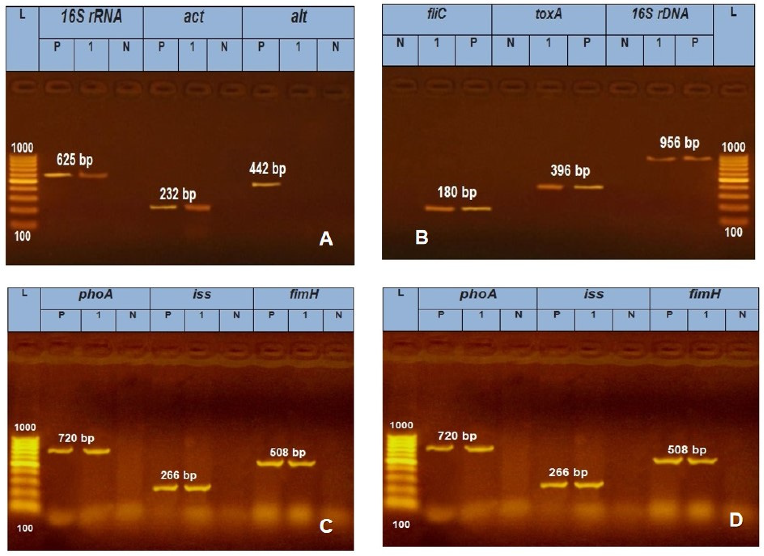
Figure 1. PCR amplification for identification and virulence related genes specific for A. hydrophila (A), P. aeruginosa (B), Salmonella (C) and E. coli (D) isolates. Lane (L): 100 bp Ladder ''Marker'', Lanes (1): examined samples, Lane Pos: Positive control, Lane Neg: Negative control.
Preprints 91365 g001
Figure 2. FTIR of the ZnO nanoparticles (a), Zeolite nanoparticles (b) and ZnO-Z nanocomposite.
Preprints 91365 g002
Figure 3. XRD of ZnO NPs(a), zeolite NPs(b), ZnO-Z nanocomposite (c).
Preprints 91365 g003
Figure 5. SEM of ZnO nanoparticle (a, b), Zeolite nanoparticles (c), ZnO-Z. nanocomposite (d).
Preprints 91365 g004
Figure 6. Histopathological investigation of ZnO NPs (A1, B1, & C1), Zeolite (A2, B2, & C2) and their nanocomposites (A3, B3, & C3). At different body organs (brain (A) Liver (B), Kidney (C)), all showed normal histological structure without appearance of specific pathological lesion.
Preprints 91365 g005
Table 1. Oligonucleotide sequence and virulence genes specific for A. hydrophila, P. aeruginosa, E. coli and Salmonella determined during the study.
Microbial agent Target gene Primers sequences Amplified segment (bp) Primary
denaturation
Amplification (35 cycles) Final extension Reference
Secondary denaturation Annealing Extension
A.. hydrophila
16S rRNA GAAAGGTTGATGCCTAATACGTA 625 94˚C
5 min.
94˚C
30 sec.
50˚C
40 sec.
72˚C
45 sec.
72˚C
10 min.
Gordon et al. [26]
CGTGCTGGCAACAAAGGACAG
Act AGAAGGTGACCACCACCAAGAACA 232 94˚C
5 min.
94˚C
30 sec.
55˚C
30 sec.
72˚C
30 sec.
72˚C
7 min.
Nawaz et al. [27]
AACTGACATCGGCCTTGAACTC
alt TGACCCAGTCCTGGCACGGC 442 94˚C
5 min.
94˚C
30 sec.
55˚C
40 sec.
72˚C
40 sec.
72˚C
10 min.
GGTGATCGATCACCACCAGC
P. aeruginosa 16S rDNA GGGGGATCTTCGGACCTCA 956 94˚C
5 min.
94˚C
30 sec.
52˚C
40 sec.
72˚C
45 sec.
72˚C
10 min.
Spilkeret al. [28]
TCCTTAGAGTGCCCACCCG
toxA GACAACGCCCTCAGCATCACCAGC 396 94˚C
5 min.
94˚C
30 sec.
55˚C
40 sec.
72˚C
40 sec.
72˚C
10 min.
Mataret al. [29]
CGCTGGCCCATTCGCTCCAGCGCT
fliC TGAACGTGGCTACCAAGAACG 180 94˚C
5 min.
94˚C
30 sec.
56.2˚C
30 sec.
72˚C
30 sec.
72˚C
7 min.
Ghadaksazet al. [30] 2015
TCTGCAGTTGCTTCACTTCGC
E. coli phoA CGATTCTGGAAATGGCAAAAG 720 94˚C
5 min.
94˚C
30 sec.
55˚C
40 sec.
72˚C
45 sec.
72˚C
10 min.
Hu et al. [31]
CGTGATCAGCGGTGACTATGAC
Iss ATGTTATTTTCTGCCGCTCTG 266 94˚C
5 min.
94˚C
30 sec.
54˚C
30 sec.
72˚C
30 sec.
72˚C
7 min.
Yaguchiet al. [32]
CTATTGTGAGCAATATACCC
fimH TGCAGAACGGATAAGCCGTGG 508 94˚C
5 min.
94˚C
30 sec.
50˚C
40 sec.
72˚C
45 sec.
72˚C
10 min.
Ghanbarpour and Salehi [33]
GCAGTCACCTGCCCTCCGGTA
S. tTphimurium STM4495 GGT GGC AAG GGA ATG AA 915 94˚C
5 min.
94˚C
30 sec.
50˚C
40 sec.
72˚C
50 sec.
72˚C
10 min.
Liu et al. [34]
CGC AGC GTA AAG CAA CT
Stn TTG TGT CGC TAT CAC TGG CAA CC 617 94˚C
5 min.
94˚C
30 sec.
59˚C
40 sec.
72˚C
45 sec.
72˚C
10 min.
Murugkaret al. [35]
ATT CGT AAC CCG CTC TCG TCC
sopB TCA GAA GRC GTC TAA CCA CTC 517 94˚C
5 min.
94˚C
30 sec.
58˚C
40 sec.
72˚C
45 sec.
72˚C
10 min.
Huehnet al. [36]
TAC CGT CCT CAT GCA CAC TC
Table 2. Prevalence of the pathogenic bacteria isolated from different water sources in the current study.
Water source Examined samples No. (%) of positive samples No. (%) of bacterial isolates
E. coli Salmonella Spp. P. aeruginosa A. hydrophila
Tap water
Main source 10 1(10.0) 0 (0.0) 0 (0.0) 1(100.0) 0 (0.0)
Drinkers 30 24 (80.0) 10 (41.7) 6 (25.0) 3 (12.5) 5 (20.8)
Hand pump
Main source 10 0 (0.0) 0 (0.0) 0 (0.0) 0 (0.0) 0 (0.0)
Drinkers 30 12 (40.0) 5 (41.6) 2 (16.7) 3 (25.0) 2 (16.7)
Surface water

Main source
10 10 (100.0) 4 (40.0) 4 (40.0) 1 (10.0) 1(10.0)
Drinkers 30 28 (93.3) 15 (53.0) 9 (32.1) 2 (7.1) 2 (7.1)
Total 120 75 (62.5) 34 (45.3) 21 (28.0) 10 (13.3) 10 (13.3)
X2= 56.250    P>0.000 X2=20,840        P>0.000
Table 3. Serological identification and percentage of E. coli and Salmonella spp. obtained from the examined water sources.
Bacteria isolated Serogroups No. (%)


E. coli
O157
O26
O144
Ountyped
16 (47.1)
4 (11.8)
8 (23.5)
6 (17.6)
Total 34 (45.3)


Salmonella spp.
S. Kentucky
S. typhimurium
S. infantis
S. ferruch
S. kottobus
10 (47.6)
5 (23.8)
4 (19.0)
1 (4.8)
1 (4.8)
Total 21 (28.0)
X2= 29.273, P >0.000
Table 4. Biocidal activity of the tested disinfectants at different concentration against the isolated bacterial traits.
Disinfectant tested Conc.mg/l Biocidal activity of tested disinfectant against bacterial isolates at different contact times
E. coli (20) Salmonella spp. (20) P.Aerogenosa (10) A. hydrophila(10)
30min 2h 24h 30min 2h 24h 30min 2h 24h 30min 2h 24h
S R S R S R S R S R S R S R S R S R S R S R S R
Iodine 0.5 0 100.0 0 100.0 15.0 85.0 5.0 95.0 15.0 85.0 30.0 70.0 5.0 95.0 20.0 80.0 35.0 65.0 0 100.0 10.0 90.0 25.0 75.0
1.0 5.0 95.0 10.0 90.0 25.0 75.0 5.0 95.0 20.0 80.0 25.0 75.0 5.0 95.0 25.0 75.0 50.0 50.0 0 100.0 15.0 85. 0 30.0 70.0
H2O2 3.0 5.0 95.0 25.0 75.0 30.0 70.0 10.0 90.0 20.0 80.0 25.0 75.0 5.0 95.0 20.0 80.0 40.0 60.0 0 100.0 0 10.0 20.0 80.0
5.0 15.0 85.0 45.0 55.0 55.0 45.0 20.0 80.0 30.0 70.0 30.0 70.0 10.0 90.0 35.0 65.0 40.0 60.0 5.0 95.0 15.0 85.0 30.0 70.0
Terminator 0.3 10.0 90.0 25.0 75.0 45.0 55.0 0 100.0 5.0 95.0 20.0 80.0 15.0 85.0 25.0 75.0 45.0 55.5 5.0 95.0 20.0 80.0 50.0 50.0
0.5 15.0 85.0 40.0 60.0 50.0 50.0 15.0 85.0 25.0 75.0 25.0 75.0 20.0 80.0 30.0 70.0 60.0 40.0 10.0 90.0 25.0 75.0 60.0 40.0
P.value X2=522.7, P=0.000 X2=734.7, P=0.000 X2=382.7, P=0.000 X2=669.5, P=0.000
Table 5. Biocidal activity of the tested Nano materials (Zeolite NPs, ZnO NPs and ZnO-Z NPs) at different contact times against the isolated bacterial traits.
Bacteria spp. Zeolite nanoparticles Zinc oxide nanoparticles Zinc oxide-zeolite nanocomposites
30min 2hr 24hr 30 2hr 24hr 30min 2hr 24hr
S
%
I
%
R
%
S
%
I
%
R
%
S
%
I
%
R
%
S
%
I
%
R
%
S
%
I
%
R
%
S
%
I
%
R
%
S
%
I
%
R
%
S
%
I
%
R
%
S
%
I
%
R
%
E. coli 0.0 2.0 98.0 3.0 5.0 92.0 8.0 9.0 83.0 19.0 10.0 71.0 36.0 11.0 53.0 40.0 12.0 48.0 42.0 11.0 47.0 50.0 15.0 35.0 73.0 15.0 12.0
Salmonellaspp. 1.0 2.0 97.0 2.0 4.0 94.0 2.0 7.0 91.0 36.0 19.0 45.0 41.0 12.0 39.0 43.0 13.0 40.0 41.0 15.0 36.0 52.0 20.0 28.0 76.0 15.0 9.0
p.aeroginosa 2.0 3.0 95.0 5.0 2.0 93.0 5.0 5.0 90.0 39.0 12.0 49.0 29.0 15.0 56.0 42.0 19.0 39.0 57.0 12.0 31.0 43.0 27.0 30.0 69.0 18.0 13.0
A.hydrophila 2.0 5.0 93.0 5.0 4.0 91.0 8.0 3.0 88.0 31.0 19.0 50.0 31.0 14.0 55.0 48.0 17.0 35.0 52.0 10.0 38.0 45.0 29.0 26.0 63.0 22.0 15.0
P-value X2=864.004, P=0.000 X2=214.914, P=0.000 X2=281.951, P=0.000
Table 6. Different doses (mg/kg b.wt.), total number of animals (9), and dead animal's number in different treatments (ZnO NPs, Zeolite ZnO NPs and their combinations.
Dose ZnO-Z nanocomposite Z NP ZnO NP No. of animals
(mg/kgb.wt.) No. of dead animals/ group No. of animals
/ group
No. of dead animals/ group / group
200 0 0 0 9
400 0 0 0 9
600 0 0 0 9
800 0 0 0 9
1000 0 0 0 9
1500 1 0 0 9
2000 2 2 0 9
3000 5 3 2 9
4000 8 6 5 9
5000 10 8 8 9
10000 10 10 10 9
Table 7. LD50 and LD90 estimation of Zeolites, ZnO NPs and their nanocomposites.
Treatment LD50
(%)
(LC50)
95% CL LD90
or
LC50
95% CL X2
(df = 8)
P*
LCL UCL LCL UCL
Zeolite 3251 2675 4054 8476 5929 21737 1.63 0.99
ZnO NPs 3709 3089 4514 7620 5667 21379 0.53 0.05
Nanocomposite 2658 2187 3386 6636 4627 1791 0.39 0.99
LCL: lower confidential limit, UCL: upper confidential limit, X2: Chi-square,df: degree of freedom, LC50 and LC90 were lethal concentration at which 50% and 90% population dies respectively. * p> 0.05 is non-significant.
Table 8. Comparative presentation of LD0, LD20, LD50, LD90 and LD100values obtained by probit analysis of zeolite, ZnO NPs and their nanocomposites.
Parameters Results
Zeolite (mg/kg)
LD0 1247
LD20 1396
LD50 3251
LD90 5508
LD100 8467
ZnO NPs (mg/kg)
LD0 1805
LD20 1964
LD50 3709
LD90 5515
LD100 7620
Nanocomposite (mg/kg)
LD0 1046
LD20 1185
LD50 2658
LD90 4400
LD100 6636
Disclaimer/Publisher’s Note: The statements, opinions and data contained in all publications are solely those of the individual author(s) and contributor(s) and not of MDPI and/or the editor(s). MDPI and/or the editor(s) disclaim responsibility for any injury to people or property resulting from any ideas, methods, instructions or products referred to in the content.
Copyright: This open access article is published under a Creative Commons CC BY 4.0 license, which permit the free download, distribution, and reuse, provided that the author and preprint are cited in any reuse.
Prerpints.org logo

Preprints.org is a free preprint server supported by MDPI in Basel, Switzerland.

Subscribe

© 2024 MDPI (Basel, Switzerland) unless otherwise stated