Preprint
Article

Protective Effects of Dietary Vitamin D3, Turmeric Powder and Their Combination Against Gasoline Intoxication in Rats

Altmetrics

Downloads

138

Views

29

Comments

0

A peer-reviewed article of this preprint also exists.

Submitted:

24 November 2023

Posted:

27 November 2023

You are already at the latest version

Alerts
Abstract
Inhalation of gasoline vapors (GV) is associated with developing various pathologies. Particularly, oil refinery and gas station workers are at greater risk of developing lung cancer, kidney cancer, bladder cancer, and hematological disorders, including acute myeloid leukemia. Therefore, preventing the harmful effects of GV and alleviating their consequences appear to be an important and timely issue. In this study, we investigated the potential of vitamin D3, turmeric powder and their combination to ameliorate the toxicity of gasoline fumes in rats. Separate groups of animals fed with a standard rodent diet, with or without supplementation of vitamin D3 (750 IU/kg of body weight) and/or turmeric powder (0.5%, w/w, in food), were untreated or treated with GV (11.5±1.3 cm3/h/m3/day) for 30, 60 or 90 days. Changes in the body weight were monitored weekly. Histological, biochemical, and hematological parameters were determined at the end of each treatment period. While exposure of rats to GV resulted in a time-dependent reduction in body weight, supplementation with vitamin D3, but not with turmeric root powder or their combination, partially prevented weight loss. Macroscopical and histological analyses showed pronounced time-dependent changes in the organs and tissues of GV-treated rats. These included alveolar wall collapse in the lungs, destruction of the lobular structure and hepato-cytolysis in the liver, shrinkage and fragmentation of glomeruli in the kidneys, and disorganization of the lymphoid follicles in the spleen. However, co-treatment with the nutritional supplements tested, especially vitamin D3, noticeably alleviated the above conditions. This was accompanied by a significant improvement in blood chemistry and hematological parameters. Collectively, our results demonstrate that the harmful effects of environmental exposure to GV can be reduced upon supplementation of vitamin D3. The fact that the protective activity of vitamin D3 alone was higher than that of turmeric root powder or the combined treatment suggests that combinations of these supplements may not always be more beneficial than each agent applied separately.
Keywords: 
Subject: Biology and Life Sciences  -   Toxicology

1. Introduction

Gasoline is the most consumed product of crude oil refining, which contains at least 150 hydrocarbons, including alkanes (60-70%), aromatics (25-30%) and alkenes (6-9%). Gasoline vapors (GV) have been linked to various pathologies, such as lung disorders, hematotoxicity, and encephalopathies [1,2,3,4]. The results of many studies have also shown that employees of gas stations and refineries who are chronically exposed to GV are at high risk of developing lung, kidney, and bladder cancers [5,6,7], as well as hematological malignancies [8].
To date, several studies have examined the protective actions of some natural agents in preventing or mitigating the harmful effects of GV in rat models in vivo. For instance, supplementing a standard rodent diet with fenugreek seed powder was shown to alleviate pathological changes in the liver and lung biochemical and histological parameters and suppress oxidative stress and inflammation in GV-exposed rats [9,10]. In a similar study, consuming green tea extract as drinking water or dietary curcumin, a major active component of turmeric (Curcuma Longa root powder), reduced DNA fragmentation in the spleen and liver of mice subjected to GV inhalation [11]. Uboh et al. [12,13] have demonstrated that in the rat model of GV toxicity, the hepatoprotective activity of vitamin E was higher than that of vitamin A or C.
Vitamin D is a multifunctional nutrient produced in the skin through exposure to sunlight and can also be obtained from food. It is becoming increasingly clear that active vitamin D metabolites play a critical role in human health [14]. Vitamin D has been extensively studied for its beneficial effects on various pathologies, including cardiovascular diseases [15], diabetes [16] and infectious diseases [17]. In addition, a number of animal studies have shown that vitamin D protects against the harmful influence of different environmental factors, toxic compounds, and drugs, e.g., lead [18,19], cadmium [20], carbon tetrachloride [21], and paracetamol [22]. However, to the best of our knowledge, the protective activity of vitamin D against the toxic effects of GV has not yet been reported.
In the present study, we investigated whether supplementation with vitamin D3 (750 IU/kg of body weight), turmeric powder (0.5%, w/w, in food) or their combination could alleviate pathological changes in rats exposed to GV for 30-90 days. Our results demonstrated that consumption of vitamin D3 significantly improved the macroscopical organ appearance, as well as histological, biochemical, and hematological parameters in GV-exposed rats, whereas the protective effects of turmeric or its combination with vitamin D3 were found to be less pronounced. These findings suggest that the harmful effects of environmental exposure to GV can be reduced upon supplementing the diet with vitamin D3.

2. Results

2.1. Changes in the Body Weight of Rats Supplemented with Vitamin D3, Turmeric Powder or Their Combination, with and without Exposure to Gasoline Vapors
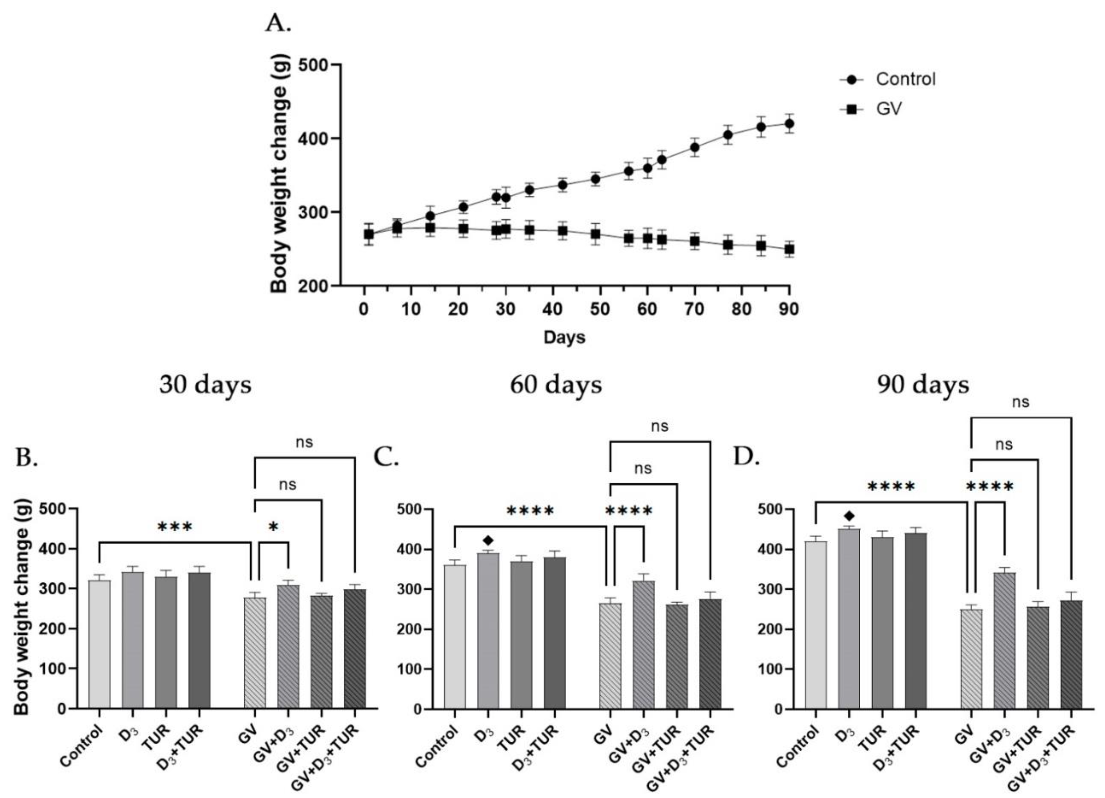
As demonstrated in Figure 1A–D, exposure of rats to GV resulted in a time-dependent reduction in body weight. In particular, 15.2%, 31.0%, and 41.4% reduction was observed following 30, 60 and 90 days of exposure, respectively, as compared with the control group (Figure 1B–D). GV-treated rats supplemented with vitamin D3 alone, but not with turmeric powder or their combination, exhibited a significantly less pronounced weight loss than the GV alone group. Interestingly, vitamin D3 treatment without GV exposure also resulted in a small but significant increase in body weight compared to the control (Figure 1B–D). These data indicate that, among the supplements used, only vitamin D3 had a beneficial effect against GV-induced weight loss.
Figure 1. Changes in the body weight of control and GV-treated rats following supplementation with vitamin D3, turmeric powder or their combination. Rats from all groups (15 rats/group) were weighted weekly starting from Day 0. (A) Comparison of the body weight gain between control and GV alone-treated group. (B–D) Five rats from the indicated groups were weighed on days 30, 60, and 90. Data are mean±SD. One-way ANOVA followed by Tukey's post hoc multiple comparisons test. *, p <0.05; ***, p <0.001; ****, p<0.0001 significant differences between the indicated groups; ♦, p <0.05 vs untreated control group; ns – not significant.
Preprints 91439 g001

2.2. Macroscopical and Histological Features of the Lungs, Liver, Kidneys, and Spleen of Rats Supplemented with Vitamin D3, Turmeric Powder, or Their Combination, with and without Exposure to Gasoline Vapors

Organs were excised from all groups of rats (5 rats/group) following 30, 60, and 90 days of treatments, photographed, and fixed in neutral buffered formalin for further histological examination. Hematoxylin and eosin (H&E)-stained tissue sections were analyzed under a light microscope. The results of macroscopical and histological analyses are exemplified in Figure 2 and Figure 3, respectively (data obtained on days 30 and 90 are not shown).

2.2.1. Macroscopical Analysis

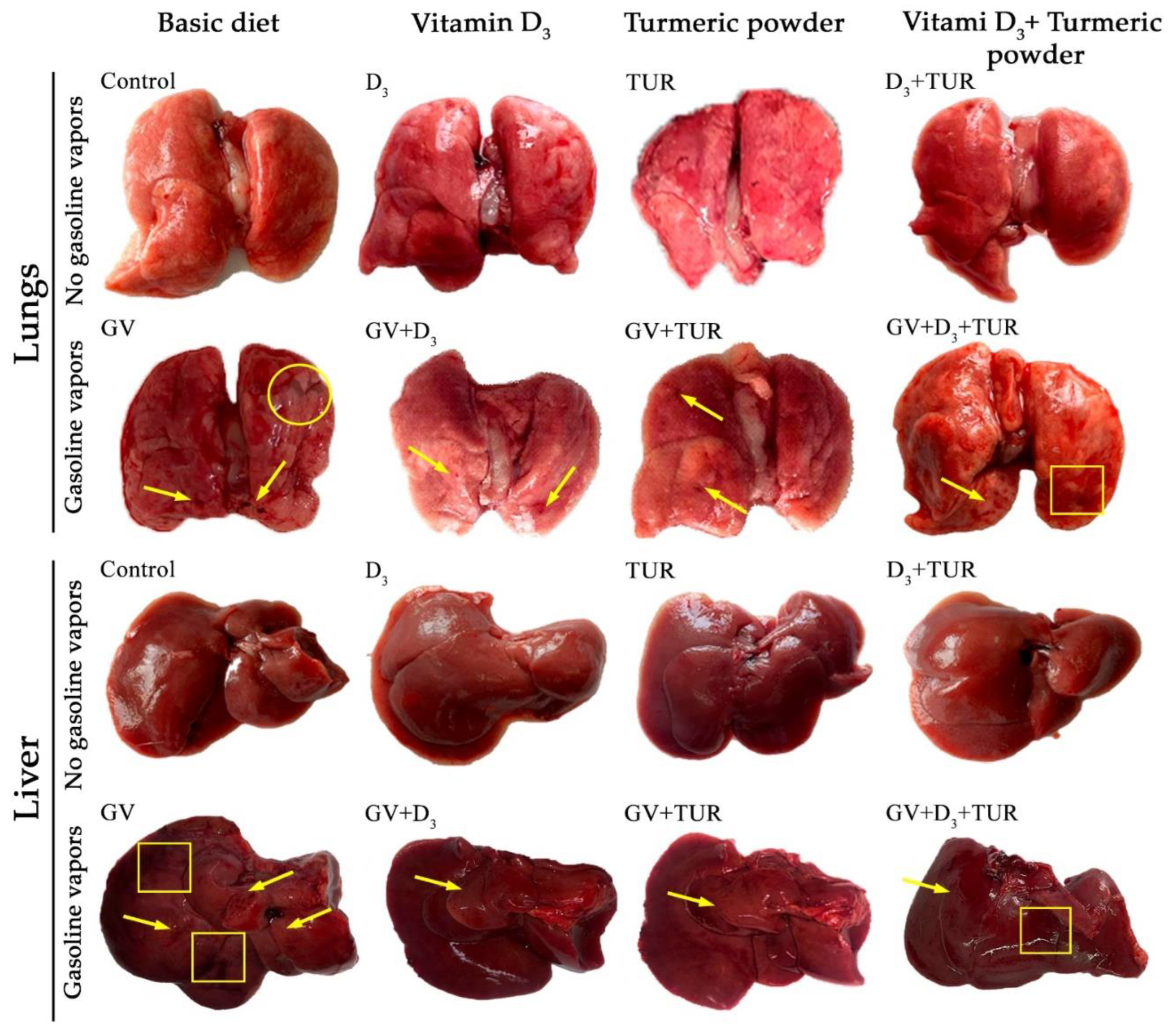
In rats exposed to GV alone, the lungs were swollen, with uneven surfaces and a brownish tint (Figure 2, lungs). Multiple hemorrhages appeared in both lungs (arrows), and most animals had pleural exudates (circles). Administration of vitamin D3, turmeric powder or their combination largely protected the lungs from the edema, though small sporadic hemorrhages (arrows) could still be observed. In contrast to the rats supplemented with vitamin D3 alone, the lungs of the animals receiving turmeric powder or its combination with vitamin D3 displayed some surface unevenness and darkened areas (squares). The areas of discoloration were more frequent in the lungs of the combination-treated animals compared to the turmeric powder group.
Similar to the changes observed in the lungs, the liver in GV-treated rats was swollen and had a rough dark-brown surface without a natural luster (Figure 2, liver). Multiple hemorrhages could primarily be seen in the left and right lateral lobes and the caudate lobe (arrows). Areas of unusually darkened tissue were noticed mainly in the right and medial lobes (squares). Rats receiving dietary supplements displayed less dramatic overall changes in the liver compared to the GV-only group. Particularly, organ edema was less pronounced, and only small petechial hemorrhages were noticed, primarily in the caudal lobe (arrows). The livers from the combination-treated group still displayed some darkened surface regions (squares).
Macroscopical examination of the kidneys showed that treatment with GV resulted in significant kidney swelling, with a less noticeable bean-like shape compared to the untreated control group. In most rats of this group, the left kidney was more enlarged than the right one (Supplementary Figure S1, kidneys). Both kidneys had an indurated texture and a rough, dark-brown surface. Supplementation with vitamin D3 alone markedly reduced swelling and tissue density and normalized the surface color of the kidneys. The improvement in the groups receiving turmeric powder or its combination with vitamin D3 was somewhat less evident compared to vitamin D3 alone-treated rats. Exposure to GV led to enlargement, swelling, loss of the ribbon-like shape, and darkening of the spleens. However, dietary supplements, particularly vitamin D3 alone, partially protected from this harmful impact of GV (Supplementary Figure S1, spleen).
In the absence of GV, supplementation with vitamin D3, turmeric powder or their combination did not significantly affect the macroscopical features of the organs evaluated in this study (Figure 2 and Supplementary Figure S1). Of note, in both GV-treated and –untreated animals supplemented with turmeric root powder or its combination with vitamin D3, all the organs, except the spleen, had a yellowish tint (presumably due to curcuminoid-enriched diet).
Figure 2. Changes in the macroscopical appearance of the lungs and the liver of control and GV-treated rats supplemented with vitamin D3, turmeric powder or their combination. Following the indicated treatments, the organs were excised on day 60 and photographed. Representative images of the organs from one out of five rats in each group are shown. Arrows - tissue hemorrhages; squares – tissue discoloration; circles - pleural exudate.
Preprints 91439 g002

2.2.2. Histological Analysis

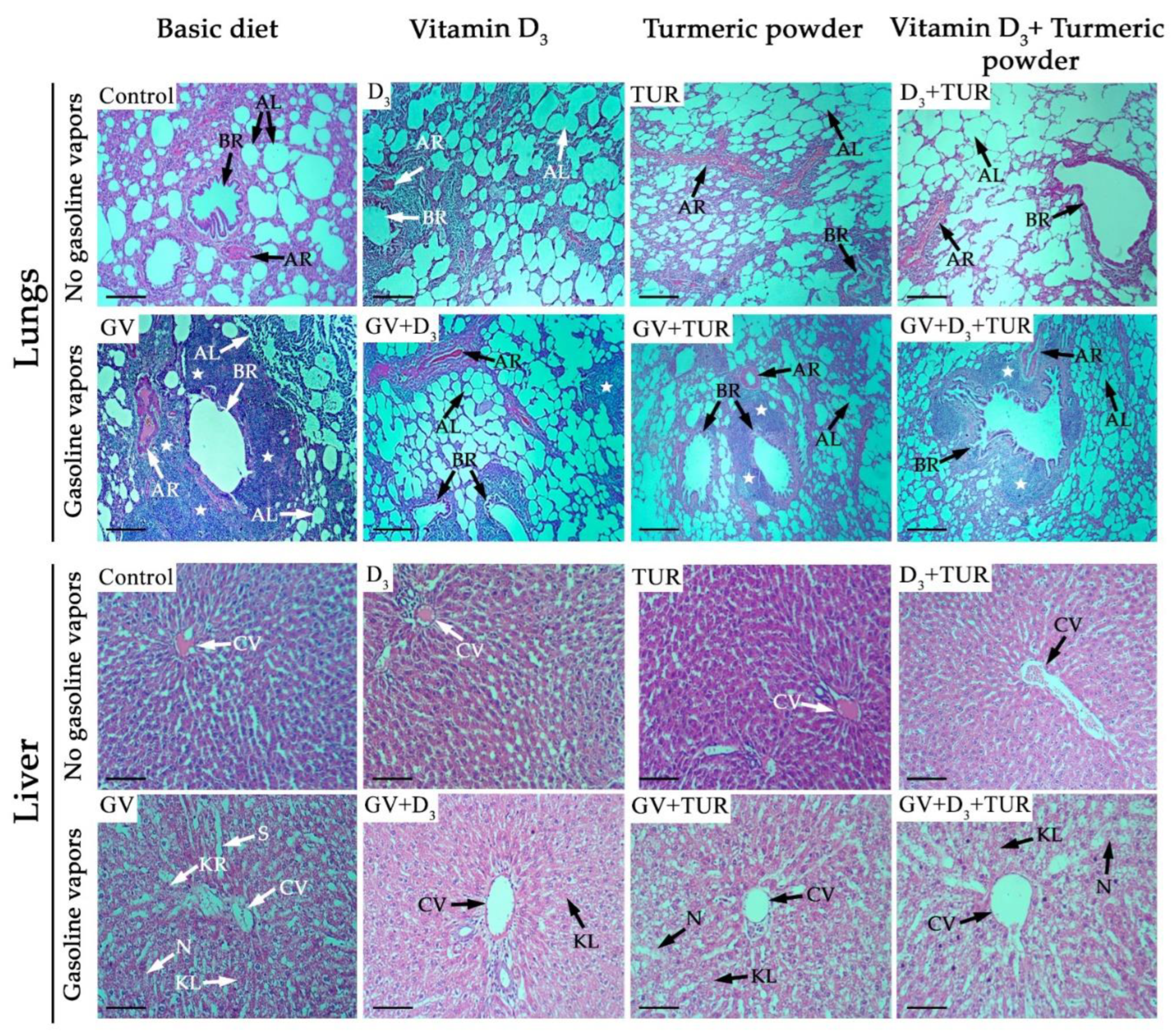
To investigate the influence of GV and dietary supplements on the lungs, liver, kidneys, and spleen in more detail, we performed a histological analysis of hematoxylin and eosin (H&E)-stained tissue sections of the above organs (Figure 3 and Supplementary Figure S2). The following changes were detected in the lung tissue of GV-exposed rats compared to the control group (Figure 3, lungs). A large part of the alveoli appeared to be collapsed (arrow AL). The inner wall of some of the bronchioles was found to be detached (arrow BR), and arteriole walls were thickened, resulting in the narrowing of the lumen (arrow AR). In addition, there was massive cellular, likely leukocytic, infiltration in peribronchial and perivascular areas (stars), which was associated with the loss of alveolar architecture. Supplementation with vitamin D3, turmeric powder or their combination partially improved the structure of the lung tissue to a varying extent compared to the GV alone-treated group. Specifically, the rats receiving vitamin D3 alone displayed healthier alveoli and a better-preserved wall structure of the bronchioles compared to the groups supplemented with turmeric powder or its combination with vitamin D3 (Figure 3, lungs). Furthermore, there were smaller areas of leukocytic infiltration in the lungs of vitamin D3-supplemented rats compared to the other groups.
Liver tissue sections from the rats exposed to GV (Figure 3, liver) demonstrated profound structural changes, such as disorganized regular radiating rows of hepatocytes, the appearance of necrotic areas (arrow N), karyolysis (arrow KL), and karyorrhexis (arrow KR). In addition, we observed pronounced damage to the central vein walls (arrow CV) and sinusoidal dilatation (arrow S). However, administration of vitamin D3 largely alleviated the above pathological changes. The addition of turmeric powder and, mainly, its combination with vitamin D3 was less effective in ameliorating GV toxicity compared to the vitamin D3 alone group (Figure 3, liver).
Figure 3. Histological changes in the lungs and the liver of control and GV-treated rats supplemented with vitamin D3, turmeric powder or their combination. H&E stained tissue sections were prepared from the rats subjected to the indicated treatments for 60 days. Representative images of the sections from one out of five rats in each group are shown. AL - alveoli; BR - bronchioles; AR – arterioles; CV – central vein; N - necrosis; KR - karyorrhexis; KL - karyolysis; S – sinusoids; stars – cellular infiltrates. Magnification ×100. Scale bars, 50 µm.
Preprints 91439 g003
Unlike the kidney tissue sections of control rats (Supplementary Figure S2, kidneys), the ones from GV-exposed rats showed shrinkage and fragmentation of glomeruli (arrow G) with a dramatic reduction in the Bowman's capsule space (arrow BC). Furthermore, the renal tubules were much narrower than those of the control group (arrow RT), and interstitial hemorrhages could be seen (arrow H). Remarkably, supplementation with vitamin D3 led to a marked protection of the tissue structure that appeared almost normal, except for occasional small hemorrhages. A certain amelioration of GV-induced changes was also observed in rats receiving turmeric powder or its combination with vitamin D3, but the tissue structure was less preserved compared to the vitamin D3 alone group (Supplementary Figure S2, kidneys).
The analysis of the spleen sections (Supplementary Figure S2, spleen) showed that treatment with GV resulted in an extensive disruption of the lymphoid follicle (LF) structure with poorly distinguishable mantle region (arrow M) and marginal zone (MZ). The lymphoid follicles were barely distinct from the red pulp (RP) and periarterial lymphocytic sheath (P). Administration of vitamin D3 or turmeric powder led to a better-preserved structure of the lymphoid follicles with more distinct mantle regions, while in the combination-treated rats, the protection of the spleen tissue was less noticeable (Supplementary Figure S2, spleen).
In summary, exposure to GV resulted in time- and organ-dependent pathological changes at the histological level that were ameliorated to varying degrees upon supplementation with vitamin D3, turmeric powder or their combination. No significant differences in tissue structure were observed between the control rats and those administered with the tested supplements in the absence of GV.

2.3. Blood Chemistry Analysis of Rats Supplemented with Vitamin D3, Turmeric Powder or Their Combination, with and without Exposure to Gasoline Vapors
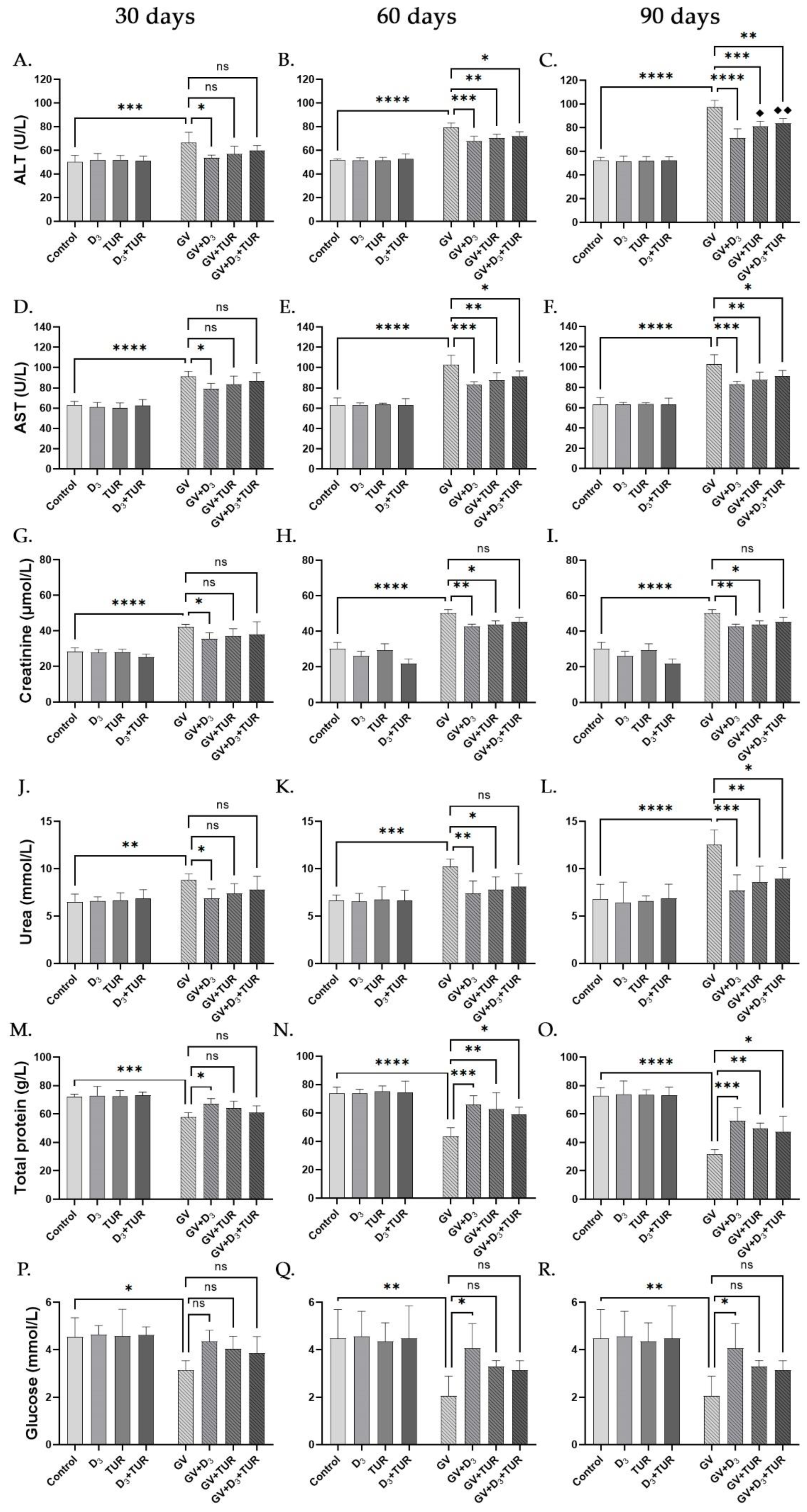
To further explore the influence of GV and dietary supplements on the liver and kidneys, we determined the functional status of these organs by measuring serum concentrations of corresponding biomarkers. Increased serum levels of alanine aminotransferase (ALT) and aspartate aminotransferase (AST) indicate liver damage, while increased creatinine and urea concentrations point to deterioration of the kidney function. Decreases in serum total protein and glucose levels may result from malnutrition and impaired liver and kidney functions.
The exposure of rats to GV for 30, 60, and 90 days resulted in a marked and time-dependent elevation of the serum levels of the liver markers, ALT (by 32.6%, 53.1%, 86.7%, respectively; Figure 4A–C) and AST (45.1%, 63.1%, 85.2%, respectively; Figure 4D–F), as compared with the corresponding control group. However, administration of vitamin D3 alone led to a significant decline in serum levels of both ALT (by 19.3%, 14.6%, 26.9%, respectively; Figure 4A–C) and AST (by 13.4%, 19.6%, 17.2% respectively; Figure 4D–F), compared to the corresponding GV group. Supplementation with turmeric powder or its combination with vitamin D3 tended to be less effective than vitamin D3 alone (e.g., Figure 4C,F).
Time-dependent increases in creatinine and urea serum levels also characterized GV-exposed rats. Specifically, following 30, 60, and 90 days of treatment, creatinine and urea levels were elevated by 49.3%, 66.3%, 102.9% and by 35.6%, 54.3%, 84.2%, respectively, relative to the corresponding control group (Figure 4G–L). Co-administration of the supplements and their combination significantly improved the levels of both protein metabolites, the combined treatment tending to be the least effective. Treatment with vitamin D3 resulted in a minor (by 13-16%) but significant reduction in creatinine levels at all the time points (Figure 4G–I), while the decreases in urea levels in this group were time-dependent and more pronounced (by 22.3%, 27.8%, 38.7% vs GV group, respectively; Figure 4J–L).
Exposure to GV for 30, 60, and 90 days led to a substantial reduction in the serum concentrations of the total protein (by 19.2%, 40.8%, and 56.5%, respectively) and glucose (by 31.0%, 54.1%, and 69.2%, respectively) relative to the corresponding controls. However, supplementation with vitamin D3 alone significantly reversed these harmful effects by elevating both the total protein levels (by 16.0%, 50.7%, 74.1%, respectively; Figure 4M–O) and glucose levels (by 39.0%, 98.0%, 140.2%; respectively; Figure 4P–R) compared to the corresponding GV group. The diets fortified with turmeric powder or its combination with vitamin D3 had similar, though less efficient, protection (Figure 4M–R). Of note, vitamin D3 supplementation was capable of essentially restoring serum urea and glucose concentrations in GV-treated rats approximately up to the untreated control levels throughout the entire experimental period (Figure 4J–L and P–R).
Figure 4. Biochemical changes in the blood of control and GV-treated rats supplemented with vitamin D3, turmeric powder or their combination. Peripheral blood samples from five rats of the indicated groups collected on days 30, 60, and 90 were analyzed. Data are mean±SD. One-way ANOVA followed by Tukey's post hoc multiple comparisons test. *, p <0.05; **, p <0.01; ***, p <0.001; ****, p<0.0001 significant differences between the indicated groups; ♦, p <0.05; ♦♦, p <0.01; vs GV+D3 group; ns – not significant.
Preprints 91439 g004

2.4. Hematological Analysis of Rats Supplemented with Vitamin D3, Turmeric Powder or Their Combination, with and without Exposure to Gasoline Vapors
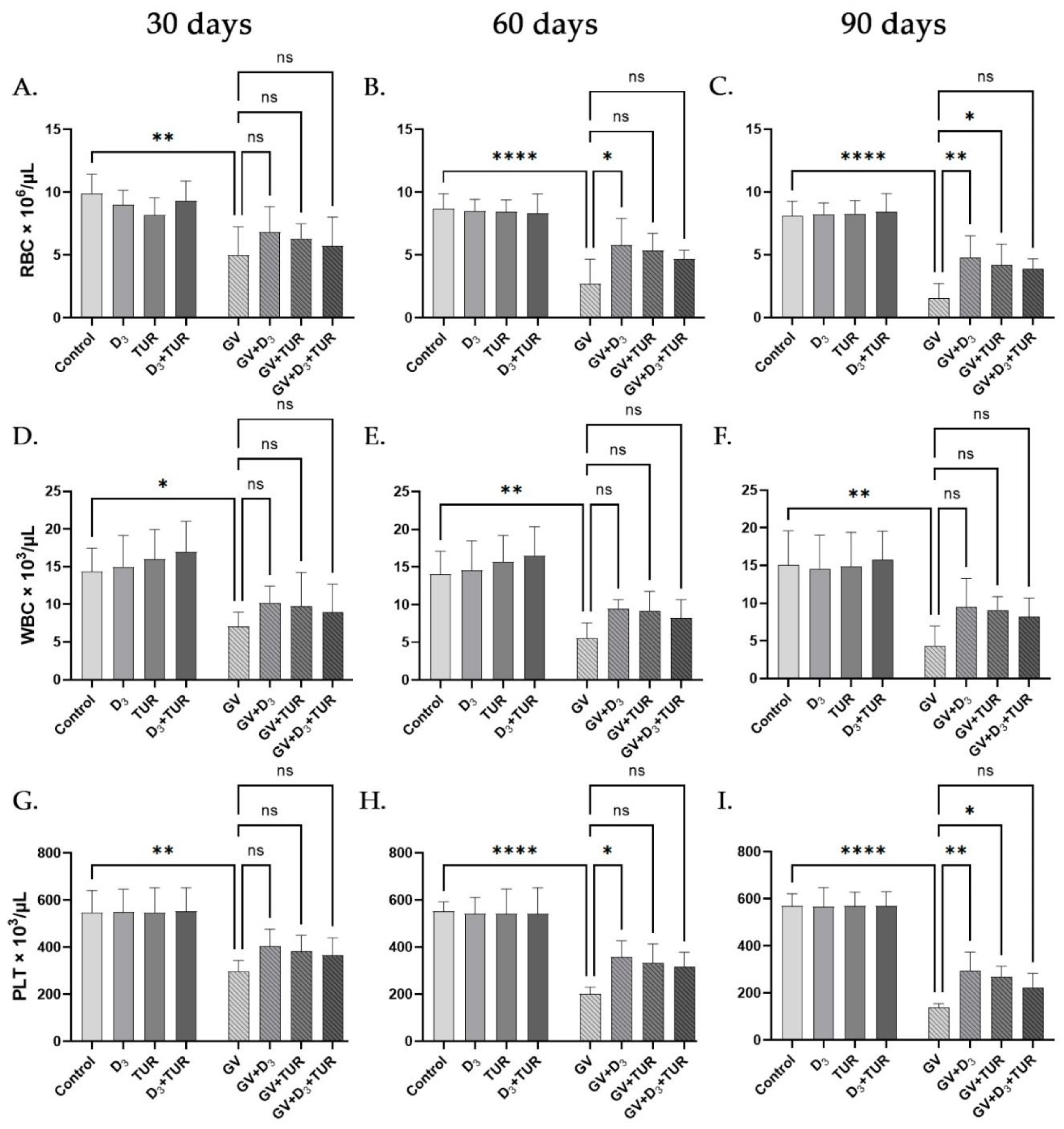
A complete blood count (CBC) was performed in the peripheral blood samples to examine the influence of GV inhalation and dietary supplements on the hematological parameters of experimental rats. GV exposure for 30, 60, and 90 days resulted in a highly significant and time-dependent decrease in the red blood cells (RBC) by 49.8%, 68.6%, and 81.2%, respectively, compared to the control group (Figure 5A–C). Similar data were obtained by measuring hematocrit (HCT) and hemoglobin (HGB) levels (Supplementary Figures S3A–C and S3D–F). Importantly, all the above parameters were partially recovered as a result of vitamin D3 supplementation in a time-dependent manner, as follows. The levels of RBC were elevated by 36.9%, 111.7%, 214.5% (Figure 5A–C); HCT by 29.0%, 136.8%, 207.2% (Supplementary Figure S3A–C), and HGB by 39.7%, 147.6%, 243.2% (Supplementary Figure S3D–F) after 30, 60 and 90 days, respectively, compared to the corresponding GV group.
GV inhalation also caused significant and time-dependent decreases in white blood cell (WBC) counts, including granulocyte (GRA) and lymphocyte (LYM) percentages and platelet counts (Figure 5D–F and Figure 5G–I; Supplementary Figure S3G–L). For instance, after 30, 60, and 90 days, WBC counts were reduced by 50.7%, 60.5%, and 71.1%, respectively (Figure 5D–F), and platelet counts by 45.6%, 63.5%, and 75.7%, respectively (Figure 5G–I), compared to the control group. Similar to the protective action of vitamin D3 on the red blood cell parameters, we observed a significant increase in WBC counts (by 43.8%, 71.1%, and 119%; Figure 5D–F) and platelet counts (by 36.0%, 77.2%, and 114.5%; Figure 5G–I), respectively, in vitamin D3-treated groups compared to the corresponding GV groups. Comparable reversal of GV-induced granulocytopenia (Supplementary Figure S3G–I) and lymphopenia (Supplementary Figure S3J–L) by vitamin D3 supplementation was also detected. Dietary administration of turmeric powder or its combination with vitamin D3 had similar but less noticeable effects on the above hematological parameters tested in GV-exposed rats. Neither supplementation type significantly influenced GV-untreated rats (Figure 5 and Supplementary Figure S3).
Figure 5. Changes in complete blood counts of control and GV-treated rats supplemented with vitamin D3, turmeric powder or their combination. Peripheral blood samples from five rats of the indicated groups collected on days 30, 60, and 90 were analyzed. Data are mean±SD. One-way ANOVA followed by Tukey's post hoc multiple comparisons test. *, p <0.05; **, p <0.01; ***, p <0.001; ****, p<0.0001 significant differences between the indicated groups; ns – not significant.
Preprints 91439 g005

3. Discussion

In this study, we investigated the potential of oral supplementation with vitamin D3, turmeric powder and their combination to reduce chronic GV toxicity in rats. Our findings demonstrated for the first time that administration of vitamin D3 at 750 IU/kg daily dose had a marked protective activity against the harmful effects of GV, as monitored by macroscopical, histological, biochemical, and hematological analyses for the period up to 90 days. Similar but less pronounced protection was observed following supplementation with turmeric powder at 0.5%, w/w, in food. Despite our expectation of positive cooperation between vitamin D3 and turmeric powder in alleviating GV toxicity, the combined treatment appeared to be as effective as turmeric powder alone or even less so.
Exposure to GV poses a severe health hazard to humans and animals, causing various pathologies, including lung, kidney, and bladder cancers [5,6,7], as well as hematological malignancies [8]. Therefore, it is essential to develop protective and preventive measures against the toxic effects of GV. Still, little attention has been paid to this issue, and so far, only few publications have described the protective activity of some natural agents. Abdrabouh [9,10] has recently reported that rats exposed to gasoline fumes for 6 h daily, 6 days a week for 10 weeks, displayed marked pathological changes in the liver [9] and the lungs [10]. This was manifested by elevated levels of liver enzymes in the blood [9] and oxidative stress and inflammatory markers in tissue homogenates [9,10]. These changes were alleviated by adding fenugreek seed powder (5%, w/w) to a standard diet [9,10]. In a similar study using a mouse model, Elsayed [11] has demonstrated that consumption of green tea extract (1.5% in drinking water) or purified curcumin (3%, w/w, in food) protected the liver and spleen tissues from DNA fragmentation in animals exposed to GV for 2 h/day for 3 weeks.
Although, to our knowledge, the protective activity of whole turmeric root powder against GV toxicity has not been reported, several studies have shown its ability to alleviate the harmful effects of other environmental agents and drugs. In particular, the inclusion of turmeric root powder (200 mg/kg body weight) in drinking water [23] or by intragastric gavage [24] was found to protect rats from hepatotoxicity induced by cadmium or carbon tetrachloride, respectively. Likewise, dietary supplementation of turmeric root powder at 2% or 4% (w/w) reduced renal damage caused by gentamycin. This was associated with decreased plasma levels of renal function markers and improved antioxidant status in kidney homogenates [25]. In another study, adding turmeric powder to food at 1%, 2% or 5% (w/w) attenuated oxidative stress in the gastric, liver, kidney, and heart tissues of rats treated with an ulcerogenic dose of indomethacin [26].
It has been shown that oral vitamin E (200 IU/kg or 400 IU/kg) [12,13], A (400 IU/kg) [12] or C (200 mg/kg) [13] significantly lowered serum levels of liver enzymes and ameliorated liver damage in rats subjected to GV inhalation for 6 h/day, 5 days a week during ten [13] or twenty [12] weeks. However, surprisingly, the beneficial activity of vitamin D against GV toxicity has not been reported so far despite its well-documented ability to protect from other toxicants, such as heavy metals and poisonous chemicals [27]. For instance, intramuscular injections of vitamin D3 (1000 IU/kg, 3 days a week) alleviated the damage to the liver [18], kidneys and testicles [19] in rats consuming lead in the drinking water. This was accompanied by lowering the levels of oxidative stress and pro-inflammatory markers and increasing the expression of antioxidant and anti-inflammatory markers and vitamin D- and Ca2+-related regulatory molecules in the damaged tissues [18,19]. In a similar study, intramuscular injections of vitamin D3 (600 IU/kg, 3 times a week) and/or oral supplementation of calcium (100 mg/kg, 5 times a week) protected rats from cadmium hepatotoxicity. Notably, the two agents positively cooperated when applied together [20]. It has also been reported that intraperitoneal administration of vitamin D3 (20 IU/kg, daily) had a protective effect against carbon tetrachloride-induced nephrotoxicity in rats. This was manifested by restoration of serum levels of renal markers (urea and creatinine) and recovery of histopathological lesions in the kidneys [21]. El-Boshy et al. [22] have investigated prophylactic and therapeutic activities of intraperitoneally administered vitamin D3 against paracetamol-induced hepatorenal damage. They reported that two rounds of vitamin D3 injections at 1000 IU/kg/day (5 days/week) before and another round after paracetamol poisoning showed better protective effects compared to a single round of vitamin D3 at a higher dose (3000 IU/kg/day; 5 days) just post-paracetamol intoxication.
In the present study, we demonstrated for the first time that oral treatment with vitamin D3 at a moderate dose of 750 IU/kg (6 days/week) for 30-90 days alleviated GV-induced toxicity in rats. This was associated with an improvement in the general condition of the animals manifested by a better appearance (data not shown) and less pronounced reduction in the body weight, which appeared to stabilize over the time course of supplementation relative to the continuing weight loss in GV-treated animals (Figure 1). Vitamin D3 treatment resulted in at least a partial preservation of intact organ appearance (Figure 2 and Supplementary Figure S1) and tissue structure (Figure 3 and Supplementary Figure S2) of the lungs, liver, kidneys and spleen. Furthermore, there was a significant improvement in serum levels of liver and kidney functional biomarkers and glucose (Figure 4), as well as a partial restoration of hematological parameters, such as red blood cell, white blood cell and platelet counts (Figure 5 and Supplementary Figure S3). A similar but less effective protection was observed following dietary supplementation with turmeric powder at a relatively lower dose (0.5%, w/w; 6 days/week) compared to those used in the above-referenced studies (e.g., [25,26]). These protective effects of vitamin D3 and turmeric are consistent with their known antioxidant, anti-inflammatory and immunomodulatory activities [28,29,30,31,32].
Although combined effects of vitamin D3 and turmeric/curcumin on GV toxicity have not been previously determined, several studies have reported a positive cooperative activity of these compounds in animal models of other pathologies. In particular, oral administration of a nanoencapsulated combination of vitamin D3 (16 IU/day) and curcumin (4 mg/kg) was found to be an effective anti-inflammatory adjuvant treatment of rheumatoid arthritis in rats [33]. In a similar model in mice, a diet enriched with vitamin D3 (10,000 IU/kg of food) and omega-3-fatty acids (10 g/kg of food) combined with oral supplementation of a highly bioavailable form of curcumin (100 mg/kg) markedly reduced the severity of collagen-induced arthritis, delayed the onset and slowed the progression of the disease [34]. In another study, oral administration of the formulation containing 33.26% total curcuminoids, 3.47% lutein, 0.7% zeaxanthin, and 930 IU vitamin D3 (200 mg/kg) alleviated the symptoms of dry eye condition in rats [35]. Attia et al. [36] have reported that the combination of curcumin, the active hormonal form of vitamin D3 (1α,25-dihydroxivitamin D3; 1,25D3), and the anticancer drug paclitaxel produced a synergistic cytotoxic effect on human MCF-7 breast cancer cells in vitro. This study also showed that oral treatment with 50 mg/kg of curcumin and 5000 IU/kg of vitamin D3 (3 times/week) resulted in a cooperative reduction in murine Ehrlich ascites carcinoma tumor size in vivo. Additionally, we have previously reported that the combination of 1,25D3 and curcumin synergistically induced cell differentiation and a partial G0/G1 cell cycle arrest in acute myeloid leukemia cells in vitro [37,38].
In contrast to the beneficial effects of the vitamin D3 and turmeric/curcumin combinations described above, we unexpectedly observed either no cooperation between these agents or even less adequate protection by their combination relative to the effects of single treatments. In some cases, co-administration of turmeric powder appeared to diminish or even abolish the protective effect of vitamin D3 (e.g., Figure 1, Figure 4, Figure 5 and Supplementary Figure S3). At this stage, the reason for this apparent antagonism is unclear, even though we used relatively low doses of the two components, which, individually or in combination, had no toxic influence on healthy control rats. One explanation might be that curcumin, known as an antioxidant and anti-inflammatory agent, can also exhibit a dose-dependent pro-oxidant activity under specific conditions [39,40].
Interestingly, data from several large-scale human intervention studies also demonstrated the adverse effects of the known antioxidant β-carotene, alone or in combination with vitamin A or E, on cancer incidence and all-cause mortality. For instance, the Alpha-Tocopherol, Beta-Carotene Cancer Prevention (ATBC) and the Beta-Carotene and Retinol Efficacy Trial (CARET) studies demonstrated that cigarette smokers given supplements of β-carotene and either vitamin E [41] or vitamin A [42,43] had increased lung cancer incidence as well as the overall and cardiovascular mortality rates. Further, a meta-analysis of data from 68 randomized trials revealed that supplementation with β-carotene, vitamin A or vitamin E, alone or in combination, was associated with a significantly increased risk of colon cancer and overall mortality [44]. The cause of the increased mortality in the studies reported above remains unclear, though it was suggested that at the applied doses, some antioxidant compounds, such as β-carotene and vitamin C, might act as pro-oxidants [42,43]. The fact that in both ATBC and CARET studies, similar negative consequences occurred in the β-carotene-containing arms suggests that β-carotene was the agent responsible for the adverse effects [43].
More studies are needed to characterize further the protective action of vitamin D3 against GV toxicity and to determine the mechanism of this effect. Although our data showed a certain antagonistic relationship between vitamin D3 and turmeric powder supplements, this multifaceted vitamin might positively cooperate with other natural agents against GV toxicity.

4. Materials and Methods

4.1. Experimental Rats

This study was carried out in the animal facility of the al-Farabi Kazakh National University in accordance with the protocol approved by the ethical commission of the RSE "Institute of Human and Animal Physiology" CS MES, Republic of Kazakhstan (No. 12-28 of 03.02.2023). In the experiment, 120 inbred male albino rats (3-month-old) with an initial weight of 270.8±12.6 g were used. The animals were housed at 15 rats/cage and had free access to drinking water and a standard rodent diet (SS R 50258-92, Krupy Vostoka, Oskemen, Kazakhstan) containing 200 IU/kg vitamin D. Before the experiment, the animals were quarantined for 14 days.

4.2. Experimental Protocols and Sample Collection

Rats were randomly divided into 8 groups (15 rats/group) and treated as follows:
Gr.1 – Control: untreated.
Gr.2 – D3: oral administration of vitamin D3 (Detrimax® Baby, Curtis Health Caps, Przeźmierowo, Poland) at a daily dose of 200 IU/rat (~750 IU/kg) using a dosing pump.
Gr.3 – TUR: turmeric powder (Kevala, Dallas, TX, USA) mixed with standard diet at 0.5% (w/w).
Gr.4 - D3+TUR: oral vitamin D3 and dietary turmeric powder.
Gr.5 - GV: exposure to GV (11.5±1.3 cm3/h/m3/day).
Gr.6 - GV+D3: exposure to GV and oral vitamin D3.
Gr.7 - GV+TUR: exposure to GV and dietary turmeric powder.
Gr.8 - GV+D3+TUR: exposure to GV, oral vitamin D3, and dietary turmeric powder.
The rats were followed for 90 days. Changes in the body weight were recorded weekly. Five rats from each group were subjected to laboratory tests after 30, 60 and 90 days as follows. Animals were anesthetized by inhalation of a lethal dose of ether. Blood (~1 ml) was immediately sampled from the superior vena cava into vacuum tubes for blood chemistry tests (without anticoagulant) and hematological analysis (with ethylenediaminetetraacetic acid as an anticoagulant). Following termination, rats were autopsied, and internal organs were visually examined. Liver, lungs, kidneys and spleen were then excised, macroscopically evaluated, photographed by a Canon Zoemini S2 digital camera, and fixed in 10% neutral buffered formalin for the following histopathological analysis.

4.3. Exposure to Gasoline Vapor

Rats (Groups 5-8) were subjected to GV inhalation using the protocol described by Uboh et al. [12]. Briefly, 4 cages (15 rats/cage) were placed in separate exposure chambers (110 cm x 90 cm x 110 cm; 1.089 m3). Each chamber included two 1000 cm3 glass beakers containing 500 cm3 liquid gasoline that could freely evaporate at ambient temperature. Animals were exposed to GV for 6 h/day (11.5±1.3 cm3/h/m3/day), 6 days a week [12], for 30-90 days.

4.4. Histopathological Analysis

Formalin-fixed tissue samples were embedded in paraffin blocks. Five to six μm sections were cut by a Technom MZP-01 microtome (Technom, Ekaterinburg, Russia). The tissue slices were dehydrated in a series of decreasing alcohol concentrations and stained with hematoxylin-eosin (H&E; BioVitrum, Saint-Petersburg, Russia). Three random non-overlapping fields of each section were analyzed at 100× magnification on a MicroOptix MX 300 T light microscope equipped with a Vision СAM® V500 digital camera (MicroOptix, Wiener Neudorf, Austria).

4.5. Blood Chemistry Analysis

Blood samples were left for 3 hours at room temperature for coagulation, followed by centrifugation at 10,000 rpm for 5 minutes. Serum was then analyzed for glucose, creatinine, urea, total protein, alanine transaminase (ALT), and aspartate aminotransferase (AST) using a HumaStar 100 analyzer (Human Diagnostics Worldwide, Wiesbaden, Germany).

4.6. Hematological Analysis

Blood samples were analyzed in an Advia-2120i hematology analyzer (Siemens, Munich, Germany) for erythrocyte, white blood cell, lymphocyte, neutrophil and platelet counts, and hemoglobin and hematocrit levels.

4.7. Statistical Analysis.

The significance of differences between the means was determined using one-way ANOVA followed by Tukey's post hoc multiple comparisons test. Differences were considered statistically significant at p<0.05. Statistical analysis was carried out using GraphPad Prism 6.0 software (GraphPad, San Diego, USA).

5. Conclusions

The results of this study show for the first time that supplementation of vitamin D3 for up to 90 days significantly ameliorated GV toxicity in rats. Turmeric root powder alone caused a similar but less effective protection. However, when both supplements were applied together, turmeric root powder appeared to antagonize some of the protective effects of vitamin D3. These data suggest that the combined use of these two agents may not always provide an enhanced beneficial activity, at least against harmful consequences of GV exposure.

Supplementary Materials

The following supporting information can be downloaded at: Preprints.org, Figure S1: Changes in the macroscopical appearance of the kidneys and the spleen of control and GV-treated rats supplemented with vitamin D3, turmeric powder or their combination; Figure S2: Histological analysis of the kidney and the spleen tissue sections of control and GV-treated rats supplemented with vitamin D3, turmeric powder or their combination; Figure S3: Changes of the complete blood count in the blood of rats supplemented with vitamin D3, turmeric powder or their combination, with and without exposure to gasoline vapors.

Author Contributions

Conceptualization, M.D. and Z.Y.; methodology, G.Y., Z.Y., and M.D.; validation, M.D. and G.Y.; formal analysis, G.Y.; investigation, G.Y. and Z.Y.; resources, Z.Y.; writing—original draft preparation, G.Y. and M.D.; writing—review and editing, M.D., Z.Y. and G.Y.; visualization, G.Y. and M.D.; supervision, Z.Y. and M.D.; project administration, Z.Y.; funding acquisition, Z.Y. All authors have read and agreed to the published version of the manuscript.

Funding

This research was funded by Limited Liability Partnership NPTC Zhalyn, grant number BR05236520.

Institutional Review Board Statement

The animal study protocol was approved by the ethical commission of the RSE "Institute of Human and Animal Physiology" CS MES RK (No. 12-28 of 03.02.2023).

Data Availability Statement

All the experimental data are presented in this manuscript and Supplementary Materials.

Acknowledgments

We acknowledge the administration of the Faculty of Biology and Biotechnology of al-Farabi Kazakh National University for their support in conducting this study.

Conflicts of Interest

The authors declare no conflict of interest.

References

  1. Rahimi Moghadam, S.; Afshari, M.; Moosazadeh, M.; Khanjani, N.; Ganjali, A. The effect of occupational exposure to petrol on pulmonary function parameters: a review and meta-analysis. Rev Environ Health 2019, 34, 377–390. [Google Scholar] [CrossRef]
  2. Cairney, S.; Maruff, P.; Burns, C.B.; Currie, J.; Currie, B.J. Saccade dysfunction associated with chronic petrol sniffing and lead encephalopathy. J Neurol Neurosurg Psychiatry 2004, 75, 472–476. [Google Scholar] [CrossRef]
  3. Teklu, G.; Negash, M.; Asefaw, T.; Tesfay, F.; Gebremariam, G.; Teklehaimanot, G.; Wolde, M.; Tsegaye, A. Effect of Gasoline Exposure on Hematological Parameters of Gas Station Workers in Mekelle City, Tigray Region, Northern Ethiopia. J Blood Med 2021, 12, 839–847. [Google Scholar] [CrossRef]
  4. Uboh, F.E.; Eteng, M.U.; Ebong, P.E.; Umoh, I.B. Vitamins A and E reverse gasoline vapors-induced hematotoxicity and weight loss in female rats. Toxicol Ind Health 2010, 26, 559–566. [Google Scholar] [CrossRef]
  5. Warden, H.; Richardson, H.; Richardson, L.; Siemiatycki, J.; Ho, V. Associations between occupational exposure to benzene, toluene and xylene and risk of lung cancer in Montréal. Occup Environ Med 2018, 75, 696–702. [Google Scholar] [CrossRef]
  6. Enterline, P.E. Review of new evidence regarding the relationship of gasoline exposure to kidney cancer and leukemia. Environ Health Perspect 1993, 101 Suppl 6, 101–103. [Google Scholar] [CrossRef]
  7. Hadkhale, K.; Martinsen, J.I.; Weiderpass, E.; Kjaerheim, K.; Sparen, P.; Tryggvadottir, L.; Lynge, E.; Pukkala, E. Occupational exposure to solvents and bladder cancer: A population-based case control study in Nordic countries. Int J Cancer 2017, 140, 1736–1746. [Google Scholar] [CrossRef]
  8. Talbott, E.O.; Xu, X.; Youk, A.O.; Rager, J.R.; Stragand, J.A.; Malek, A.M. Risk of leukemia as a result of community exposure to gasoline vapors: a follow-up study. Environ Res 2011, 111, 597–602. [Google Scholar] [CrossRef]
  9. Abdrabouh, A.E. Liver disorders related to exposure to gasoline fumes in male rats and role of fenugreek seed supplementation. Environ Sci Pollut Res Int 2019, 26, 8949–8957. [Google Scholar] [CrossRef]
  10. Abdrabouh, A.E. Inflammatory and proapoptotic effects of inhaling gasoline fumes on the lung and ameliorative effects of fenugreek seeds. Sci Rep 2022, 12, 14446. [Google Scholar] [CrossRef]
  11. Elsayed, A.S.I. DNA fragmentation and apoptosis caused by gasoline inhalation, and the protective role of green tea and curcumin. Pyrex Journal of Biomedical Research. 2015, 1. №. 6, 68–73. [Google Scholar]
  12. Uboh, F.E.; Akpanabiatu, M.I.; Alozie, Y. Comparative Effect of Gasoline Vapours on Renal Functions in Male and Female Albino Wistar Rats. Journal of Pharmacology and Toxicology 2008, 3, 478–484. [Google Scholar] [CrossRef]
  13. Uboh, F.E.; Ebong, P.E.; Akpan, H.D.; Usoh, I.F. Hepatoprotective effect of vitamins C and E against gasoline vapor-induced liver injury in male rats. Turkish Journal of Biology 2012, 36, 217–223. [Google Scholar] [CrossRef]
  14. Charoenngam, N.; Holick, M.F. Immunologic Effects of Vitamin D on Human Health and Disease. Nutrients 2020, 12. [Google Scholar] [CrossRef]
  15. Zittermann, A.; Frisch, S.; Berthold, H.K.; Götting, C.; Kuhn, J.; Kleesiek, K.; Stehle, P.; Koertke, H.; Koerfer, R. Vitamin D supplementation enhances the beneficial effects of weight loss on cardiovascular disease risk markers. Am J Clin Nutr 2009, 89, 1321–1327. [Google Scholar] [CrossRef]
  16. von Hurst, P.R.; Stonehouse, W.; Coad, J. Vitamin D supplementation reduces insulin resistance in South Asian women living in New Zealand who are insulin resistant and vitamin D deficient - a randomised, placebo-controlled trial. Br J Nutr 2010, 103, 549–555. [Google Scholar] [CrossRef]
  17. Ismailova, A.; White, J.H. Vitamin D, infections and immunity. Rev Endocr Metab Disord 2022, 23, 265–277. [Google Scholar] [CrossRef]
  18. Almasmoum, H.; Refaat, B.; Ghaith, M.M.; Almaimani, R.A.; Idris, S.; Ahmad, J.; Abdelghany, A.H.; BaSalamah, M.A.; El-Boshy, M. Protective effect of Vitamin D3 against lead induced hepatotoxicity, oxidative stress, immunosuppressive and calcium homeostasis disorders in rat. Environ Toxicol Pharmacol 2019, 72, 103246. [Google Scholar] [CrossRef]
  19. BaSalamah, M.A.; Abdelghany, A.H.; El-Boshy, M.; Jawwad, A.; Shakir, I.; Bassem, R. Vitamin D alleviates lead induced renal and testicular injuries by immunomodulatory and antioxidant mechanisms in rats. Scientific Reports, 2018; 8, 4853. [Google Scholar] [CrossRef]
  20. El-Boshy, M.; Refaat, B.; Almaimani, R.A.; Abdelghany, A.H.; Ahmad, J.; Idris, S.; Almasmoum, H.; Mahbub, A.A.; Ghaith, M.M.; BaSalamah, M.A. Vitamin D. J Biochem Mol Toxicol 2020, 34, e22440. [Google Scholar] [CrossRef]
  21. Elmubarak, S.; Özsoy, N. Histoprotective effect of vitamin D against carbon tetrachloride nephrotoxicity in rats. Hum Exp Toxicol 2016, 35, 713–723. [Google Scholar] [CrossRef]
  22. El-Boshy, M.; BaSalamah, M.A.; Ahmad, J.; Idris, S.; Mahbub, A.; Abdelghany, A.H.; Almaimani, R.A.; Almasmoum, H.; Ghaith, M.M.; Elzubier, M.; et al. Vitamin D protects against oxidative stress, inflammation and hepatorenal damage induced by acute paracetamol toxicity in rat. Free Radic Biol Med 2019, 141, 310–321. [Google Scholar] [CrossRef]
  23. El-Mansy, A.A.; Mazroa, S.A.; Hamed, W.S.; Yaseen, A.H.; El-Mohandes, E.A. Histological and immunohistochemical effects of Curcuma longa on activation of rat hepatic stellate cells after cadmium induced hepatotoxicity. Biotech Histochem 2016, 91, 170–181. [Google Scholar] [CrossRef] [PubMed]
  24. Serairi Beji, R.; Ben Mansour, R.; Bettaieb Rebey, I.; Aidi Wannes, W.; Jameleddine, S.; Hammami, M.; Megdiche, W.; Ksouri, R. Does. Food Sci Biotechnol 2019, 28, 181–189. [Google Scholar] [CrossRef] [PubMed]
  25. Ademiluyi, A.O.; Oboh, G.; Ogunsuyi, O.B.; Akinyemi, A.J. Attenuation of gentamycin-induced nephrotoxicity in rats by dietary inclusion of ginger (Zingiber officinale) and turmeric (Curcuma longa) rhizomes. Nutr Health 2012, 21, 209–218. [Google Scholar] [CrossRef] [PubMed]
  26. Oluwafemi, A.G.; Ajayi, O.B.; Aluko, B.T. Defensive potential and deleterious impact of turmeric (Curcuma longa) L rhizome powder supplemented diet on antioxidant status of indomethacin-induced ulcerated wistar rats. J Food Biochem 2022, 46, e14019. [Google Scholar] [CrossRef]
  27. Schwalfenberg, G.K.; Genuis, S.J. Vitamin D, Essential Minerals, and Toxic Elements: Exploring Interactions between Nutrients and Toxicants in Clinical Medicine. ScientificWorldJournal 2015, 2015, 318595. [Google Scholar] [CrossRef] [PubMed]
  28. Santa, K.; Watanabe, K.; Kumazawa, Y.; Nagaoka, I. Phytochemicals and Vitamin D for a Healthy Life and Prevention of Diseases. Int J Mol Sci 2023, 24. [Google Scholar] [CrossRef]
  29. Chen, J.; Tang, Z.; Slominski, A.T.; Li, W.; Żmijewski, M.A.; Liu, Y. Vitamin D and its analogs as anticancer and anti-inflammatory agents. Eur J Med Chem 2020, 207, 112738. [Google Scholar] [CrossRef] [PubMed]
  30. Sassi, F.; Tamone, C.; D'Amelio, P. Vitamin D: Nutrient, Hormone, and Immunomodulator. Nutrients 2018, 10. [Google Scholar] [CrossRef]
  31. El-Saadony, M.T.; Yang, T.; Korma, S.A.; Sitohy, M.; Abd El-Mageed, T.A.; Selim, S.; Al Jaouni, S.K.; Salem, H.M.; Mahmmod, Y.; Soliman, S.M.; et al. Impacts of turmeric and its principal bioactive curcumin on human health: Pharmaceutical, medicinal, and food applications: A comprehensive review. Front Nutr 2022, 9, 1040259. [Google Scholar] [CrossRef]
  32. Jakubczyk, K.; Drużga, A.; Katarzyna, J.; Skonieczna-Żydecka, K. Antioxidant Potential of Curcumin-A Meta-Analysis of Randomized Clinical Trials. Antioxidants (Basel) 2020, 9. [Google Scholar] [CrossRef]
  33. da Silva, J.L.G.; Passos, D.F.; Bernardes, V.M.; Cabral, F.L.; Schimites, P.G.; Manzoni, A.G.; de Oliveira, E.G.; de Bona da Silva, C.; Beck, R.C.R.; Jantsch, M.H.; et al. Co-Nanoencapsulation of Vitamin D. Inflammation 2019, 42, 1595–1610. [Google Scholar] [CrossRef]
  34. Hemshekhar, M.; Anaparti, V.; El-Gabalawy, H.; Mookherjee, N. A bioavailable form of curcumin, in combination with vitamin-D- and omega-3-enriched diet, modifies disease onset and outcomes in a murine model of collagen-induced arthritis. Arthritis Res Ther 2021, 23, 39. [Google Scholar] [CrossRef]
  35. Muz, O.E.; Orhan, C.; Erten, F.; Tuzcu, M.; Ozercan, I.H.; Singh, P.; Morde, A.; Padigaru, M.; Rai, D.; Sahin, K. A Novel Integrated Active Herbal Formulation Ameliorates Dry Eye Syndrome by Inhibiting Inflammation and Oxidative Stress and Enhancing Glycosylated Phosphoproteins in Rats. Pharmaceuticals (Basel) 2020, 13. [Google Scholar] [CrossRef]
  36. Attia, Y.M.; El-Kersh, D.M.; Ammar, R.A.; Adel, A.; Khalil, A.; Walid, H.; Eskander, K.; Hamdy, M.; Reda, N.; Mohsen, N.E.; et al. Inhibition of aldehyde dehydrogenase-1 and p-glycoprotein-mediated multidrug resistance by curcumin and vitamin D3 increases sensitivity to paclitaxel in breast cancer. Chem Biol Interact 2020, 315, 108865. [Google Scholar] [CrossRef]
  37. Danilenko, M.; Wang, Q.; Wang, X.; Levy, J.; Sharoni, Y.; Studzinski, G.P. Carnosic acid potentiates the antioxidant and prodifferentiation effects of 1alpha,25-dihydroxyvitamin D3 in leukemia cells but does not promote elevation of basal levels of intracellular calcium. Cancer Res 2003, 63, 1325–1332. [Google Scholar]
  38. Wang, Q.; Salman, H.; Danilenko, M.; Studzinski, G.P. Cooperation between antioxidants and 1,25-dihydroxyvitamin D3 in induction of leukemia HL60 cell differentiation through the JNK/AP-1/Egr-1 pathway. J Cell Physiol 2005, 204, 964–974. [Google Scholar] [CrossRef]
  39. Aggeli, I.K.; Koustas, E.; Gaitanaki, C.; Beis, I. Curcumin acts as a pro-oxidant inducing apoptosis via JNKs in the isolated perfused Rana ridibunda heart. J Exp Zool A Ecol Genet Physiol 2013, 319, 328–339. [Google Scholar] [CrossRef]
  40. Wolnicka-Glubisz, A.; Wisniewska-Becker, A. Dual Action of Curcumin as an Anti- and Pro-Oxidant from a Biophysical Perspective. Antioxidants (Basel) 2023, 12. [Google Scholar] [CrossRef]
  41. Alpha-Tocopherol, B.t.C.C.P.S.G. The effect of vitamin E and beta carotene on the incidence of lung cancer and other cancers in male smokers. N Engl J Med 1994, 330, 1029–1035. [Google Scholar] [CrossRef]
  42. Omenn, G.S.; Goodman, G.E.; Thornquist, M.D.; Balmes, J.; Cullen, M.R.; Glass, A.; Keogh, J.P.; Meyskens, F.L.; Valanis, B.; Williams, J.H.; et al. Effects of a combination of beta carotene and vitamin A on lung cancer and cardiovascular disease. N Engl J Med 1996, 334, 1150–1155. [Google Scholar] [CrossRef]
  43. Goodman, G.E.; Thornquist, M.D.; Balmes, J.; Cullen, M.R.; Meyskens, F.L.; Omenn, G.S.; Valanis, B.; Williams, J.H. The Beta-Carotene and Retinol Efficacy Trial: incidence of lung cancer and cardiovascular disease mortality during 6-year follow-up after stopping beta-carotene and retinol supplements. J Natl Cancer Inst 2004, 96, 1743–1750. [Google Scholar] [CrossRef]
  44. Bjelakovic, G.; Nikolova, D.; Gluud, L.L.; Simonetti, R.G.; Gluud, C. Mortality in randomized trials of antioxidant supplements for primary and secondary prevention: systematic review and meta-analysis. JAMA 2007, 297, 842–857. [Google Scholar] [CrossRef]
Disclaimer/Publisher’s Note: The statements, opinions and data contained in all publications are solely those of the individual author(s) and contributor(s) and not of MDPI and/or the editor(s). MDPI and/or the editor(s) disclaim responsibility for any injury to people or property resulting from any ideas, methods, instructions or products referred to in the content.
Copyright: This open access article is published under a Creative Commons CC BY 4.0 license, which permit the free download, distribution, and reuse, provided that the author and preprint are cited in any reuse.
Prerpints.org logo

Preprints.org is a free preprint server supported by MDPI in Basel, Switzerland.

Subscribe

© 2024 MDPI (Basel, Switzerland) unless otherwise stated