1. Introduction
Over the past decades, numerous techniques have emerged for correcting the contours of the lower third of the face [
1]. Modern patients are well-informed about the capabilities of plastic surgery and demonstrate an increased interest in rejuvenation methods. According to statistics in our practice, approximately 400 patients seek facial rejuvenation at our clinic annually, including the cervico-mental area, with about 20% of them being over 60 years old.
The term “age-related changes in the lower third of the face” commonly refers to the sagging of the cervico-mental region, leading to the formation of jowls, the deepening of perioral wrinkles, and “marionette lines.” Aging manifests as ptosis, skin laxity in the anterior neck region, accumulation of submental fat, deformation of the “witch’s chin,” and weakness of the platysma muscle [
2].
The aim of this study is to employ a comprehensive and individualized approach in addressing age-related changes in the lower third of the face using CT scan as key part of the preoperative assessment
2. Methods of Evaluation for Cervico-mental Area Correction
The initial assessment of the face begins with a visual examination. The facial shape of an individual is primarily determined by the underlying bone structure, which is covered by soft tissues that possess distinct characteristics. Bone tissue generally undergoes minimal changes with age, such as a decrease in density and, in some cases, volume. These changes can be addressed through the use of implants or, to a certain extent, by augmenting the soft tissues [
3]. However, facial aesthetic surgery primarily focuses on the manipulation of soft tissues. Currently, there are various advanced technological methods employed in the field of medicine.
Contemporary correction methods necessitate a comprehensive and individualized approach for each patient. Factors such as the size and shape of the lower jaw, neck fat distribution, the position of the hyoid bone, and the thickness and anatomical variations of the platysma muscle fibers change due to aging. To achieve optimal aesthetic results, a meticulous analysis of the patient’s parameters is essential [
4,
5].
A notable advantage of selecting a correction method is that aesthetic surgery is not standardized. Similar to all forms of art, it is contingent upon the subjective judgments of both the patient and surgeon. Consequently, existing approaches for treating the aging neck, including neck liposuction, bilateral platysma plication, midline platysma plication with distal fiber crossing, neck lift with skin and soft tissue excision, and botulinum toxin injections for platysma relaxation, cater to specific patient categories. It is crucial for the surgeon to possess a thorough understanding of all the aforementioned approaches to ensure the most favorable aesthetic outcomes for each individual patient.
CT scan is performed to determine the hyoid bone elevation level, the volume of soft tissue structures in the cervico-mental area, ptosis of subplatysmal structures, and the volume of submandibular salivary glands [
6]. Ultrasound study of the submental projection at the preoperative stage can reveal insufficient projection in this area, while postoperative assessment can determine the degree of fat subplatysmal autograft survival, the use of which increased chin projection and harmonized the contours of the lower third.
3. Clinical Cases
3.1. Case 1
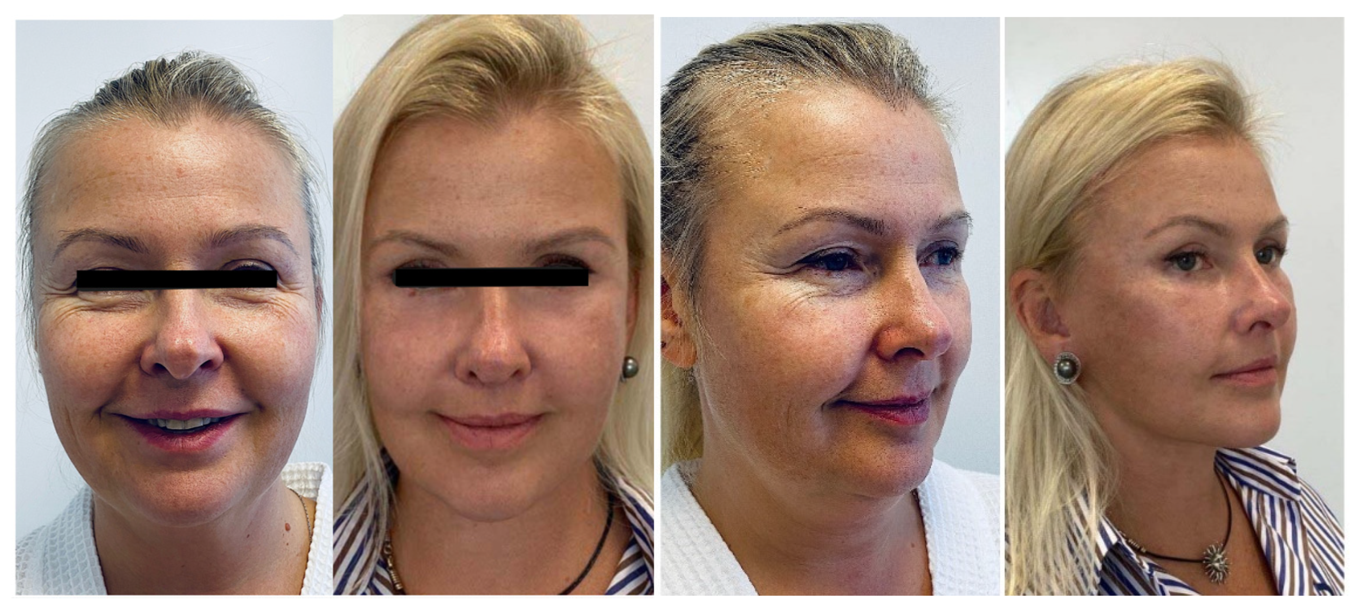
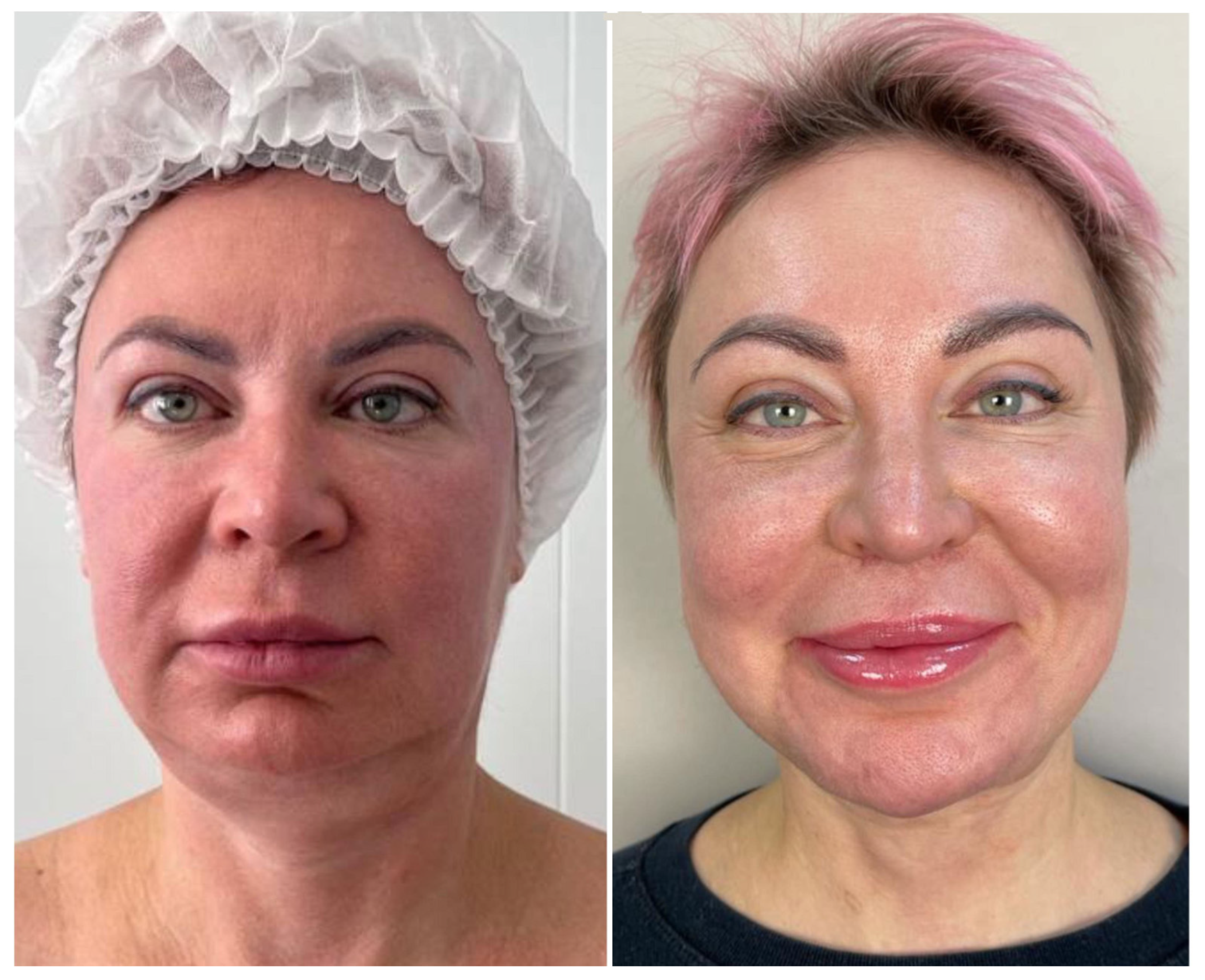
A 47-year-old woman, with no concomitant diseases, presented with slight gravitational changes in the lower third, violation of the cervico-mental angle, deep nasolabial folds and jaws. Along with this, she presented drooping of the eyebrow tails, overhanging skin in the upper eyelid area, bags in the lower eyelid area and fine mimic wrinkles. When looking at the frontal view photos, the lack of proportions of the lower third is evident (
Photo 1).
Photo 1.
Patient before and 6 months after complete rejuvenation of the face.
Photo 1.
Patient before and 6 months after complete rejuvenation of the face.
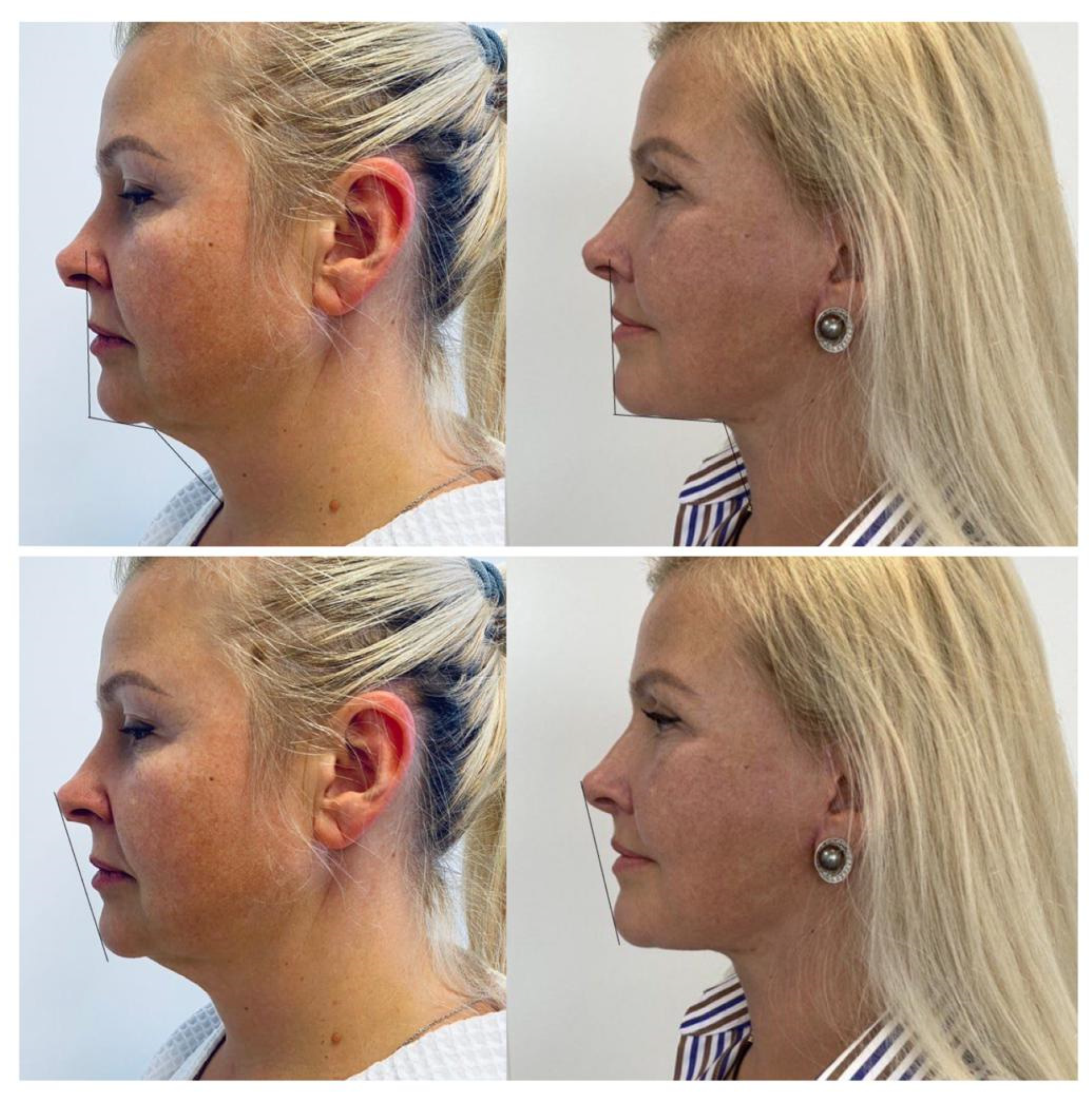
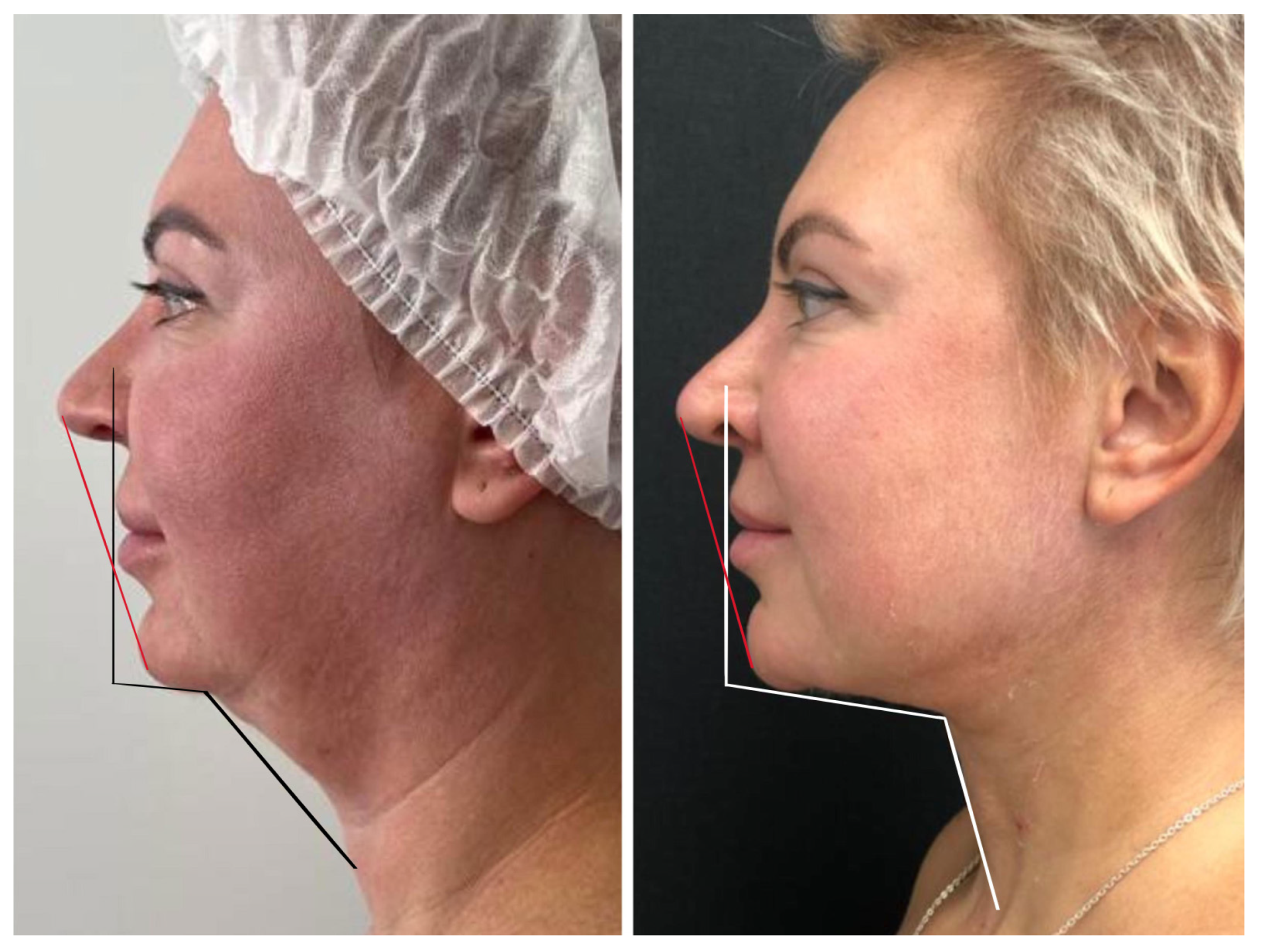
In the lateral view, according to the Riedel-Rikkitz lines, the projection of the chin protrusion is missing (
Photo 2). This way, it highlights the lack of support for the midface.
Photo 2.
Patient before and 6 months after complete rejuvenation of the face. Riedel plane and Rickett’s line show improvement in the chin projection.
Photo 2.
Patient before and 6 months after complete rejuvenation of the face. Riedel plane and Rickett’s line show improvement in the chin projection.
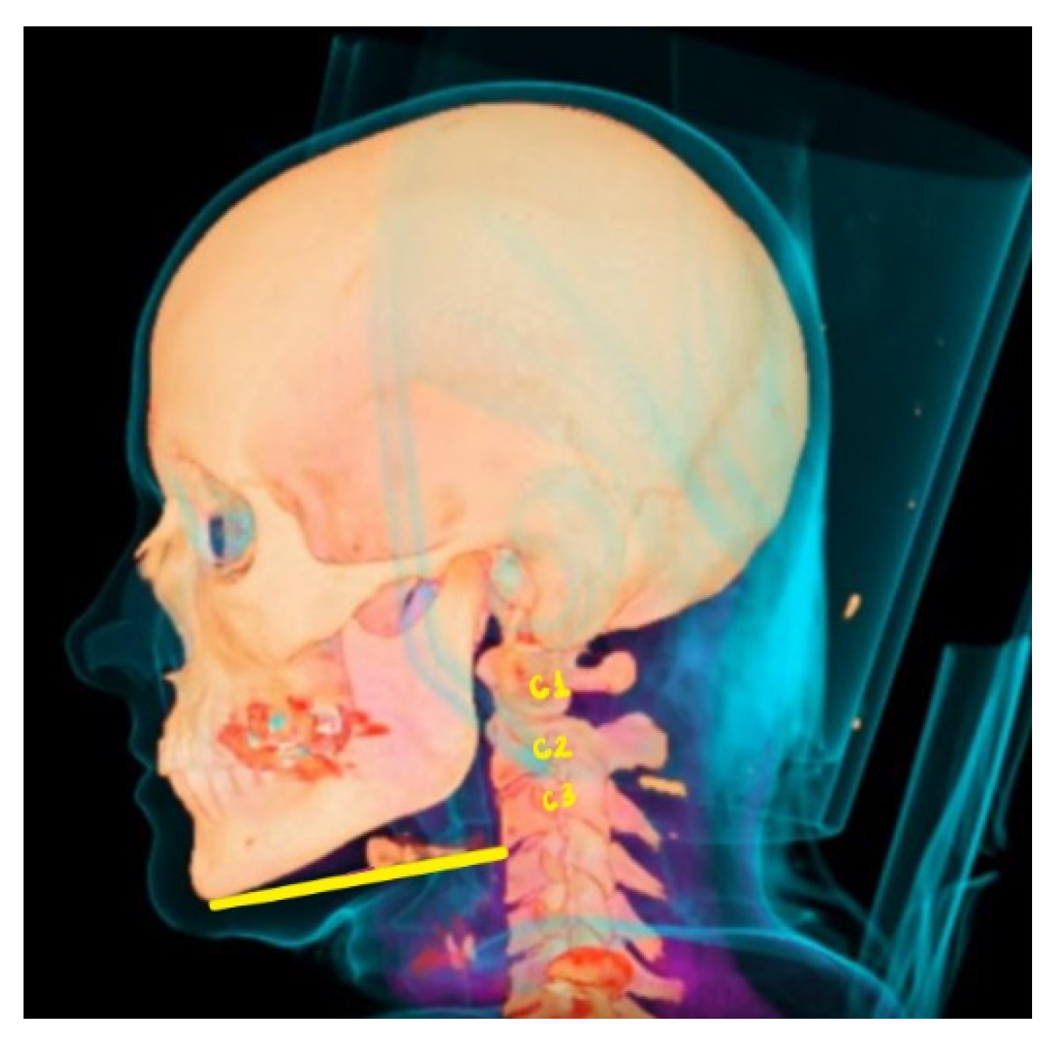
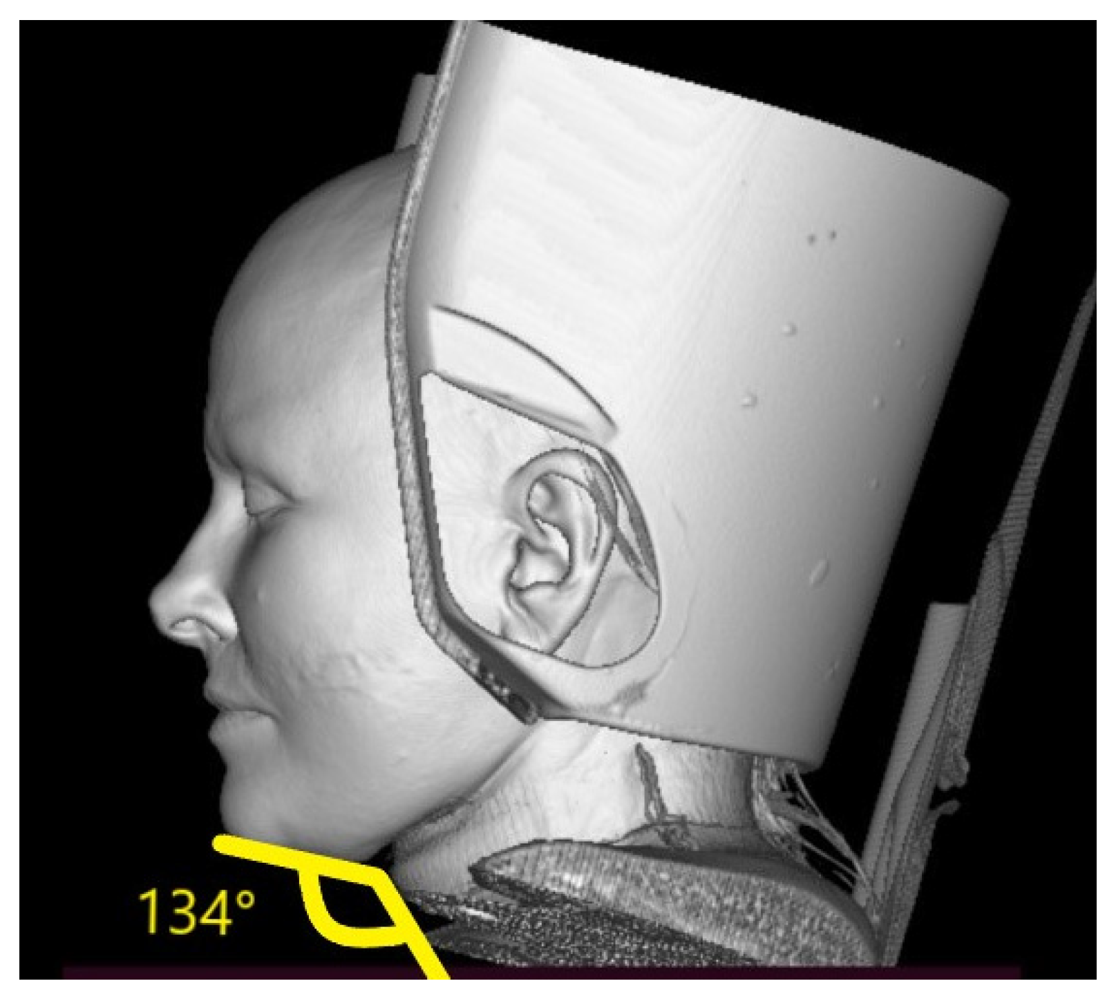
On the CT scan, we assess the level of the hyoid bone so we can guarantee the formation of a beautiful line of the chin-neck angle. In this case, the hyoid bone was quite high. Also, determine the presence submandibular gland ptosis is key to achieve an optimal result. For the stability of the results, on the sagittal view of CT, we see the angle and plan a slight extension to harmonize the face (
Photo 3 and
Photo 4).
Photo 3.
Computed Tomography, Rocabado plane shows high hyoid bone, a favorable situation to achieve excellent results with platysmaplasty.
Photo 3.
Computed Tomography, Rocabado plane shows high hyoid bone, a favorable situation to achieve excellent results with platysmaplasty.
Photo 4.
The soft tissues of the neck show an obtuse angle, far from the 90° that is considered aesthetically ideal.
Photo 4.
The soft tissues of the neck show an obtuse angle, far from the 90° that is considered aesthetically ideal.
Surgery plan: complex rejuvenation in the volume of DEEP SMAS, temporal lifting, upper + lower blepharoplasty, medial and lateral platysmaplasty with resection of the subplatysmal volume of adipose tissues and a slight extension of the chin with fat autografting, bullhorn lip lift.
3.2. Case 2
A female patient, 45 years-old, normasthenic, height 165, weight 67 BMI: 24, smoker, presenting with dysmorphic-edematous type of aging. The lower third is unsatisfactory, when analyzing the photos, a straight line is drawn showing the insufficiency of the lower third. In the lateral projection, it is evident that the chin protrusion is not enough, there is malocclusion, with microgenia and micrognathia (
Photo 5). From the anamnesis, chin liposuction + cryotherapy-cryolipolysis was performed twice with an unsatisfactory result. Patient’s main complaint was chin-neck contour.
Photo 5.
Riedel plane and Rickett’s line show insufficiency in the lower third of the face, poor chin projection and obtuse angle of the soft tissues. After surgery, there’s improvement in the lower third of the face.
Photo 5.
Riedel plane and Rickett’s line show insufficiency in the lower third of the face, poor chin projection and obtuse angle of the soft tissues. After surgery, there’s improvement in the lower third of the face.
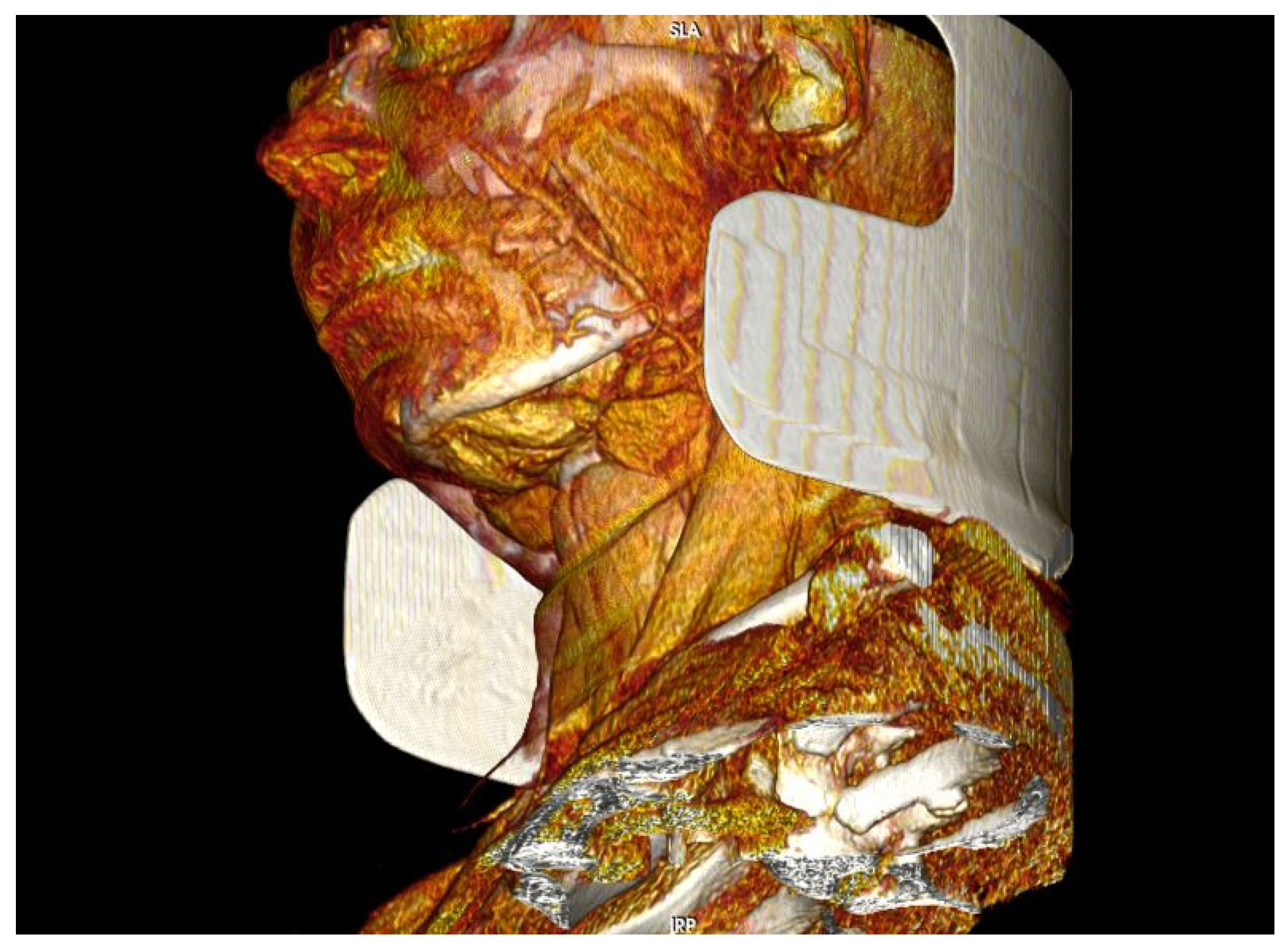
According to the CT scan, the glands are lower than the level of the lower jaw and shows how hypertrophied in volume are the anterior abdomens of the digastric muscles (
Photo 6). The level of the hyoid bone is very low, the cervico-mental angle is 145 degrees, the bone level can shift during the resection of the subplatysmal tissues.
Photo 6.
Ct scan before surgery. In this projection, the ptosis of the submandibular glands and the hypertrophic digastric muscles are evident.
Photo 6.
Ct scan before surgery. In this projection, the ptosis of the submandibular glands and the hypertrophic digastric muscles are evident.
Surgery plan: Medial platysmaplasty with extended resection of subplatysmal tissues, resection of fat, bilateral digastric muscles by squamous resection, as well as submental glands, closure with medial platysmoplasty with suturing behind the hyoid bone, fixations to raise it to an aesthetically advantageous position and lateral platysmaplasty and vertical midface lifting with minimal access, upper lower blepharoplasty, bullhorn lip lift. Chin augmentation using subplatysmal fat autografting to strengthen the support of the middle zone. (
Photo 7)
Photo 7.
Before and after surgery, it is evident how the proportions of the face are now harmonized, the result was possible thanks to the integral management of soft tissues in the neck, identified in the CT scan.
Photo 7.
Before and after surgery, it is evident how the proportions of the face are now harmonized, the result was possible thanks to the integral management of soft tissues in the neck, identified in the CT scan.
4. Discussion
Facial and neck rejuvenation has been, and continues to be, one of the most critical aspects of plastic surgery. Age-related changes in the cervical submental area are characterized by the development of an obtuse cervico-mental angle, the disappearance of well-defined lower jaw borders, the presence of jowls, the emergence of vertical platysmal bands, localized submental fat deposits, and excess skin [
7]. An “ideal” surgical rejuvenation method for the face and neck should address all the aforementioned age-related changes with minimal risk of complications and a short recovery period [
8]. A satisfactory result is considered to be one that allows the patient to perceive a reduction in gravitational changes on the face, a biological age younger than their actual age, attractiveness, and a desire to radiate positive energy.
Attractiveness or the patient’s biological (external) age is closely linked to various factors, such as skin quality, which is influenced by parameters like harmful habits, improper nutrition, sleep patterns, stress, and more. Our comprehensive rehabilitation measures include promoting a healthy lifestyle and cosmetological skin care.
In our practice, we pay attention to the following significant criteria for age-related changes:
Decreased skin turgor and tone, the appearance of excess skin
Involutional changes in facial skeletal structures
Excess volume in the lower third of the face
Eyebrow ptosis of the eyebrow tail and displacement of soft tissues in the temporal area
Age-related changes in periocular soft tissues
Hypertonicity or hypotonicity of the chin area
Presence of jowls
Presence of an obtuse cervico-mental angle
Ptosis of the upper lip
Manifestation of deep facial wrinkles
These age-related changes must be approached individually, assessing their specific anatomic characteristics, therefore, we proposed the CT scan as mandatory measure for the preoperative evaluation and surgical planning. In one study, patients were divided by age, type of aging, and BMI. Depending on the individual characteristics of patients and the understanding of the mechanisms underlying the processes of tissue involution and age-related changes in the face, the choice of effective methods for their elimination were determined [
9].
5. Conclusion
Thus, in order to obtain a harmonious result of surgical correction of age-related changes in the face and neck, it is necessary to take into account the anatomical features of the patient and the severity of age-related changes. In addition, a comprehensive analysis of the face, taking into account the type of facial aging and with the help of ultrasound examination of the soft tissues of the chin area, aid in drawing up an individual plan to obtain the ideal result that the patient imagines and can be implemented by the surgeon. The older the patient, the closer cosmetology and plastic surgery should cooperate, complementing each other, in order to obtain a rejuvenating effect of the skin both in terms of shape and volume, and in terms of tissue quality. To achieve long-term results in the correction of an aging face and neck, it is necessary to master a whole arsenal of various techniques.
Type of aging, BMI, concomitant diseases, patients expectative and anatomical characteristics are key for preoperative planning and achieve harmony and proportions, golden proportions and not only prevent aging, but enhanced the beauty.
Author Contributions
Conceptualization, A.B. and V.S.; methodology, A.B., S.A. and N.B. software 3D modeling and analysis, N.B. and A.A.; investigation, A.A. and F. O.; resources, V.S.; writing—original draft preparation, A.B.; writing—review and editing, V.S.; visualization, N.B. and A.A.; supervision, V.S.; All authors have read and agreed to the published version of the manuscript.
Funding
This research received no external funding
Informed Consent Statement
Written informed consent has been obtained from the patients to publish this paper.
Conflicts of Interest
The authors declare no conflict of interest.
References
- Sinno S, Thorne CH. Cervical Branch of Facial Nerve: An Explanation for Recurrent Platysma Bands Following Necklift and Platysmaplasty. Aesthetic Surgery Journal. 2018;39(1):1-7. [CrossRef]
- Frisenda J, Nassif P. Correction of the Lower Face and Neck. Facial Plastic Surgery. 2018;34(5):480-487. [CrossRef]
- Mendelson B, Wong CH. Changes in the facial skeleton with aging: implications and clinical applications in facial rejuvenation. Aesthetic Plast Surg. 2012;36(4):753-760. [CrossRef]
- Alimova S.M., Sharobaro V.I., Telnova A.V., Stepanyan E.E. Planning methods for surgical correction of soft tissues of the face and neck. Medical Imaging. 2021;25(4):47-52. [CrossRef]
- Alimova SM, Sharobaro VI, Telnova AV, Stepanyan EE. Planning of methods of surgical correction of soft tissues of the face and neck. Medical Visualization. 2021;25(4):47-52. (In Russ.). [CrossRef]
- Raveendran SS, Anthony DJ, Ion L. An Anatomic Basis for Volumetric Evaluation of the Neck. Aesthetic Surgery Journal. 2012;32(6):685-691. [CrossRef]
- Langsdon, P. R.; Velargo, P. A.; Rodwell, D.; Douglas, D. Submental Muscular Medialization and Suspension. 2013, 33 (7), 953–966. [CrossRef]
- Giampapa, V. C.; Mesa, J. M. Neck Rejuvenation with Suture Suspension Platysmaplasty Technique. 2014, 41 (1), 109–124. [CrossRef]
- Dikarev A.S., Mantardzhiev D.V., Tsinenko D.I. Facial contouring with porous implants as an auxiliary measure to optimize the results of surgical rejuvenation. Plastic Surgery and Aesthetic Medicine. 2021; (1):3643.
|
Disclaimer/Publisher’s Note: The statements, opinions and data contained in all publications are solely those of the individual author(s) and contributor(s) and not of MDPI and/or the editor(s). MDPI and/or the editor(s) disclaim responsibility for any injury to people or property resulting from any ideas, methods, instructions or products referred to in the content. |
© 2023 by the authors. Licensee MDPI, Basel, Switzerland. This article is an open access article distributed under the terms and conditions of the Creative Commons Attribution (CC BY) license (http://creativecommons.org/licenses/by/4.0/).
