Preprint
Article

Endometrial CAFs Resist Effect of Antitumor Drugs In A Patient-Derived 2-Cell Hybrid Co-Culture Model

Altmetrics

Downloads

116

Views

42

Comments

0

A peer-reviewed article of this preprint also exists.

Submitted:

20 November 2023

Posted:

01 December 2023

You are already at the latest version

Alerts
Abstract
Drug resistance in tumor cells is a significant roadblock in the clinical management of advanced or recurrent diseases in endometrial cancers. As a part of the tumor-stromal ecosystem, tumor cells are ecologically connected to the cancer-associated fibroblasts (CAFs), which form contributory elements of the tumor microenvironment (TME). We recently reported a novel model of patient-derived CAF-based 2-cell Hybrid Co-Culture (HyCC) to evaluate CAFs' role in developing drug resistance and understanding personalized tumor cell-CAF dialogue. Using our 2-cell HyCC model of patient-derived endometrial CAFs, we present data to demonstrate the direct counter-inhibitory effect of CAFs to combinations of cytotoxic and targeted drugs in developing drug resistance. CAFs derived from resected endometrial tumor samples were first passage-wise characterized before and after freeze-thaw for their positive and negative CAF markers expression pattern. Paclitaxel and its combination with copanlisib, TAK228, lenvatinib, and trametinib were used to test the 3D clonogenic growth of endometrial AN3CA cells on endometrial CAFs. We demonstrate that CAF-mediated resistance to antitumor drugs occurs via direct and indirect contact with CAFs in HyCC. Our data established the strength of the 2-cell HyCC model of patient-derived CAFs in solid tumors and provided a model of resistance to antitumor drugs tailored on a patient-to-patient basis.
Keywords: 
Subject: Medicine and Pharmacology  -   Oncology and Oncogenics

1. Introduction

The development of drug resistance and the failure to mitigate the resistance are directly associated with progression and adverse outcomes in gynecological cancers. There are limitations associated with the chemotherapeutic drugs used to garner the management of advanced and recurrent endometrial cancers. The resistance to drugs in the adjuvant setting is challenging as more than 25% of patients with a stage > 1 invasive disease subsequently progress as a metastatic disease in endometrial cancers [1]. Refractory and resistant tumor cells present within the residual disease have thus been linked with primary adverse outcomes. In advanced staged endometrial cancers, relapse occurs despite surgery, hormone therapy, and chemotherapy [2].
Tumor cells within the tumor mass are known to be part of the tumor-TME (tumor microenvironment) ecosystem. Although the mutational make-up of tumor cells is the primary determinant of treatment outcome and predicts patient survival, recent studies prove that tumor evolution in space, time, and treatment depends on the close interaction of mutagenized tumor cells with their TME [3]. Cancer-associated fibroblasts are critical features of TME [4]. The essential role of cancer-associated fibroblasts (CAFs) within the tumor stroma has been associated with developing resistance to drugs in various solid tumors, including endometrial cancers, and is an emerging field in cancer biology [5,6]. CAF tumor cell crosstalk and CAF’s crosstalk with the rest of TME components, angiogenic and immunogenic, is a bête noire of therapy and determines disease prognosis [7].
Thus, understanding the mechanism of CAF-mediated drug resistance holds power to address the challenge of resistance via disrupting the CAF-tumor dialogue. The key to knowing how to disrupt the CAF-tumor dialogue is gaining knowledge about the conversation in a patient-specific manner. We have recently designed a patient-derived CAF-based 2-cell Hybrid Co-Culture (HyCC) model to test the effect of CAF on endometrial tumor cells [8]. Using HyCC, we present the data to demonstrate the direct counter-inhibitory effect of CAFs on combinations of cytotoxic and targeted drugs.

2. Materials & Methods

2.1. Patient Consent & Tissue Collection

The institutional and/or licensing committee approved all experimental protocols. Informed consent(s) (IRB approved: Protocol Number Study: 2017.053-100399_ExVivo001) was obtained from patients. Resected unfixed tumor tissue(s) were received from the pathology department. The tissues were collected during surgery in designated collection media as per the guidelines and relevant regulations and provided by the pathologist, depending upon the availability of the tissue on a case-to-case basis. The primary cultures of CAFs were set up from the resected samples of tumors from patients with endometrial cancers, as per the guidelines and relevant regulations provided by the pathologist who performed the grossing. Samples were collected in DMEM/F-12 + Glutamax 500mL (base) supplemented with HyClone Penicillin-Streptomycin 100X 100mL (1%).

2.2. Cell Lines and Reagents

Fibroblast cells (Human uterine fibroblasts HUF; Primary Uterine Fibroblasts, Cat # PCS-460-010) and endometrial tumor cells (RL-95-2 and AN3CA) were bought from AATC and were culture according to the ATCC recommended method. Antibodies for ICC were from Cell Marque, NOVUS, Abcam, Agilent-Dako, and Cell Signaling. All cells within mid-passages (7-8) used for the study were tested negative for mycoplasma. The antibodies for WB were bought from Cell Signaling, USA.

2.3. Establishment of Patient-specific Ex Vivo Tumor-Endometrial CAF-2-Cell Model of Hybrid Co-Culture (HyCC)

We established and characterized the primary culture of CAF from the resected tumor tissues from patients (TCAF here off used interchangeably with CAF) [9]. As mentioned, we established a novel matrigel-based On-Top 2-cell HyCC model [8]. The DiO and DiI, were used for the 3D matrigel On-Top clonogenic assays. HyCC consisted of preparatory and experiment phases, as mentioned earlier. In short, the experiment phase included (1) 24 hours plating of DiO-CAFs, (2) plating of DiI-stained tumor cells on DiO-CAFs with and without paclitaxel and five combos (Combo A, Combo B, Combo C, Combo D, Combo E) as mentioned in the Table 1 in 3D formats. A parallel single-cell culture was set up separately to test the effect of the individual drug combo(s) on CAF and AN3CA cells. The HyCC was set up in quadruplicates. The media was changed every 2 days. The double-fluorescence signals from the live cells in cultures were recorded along with bright field photomicrographs at different time points using dry-objectives of Olympus BX43 Microscope using cellSens 1.18 LIFE SCIENCE IMAGING SOFTWARE (OLYMPUS CORPORATION). Photomicrographs were taken on day 0+ (within 4 hours for the 3D format as pictures are difficult to focus at zero hours in 3D) and on day 7. Semi-quantification was performed based on the fluorescence intensities of DiI tumor cells on DiO-TCAFs/NCAFs from 5–6 random microscopic fields of independent experiments (performed in quadruplicates). Statistical significance was determined by calculating Student’s t-test at p < 0.05.

2.4. Effect of CAFs On Paclitaxel Alone and In Combination with Targeted Antitumor Drugs Using 2-Cell Hybrid Co-culture Model

We then evaluated the effect of CAF on the tumoricidal effect of paclitaxel and its combination with targeted drugs (Table 1) by plating AN3CA in HyCC on TCAF in 3D format. To test the autocrine and paracrine effect of CAF in resisting the effect of paclitaxel and targeted drugs in HyCC, we evaluated the effect of drugs on both (1) direct contact with the CAF and (2) non-direct contact with CAF. Direct contact with the CAF and tumor cells was tested by plating AN3CA cells on the CAF-coated coverslips. In contrast, a non-direct contact with CAF comprised of AN3CA cells was placed on the plate outside the circumference of CAF-coated coverslips in the same HyCC wells as schematically presented in Figure 1A.

3. Results

3.1. Patient Information

Our cohort involved a total of 53 patients with endometrial cancers who participated in the study and provided informed consent. The information about the patients included in the study has been presented earlier [9]. In short, CAFs were established from samples received at the time of surgery. Out of 53 tissue samples, a total of 44 tissue samples were used for establishing CAF cultures.

3.2. Ex Vivo Primary Culture Of CAFs and Marker-based Verification of CAF

Characterization and validation of the primary culture of TCAF in the ex vivo culture were performed based on expression (ICC, qRT-PCR, WB, and flow cytometry) of positive and negative markers, as mentioned earlier [9]. In short, TCAFs were positive markers of CAF, including SMA, FAP, TE-7, and S100A4, , while they were always negative for EpCAM and CK 8,18 (a positive marker of epithelial tumor cells), CD31 (endothelial marker) and CD45 (LCA marker for leucocytes).

3.3. Patient-specific Ex Vivo Model of Hybrid Co-Culture (HyCC)

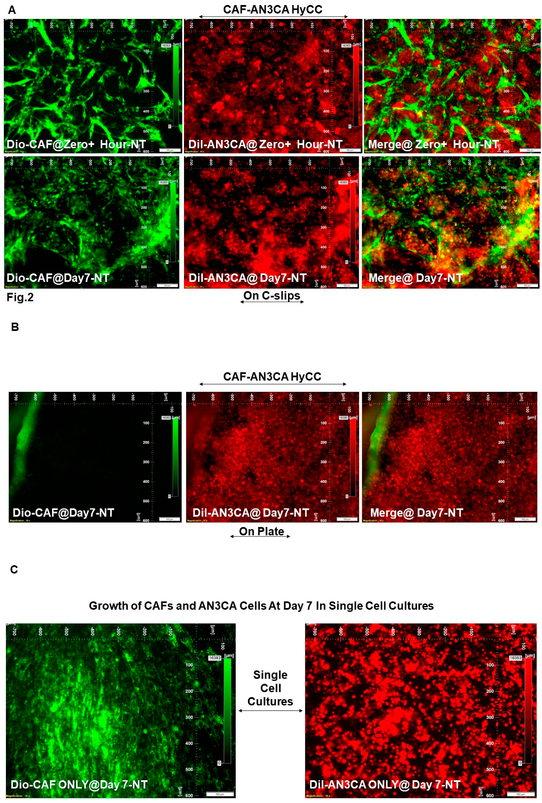
We first evaluated the growth pattern of endometrial tumor cells on TCAFs in two 3D formats of HyCC, (1) a direct contact format and (2) a non-contact format, as diagrammatically presented in Figure 1. We observed that growth of (1) AN3CA cells was higher on day 7 as compared to day 0+ in both the formats of HyCC (Figure 2A & B) and (2) both AN3CA and CAFs are enhanced on day 7 of the single cell culture (Figure 2C). Photomicrographs of Figure 2 show the clonogenic growth of AN3CA is significantly higher on day 7 compared to Day 0+.
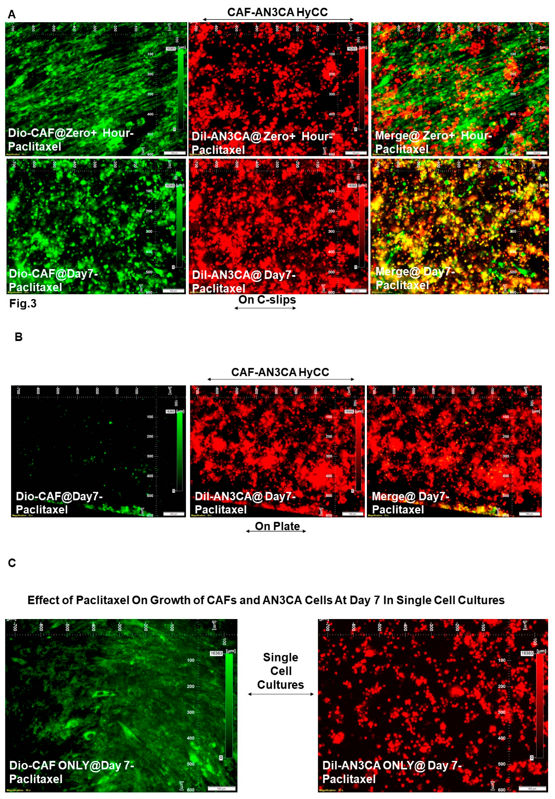
The treatment with paclitaxel decreased the growth of AN3CA cells in a single-cell AN3CA-only culture. Since we have previously reported the effect of paclitaxel on 3D Matrigel growth of AN3CA cells plated on CAF [9], the validity of the HyCC formats here was confirmed by testing the effect CAF on the effect of paclitaxel (Figure 3) as an internal validation control of drug effect as compared to the non-treated vehicle control (Figure 2) at day 7.

3.4. Effect of CAFs in Resisting Paclitaxel in Combination with Pathway-Targeted Drugs
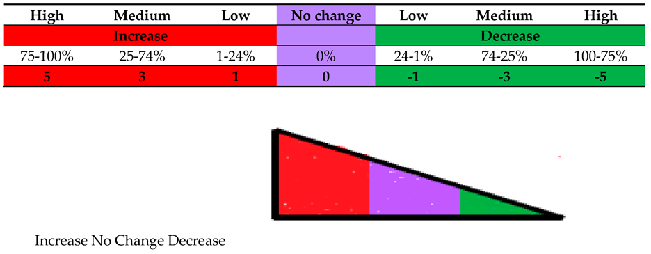
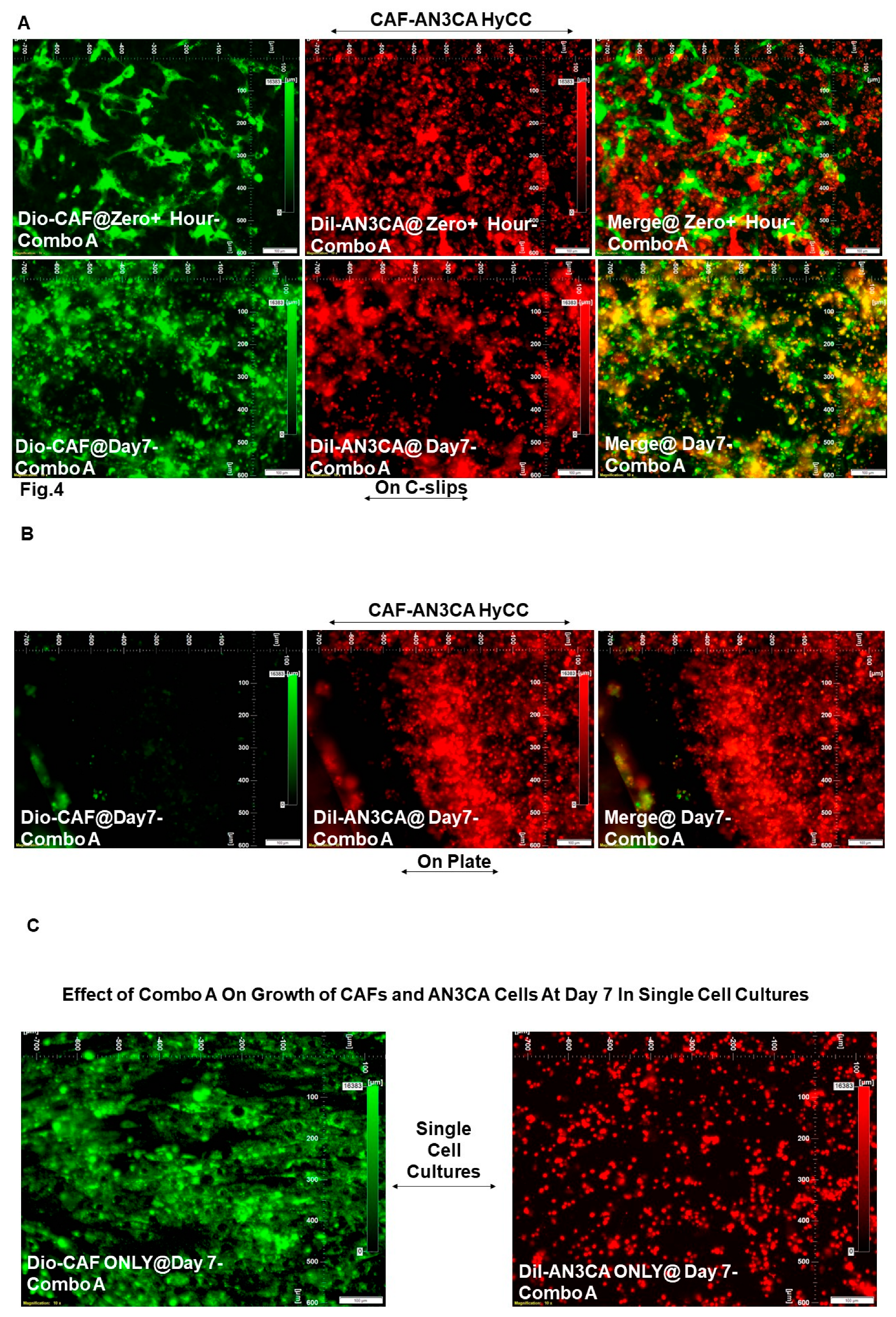
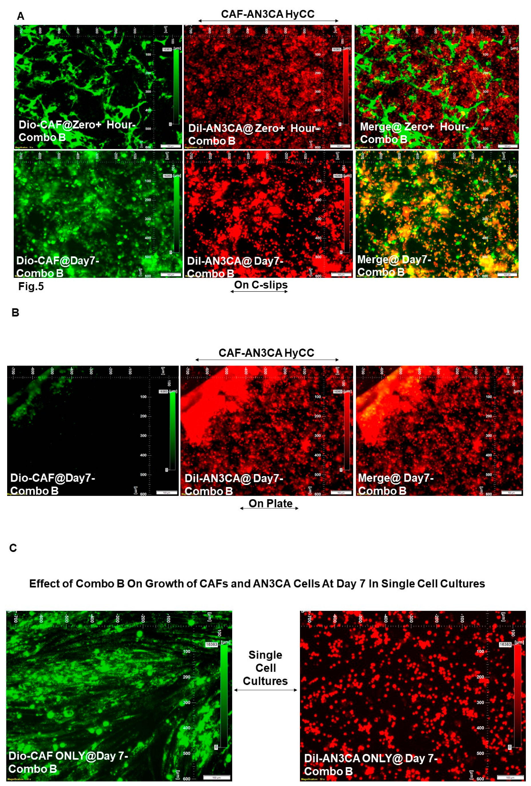
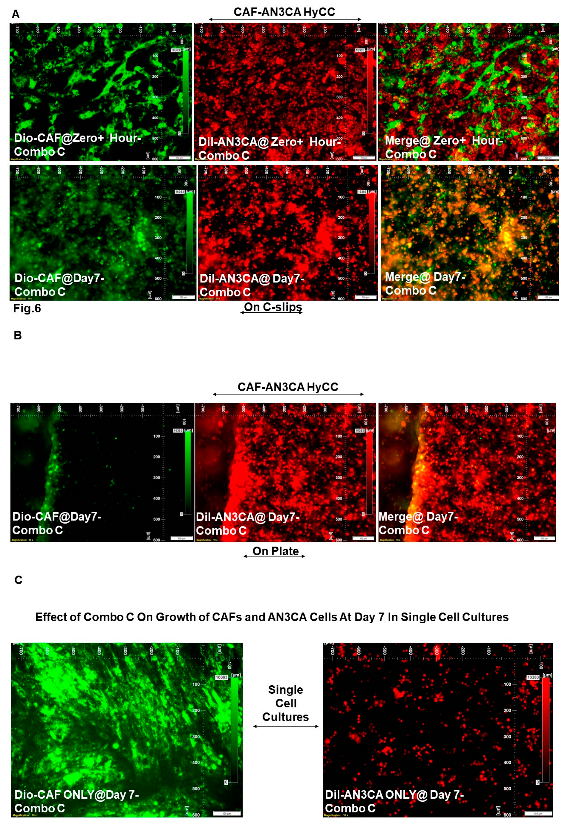
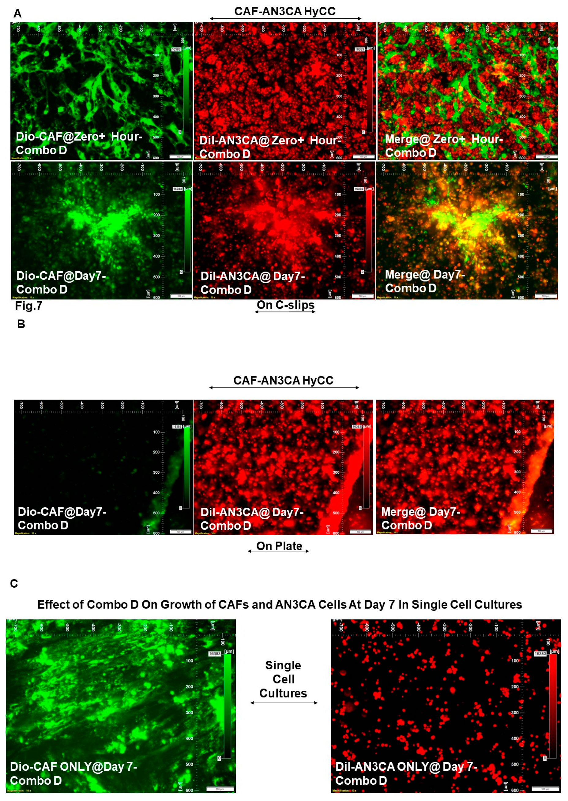
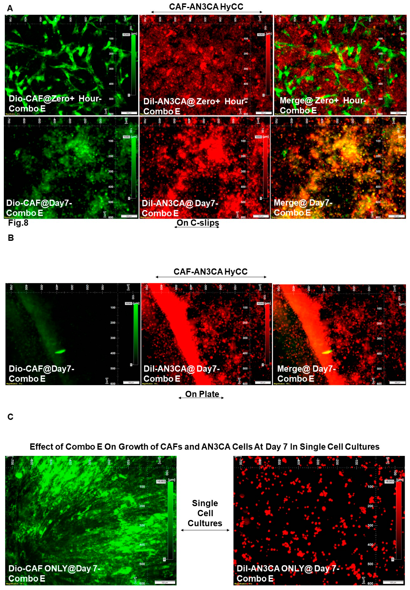
We used two separate culture platforms, HyCC and a parallel single-cell culture platform. The design included (1) two types of cell types, DiO-CAF and DiI-AN3CA cells, (2) five treatment combos (Combo A, Combo B, Combo C, Combo D, and Combo E; Table 1), (3) two culture modes of HyCC, HyCC On-Coverslips, and HyCC On-Plates and (4) two-time points, Zero+ hour and 7 days. The growth inhibitory effect of paclitaxel was used as the baseline in measuring the role of CAF in resisting paclitaxel’s growth inhibitory effect in combination with pathway-targeted drugs Combo A, Combo B, Combo C, Combo D, and Combo E on endometrial tumor cells. Figure 4 shows that although treatment with paclitaxel in combination with copanlisib (Combo A) blocked the growth of AN3CA cells on day 7 in the single cell culture, the presence of CAF significantly blocked the growth inhibitory effect of the combination. The growth of CAF in the single cell culture is not altered. Likewise, treatment with paclitaxel in combination with TAK228 (Combo B), lenvatinib (Combo C), and trametinib (Combo D) as well as copanlisib only (Combo E), although blocked the growth of AN3CA in single cell culture, had failed to inhibit the growth of AN3CA in HyCC with CAF in both on-coverslip and on-plate formats (Figure 5, Figure 6, Figure 7 and Figure 8). The summarized results with the conditional formatted semi-quantified data are presented in Table 2. The result indicated that the effect of CAF is mediated not via direct contact with the AN3CA cells but via the paracrine secretory mechanism. Table 2 semi-quantitatively demonstrated that a HyCC with CAF had a similar protecting effect on paclitaxel and pathway-targeted drugs on clonogenic growth of AN3CA, while none of the drugs had significant growth inhibitor effects on CAF in a single cell culture. In summary, our data show that endometrial CAF confers a protective effect on endometrial tumor cells against the chemotherapy drug paclitaxel in combination with different pathway-targeted drugs in HyCC.
Scale for Table 2. Arbitrary Scale Based On % Change In The Relative Fluorescence Intensity (DiI-AN3CA & DiO-CAF).
Preprints 91022 i001

4. Discussion

The Cancer Genome Atlas (TCGA) presents a classification of endometrial cancers into four molecular subtypes whose treatment consideration is now recommended [10]. The molecular characterization of endometrial cancer has paved the way for the develop,ent of novel combinations of immunotherapies and pathway-targeted therapies, leading to the approval of immune checkpoint inhibitors (combination of lenvatinib plus pembrolizumab) [11].
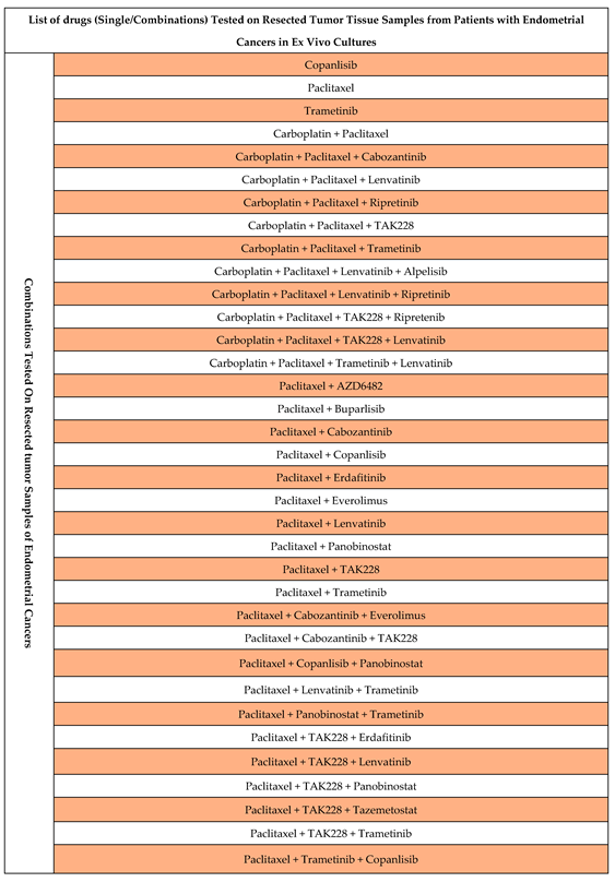
Our cohort consisted predominantly of patients with endometrioid adenocarcinoma, with fewer patients with high-grade serous carcinomas of both papillary and mixed types (see Table 1 of [9]). Paclitaxel is routinely used (weekly) as a single agent to patients with recurrent or metastatic disease is the most commonly practiced approach in the clinics [12] (https://www.cancer.org/). Carboplatin plus paclitaxel is the established frontline treatment for advanced/recurrent disease [11]. We have chosen the combinations of paclitaxel with pathway-targeted drugs based on the primary pathway alterations in these cancers and the list of effective combinations from our ex vivo testing of drug effects in endometrial cancers (Table 3). Considering the recent knowledge about the role of TME-CAF in mediating drug resistance in solid tumors, we hypothesized that the drug (adjuvant) mediated resistance in endometrial cancers is contributed by the CAF component of TME. We tested the effect of drug combinations on endometrial tumor samples in ex vivo cultures. Out of several combinations tested, we observed anti-proliferative and pro-apoptotic effects in certain combinations (submitted for publication). Hence, in this study, we tested the function of CAF from the tumor samples, which was affected by our study’s ex vivo drug treatment. Our data provides experimental proof of “Deadlock game” as explained in non-small cell lung cancer [13]. Our results demonstrate that drug treatment decreased the 3D growth of endometrial tumor cells in matrigel in a single-cell culture, while the presence of CAF in HyCC resisted the growth inhibitor effect of the drugs, alone or in combinations (Table 2). We present the data of a direct-contact effect versus a non-direct-contact effect of CAF using HyCC using (Figure 1B). A direct-contact effect was tested by taking the photomicrographs focused on the Dil-CAF coated coverslips within the HyCC. A non-direct-contact effect was tested by taking the photomicrographs focused on the plate area outside the Dil-CAF-coated cover-clips within the HyCC.
Although the growth of CAF was not significantly altered following the treatment of drugs in HyCC or single cell culture, we observed a characteristic morphological pattern of the CAFs in HyCC. The fact that the protective effect of CAF was observed simultaneously both in a direct-contact format and an indirect-contact format of HyCC proves that the mechanism of function of CAF involves a paracrine action. In fact, several studies have reported the role of CAF-derived exosomes in cancer progression [14] by regulating the survival and proliferation of cancer cells [15]. Moreover, crosstalk between CAF and immune cells of TME [16] has been implicated as the basis of CAF’s clinical and therapeutic relevance [17].
CAFs have been beginning to present a deterministic role in influencing disease outcomes [17]. CAF-inclusive treatment is just beginning to broaden drug design and target the TME [6]. A number of studies provided evidence in favor of a CAF-inclusive therapy for clinical management of the disease, tumor growth, disease progression, therapeutic resistance, immune therapy, and angiogenesis [17,18,19,20]. In line with the above fact, several CAF-directed/inclusive clinical trials have been currently instituted and are ongoing in the NIH (https://clinicaltrials.gov/ct2/home). Although a majority of these clinical trials are initiated in advanced/metastatic tumors (ClinicalTrials.gov Identifier: NCT05547321), including hepatocellular carcinomas and neoplasms of the breast, colon, prostate, lung, ovary, and PDAC (ClinicalTrials.gov Identifier: NCT05262855), CAF-directed clinical trials in endometrial cancers are rare. Recently, we have studied the role of CAFs in the development of resistance to anti-angiogenic drugs in ovarian cancers. We have recently published the first report demonstrating a correlation between the post-surgical event and the aggressive nature of CAFs in endometrial cancers, providing an undeniable reason to study the in-depth mechanism of CAF function toward developing treatment resistance in endometrial cancers [9]. Here, we tested the hypothesis that the addition of an essential TME component, CAF, which is known to be involved in the poor prognosis, could provide more predictive models. Indeed, the result confirmed the role of CAF on tumor cell survival in the face of antitumor drug combinations. Our data collectively provides a compelling experimental proof-of-concept for the protective role of CAF towards tumor cells against anti-angiogenic and antitumor drugs in gynecological cancers.

Funding

The study was funded entirely by Avera Cancer Institute.

Authors’ Contribution

RS: The pathologist provided the evaluation of CAF based on IHC staining. JCA: JCA performed the flow cytometry work. JCA supervised the ex vivo study logistics and helped in the EndNote library. XL: Research Assistant Lead standardized and performed tissue processing, ICC staining for CAF markers, H&E stains, and IHC. XL maintained the record of the histology work. AD: Research Associate obtained consent from patients and provided technical assistance in record keeping. AD helped in the preparation of figures, tables, and statistical correlations. KG: Provided insight into the overall logistical management of the ex vivo study and coordinated the clinical logistics of the study. LRE: The surgeon provided clinical insight into endometrial and ovarian tumors and corresponding blood samples. DS: The surgeon provided clinical insight into endometrial and ovarian tumors and corresponding blood samples. PD: Senior Scientist interrogated the genomic alteration of each tumor sample and matched the genomic alteration with the drug combination for the testing. PD helped in the analysis of data and writing the MS. ND: Senior Scientist conceptualized and supervised the study, wrote the MS, and analyzed the data.

Institutional Review Board Statement

Anonymized tissue samples were collected at surgery from patients with endometrial cancers following their Informed (IRB approved: Protocol Number Study: 2017.053-100399_ExVivo001) consent.

Informed Consent Statement

Informed (IRB approved: Protocol Number Study: 2017.053-100399_ExVivo001) consents for receiving resected tissue were obtained from 72 enrolled patients with endometrial cancers.

Acknowledgments

We acknowledge Avera Cancer Institute for funding the entire study. We acknowledge every patient and their family for their participation in the ex vivo study at the Avera Cancer Institute.

Patent Status

The study presented in the MS is part of a patent application (United States Patent and Trademark Office; Application number 16/875,910).

References

  1. Brooks, R.A.; Fleming, G.F.; Lastra, R.R.; Lee, N.K.; Moroney, J.W.; Son, C.H.; Tatebe, K.; Veneris, J.L. Current recommendations and recent progress in endometrial cancer. CA Cancer J Clin 2019, 69, 258–279. [Google Scholar] [CrossRef] [PubMed]
  2. Connor, E.V.; Rose, P.G. Management Strategies for Recurrent Endometrial Cancer. Expert Rev Anticancer Ther 2018, 18, 873–885. [Google Scholar] [CrossRef] [PubMed]
  3. de Visser, K.E.; Joyce, J.A. The evolving tumor microenvironment: From cancer initiation to metastatic outgrowth. Cancer Cell 2023, 41, 374–403. [Google Scholar] [CrossRef] [PubMed]
  4. Chhabra, Y.; Weeraratna, A.T. Fibroblasts in cancer: Unity in heterogeneity. Cell 2023, 186, 1580–1609. [Google Scholar] [CrossRef] [PubMed]
  5. Winterhoff, B.; Konecny, G.E. Targeting fibroblast growth factor pathways in endometrial cancer. Curr Probl Cancer 2017, 41, 37–47. [Google Scholar] [CrossRef] [PubMed]
  6. Dzobo, K.; Dandara, C. Broadening Drug Design and Targets to Tumor Microenvironment? Cancer-Associated Fibroblast Marker Expression in Cancers and Relevance for Survival Outcomes. OMICS 2020, 24, 340–351. [Google Scholar] [CrossRef] [PubMed]
  7. Herrera, M.; Berral-Gonzalez, A.; Lopez-Cade, I.; Galindo-Pumarino, C.; Bueno-Fortes, S.; Martin-Merino, M.; Carrato, A.; Ocana, A.; De La Pinta, C.; Lopez-Alfonso, A.; et al. Cancer-associated fibroblast-derived gene signatures determine prognosis in colon cancer patients. Mol Cancer 2021, 20, 73. [Google Scholar] [CrossRef] [PubMed]
  8. Sulaiman, R.; De, P.; Aske, J.C.; Lin, X.; Dale, A.; Gaster, K.; Espaillat, L.R.; Starks, D.; Dey, N. A CAF-Based Two-Cell Hybrid Co-Culture Model to Test Drug Resistance in Endometrial Cancers. Biomedicines 2023, 11. [Google Scholar] [CrossRef] [PubMed]
  9. Sulaiman, R.; De, P.; Aske, J.C.; Lin, X.; Dale, A.; Gaster, K.; Espaillat, L.R.; Starks, D.; Dey, N. Characterization and Clinical Relevance of Endometrial CAFs: Correlation between Post-Surgery Event and Resistance to Drugs. Int J Mol Sci 2023, 24. [Google Scholar] [CrossRef] [PubMed]
  10. Karpel, H.C.; Slomovitz, B.; Coleman, R.L.; Pothuri, B. Treatment options for molecular subtypes of endometrial cancer in 2023. Curr Opin Obstet Gynecol 2023, 35, 270–278. [Google Scholar] [CrossRef] [PubMed]
  11. Rubinstein, M.; Shen, S.; Monk, B.J.; Tan, D.S.P.; Nogueira-Rodrigues, A.; Aoki, D.; Sehouli, J.; Makker, V. Looking beyond carboplatin and paclitaxel for the treatment of advanced/recurrent endometrial cancer. Gynecol Oncol 2022, 167, 540–546. [Google Scholar] [CrossRef] [PubMed]
  12. Markman, M.; Fowler, J. Activity of weekly paclitaxel in patients with advanced endometrial cancer previously treated with both a platinum agent and paclitaxel. Gynecol Oncol 2004, 92, 180–182. [Google Scholar] [CrossRef] [PubMed]
  13. Kaznatcheev, A.; Peacock, J.; Basanta, D.; Marusyk, A.; Scott, J.G. Fibroblasts and alectinib switch the evolutionary games played by non-small cell lung cancer. Nat Ecol Evol 2019, 3, 450–456. [Google Scholar] [CrossRef] [PubMed]
  14. Li, C.; Teixeira, A.F.; Zhu, H.J.; Ten Dijke, P. Cancer associated-fibroblast-derived exosomes in cancer progression. Mol Cancer 2021, 20, 154. [Google Scholar] [CrossRef] [PubMed]
  15. Richards, K.E.; Zeleniak, A.E.; Fishel, M.L.; Wu, J.; Littlepage, L.E.; Hill, R. Cancer-associated fibroblast exosomes regulate survival and proliferation of pancreatic cancer cells. Oncogene 2017, 36, 1770–1778. [Google Scholar] [CrossRef] [PubMed]
  16. Mao, X.; Xu, J.; Wang, W.; Liang, C.; Hua, J.; Liu, J.; Zhang, B.; Meng, Q.; Yu, X.; Shi, S. Crosstalk between cancer-associated fibroblasts and immune cells in the tumor microenvironment: new findings and future perspectives. Mol Cancer 2021, 20, 131. [Google Scholar] [CrossRef] [PubMed]
  17. Chen, Y.; McAndrews, K.M.; Kalluri, R. Clinical and therapeutic relevance of cancer-associated fibroblasts. Nat Rev Clin Oncol 2021, 18, 792–804. [Google Scholar] [CrossRef] [PubMed]
  18. Orimo, A.; Gupta, P.B.; Sgroi, D.C.; Arenzana-Seisdedos, F.; Delaunay, T.; Naeem, R.; Carey, V.J.; Richardson, A.L.; Weinberg, R.A. Stromal fibroblasts present in invasive human breast carcinomas promote tumor growth and angiogenesis through elevated SDF-1/CXCL12 secretion. Cell 2005, 121, 335–348. [Google Scholar] [CrossRef] [PubMed]
  19. Xu, Y.; Li, W.; Lin, S.; Liu, B.; Wu, P.; Li, L. Fibroblast diversity and plasticity in the tumor microenvironment: roles in immunity and relevant therapies. Cell Commun Signal 2023, 21, 234. [Google Scholar] [CrossRef] [PubMed]
  20. Ravichandra, A.; Bhattacharjee, S.; Affo, S. Cancer-associated fibroblasts in intrahepatic cholangiocarcinoma progression and therapeutic resistance. Adv Cancer Res 2022, 156, 201–226. [Google Scholar] [CrossRef] [PubMed]
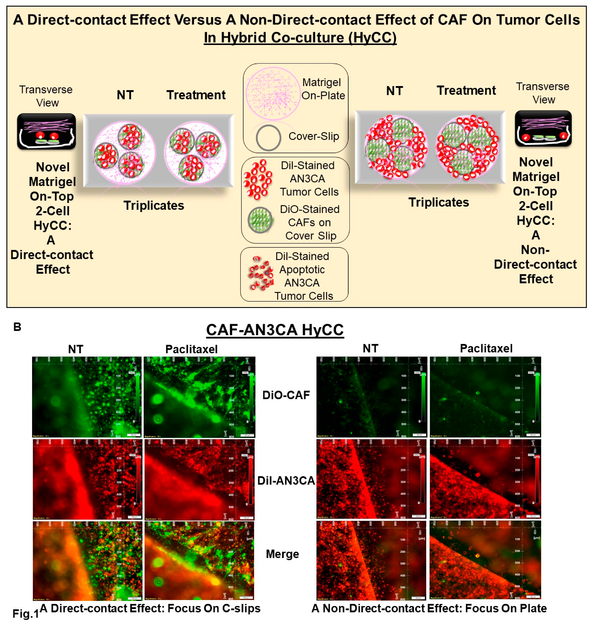
Figure 1. Schematic plan to demonstrate a direct-contact effect versus a non-direct-contact effect of CAF using Hybrid Co-culture (HyCC): The model was used to test the effect of the antitumor and anti-angiogenic drugs (A). We present the results of a direct-contact effect versus a non-direct-contact effect of CAF using Hybrid Co-culture using the effect of paclitaxel (B). A direct-contact effect was tested by taking the photomicrographs focused on the Dil-CAF coated cover-clips within the HyCC (Left two panels of Figure 1B). A non-direct-contact effect was tested by taking the photomicrographs focused on the plate area outside the Dil-CAF coated cover-clips within the HyCC (Right two panels of Figure 1B.
Preprints 91022 g001
Figure 2. Growth of AN3CA cells on patient-derived CAF for 7 days in 3D Matrigel HyCC: CAF derived from the primary culture of resected tumor tissue samples from patients with endometrial cancers were stained with DiO-fluorescence stain. DiI-stained AN3CA cells were plated 24 hours after on DiO-CAF-coated coverslips. Photomicrographs were taken on zero+ hours (upper panel of A) of the plating of AN3CA cells on DiO-CAF coated coverslips and (lower panel of A) as well as on the cells on-plate (panel of B) on day 7. The growth of AN3CA cells in 3D on-matrigel alone on day 7 is presented (C right photomicrograph) compared to the growth of DiO-CAF (C left photomicrograph).
Preprints 91022 g002
Figure 3. Effects of patient-derived primary CAF on the antitumor effect of paclitaxel in Hybrid Co-Culture: Protective effect of CAF (DiO-stained; green fluorescent color) against the tumoricidal effect of paclitaxel on AN3CA cells (DiI-stained; orange-red fluorescent color) is demonstrated from the photomicrographs of live cells plated on CAF-coated coverslips (direct contact with CAF) (A) as well as on-plate (non-direct contact with CAF) (B) in 3D matrigel HyCC. The growth inhibitory effect of paclitaxel in 3D on-matrigel growth of DiI-AN3CA alone on day 7 is presented in the picture (C right photomicrograph) as compared to the no-effect on the growth of DiO-CAF (C left photomicrograph).
Preprints 91022 g003
Figure 4. Effects of patient-derived primary CAF on the antitumor effect of paclitaxel in combination with Copanlisib (Combo A) in Hybrid Co-Culture: Protective effect of CAF (DiO-stained; green fluorescent color) against the tumoricidal effect of paclitaxel in combination with Copanlisib (Combo A) on AN3CA cells (DiI-stained; orange-red fluorescent color) is demonstrated from the photomicrographs of live cells plated on CAF-coated coverslips (direct contact with CAF) (A) as well as on-plate (non-direct contact with CAF) (B) in 3D matrigel HyCC. The growth inhibitory effect of paclitaxel in combination with Copanlisib (Combo A) in 3D on-matrigel growth of DiI-AN3CA alone on day 7 is presented in the picture (C right photomicrograph) as compared to the no-effect on the growth of DiO-CAF (C left photomicrograph). CAF resisted the growth inhibitory effect of paclitaxel in combination with Copanlisib (Combo A) on AN3CA in a HyCC 3D format.
Preprints 91022 g004
Figure 5. Effects of patient-derived primary CAF on the antitumor effect of paclitaxel in combination with TAK228 (Combo B) in Hybrid Co-Culture: Protective effect of CAF (DiO-stained; green fluorescent color) against the tumoricidal effect of paclitaxel in combination with TAK228 (Combo B) on AN3CA cells (DiI-stained; orange-red fluorescent color) is demonstrated from the photomicrographs of live cells plated on CAF-coated coverslips (direct contact with CAF) (A) as well as on-plate (non-direct contact with CAF) (B) in 3D matrigel HyCC. The growth inhibitory effect of paclitaxel in combination with TAK228 (Combo B) in 3D on-matrigel growth of DiI-AN3CA on day 7 is presented in the picture (C right photomicrograph) as compared to the no-effect on the growth of DiO-CAF (C left photomicrograph). CAF resisted the growth inhibitory effect of paclitaxel in combination with TAK228 (Combo B) on AN3CA in a HyCC 3D format.
Preprints 91022 g005
Figure 6. Effects of patient-derived primary CAF on the antitumor effect of paclitaxel in combination with Lenvatinib (Combo C) using Hybrid Co-Culture: Protective effect of CAF (DiO-stained; green fluorescent color) against the tumoricidal effect of paclitaxel in combination with Lenvatinib (Combo C) on AN3CA cells (DiI-stained; orange-red fluorescent color) is demonstrated from the photomicrographs of live cells plated on CAF-coated coverslips (direct contact with CAF) (A) as well as on-plate (non-direct contact with CAF) (B) in 3D matrigel HyCC. The growth inhibitory effect of paclitaxel in combination with Lenvatinib (Combo C) in 3D on-matrigel growth of DiI-AN3CA alone on day 7 is presented in the picture (C right photomicrograph) as compared to the no-effect on the growth of DiO-CAF (C left photomicrograph). CAF resisted the growth inhibitory effect of paclitaxel in combination with Lenvatinib (Combo C) on AN3CA in a HyCC 3D format.
Preprints 91022 g006
Figure 7. Effects of patient-derived primary CAF on the antitumor effect of paclitaxel in combination with Trametinib (Combo D) in Hybrid Co-Culture: Protective effect of CAF (DiO-stained; green fluorescent color) against the tumoricidal effect of paclitaxel in combination with Trametinib (Combo D) on AN3CA cells (DiI-stained; orange-red fluorescent color) is demonstrated from the photomicrographs of live cells plated on CAF-coated coverslips (direct contact with CAF) (A) as well as on-plate (non-direct contact with CAF) (B) in 3D matrigel HyCC. The growth inhibitory effect of paclitaxel in combination with Trametinib (Combo D) in 3D on-matrigel growth of DiI-AN3CA alone on day 7 is presented in the picture (C right photomicrograph) as compared to the no-effect on the growth of DiO-CAF (C left photomicrograph). CAF resisted the growth inhibitory effect of paclitaxel in combination with Trametinib (Combo D) on AN3CA in a HyCC 3D format.
Preprints 91022 g007
Figure 8. Effects of patient-derived primary CAF on the antitumor effect of copanlisib (Combo E) in Hybrid Co-Culture: Protective effect of CAF (DiO-stained; green fluorescent color) against the tumoricidal effect of copanlisib (Combo E) on AN3CA cells (DiI-stained; orange-red fluorescent color) is demonstrated from the photomicrographs of live cells plated on CAF-coated coverslips (direct contact with CAF) (A) as well as on-plate (non-direct contact with CAF) (B) in 3D matrigel HyCC. The growth inhibitory effect of copanlisib (Combo E) in 3D on-matrigel growth of DiI-AN3CA on day 7 is presented in the picture (C right photomicrograph) as compared to the no-effect on the growth of DiO-CAF (C left photomicrograph). CAF resisted the growth inhibitory effect of copanlisib (Combo E) on AN3CA in a HyCC 3D format.
Preprints 91022 g008
Table 1. List of the drug combinations tested to demonstrate the protective effect of endometrial CAFs on the 3D Matrigel growth of endometrial cancer cells in HyCC.
Combination* # Drugs used for the Combination
A Paclitaxel plus Copanlisib
B Paclitaxel plus TAK228
C Paclitaxel plus Lenvatinib
D Paclitaxel plus Trametinib
E Copanlisib
*The combinations are referred in the text as Combo A, Combo B, Combo C, Combo D, and Combo E.
Table 2. Conditional Formatting of Grades based on the Arbitrary Scale of the relative % of Fluorescence Intensity of DiI-AN3CA Cells and DiO-CAF. DiO-CAFs are plated on coverslips.
Cell Type(s) Involved in the Experi-ment Treat-ment Type (s) Culture Platform Type(s) End- Time of the Experi-ment Comparison Type(s) Photo-micrographs Presented in Figure # Grades* (Arbitrary Scale Based on the % of Fluorescence Intensity of DiI-AN3CA) Grades* (Arbitrary Scale Based on the % of Fluorescence Intensity of DiO-CAF)
DiO-CAF & DiI-AN3CA Vehicle Control (NT) HyCC On Cover slips (DiO-CAF) Day 7 Day Zero+ vs Day7 Figure 2A 5 3
DiI-AN3CA HyCC On Plates Day7 as compared to Day Zero+ Figure 2B 5 Not Applicable (Focused On-Plate)
DiI-AN3CA Single Cell Cultures Day7 as compared to Day Zero+ Figure 2C (Right Panel) 5 Not Applicable
DiO-CAF Day7 as compared to Day Zero+ Figure 2C (Left Panel) Not Applicable 5
Cell Type(s) Involed in the Experi-ment Treat-ment Type (s) Culture Platform Type(s) End- Time of the Experi-ment Comparison Type(s) Photo-micrographs Presented in Figure # Grades* (Arbitrary Scale Based on the % of Fluorescence Intensity of DiI-AN3CA) Grades* (Arbitrary Scale Based on the % of Fluorescence Intensity of DiO-CAF)
DiO-CAF & DiI-AN3CA Pacli-taxel HyCC On Cover slips (DiO-CAF) Day 7 Drug Treatment
Vs.
No Treatment
Figure 3A
vs.
Figure 2A
0 0
DiI-AN3CA HyCC On Plates Figure 3B vs. Figure 2B 0 Not Applicable (Focused On-Plate)
DiI-AN3CA Single Cell Cultures Figure 3C
(Right Panel)
vs.
Figure 2C (Right Panel)
-5 Not Applicable
DiO-CAF Figure 3C (Left Panel)
vs
Figure 2C (Left Panel)
Not Applicable 0
Cell Type(s) Involed in the Experi-ment Treat-ment Type (s) Culture Platform Type(s) End- Time of the Experi-ment Comparison Type(s) Photo-micrographs Presented in Figure # Grades* (Arbitrary Scale Based on the % of Fluorescence Intensity of DiI-AN3CA) Grades* (Arbitrary Scale Based on the % of Fluorescence Intensity of DiO-CAF)
DiO-CAF & DiI-AN3CA Pacli-taxel + Copan-lisib
(Combo A)
HyCC On Cover slips (DiO-CAF) Day 7 Drug Treatment
Vs.
No Treatment
Figure 4A vs.
Figure 2A
0 0
DiI-AN3CA HyCC On Plates Figure 4B vs. Figure 2B 0 Not Applicable (Focused On-Plate)
DiI-AN3CA Single Cell Cultures Figure 4C (Right Panel)
vs.
Figure 2C (Right Panel)
-5 Not Applicable
DiO-CAF Figure 4C (Left Panel)
vs
Figure 2C (Left Panel)
Not Applicable 0
Cell Type(s) Involed in the Experi-ment Treat-ment Type (s) Culture Platform Type(s) End- Time of the Experi-ment Comparison Type(s) Photo-micrographs Presented in Figure # Grades* (Arbitrary Scale Based on the % of Fluorescence Intensity of DiI-AN3CA) Grades* (Arbitrary Scale Based on the % of Fluorescence Intensity of DiO-CAF)
DiO-CAF & DiI-AN3CA Pacli-taxel + TAK 228 (Combo B) HyCC On Cover slips (DiO-CAF) Day 7 Drug Treatment
Vs.
No Treatment
Figure 5A vs.
Figure 2A
0 0
DiI-AN3CA HyCC On Plates Figure 5B vs. Figure 2B 0 Not Applicable (Focused On-Plate)
DiI-AN3CA Single Cell Cultures Figure 5C (Right Panel)
vs.
Figure 2C (Right Panel)
-5 Not Applicable
DiO-CAF Figure 5C (Left Panel)
vs
Figure 2C (Left Panel)
Not Applicable 0
Cell Type(s) Involed in the Experi-ment Treat-ment Type (s) Culture Platform Type(s) End- Time of the Experi-ment Comparison Type(s) Photo-micrographs Presented in Figure # Grades* (Arbitrary Scale Based on the % of Fluorescence Intensity of DiI-AN3CA) Grades* (Arbitrary Scale Based on the % of Fluorescence Intensity of DiO-CAF)
DiO-CAF & DiI-AN3CA Pacli-taxel + Lenva-tinib
(Combo C)
HyCC On Cover slips (DiO-CAF) Day 7 Drug Treatment
Vs.
No Treatment
Figure 6A vs.
Figure 2A
0 0
DiI-AN3CA HyCC On Plates Figure 6B vs. Figure 2B 0 Not Applicable (Focused On-Plate)
DiI-AN3CA Single Cell Cultures Figure 6C (Right Panel)
vs.
Figure 2C (Right Panel)
-5 Not Applicable
DiO-CAF Figure 6C (Left Panel)
vs
Figure 2C (Left Panel)
Not Applicable 0
Cell Type(s) Involed in the Experi-ment Treat-ment Type (s) Culture Platform Type(s) End- Time of the Experi-ment Comparison Type(s) Photo-micrographs Presented in Figure # Grades* (Arbitrary Scale Based on the % of Fluorescence Intensity of DiI-AN3CA) Grades* (Arbitrary Scale Based on the % of Fluorescence Intensity of DiO-CAF)
DiO-CAF & DiI-AN3CA Pacli-taxel + Trame-tinib
(Combo D)
HyCC On Cover slips (DiO-CAF) Day 7 Drug Treatment
Vs.
No Treatment
Figure 7A vs.
Figure 2A
0 0
DiI-AN3CA HyCC On Plates Figure 7B vs. Figure 2B 0 Not Applicable (Focused On-Plate)
DiI-AN3CA Single Cell Cultures Figure 7C (Right Panel)
vs.
Figure 2C (Right Panel)
-5 Not Applicable
DiO-CAF Figure 7C (Left Panel)
vs
Figure 2C (Left Panel)
Not Applicable 0
Cell Type(s) Involed in the Experi-ment Treat-ment Type (s) Culture Platform Type(s) End- Time of the Experi-ment Comparison Type(s) Photo-micrographs Presented in Figure # Grades* (Arbitrary Scale Based on the % of Fluorescence Intensity of DiI-AN3CA) Grades* (Arbitrary Scale Based on the % of Fluorescence Intensity of DiO-CAF)
DiO-CAF & DiI-AN3CA Copan-lisib (Combo E) HyCC On Cover slips (DiO-CAF) Day 7 Drug Treatment
Vs.
No Treatment
Figure 8A vs.
Figure 2A
0 0
DiI-AN3CA HyCC On Plates Day 7 Figure 8B vs. Figure 2B 0 Not Applicable (Focused On-Plate)
DiI-AN3CA Single Cell Cultures Day 7 Figure 8C (Right Panel)
vs.
Figure 2C (Right Panel)
-5 Not Applicable
DiO-CAF Day 7 Figure 8C (Left Panel)
vs
Figure 2C (Left Panel)
Not Applicable 0
* Semi-quantification was performed based on the % fluorescence intensities of DiI-AN3CA cells and DiO-CAF from 5–6 random microscopic fields of independent experiments (performed in quadruplicates) at different times. Statistical significance was determined by calculating the Student’s t-test at p < 0.05. Conditional formatting was performed using a 3-color scale, red being the highest/maximum, violet being the no change, and green being the lowest/minimum values in the arbitrary scale. Positive numbers in combination with the red color indicated a high (5), medium (3), and low (1) percentage of increase. In contrast, negative numbers in combination with the green color indicated a high (-5), medium (-3), and low (-1) percentage of decrease.
Table 3. List of drugs (Single/Combinations) tested in ex vivo culture on tumor samples resected from patients with endometrial cancers.
Preprints 91022 i002
Disclaimer/Publisher’s Note: The statements, opinions and data contained in all publications are solely those of the individual author(s) and contributor(s) and not of MDPI and/or the editor(s). MDPI and/or the editor(s) disclaim responsibility for any injury to people or property resulting from any ideas, methods, instructions or products referred to in the content.
Copyright: This open access article is published under a Creative Commons CC BY 4.0 license, which permit the free download, distribution, and reuse, provided that the author and preprint are cited in any reuse.
Prerpints.org logo

Preprints.org is a free preprint server supported by MDPI in Basel, Switzerland.

Subscribe

© 2024 MDPI (Basel, Switzerland) unless otherwise stated