Submitted:
02 December 2023
Posted:
04 December 2023
You are already at the latest version
Abstract
Keywords:
Introduction
Results
Herbal Extracts Unveil Nematocidal Potency
Dose-Dependent Nematocidal Effects of Herbal Extracts: Correlation and Phenotypic Observations
Impact of Herb Extracts on Nematode Survival and Bacterial Growth
Herbal extracts cause defective germline progression
Herbal extracts activate DNA damage checkpoint pathways: ATM, ATR, CHK1, and apoptosis.
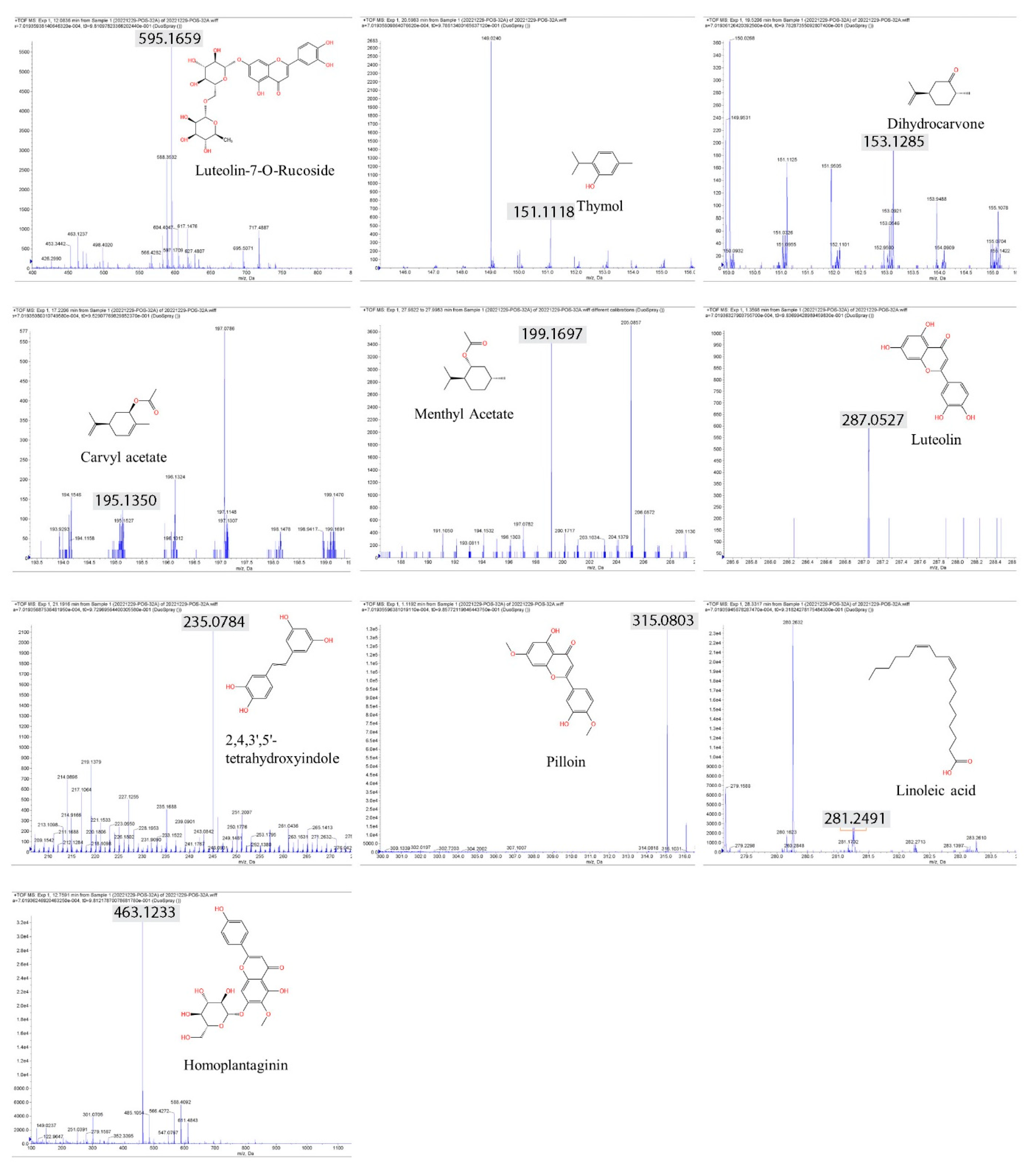
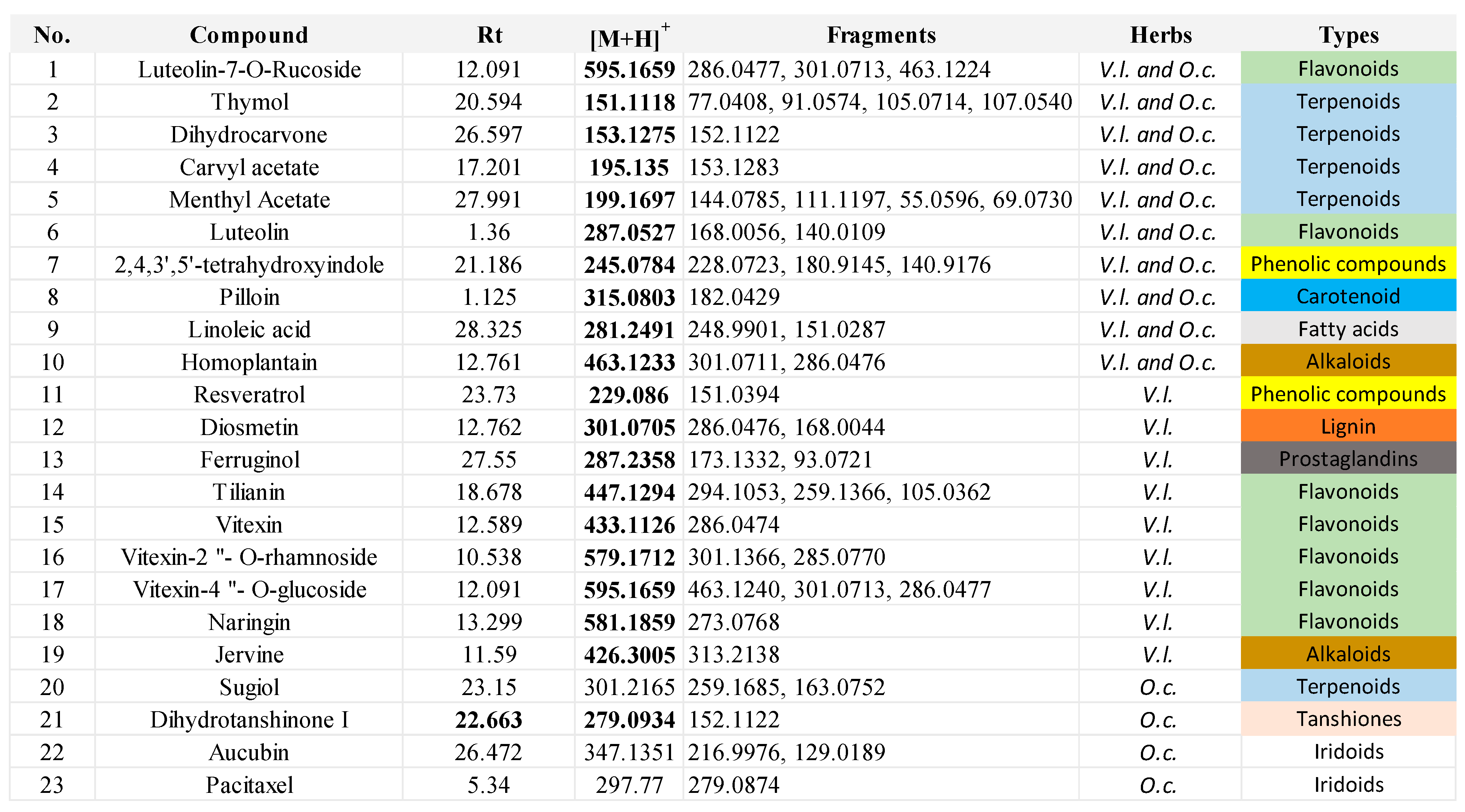
LC‒MS analysis identified anticancer compounds
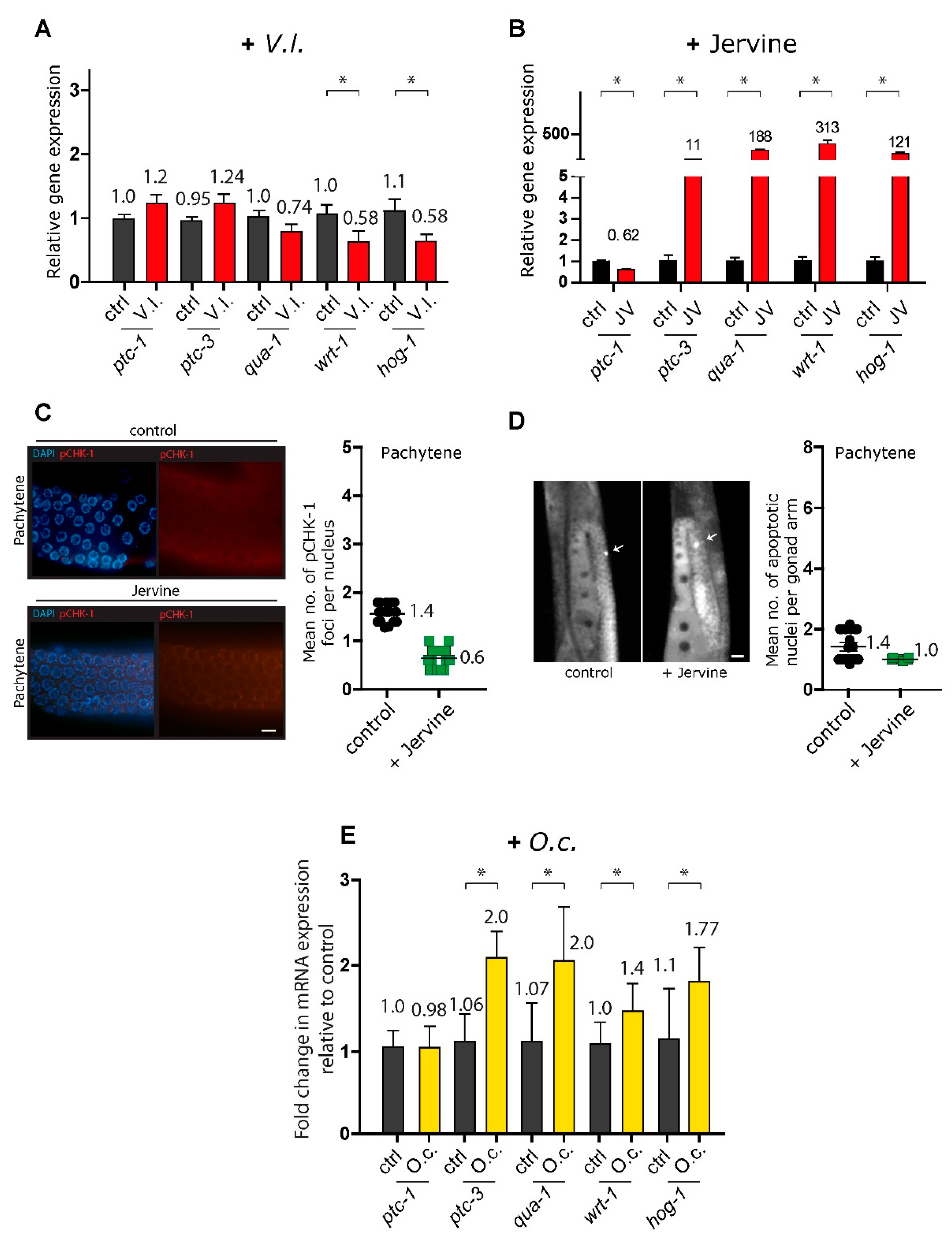
V. lobelianum and the Hedgehog pathway
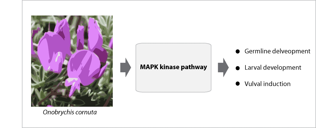
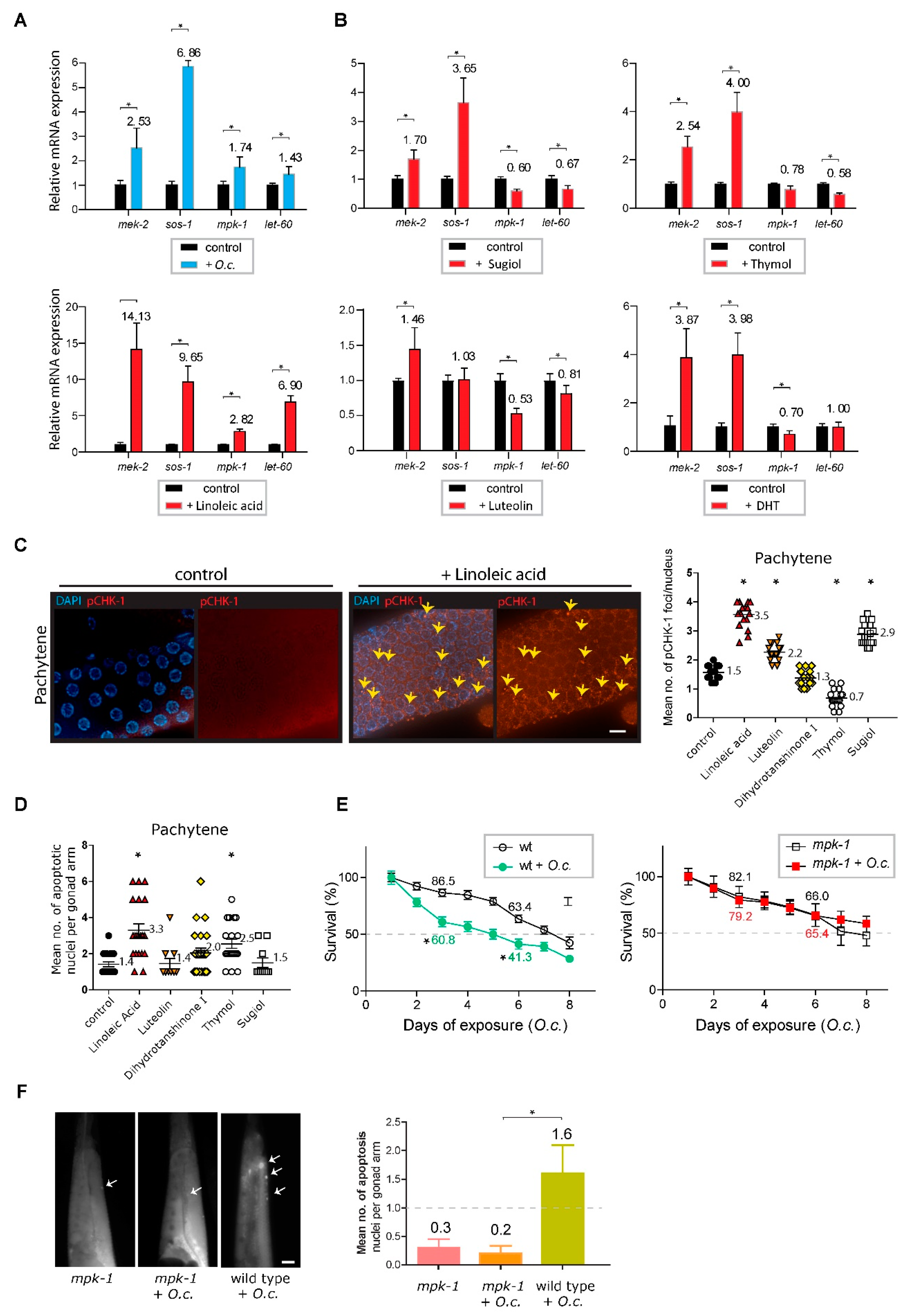
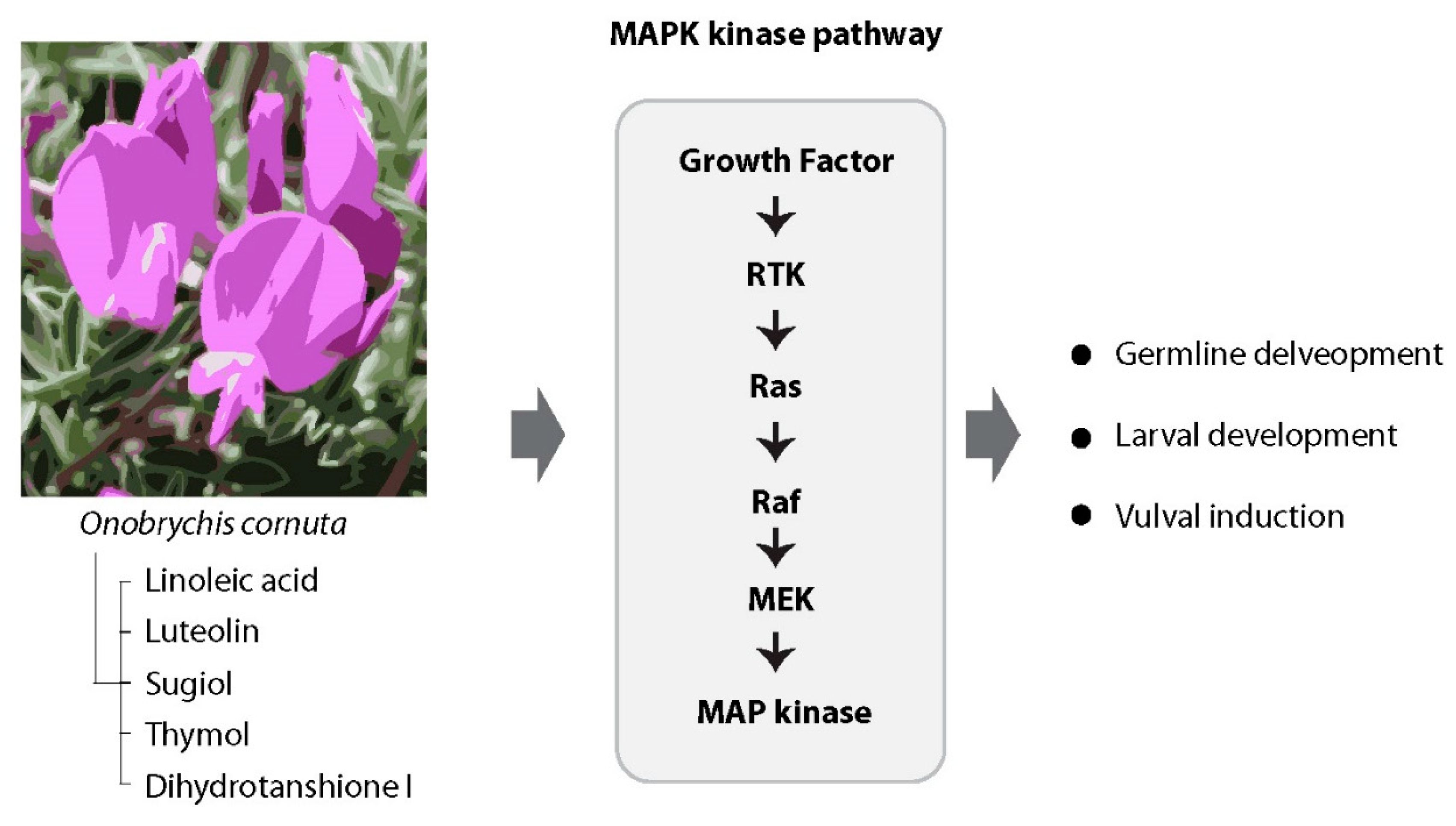
O. cornuta and the MAPK kinase pathway
Discussion
V. lobelianum
O. cornuta
V. lobelianum and O. cornuta Herbal Extracts: Balancing Promise and Toxicity
Materials and Methods
Strains and alleles
Herb extraction
Survival, larval arrest/lethality and HIM
Cumulative survival
Preparation of worm lysates for mass spectrometric analysis
LC‒MS analysis
Monitoring the growth of E. coli
Immunofluorescence staining
Quantitative analysis of pCHK-1 foci
Quantitation of germline apoptosis
Acknowledgments
References
- Report, M.A. Medical Foods Market Size, Share & Trends Analysis Report By Route of Administration (Oral, Enteral), By Product (Pills, Powder, Liquid), By Application, By Sales Channel, And Segment Forecasts, 2022 - 2030. 2020.
- Shaul, N.C.; Jordan, J.M.; Falsztyn, I.B.; Baugh, L.R. Insulin/IGF-dependent Wnt signaling promotes formation of germline tumors and other developmental abnormalities following early-life starvation in Caenorhabditis elegans. Genetics. 2022. [CrossRef]
- Lui, D.Y.; Colaiacovo, M.P. Meiotic development in Caenorhabditis elegans. Advances in experimental medicine and biology 2013, 757, 133–170. [Google Scholar]
- Duronio, R.J.; O’Farrell, P.H.; Sluder, G.; Su, T.T. Sophisticated lessons from simple organisms: appreciating the value of curiosity-driven research. Disease models & mechanisms 2017, 10, 1381–1389. [Google Scholar]
- Wang, Q.; Yang, F.; Guo, W.; Zhang, J.; Xiao, L.; Li, H.; et al. Caenorhabditis elegans in Chinese medicinal studies: making the case for aging and neurodegeneration. Rejuvenation Res. 2014, 17, 205–208. [Google Scholar] [CrossRef] [PubMed]
- Matsunami, K. Frailty and Caenorhabditis elegans as a Benchtop Animal Model for Screening Drugs Including Natural Herbs. Front Nutr. 2018, 5, 111. [Google Scholar] [CrossRef]
- Choi, J.; Ahn, A.; Kim, S.; Won, C.W. Global Prevalence of Physical Frailty by Fried’s Criteria in Community-Dwelling Elderly With National Population-Based Surveys. J Am Med Dir Assoc. 2015, 16, 548–550. [Google Scholar] [CrossRef] [PubMed]
- David, D.C.; Ollikainen, N.; Trinidad, J.C.; Cary, M.P.; Burlingame, A.L.; Kenyon, C. Widespread protein aggregation as an inherent part of aging in C. elegans. PLoS Biol. 2010, 8, e1000450. [Google Scholar] [CrossRef] [PubMed]
- Moliner, C.; López, V.; Barros, L.; Dias, M.I.; Ferreira, I.C.; Langa, E.; et al. Rosemary flowers as edible plant foods: phenolic composition and antioxidant properties in Caenorhabditis elegans. Antioxidants. 2020, 9, 811. [Google Scholar] [CrossRef] [PubMed]
- Sayed, S.M.; Siems, K.; Schmitz-Linneweber, C.; Luyten, W.; Saul, N. Enhanced Healthspan in Caenorhabditis elegans Treated With Extracts From the Traditional Chinese Medicine Plants Cuscuta chinensis Lam. and Eucommia ulmoides Oliv. Frontiers in Pharmacology. 2021, 12, 604435. [Google Scholar] [CrossRef]
- Anjaneyulu, J.; Vidyashankar, R.; Godbole, A. Differential effect of Ayurvedic nootropics on C. elegans models of Parkinson’s disease. Journal of Ayurveda and Integrative Medicine. 2020, 11, 440–447. [Google Scholar] [CrossRef]
- Hodgkin, J.; Horvitz, H.R.; Brenner, S. Nondisjunction Mutants of the Nematode CAENORHABDITIS ELEGANS. Genetics. 1979, 91, 67–94. [Google Scholar] [CrossRef] [PubMed]
- Cinquin, O.; Crittenden, S.L.; Morgan, D.E.; Kimble, J. Progression from a stem cell-like state to early differentiation in the C. elegans germ line. Proc Natl Acad Sci U S A. 2010, 107, 2048–2053. [Google Scholar] [CrossRef] [PubMed]
- Dernburg, A.F.; McDonald, K.; Moulder, G.; Barstead, R.; Dresser, M.; Villeneuve, A.M. Meiotic recombination in C. elegans initiates by a conserved mechanism and is dispensable for homologous chromosome synapsis. Cell. 1998, 94, 387–398. [Google Scholar] [CrossRef] [PubMed]
- Gisselsson, D. Classification of chromosome segregation errors in cancer. Chromosoma. 2008, 117, 511–519. [Google Scholar] [CrossRef] [PubMed]
- Csankovszki, G.; Collette, K.; Spahl, K.; Carey, J.; Snyder, M.; Petty, E.; et al. Three distinct condensin complexes control C. elegans chromosome dynamics. Curr Biol. 2009, 19, 9–19. [Google Scholar] [CrossRef] [PubMed]
- Girard, C.; Roelens, B.; Zawadzki, K.A.; Villeneuve, A.M. Interdependent and separable functions of Caenorhabditis elegans MRN-C complex members couple formation and repair of meiotic DSBs. Proc Natl Acad Sci U S A. 2018, 115, E4443–E4452. [Google Scholar] [CrossRef] [PubMed]
- Hofmann, E.R.; Milstein, S.; Boulton, S.J.; Ye, M.; Hofmann, J.J.; Stergiou, L.; et al. Caenorhabditis elegans HUS-1 is a DNA damage checkpoint protein required for genome stability and EGL-1-mediated apoptosis. Curr Biol. 2002, 12, 1908–1918. [Google Scholar] [CrossRef] [PubMed]
- Kim, H.M.; Colaiacovo, M.P. ZTF-8 Interacts with the 9-1-1 Complex and Is Required for DNA Damage Response and Double-Strand Break Repair in the C. elegans Germline. PLoS Genet. 2014, 10, e1004723. [Google Scholar] [CrossRef] [PubMed]
- Marechal, A.; Zou, L. DNA damage sensing by the ATM and ATR kinases. Cold Spring Harb Perspect Biol. 2013, 5. [Google Scholar] [CrossRef]
- Kim, H.M.; Colaiacovo, M.P. New Insights into the Post-Translational Regulation of DNA Damage Response and Double-Strand Break Repair in Caenorhabditis elegans. Genetics. 2015, 200, 495–504. [Google Scholar] [CrossRef]
- Gartner, A.; Milstein, S.; Ahmed, S.; Hodgkin, J.; Hengartner, M.O. A conserved checkpoint pathway mediates DNA damage--induced apoptosis and cell cycle arrest in C. elegans. Mol Cell. 2000, 5, 435–443. [Google Scholar] [CrossRef]
- Choudhary, R. New Steroidal Alkaloids from Rhizomes of Veratrum album. Journal of Natural Products 1992, 55, 565–570. [Google Scholar]
- Taldaev, A.; Terekhov, R.P.; Melnik, E.V.; Belova, M.V.; Kozin, S.V.; Nedorubov, A.A.; et al. Insights into the Cardiotoxic Effects of Veratrum Lobelianum Alkaloids: Pilot Study. Toxins. 2022, 14, 490. [Google Scholar] [CrossRef] [PubMed]
- Tang, J.; Li, H.L.; Shen, Y.H.; Jin, H.Z.; Yan, S.K.; Liu, X.H.; et al. Antitumor and antiplatelet activity of alkaloids from veratrum dahuricum. Phytother Res. 2010, 24, 821–826. [Google Scholar] [CrossRef]
- Tang, J.; Li, H.L.; Shen, Y.H.; Jin, H.Z.; Yan, S.K.; Liu, R.H.; et al. Antitumor activity of extracts and compounds from the rhizomes of Veratrum dahuricum. Phytother Res. 2008, 22, 1093–1096. [Google Scholar] [CrossRef] [PubMed]
- Chen, J.; Wen, B.; Wang, Y.; Wu, S.; Zhang, X.; Gu, Y.; et al. Jervine exhibits anticancer effects on nasopharyngeal carcinoma through promoting autophagic apoptosis via the blockage of Hedgehog signaling. Biomedicine & Pharmacotherapy. 2020, 132, 110898. [Google Scholar]
- Burglin, T.R.; Kuwabara, P.E. Homologs of the Hh signalling network in C. elegans. WormBook 2006, 1–14. [Google Scholar] [CrossRef]
- Raleigh, D.R.; Reiter, J.F. Misactivation of Hedgehog signaling causes inherited and sporadic cancers. J Clin Invest. 2019, 129, 465–475. [Google Scholar] [CrossRef] [PubMed]
- Wu, Y.; Han, M.; Guan, K.L. MEK-2, a Caenorhabditis elegans MAP kinase kinase, functions in Ras-mediated vulval induction and other developmental events. Genes Dev. 1995, 9, 742–755. [Google Scholar] [CrossRef]
- Zhang, W.; Liu, H.T. MAPK signal pathways in the regulation of cell proliferation in mammalian cells. Cell Res. 2002, 12, 9–18. [Google Scholar] [CrossRef]
- Lackner, M.R.; Kim, S.K. Genetic analysis of the Caenorhabditis elegans MAP kinase gene mpk-1. Genetics. 1998, 150, 103–117. [Google Scholar] [CrossRef]
- W-J Liao Y-MYaD-YZ. Biogeography and evolution of flower color in Veratrum (Melanthiaceae) through inference of a phylogeny based on multiple DNA markers. Pl Syst Evol 2007, 267, 177–190. [CrossRef]
- Melnik, E.; Belova, M.; Tyurin, I.; Ramenskaya, G. Quantitative Content Parameter in the Standardization of Veratrum Aqua, Veratrum Lobelianum Bernh. Based Drug. Drug development & registration. 2021, 10, 107–113. [Google Scholar]
- Yakan S, Aydin T, Gulmez C, Ozden O, Eren Erdogan K, Daglioglu YK, et al. The protective role of jervine against radiation-induced gastrointestinal toxicity. Journal of Enzyme Inhibition and Medicinal Chemistry. 2019, 34, 789–798. [Google Scholar] [CrossRef] [PubMed]
- Zomlefer, W.B.; Williams, N.H.; Whitten, W.M.; Judd, W.S. Generic circumscription and relationships in the tribe Melanthieae (Liliales, Melanthiaceae), with emphasis on Zigadenus: evidence from ITS and trnL-F sequence data. Am J Bot. 2001, 88, 1657–1669. [Google Scholar] [CrossRef] [PubMed]
- ZOMLEFERW; WILLIAMS; JUDD An Overview of Veratrum s.l. (Liliales: Melanthiaceae) and an Infrageneric Phylogeny Based on ITS Sequence Data. Systematic Botany. 2003, 28, 250–269.
- Erkovan HI, Mehmet Kerim Gullap, Şule Erkovan and Ali Koç. HORNED SAINFOIN (ONOBRYCHIS CORNUTA (L.) DESV.): IS IT AN AMUSING OR NUISANCE PLANT FOR STEPPE RANGELANDS? Ecology & Safety. 2016, 10, 418–423. [Google Scholar]
- Joudi, L.a.G.H.B. Exploration of medicinal species of Fabaceae, Lamiaceae and Asteraceae families in Ilkhji region, Eastern Azerbaijan Province (Northwestern Iran). Journal of Medicinal Plants Research. 2010, 4, 1081–1084. [Google Scholar]
- Burcham, P.C. Genotoxic lipid peroxidation products: their DNA damaging properties and role in formation of endogenous DNA adducts. Mutagenesis. 1998, 13, 287–305. [Google Scholar] [CrossRef] [PubMed]
- Inouye, S. Site-specific cleavage of double-strand DNA by hydroperoxide of linoleic acid. FEBS Lett. 1984, 172, 231–234. [Google Scholar] [CrossRef]
- Kodama, T.N.M.K.M. Detection of DNA Damage in Cultured Human Fibroblasts Induced by Methyl Linoleate Hydroperoxide. Agric Biol Chem 1986, 50, 261–262. [Google Scholar]
- Ueda, K.; Kobayashi, S.; Morita, J.; Komano, T. Site-specific DNA damage caused by lipid peroxidation products. Biochim Biophys Acta. 1985, 824, 341–348. [Google Scholar] [CrossRef] [PubMed]
- Beeharry, N.L.J.; Hernandez, A.R.; Chambers, J.A.; Fucassi, F.; Cragg, P.J.; Green, M.H.; Green, I.C. Linoleic acid and antioxidants protect against DNA damage and apoptosis induced by palmitic acid. Mutat Res 2003, 530, 27–33. [Google Scholar] [CrossRef]
- Chang, C.; Hopper, N.A.; Sternberg, P.W. Caenorhabditis elegans SOS-1 is necessary for multiple RAS-mediated developmental signals. EMBO J. 2000, 19, 3283–3294. [Google Scholar] [CrossRef] [PubMed]
- Lee, S.T.; Panter, K.E.; Gaffield, W.; Stegelmeier, B.L. Development of an enzyme-linked immunosorbent assay for the veratrum plant teratogens: cyclopamine and jervine. J Agric Food Chem. 2003, 51, 582–586. [Google Scholar] [CrossRef] [PubMed]
- Ren, X.; Tian, S.; Meng, Q.; Kim, H.M. Histone Demethylase AMX-1 Regulates Fertility in a p53/CEP-1 Dependent Manner. Front Genet. 2022, 13, 929716. [Google Scholar] [CrossRef] [PubMed]
- Zhang X, Tian S, Beese-Sims SE, Chen J, Shin N, Colaiacovo MP, et al. Histone demethylase AMX-1 is necessary for proper sensitivity to interstrand crosslink DNA damage. PLoS Genet. 2021, 17, e1009715. [Google Scholar]
- 49. Kim HM, Colaiacovo MP. DNA Damage Sensitivity Assays in Caenorhabditis elegans. Bio-Protocol. 2015; 5.
- Webster, C.M.; Deline, M.L.; Watts, J.L. Stress response pathways protect germ cells from omega-6 polyunsaturated fatty acid-mediated toxicity in Caenorhabditis elegans. Dev Biol. 2013, 373, 14–25. [Google Scholar] [CrossRef]
- Shin, N.; Cuenca, L.; Karthikraj, R.; Kannan, K.; Colaiacovo, M.P. Assessing effects of germline exposure to environmental toxicants by high-throughput screening in C. elegans. PLoS Genet. 2019, 15, e1007975. [Google Scholar] [CrossRef]
- Sayed, S.M.A.; Siems, K.; Schmitz-Linneweber, C.; Luyten, W.; Saul, N. Enhanced Healthspan in Caenorhabditis elegans Treated With Extracts From the Traditional Chinese Medicine Plants Cuscuta chinensis Lam. and Eucommia ulmoides Oliv. Front Pharmacol. 2021, 12, 604435. [Google Scholar] [CrossRef]
- Penuelas-Urquides, K.; Villarreal-Trevino, L.; Silva-Ramirez, B.; Rivadeneyra-Espinoza, L.; Said-Fernandez, S.; de Leon, M.B. Measuring of Mycobacterium tuberculosis growth. A correlation of the optical measurements with colony forming units. Braz J Microbiol. 2013, 44, 287–289. [Google Scholar] [CrossRef] [PubMed]
- Colaiacovo MP, MacQueen AJ, Martinez-Perez E, McDonald K, Adamo A, La Volpe A, et al. Synaptonemal complex assembly in C. elegans is dispensable for loading strand-exchange proteins but critical for proper completion of recombination. Dev Cell. 2003, 5, 463–474. [Google Scholar] [CrossRef] [PubMed]
- Kelly, K.O.; Dernburg, A.F.; Stanfield, G.M.; Villeneuve, A.M. Caenorhabditis elegans msh-5 is required for both normal and radiation-induced meiotic crossing over but not for completion of meiosis. Genetics. 2000, 156, 617–630. [Google Scholar] [CrossRef] [PubMed]
- Carvalho A, Olson SK, Gutierrez E, Zhang K, Noble LB, Zanin E, et al. Acute drug treatment in the early C. elegans embryo. PLoS One. 2011, 6, e24656. [Google Scholar]
Disclaimer/Publisher’s Note: The statements, opinions and data contained in all publications are solely those of the individual author(s) and contributor(s) and not of MDPI and/or the editor(s). MDPI and/or the editor(s) disclaim responsibility for any injury to people or property resulting from any ideas, methods, instructions or products referred to in the content. |
© 2023 by the authors. Licensee MDPI, Basel, Switzerland. This article is an open access article distributed under the terms and conditions of the Creative Commons Attribution (CC BY) license (http://creativecommons.org/licenses/by/4.0/).


