1. Introduction
Hypercalcemia is defined as a serum calcium level above 2.6 mmol/L (10.7 mg/dL or 5.2 mEq/L). Patients with severe hypercalcemia may exhibit symptoms that include abdominal pain, bone pain, confusion, depression, weakness, kidney stones, or abnormal electrical cardiac rhythms, including cardiac arrest [
1]. Calcium is the most abundant extracellular cation in the human body and acts as a secondary messenger and cofactor of several enzymes. In terms of etiology, neoplasia is the most common cause of hypercalcemia in hospitalized and emergency patients, and primary hyperparathyroidism is the predominant cause in outpatient cases [
2].
According to the World Health Organization mortality database, over 700,000 people die by committing suicide each year, which makes it the fourth leading cause of death among people under the age of 30 years [
3]. A nationwide population study in East Asia revealed that suicide attempts with psychiatric illness occurred more frequently and exhibited a more serious pattern [
4]. European data indicated significant gender differences in suicide methods and lethality, with men often choosing more violent and intentional methods [
5]. Pesticide intoxication is prevalent in many Asian countries and Latin America, particularly among women, and highlights the significance of self-poisoning, including the use of pesticides or drugs [
6].
While chemical intoxication constitutes only a minor portion of poisoning cases, most ingestion-related poisonings are caused by prescription drugs [
7]. Deliberate intoxication with chemical substances, excluding certain agricultural products, is rarely reported, and commercial products like dehumidifying agents that contain calcium chloride (CaCl
2) as their main ingredient are commonly used. Although cases of CaCl
2 intoxication have been reported, most consist of gastric necrosis with or without surgical treatment and metabolic acidosis that requires continuous renal replacement therapy (CRRT) [
8,
9,
10]. Even though CaCl
2 is known as a strong stimulant of the skin and mucous membrane, massive CaCl
2 ingestion accidents sometimes occur, especially in patients with developmental disorders or dementia. We present a case of severe CaCl
2 intoxication with hematemesis and hypercalcemia and focus on the in-hospital course with an emphasis on the cardiologic manifestations.
2. Case presentation
A 39-year-old female visited the Regional Emergency Center after vomiting bloody material. She explained that she had ingested a pack of commercial desiccant with a substantial amount of tap water approximately 2 h before her visit, which indicates suicidal intent. Subsequently, she called the rescue center herself, but upon arriving at the emergency department, she was unwilling to answer the medical staff’s questions. Although her eyes remained open throughout, her reactions were extremely limited. The initial examining doctor, through several attempts at cautious history-taking, found no previous specific diseases, including psychiatric conditions. Because of the lack of next of kin or caregivers, obtaining further medical history was challenging. Our focus was primarily on detecting changes in her signs during bedside observation.
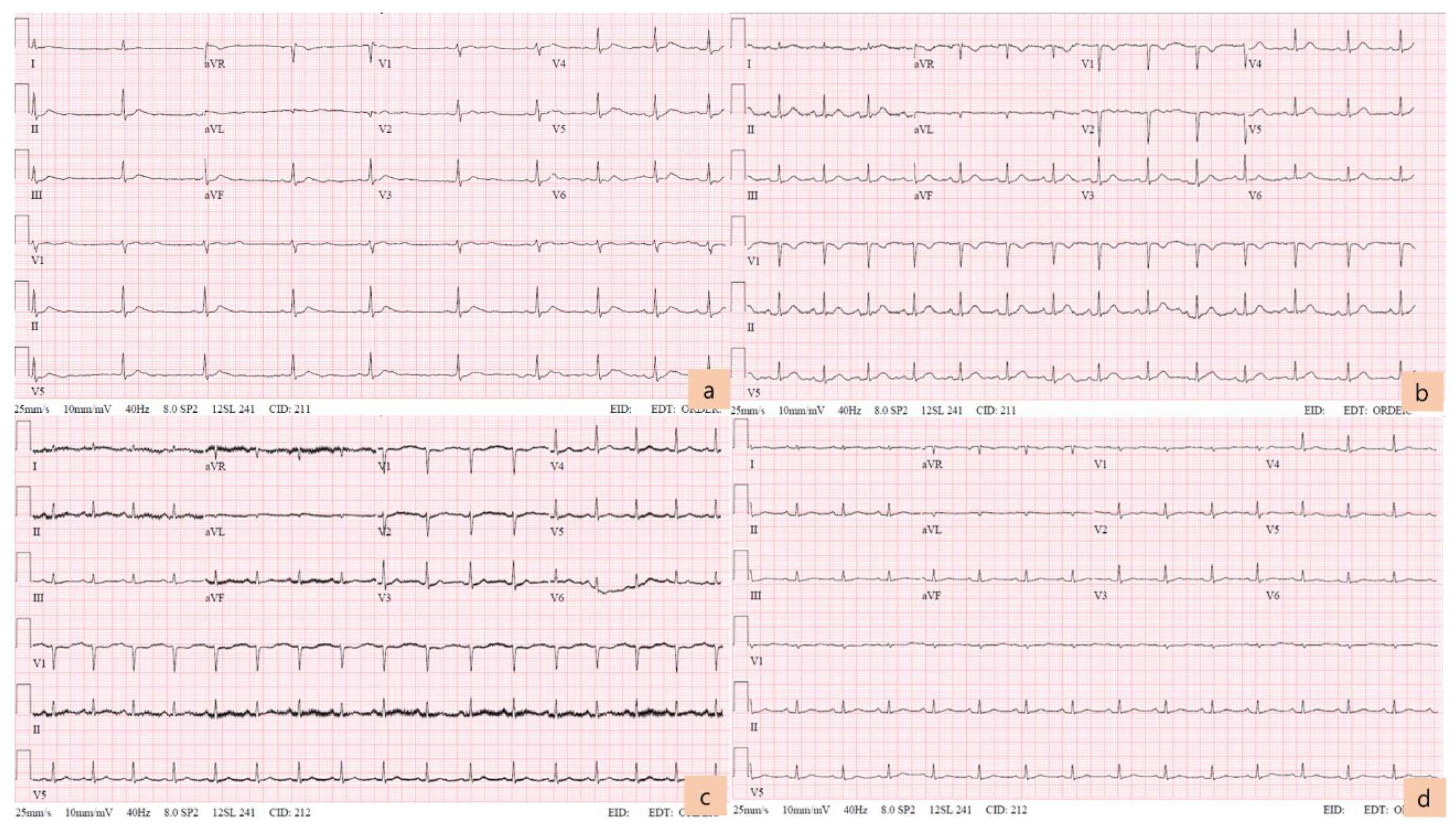
Her initial vital signs were as follows: blood pressure, 150/90 mmHg; heart rate, 51 beats/min; body temperature, 36.2°C; and respiratory rate, 18 breaths/minute. The electrocardiogram (ECG) showed a relatively short QT interval (QTc 387 ms), flattened T wave, and a subtle U wave or T-U fusion wave on the II, III, aVF, and precordial leads (
Figure 1a). Blood gas analysis revealed metabolic alkalosis with respiratory compensation (pH 7.55, pCO
2 20.5 mmHg, pO
2 177 mmHg, HCO
3- 18 Mmol/L, and SaO
2 99.6%), and hyperlactatemia (3.4 mmol/L). Cardiac enzyme levels were all elevated: hsTnI, 0.091 ng/mL (0–0.04); CK-MB, 5.8 ng/mL (0–4.7); and myoglobin, 177.5 ng/mL (0–106). A mild electrolyte imbalance was present: serum potassium was 2.7 mEq/L initially and decreased to 2.5 mEq/L at the 1-h follow up.
During observation, serial blood sampling was performed. After 12 h post-ingestion, her ionized calcium level increased to 1.58 mmol/L (1.15–1.29), and her serum calcium and phosphorus levels were 12.3 mg/dL and 19.0 ng/mL, respectively. The cardiac enzyme levels were also increased: hsTnI was 2.203 ng/mL and CK-MB was 19 ng/mL, but the potassium level normalized to 3.8 mEq/L. At that time, her ECG changed slightly. Even though the heart rate increased to 84 bpm, the corrected QT interval was within the normal range, and the shape of the T wave became more distinguishable. However, there remained a fluttering of the T-P interval in the precordial leads (
Figure 1b).
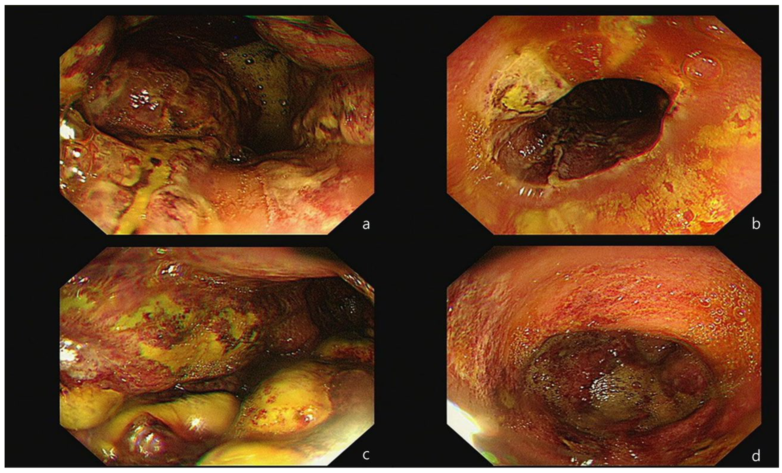
Because the bloody vomiting continued occasionally until the next morning, an esophago-gastro-duodenoscopy (EGD) was performed, under consultation, to identify the source of the ongoing bleeding and assess the severity of the intestinal injury. The procedure revealed a thick exudate with linear ulceration and hemorrhagic spots in the lower esophagus. Additionally, extensive necrosis and multiple ulcerations with brown-grayish discoloration were observed throughout the entire stomach (
Figure 2). As a result, she was diagnosed with corrosive gastritis, grade IIIB. A decision was made to admit her and maintain an NPO status until she was re-evaluated for the resumption of a normal diet. Throughout this period, her vital signs remained consistently stable.
On day 3 after admission, there were considerable changes in her blood test results. Hypocalcemia emerged inversely, and mild hyperkalemia coexisted. The ECG revealed a rather shortened QT interval (
Figure 1c), and cardiac enzymes gradually decreased. The patient complained of mild epigastric pain but did not report any additional symptoms. Definite tenderness was not prominent on the physical examination. The following day, hypocalcemia, hypophosphatemia, and hypokalemia worsened simultaneously, with the persistence of the relatively shortened QT interval on the ECG. Because of the lack of next of kin, financial constraints, and the patient’s reluctance to communicate severe negative symptoms to the medical team, further evaluation, including laboratory tests for vitamins or hormones, or a psychiatric interview, could not be conducted.
For nearly a week, hypocalcemia and hypophosphatemia persisted (
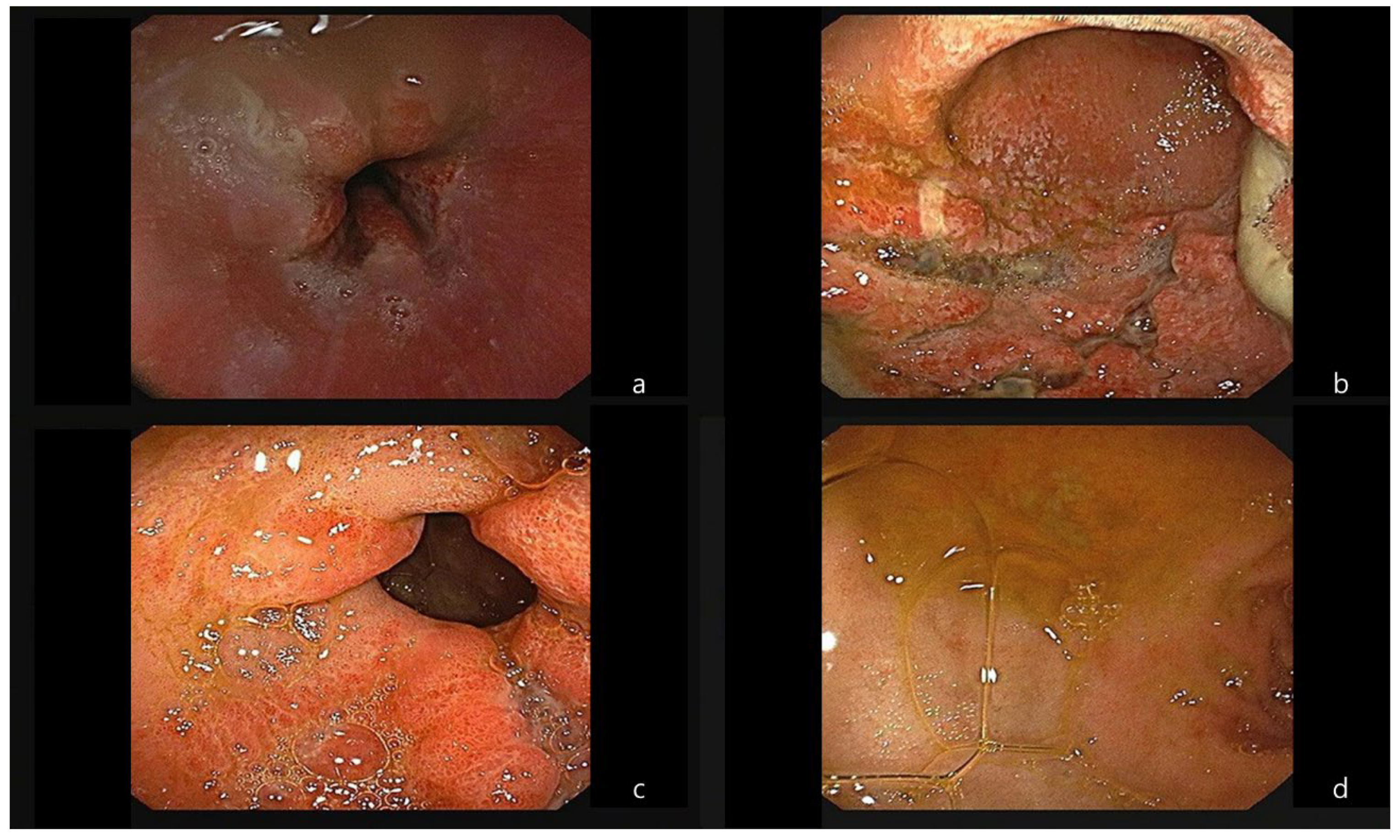
Table 1). Meanwhile, the patient complained of hunger, and although there was the possibility of intestinal lumen injury on follow-up EGD (
Figure 3), a decision was made to start her on a soft diet. There was no aggravation of abdominal pain or signs of gastrointestinal bleeding during this period. However, due to financial constraints regarding medical expenses, the patient had to be transferred.
3. Discussion
Most cases of hypercalcemia are caused by primary hyperparathyroidism or malignancy, and hypercalcemia in patients with cancer is associated with a high mortality rate [
2]. In a retrospective study, milk-alkali or calcium-alkali syndrome was found to be the third leading cause of hypercalcemia in hospitalized patients without end-stage renal disease [
11]. Numerous cases of prescription drug-induced hypercalcemia have been reported and are typically associated with various comorbidities and renal dysfunction. However, in this case, the patient was relatively healthy physically, with the only presumed serious condition being psychiatric. According to the Society for Endocrinology endocrine emergency guidance, mild hypercalcemia below 3.0 mmol/L is often asymptomatic and does not usually require urgent correction [
12]. Correspondingly, in this case, the elevated calcium levels improved with supportive care and without the need for other invasive therapeutic interventions, such as hemodialysis.
Following the initial laboratory test results, a significant elevation in the cardiac enzymes was observed. The follow-up ECGs did not reveal ST-segment or T-wave abnormalities, and definite regional wall motion restriction was not detected in the bedside ECG. A case report on an elderly patient with ischemic cardiomyopathy and renal insufficiency suggested that hypercalcemia combined with hyperkalemia (with a serum calcium level of 3.55 mmol/L) could induce an elevated ST-segment in the ECG [
13]. The authors emphasized that even in the absence of concerning symptoms and a high probability, ECG changes that resemble an ST-segment elevation myocardial infarction should be investigated for potential mimickers. In this case, the serum calcium level was not sufficiently high enough to require aggressive correction, which resulted in the subtle accompanying ECG changes. The most common ECG changes that occur with hypercalcemia are a shortened QT interval and conduction disorders, which is consistent with the aforementioned guidance [
2,
12,
13]. Elevated calcium levels lower the excitation threshold of the myocardium, decrease cardiac conduction velocity, and shorten the refractory time, especially in phase 2 of the action potential [
14].
In addition to the rapid electrolyte imbalance, stress-induced cardiomyopathy (SICMP) can be considered a major cause that explains the rise in cardiac enzyme levels in patients. Burns are one of the major causes of SICMP, along with severe medical conditions, emotional stress, and traumatic events. Left ventricular dysfunction without a definite obstructive coronary lesion, including ST changes in the ECG, can define SICMP [
15]. Based on several experimental studies, burn-induced cardiomyopathy may be triggered by mitochondrial damage, which is derived from the surge of catecholamine secretion, alteration of Ca
2+ transient proteins, and increased inflammatory responses [
16]. Chemical burns induced by CaCl
2 can cause damage to the myocytes, and it can be inferred that calcium absorption greatly contributes to worsening myocyte injury.
Due to the patient’s financial constraints, long-term follow up on her medical condition was not feasible. While the patient’s general condition improved relatively quickly, the electrolyte imbalance persisted. Sudden and extensive elevation of serum calcium levels might suppress the secretion of parathyroid hormone, which leads to bone resorption, a reduction in 1,25-dihydroxyvitamin D
3, and the subsequent decreased absorption of intestinal calcium [
17]. Although these consequences cannot be proven with laboratory results, they can be inferred from the physiological process.
Several case reports about dehumidifier-related poisoning presented with severe intestinal injuries that required surgical emergency intervention, severe metabolic acidosis necessitating CRRT, and even fatal outcomes [
8,
9,
10]. Fortunately, our patient recovered with only supportive care. We assume that vigorous vomiting immediately after she ingested a large amount of tap water might have limited the CaCl
2 absorption. This likely contributed to less severe necrosis of the intestinal mucosa and a milder elevation of serum calcium levels. Instead of many tests and expensive treatments, the close observation of the patient’s condition and conservative treatment led to a favorable outcome, which can be academically rewarding.
4. Conclusion
Sometimes, physicians can meet patients ingesting CaCl2 for suicidal attempt. Most cases of CaCl2 intoxication are related to the upper intestinal injury due to thermal burn, but some of them can experience severe electrolyte imbalance with cardiomyopathy. Therefore, we have to pay attention to the patients’ cardiac manifestion as well as gastrointestinal problems.
Author Contributions
Conceptualization, Shin SJ. and Kim YJ.; Methodology, Shin SJ.; Validation, Kim YJ.; Resources, Shin SJ.; Writing – Original Draft Preparation, Shin SJ.; Writing – Review & Editing, Kim YJ.; Visualization, Shin SJ.; Supervision, Kim YJ; Project Administration, Kim YJ.
Funding
This research received no external funding.
Institutional Review Board Statement
This study is waived to IRB approval due to no more risk than minimal level.
Informed Consent Statement
Patient consent was waived due to loss of contact, and our report is a retrospectively gathered data-based study.
Data Availability Statement
The data presented in this study are available on request from the corresponding author. The data are not publicly available due to privacy.
Conflicts of Interest
The authors declare no conflict of interest.
References
- Minisola S, Pepe J, Piemonte S, Cipriani C. The diagnosis and management of hypercalcaemia. BMJ 2015; 350. [CrossRef]
- Tonon CR, Taline Alisson Artemis Lazzarin Silva, Filipe Welson Leal Pereira, Diego Aparecido Rios Queiroz, Favero EL Jr., Martins D, et al. Review of Current Clinical Concepts in the Pathophysiology, Etiology, Diagnosis, and Management of Hypercalcemia. Med Sci Monit. 2022; 28: e935821-1–e935821-9. [CrossRef]
- World Health Organization. https://www.who.int/news-room/fact-sheets/detail/suicide [accessed on 10 Nov 2023].
- Chen CK, Chan YL, Su TH. Incidence of intoxication events and patient outcomes in Taiwan: A nationwide population-based observational study. PLoS One. 2020; 15(12): e0244438. [CrossRef]
- Mergl R, Koburger N, Heinrichs K, Székely A, Tóth M.D., Coyne J, Quintao S., Arensman E., Coffey C., Maxwell M., et al. What Are Reasons for the Large Gender Differences in the Lethality of Suicidal Acts? An Epidemiological Analysis in Four European Countries. PLoS One. 2015; 10(7): e0129062. [CrossRef]
- Ajdacic-Gross V, Weiss MG, Ring M, Hepp U, Bopp M, Gutzwiller F, Rösslera W. Methods of suicide: international suicide patterns derived from the WHO mortality database. Bull World Health Organ. 2008 Sep; 86(9): 726–732. [CrossRef]
- Zhang YJ, Yu BX, Wang NN, Li TG. Acute poisoning in Shenyang, China: a retrospective and descriptive study from 2012 to 2016. BMJ Open. 2018; 8(8): e021881. [CrossRef]
- Nakagawa YI, Maeda AY, Takahashi, Kaneoka YJ. Gastric Necrosis because of Ingestion of Calcium Chloride. ACG Case Rep J. 2020 Aug; 7(8): e00446. [CrossRef]
- Cho KH, Seo BS, Koh HS, Yang HB. Fatal case of commercial moisture absorber ingestion. BMJ Case Rep. 2018; 2018: bcr2018225121. [CrossRef]
- Jung YM, Kim HJ, Park JH, Yang KM. Death Related to the Dehumidifying Agent. Korean J Leg Med 2016; 40:133-137. [CrossRef]
- Picolos MK, Lavis VR, Orlander PR. Milk-Alkali syndrome is a major cause of hypercalcaemia among non-end-stage renal disease (non-ESRD) inpatients. Clin Endocrinol 2005; 63: 566–76. [CrossRef]
- Walsh J, Gittoes N, Selby P, the Society for Endocrinology Clinical Committee. SOCIETY FOR ENDOCRINOLOGY ENDOCRINE EMERGENCY GUIDANCE: Emergency management of acute hypercalcaemia in adult patients. Endocr Connect. 2016 Sep; 5(5): G9–G11. [CrossRef]
- Strand AO, Aung TT, Agarwal A. Not all ST-segment changes are myocardial injury: hypercalcaemia-induced ST-segment elevation. BMJ Case Rep. 2015; 2015: bcr2015211214. [CrossRef]
- Vella A, Gerber T, Hayes D, Reeder G. Digoxin, hypercalcaemia, and cardiac conduction. Postgrad Med J. 1999 Sep; 75(887): 554–556. [CrossRef]
- Yerasi C, Koifman E, Weissman G, Wang ZY, Torguson R, Gai JX, Lindsay J, Satler L.F., Pichard A.D., Waksman R., et al.. Impact of triggering event in outcomes of stress-induced (takotsubo) cardiomyopathy. Eur Heart J. Acute Cardiovasc Care. 2017; 6:280–286. [CrossRef]
- Wang MJ, Scott SR, Koniaris LG, Zimmers TA. Pathological Responses of Cardiac Mitochondria to Burn Trauma. Int J Mol Sci. 2020 Sep; 21(18): 6655. [CrossRef]
- Davies JH, Shaw NJ. Investigation and management of hypercalcaemia in children. Arch Dis Child 2012 Jun;97(6):533-8. [CrossRef]
|
Disclaimer/Publisher’s Note: The statements, opinions and data contained in all publications are solely those of the individual author(s) and contributor(s) and not of MDPI and/or the editor(s). MDPI and/or the editor(s) disclaim responsibility for any injury to people or property resulting from any ideas, methods, instructions, or products referred to in the content. |
© 2023 by the authors. Licensee MDPI, Basel, Switzerland. This article is an open access article distributed under the terms and conditions of the Creative Commons Attribution (CC BY) license (http://creativecommons.org/licenses/by/4.0/).


