Submitted:
05 January 2024
Posted:
08 January 2024
You are already at the latest version
Abstract
Keywords:
1. Introduction
2. Materials and Methods
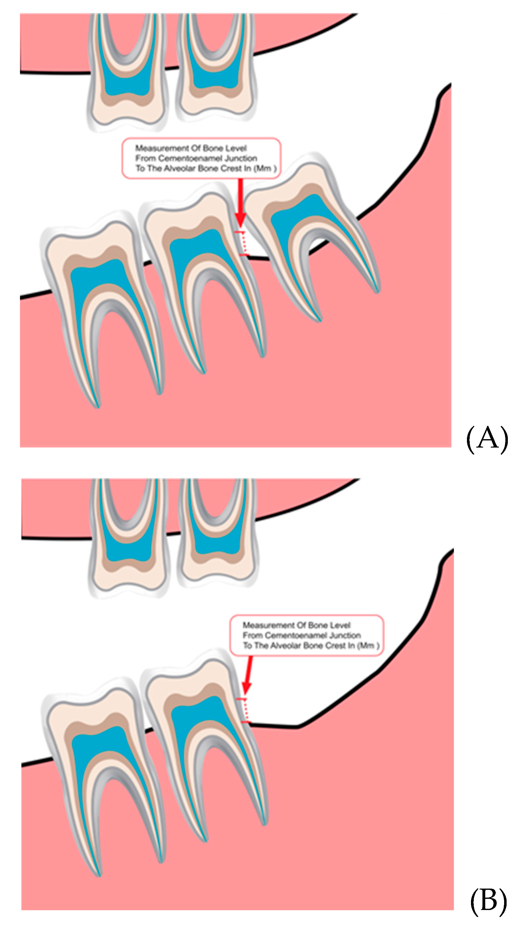
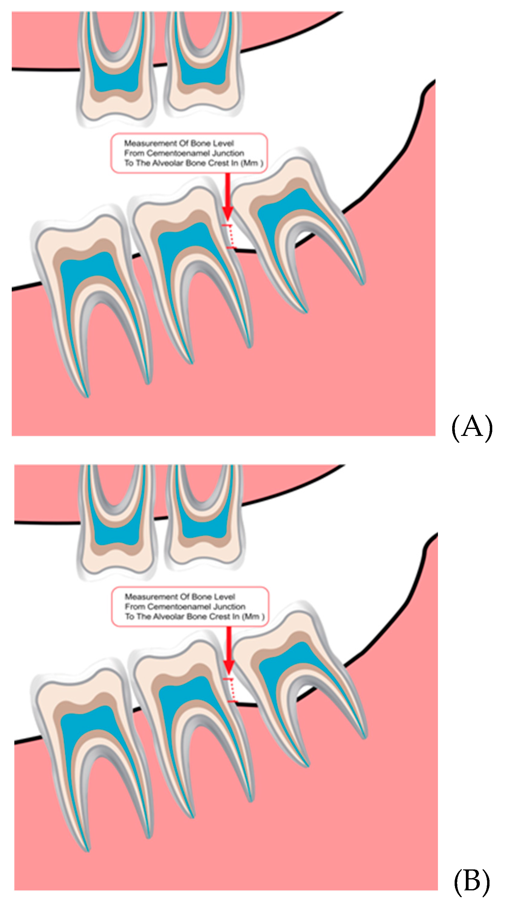
2.1. Study Description
2.2. Study Description
2.3. Inclusion and Exclusion Criteria
2.4. Inclusion and Exclusion Criteria
2.5. Statistical Analysis
3. Results
3.1. Comparison of Bone Levels among the Groups
3.2. Comparison of Bone Levels among the Groups
3.3. Bone Changes among the Two Groups Based on the Age Groups
3.4. Pattern of Impaction
3.5. Impaction Side
4. Discussion
5. Conclusions
Author Contributions
Funding
Institutional Review Board Statement
Informed Consent Statement
Data Availability Statement
Acknowledgments
Conflicts of Interest
References
- Alfadil L, Almajed E. Prevalence of impacted third molars and the reason for extraction in Saudi Arabia. Saudi Dent J. 2020 Jul;32(5):262-268. [CrossRef]
- Gupta S, Bhowate RR, Nigam N, Saxena S. Evaluation of Impacted Mandibular Third Molars by Panoramic Radiography. ISRN Dent. 2011 Sep; 2011:1–8. [CrossRef]
- Pedro FLM, Bandéca MC, Volpato LER, Marques ATC, Borba AM, Musis CR de, et al. Prevalence of impacted teeth in a Brazilian subpopulation. J Contemp Dent Pract. 2014 Mar;15(2):209–13. [CrossRef]
- Breik O, Grubor D. The incidence of mandibular third molar impactions in different skeletal face types. Aust Dent J. 2008 Dec;53(4):320–4. [CrossRef]
- Padhye MN, Dabir A V., Girotra CS, Pandhi VH. Pattern of mandibular third molar impaction in the Indian population: a retrospective clinico-radiographic survey. Oral Surg Oral Med Oral Pathol Oral Radiol. 2013 Sep;116(3):e161–6. [CrossRef]
- KalaiSelvan S, Ganesh SKN, Natesh P, Moorthy MS, Niazi TM, Babu SS. Prevalence and Pattern of Impacted Mandibular Third Molar: An Institution-based Retrospective Study. J Pharm Bioallied Sci. 2020 Aug;12(Suppl 1):S462–7. [CrossRef]
- Prajapati VK, Mitra R, Vinayak KM. Pattern of mandibular third molar impaction and its association to caries in mandibular second molar: A clinical variant. Dent Res J (Isfahan). 2017;14(2):137–42. [CrossRef]
- Hassan AH. Pattern of third molar impaction in a Saudi population. Clin Cosmet Investig Dent. 2010 Oct 11;2:109-13. [CrossRef]
- Arora A, Patil BA, Sodhi A. Validity of the vertical tube-shift method in determining the relationship between the mandibular third molar roots and the inferior alveolar nerve canal. J Korean Assoc Oral Maxillofac Surg. 2015 Apr;41(2):66-73. [CrossRef]
- Cederhag J, Truedsson A, Alstergren P, Shi XQ, Hellén-Halme K. Radiographic imaging in relation to the mandibular third molar: a survey among oral surgeons in Sweden. Clin Oral Investig. 2022 Feb;26(2):2073-2083. [CrossRef]
- Shukla S, Chug A, Afrashtehfar KI. Role of Cone Beam Computed Tomography in Diagnosis and Treatment Planning in Dentistry: An Update. J Int Soc Prev Community Dent. 2017 Nov;7(Suppl 3):S125-S136. [CrossRef]
- Ariizumi D, Sakamoto T, Yamamoto M, Nishii Y. External Root Resorption of Second Molars Due to Impacted Mandibular Third Molars during Orthodontic Retention. Bull Tokyo Dent Coll. 2022 Sep;63(3):129-138. [CrossRef]
- Jaju PP, Jaju SP. Cone-beam computed tomography: Time to move from ALARA to ALADA. Imaging Sci Dent. 2015 Dec;45(4):263-5. [CrossRef]
- Faria AI, Gallas-Torreira M, López-Ratón M. Mandibular second molar periodontal healing after impacted third molar extraction in young adults. J Oral Maxillofac Surg. 2012 Dec;70(12):2732-41. [CrossRef]
- Pham TAV, Nguyen NH. Periodontal Status of the Adjacent Second Molar after Impacted Mandibular Third Molar Surgical Extraction. Contemp Clin Dent. 2019 Apr-Jun;10(2):311-318. [CrossRef]
- McHugh ML. Interrater reliability: the kappa statistic. Biochem Med (Zagreb). 2012;22(3):276–82. [CrossRef]
- Krausz AA, Machtei EE, Peled M. Effects of lower third molar extraction on attachment level and alveolar bone height of the adjacent second molar. Int J Oral Maxillofac Surg. 2005 Oct;34(7):756-60. [CrossRef]
- Dias MJ, Franco A, Junqueira JL, Fayad FT, Pereira PH, Oenning AC. Marginal bone loss in the second molar related to impacted mandibular third molars: comparison between panoramic images and cone beam computed tomography. Med Oral Patol Oral Cir Bucal. 2020 May;25(3):e395-e402. [CrossRef]
- Fernandes IA, Galvão EL, Gonçalves PF, Falci SGM. Impact of the presence of partially erupted third molars on the local radiographic bone condition. Sci Rep. 2022 May;12(1):8683. [CrossRef]
- Tai S, Zhou Y, Pathak JL, Piao Z, Zhou L. The association of mandibular third molar impaction with the dental and periodontal lesions in the adjacent second molars. J Periodontol. 2021 Oct;92(10):1392-1401. [CrossRef]
- Tolentino PHMP, Rodrigues LG, Miranda de Torres É, Franco A, Silva RF. Extractions in Patients with Periodontal Diseases and Clinical Decision-Making Process. Acta Stomatol Croat. 2019 Jun;53(2):141-149. [CrossRef]
- Passarelli PC, Lajolo C, Pasquantonio G, D'Amato G, Docimo R, Verdugo F, D'Addona A. Influence of mandibular third molar surgical extraction on the periodontal status of adjacent second molars. J Periodontol. 2019 Aug;90(8):847-855. [CrossRef]
- Montero J, Mazzaglia G. Effect of removing an impacted mandibular third molar on the periodontal status of the mandibular second molar. J Oral Maxillofac Surg. 2011 Nov;69(11):2691-7. [CrossRef]
- Kan KW, Liu JK, Lo EC, Corbet EF, Leung WK. Residual periodontal defects distal to the mandibular second molar 6-36 months after impacted third molar extraction. J Clin Periodontol. 2002 Nov;29(11):1004-11. [CrossRef]
- Al-Dajani M, Abouonq AO, Almohammadi TA, Alruwaili MK, Alswilem RO, Alzoubi IA. A Cohort Study of the Patterns of Third Molar Impaction in Panoramic Radiographs in Saudi Population. Open Dent J. 2017 Dec 26;11:648-660. [CrossRef]
- Yilmaz S, Adisen MZ, Misirlioglu M, Yorubulut S. Assessment of Third Molar Impaction Pattern and Associated Clinical Symptoms in a Central Anatolian Turkish Population. Med Princ Pract. 2016;25(2):169-75. [CrossRef]
- Alsaegh MA, Abushweme DA, Ahmed KO, Ahmed SO. The pattern of mandibular third molar impaction and its relationship with the development of distal caries in adjacent second molars among Emiratis: a retrospective study. BMC Oral Health. 2022 Jul;22(1):306. [CrossRef]
- Eshghpour M, Nezadi A, Moradi A, Shamsabadi RM, Rezaei NM, Nejat A. Pattern of mandibular third molar impaction: A cross-sectional study in northeast of Iran. Niger J Clin Pract. 2014 Nov-Dec;17(6):673-7. [CrossRef]
- Zaman MU, Almutairi NS, Abdulrahman Alnashwan M, Albogami SM, Alkhammash NM, Alam MK. Pattern of Mandibular Third Molar Impaction in Nonsyndromic 17760 Patients: A Retrospective Study among Saudi Population in Central Region, Saudi Arabia. Biomed Res Int. 2021 Aug;2021:1880750. [CrossRef]
- Quek SL, Tay CK, Tay KH, Toh SL, Lim KC. Pattern of third molar impaction in a Singapore Chinese population: a retrospective radiographic survey. Int J Oral Maxillofac Surg. 2003 Oct;32(5):548–52. [CrossRef]


| Group | Baseline Bone Level Mean SD* |
Bone Level after Extraction/Follow-up Mean SD |
p-Value |
|---|---|---|---|
| Study group | 3.00 ± 1.68 | 2.63 ± 1.75 | 0.0001 |
| Control group | 2.73 ± 1.75 | 3.01 ± 1.98 | 0.001 |
| Groups | Gender | Baseline BL1 | Follow up BL2 | Bone Change df (mm)3 | p-Value |
|---|---|---|---|---|---|
| Study | Male | 3.51 ± 1.89 | 3.27 ± 2.17 | 0.24 ± 1.29 | 0.190 |
| Female | 2.73 ± 1.35 | 2.29 ± 1.41 | 0.44 ± 1.13 | ||
| Control | Male | 3.64 ± 1.79 | 4.12 ± 2.11 | -0.48 ± 0.66 | 0.734 |
| Female | 1.97 ± 1.32 | 2.11 ± 1.3 | -0.13 ± 0.31 |
| Groups | Age Groups | Baseline BL | Follow up BL | Bone Change df (mm) | p-Value |
|---|---|---|---|---|---|
| Study | 18–28 | 2.48 ± 1.51 | 2.18 ± 1.57 | 0.29 ± 1.07 | 0.05 |
| 29–39 | 3.25 ± 1.69 | 2.29 ± 1.51 | 0.96 ± 1.05 | ||
| 40–50 | 4.30 ± 3.53 | 5.50 ± 3.39 | -1.20 ± 0.14 | ||
| ≥51 | 3.51 ± 1.53 | 3.45 ± 1.44 | 0.05 ± 1.34 | ||
| Control | 18–28 | 2.93 ± 1.94 | 3.19 ± 2.13 | -0.26 ± 0.53 | 0.07 |
| 29–39 | 2.28 ± 1.32 | 2.45 ± 1.50 | -0.17 ± 0.25 | ||
| 40–50 | 2.20 ± 0.71 | 2.38 ± 0.46 | -0.17 ± 0.25 | ||
| ≥51 | 2.58 ± 1.72 | 3.16 ± 2.35 | -0.57 ± 0.69 |
| Classification | Inclination | Study Group (N) (F) |
Control Group (N) (F) |
Total Sample (N) (F) |
P-Value | |||
|---|---|---|---|---|---|---|---|---|
| Winter’s classification | Vertical | 31 | 44.9% | 35 | 50% | 66 | 41.25% | 0.539 |
| Mesioangular | 26 | 37.7% | 20 | 28.6% | 46 | 28.75% | ||
| Distoangular | 1 | 1.4% | 0 | 0% | 1 | 1.25% | ||
| Horizontal | 10 | 14.5% | 15 | 21.4% | 25 | 16.62% | ||
| Buccally tilted | 1 | 1.4% | 0 | 0% | 1 | 1.25% | ||
| Total | 69 | 86.25% | 70 | 87.5% | 139 | 82.28% | ||
| Impaction Side | Occurrence |
Study Group (N) (F) |
Control Group (N) (F) |
Total Sample (N) (F) |
P-Value | |||
| Right | 6 | 15% | 3 | 7.5% | 9 | 11.3% | 0.509 | |
| Left | 5 | 12.5% | 7 | 17.5% | 12 | 15% | ||
| Bilateral | 29 | 72.5% | 30 | 75% | 59 | 73.8% | ||
Disclaimer/Publisher’s Note: The statements, opinions and data contained in all publications are solely those of the individual author(s) and contributor(s) and not of MDPI and/or the editor(s). MDPI and/or the editor(s) disclaim responsibility for any injury to people or property resulting from any ideas, methods, instructions or products referred to in the content. |
© 2024 by the authors. Licensee MDPI, Basel, Switzerland. This article is an open access article distributed under the terms and conditions of the Creative Commons Attribution (CC BY) license (http://creativecommons.org/licenses/by/4.0/).


