Submitted:
08 January 2024
Posted:
09 January 2024
You are already at the latest version
Abstract
Keywords:
1. Ornamental Geophytes History
2. World Ornamental Plants Sector – Flower Bulbs situation in the sector
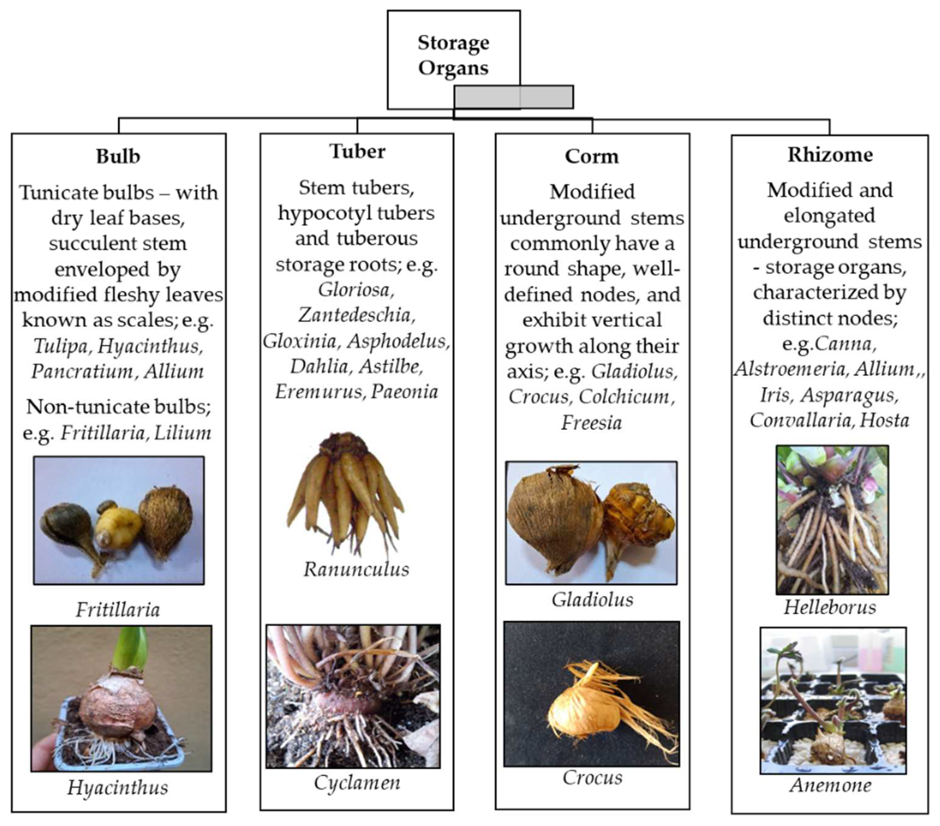
3. Flower bulbs propagation and challenges
4. Micropropagation
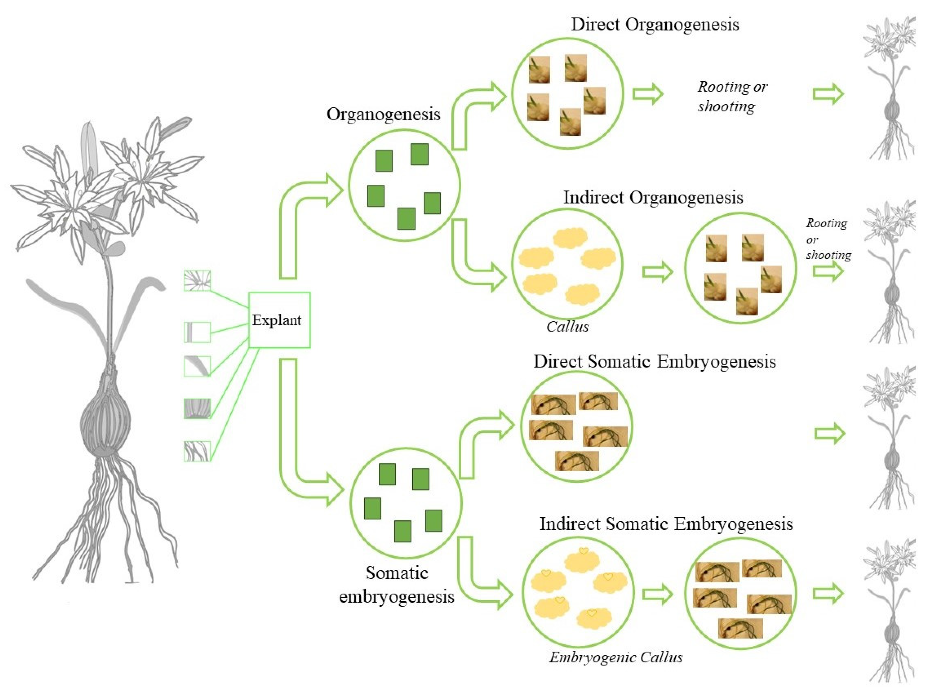
4.1. In vitro regeneration pathways
4.2. The key factors affecting micropropagation of flower bulbs
4.2.1. Explant choice
4.2.2. Culture medium
4.2.3. Environment conditions
5. Micropropagation of flower bulbs
5.1. Stage 0: Preparation of mother stock plant material
5.2. Stage 1: Establishment of aseptic culture
5.3. Stage 2: Multiplication
5.3. Stage 3: Bulb growth

5.3. Stage 4: Dormancy breaking
5.3. Stage 5: Planting
6. Somaclonal Variation
7. Conclusions and future perspectives
References
- Benschop, M.; Kamenetsky, R.; Le Nard, M.; Okubo, H.; De Hertogh, A. 1 The global flower bulb industry: Production, utilization, research. Horticultural reviews 2010, 36, 1–115. [Google Scholar]
- Correvon, H.; Massé, H. Les iris dans les jardins; Aux jardins Correvon: 1907.
- Reynolds, M.; Meachem, W. garden bulbs of spring. 1967.
- Doerflinger, F. The bulb book. (No Title) 1973.
- Margaris, N. Flowers in Greek mythology. In Proceedings of the IV International Symposium on New Floricultural Crops 541; 1999; pp. 23–29. [Google Scholar]
- Janick, J.; Kamenetsky, R.; Puttaswamy, S.H. Horticulture of the Taj Mahal: Gardens of the imagination. Chronica Horticulturae 2010, 50, 30–33. [Google Scholar]
- Bryan, J.E. Bulbs. (No Title) 1989.
- De Hertogh, A.; Schepeen, J.; Kamenetsky, R.; Le Nard, M.; Okubo, H. The Globalization of Flower Bulb Industry. Ornamental geophytes: from basic science to sustainable production 2013, 1. [Google Scholar]
- Pavord, A. The Tulip: The Story ofa Flower That Has Made Men Mad. 1999.
- Marasek-Ciolakowska, A.; Sochacki, D.; Marciniak, P. Breeding Aspects of Selected Ornamental Bulbous Crops. Agronomy 2021, 11. [Google Scholar] [CrossRef]
- Orlikowska, T.; Podwyszyńska, M.; Marasek-Ciołakowska, A.; Sochacki, D.; Szymański, R. Tulip. Ornamental crops 2018, 769–802. [Google Scholar]
- Sarac, Y.I.B., Ahmet; Deligoz, Ilyas. Lale Gece Publishing: 2021.
- Rees, A. The physiology of ornamental bulbous plants. The Botanical Review 1966, 32, 1–23. [Google Scholar] [CrossRef]
- Bailey, L.H. Manual of cultivated plants. Manual of Cultivated Plants. 1949. [Google Scholar]
- Gabellini, S.; Scaramuzzi, S. Evolving consumption trends, marketing strategies, and governance settings in ornamental horticulture: A grey literature review. Horticulturae 2022, 8, 234. [Google Scholar] [CrossRef]
- Kim, K.-W.; De Hertogh, A. Flowering Bulbs (Geophytes). Horticultural Reviews, Volume 18 1997, 18, 87. [Google Scholar]
- Pizano, M. Innovation and sustainability in South American floriculture. In Proceedings of the XXXI International Horticultural Congress (IHC2022): International Symposium on Innovations in Ornamentals: from Breeding to 1368; 2022; pp. 77–84. [Google Scholar]
- Janowska, B.; Andrzejak, R. Plant growth regulators for the cultivation and vase life of geophyte flowers and leaves. Agriculture 2023, 13, 855. [Google Scholar] [CrossRef]
- Kocak, M.; Sevindik, B.; Izgu, T.; Tutuncu, M.; Mendi, Y.Y. Synthetic seed production of flower bulbs. Synthetic Seeds: Germplasm Regeneration, Preservation and Prospects 2019, 283–299. [Google Scholar]
- Seyidoglu N., Z. M., Ayasligil Y. Position and application areas of geophytes within landscape design. African Journal of Agricultural Research 2009, 4, 1351–1357. [Google Scholar]
- Ziv, M.; Lilien-Kipnis, H. Bud regeneration from inflorescence explants for rapid propagation of geophytes in vitro. Plant Cell Reports 2000, 19, 845–850. [Google Scholar] [CrossRef] [PubMed]
- Kamenetsky, R.; Okubo, H. Ornamental geophytes: from basic science to sustainable production; CRC press: 2012.
- Marasek-Ciolakowska, A.; Sochacki, D.; Marciniak, P. Breeding aspects of selected ornamental bulbous crops. Agronomy 2021, 11, 1709. [Google Scholar] [CrossRef]
- Rees, A.R. Ornamental bulbs, corms and tubers; CAB international: 1992.
- Leeggangers, H.C.F.; Moreno-Pachon, N.; Gude, H.; Immink, R.H. Transfer of knowledge about flowering and vegetative propagation from model species to bulbous plants. Int. J. Dev. Biol. 2013, 57, 611–620. [Google Scholar] [CrossRef] [PubMed]
- Kamenetsky, R. Biodiversity of Geophytes: Phytogeography, Morphology, and Survival Strategies. In Ornamental geophytes: from basic science to sustainable production, Kamenetsky, R., Okubo, H., Eds.; CRC press: 2012; pp. 57-76.
- Dafni, A.; Cohen, D.; Noy-Mier, I. Life-cycle variation in geophytes. Annals of the Missouri Botanical Garden 1981, 652–660. [Google Scholar] [CrossRef]
- Woodhead, T.W. Ecology of woodland plants in the neighbourhood of Huddersfield. Botanical Journal of the Linnean Society 1906, 37, 333–406. [Google Scholar] [CrossRef]
- De Klerk, G. Micropropagation of bulbous crops: technology and present state. Floriculture and Ornamental Biotechnology 2012, 6, 1–8. [Google Scholar]
- Bach, A.; Sochacki, D. Ornamental Geophytes from Basic Science to Sustainable Production; Phsiology and Menagement Systems. Kamenetsky, R and Okubo, H. 2013.
- Ulus, A.; Seyidoğlu, N. Bazi Doğal Geofitlerin Doku Kültürü İle Üretimi. Journal of the Faculty of Forestry Istanbul University 2006, 56, 71–80. [Google Scholar]
- Irish, V.F. The Arabidopsis petal: a model for plant organogenesis. Trends in Plant Science 2008, 13, 430–436. [Google Scholar] [CrossRef] [PubMed]
- Langens-Gerrits, M.M.; De Klerk, G.-J.M. Micropropagation of flower bulbs: Lily and Narcissus. Plant Cell Culture Protocols 1999, 141–147. [Google Scholar]
- Luna, T. Vegetative propagation. Nursery manual for native plants: A guide for tribal nurseries 2009, 1, 153–175. [Google Scholar]
- Hartmann, H.T.; Kester, D.E. Plant propagation: principles and practice. Soil Science 1963, 95, 89. [Google Scholar] [CrossRef]
- Debergh, P.; Maene, L. A scheme for commercial propagation of ornamental plants by tissue culture. Scientia horticulturae 1981, 14, 335–345. [Google Scholar] [CrossRef]
- Debergh, P.C.; Read, P. Micropropagation. In Micropropagation: technology and application; Springer: 1991; pp. 1-13.
- Ozzambak, M.E.; Senel, U.; Gulden;, H.; Zeybekoglu, E.; Baser, S. Mikroçoğaltım teknikleri. In Süs Bitkileri Islahı (Klasik ve Biyoteknolojik Yöntemler), Yalcin Mendi, Y.K.S., Ed.; Gece Publishing: 2021; Volume 341-360.
- Podwyszyńska, M.; Orlikowska, T.; Trojak-Goluch, A.; Wojtania, A. Application and Improvement of In Vitro Culture Systems for Commercial Production of Ornamental, Fruit, and Industrial Plants in Poland. Acta Societatis Botanicorum Poloniae 2022, 91. [Google Scholar] [CrossRef]
- Bidabadi, S.S.; Jain, S.M. Cellular, molecular, and physiological aspects of in vitro plant regeneration. Plants 2020, 9, 702. [Google Scholar] [CrossRef] [PubMed]
- Ikeuchi, M.; Ogawa, Y.; Iwase, A.; Sugimoto, K. Plant regeneration: cellular origins and molecular mechanisms. Development 2016, 143, 1442–1451. [Google Scholar] [CrossRef] [PubMed]
- Long, Y.; Yang, Y.; Pan, G.; Shen, Y. New insights into tissue culture plant-regeneration mechanisms. Frontiers in plant science 2022, 13, 926752. [Google Scholar] [CrossRef] [PubMed]
- Von Arnold, S. Somatic embryogenesis. Plant propagation by tissue culture. Volume 1: the background 2008, 335-354.
- Williams, E.; Maheswaran, G. Somatic embryogenesis: factors influencing coordinated behaviour of cells as an embryogenic group. Annals of botany 1986, 57, 443–462. [Google Scholar] [CrossRef]
- Yang, X.; Zhang, X. Regulation of Somatic Embryogenesis in Higher Plants. Critical Reviews in Plant Sciences 2010, 29, 36–57. [Google Scholar] [CrossRef]
- Long, Y.; Yang, Y.; Pan, G.; Shen, Y. New Insights Into Tissue Culture Plant-Regeneration Mechanisms. Front Plant Sci 2022, 13, 926752. [Google Scholar] [CrossRef] [PubMed]
- Zimmerman, J.L. Somatic embryogenesis: a model for early development in higher plants. The plant cell 1993, 5, 1411. [Google Scholar] [CrossRef] [PubMed]
- Sharp, W.; Sondahl, M.; Caldas, L.; Maraffa, S. The physiology of in vitro asexual embryogenesis. Horticultural reviews 1980, 2, 268–310. [Google Scholar]
- Sevindik, B.; Tutuncu, M.; P;, E.C.; Y, Y.M. Somatik embryogenesis. In Süs Bitkileri Islahı (Klasik ve Biyoteknolojik Yöntemler), Yalcin Mendi, Y.K.S., Ed.; Gece Publishing: 2021; pp. 413-440.
- Von Arnold, S.; Sabala, I.; Bozhkov, P.; Dyachok, J.; Filonova, L. Developmental pathways of somatic embryogenesis. Plant cell, Tissue and Organ culture 2002, 69, 233–249. [Google Scholar] [CrossRef]
- Gahan, P.; George, E. Adventitious regeneration. Plant propagation by tissue culture. Volume 1: the background 2008, 355-401.
- George, E.F.; Hall, M.A.; De Klerk, G.-J. Plant propagation by tissue culture 3rd Edition. The Netherland, The Back Ground Springer 2008.
- Sugiyama, M. Organogenesis in vitro. Current opinion in plant biology 1999, 2, 61–64. [Google Scholar] [CrossRef] [PubMed]
- Tutuncu, M.; Basar;, S.; Yalcin Mendi, N.Y. Organogenesis. In Süs Bitkileri Islahı (Klasik ve Biyoteknolojik Yöntemler), Yalcin Mendi, Y.K.S., Ed.; Gece Publishing: 2021; pp. 361-392.
- Hicks, G.S. Patterns of organ development in plant tissue culture and the problem of organ determination. The Botanical Review 1980, 46, 1–23. [Google Scholar] [CrossRef]
- Schwarz, O.J.; Beaty, R.M. Organogenesis. In Plant Tissue Culture Concepts and Laboratory Exercises; Routledge: 2018; pp. 125-138.
- Preece, J. Stock plant physiological factors affecting growth and morphogenesis. Plant propagation by tissue culture. Volume 1: the background 2008, 403-422.
- Van Aartrijk, J.; Van der Linde, P. In vitro propagation of flower-bulb crops. In Proceedings of the Tissue culture as a plant production system for horticultural crops: Conference on Tissue Culture as a Plant Production System for Horticultural Crops, Beltsville, MD, October 20–23, 1985, 1986; pp. 317-331.
- Yasemin, S.; Koksal, N.; Buyukalaca, S. Indirect organogenesis and in vitro bulb formation of Pancratium maritimum. Plant Cell, Tissue and Organ Culture (PCTOC) 2023, 1-15. [Google Scholar] [CrossRef]
- Chu, C. Establishment of an efficient medium for another culture of rice through comparative experiments on the nitrogen sources. Scientia Sinica 1975, 18, 223–231. [Google Scholar]
- Lloyd, G.; McCown, B. Commercially-feasible micropropagation of mountain laurel, Kalmia latifolia, by use of shoot-tip culture. Commercially-feasible micropropagation of mountain laurel, Kalmia latifolia, by use of shoot-tip culture. 1980, 30, 421–427. [Google Scholar]
- Gamborg, O.L.c.; Miller, R.A.; Ojima, K. Nutrient requirements of suspension cultures of soybean root cells. Experimental cell research 1968, 50, 151–158. [Google Scholar] [CrossRef]
- George, E.; de Klerk, G. The components of plant tissue culture media I: Macro-and micro-nutrients. Plant propagation by tissue culture. Volume 1: the background 2008, 65-113.
- Thorpe, T.; Stasolla, C.; Yeung, E.; de Klerk, G.; Roberts, A.; George, E. The components of plant tissue culture media II: Organic additions, osmotic and pH effects, and support systems. Plant propagation by tissue culture. Volume 1: the background 2008, 115-173.
- Mehbub, H.; Akter, A.; Akter, M.A.; Mandal, M.S.H.; Hoque, M.A.; Tuleja, M.; Mehraj, H. Tissue Culture in Ornamentals: Cultivation Factors, Propagation Techniques, and Its Application. Plants (Basel) 2022, 11. [Google Scholar] [CrossRef] [PubMed]
- Podwyszyńska, M.; Kosson, R.; Treder, J. Polyamines and methyl jasmonate in bulb formation of in vitro propagated tulips. Plant Cell, Tissue and Organ Culture (PCTOC) 2015, 123, 591–605. [Google Scholar] [CrossRef]
- Beruto, M.; Curir, P. Effects of agar and gel characteristics on micropropagation: Ranunculus asiaticus, a case study. Floriculture, ornamental and plant biotechnology 2006, 277-284. [Google Scholar]
- George, E.; Davies, W. Effects of the physical environment. Plant propagation by tissue culture. Volume 1: the background 2008, 423-464.
- Hussey, G. The application of tissue culture to the vegetative propagation of plants. Science Progress (1933-) 1978, 185-208. [Google Scholar]
- Bornwaßer, T.; Tantau, H.-J. Evaluation of LED lighting systems in in vitro cultures. In Proceedings of the VII International Symposium on Light in Horticultural Systems 956; 2012; pp. 555–562. [Google Scholar]
- Goeden, K.; Tong, C. The effects of blue light on potato tuberisation. In Proceedings of the Abstr, 543. Poster presented for the annual meeting of the American Society Plant Biologists, 2003.
- Bach, A.; Świderski, A. The effect of light quality on organogenesis of Hyacinthus orientalis L. in vitro. Acta Biologica Cracoviensia. Series Botanica 2000, 42, 115–120. [Google Scholar]
- Bakhshaie, M.; Khosravi, S.; Azadi, P.; Bagheri, H.; van Tuyl, J.M. Biotechnological advances in Lilium. Plant cell reports 2016, 35, 1799–1826. [Google Scholar] [CrossRef] [PubMed]
- Lian, M.; Chakrabarty, D.; Paek, K. Growth and uptake of sucrose and mineral ions by bulblets of Lilium oriental hybrid ‘Casablanca’during bioreactor culture. The Journal of Horticultural Science and Biotechnology 2002, 77, 253–257. [Google Scholar] [CrossRef]
- Pałka, P.; Cioć, M.; Hura, K.; Szewczyk-Taranek, B.; Pawłowska, B. Adventitious organogenesis and phytochemical composition of Madonna lily (Lilium candidum L.) in vitro modeled by different light quality. Plant Cell, Tissue and Organ Culture (PCTOC) 2023, 152, 99–114. [Google Scholar] [CrossRef]
- Ascough, G.D.; van Staden, J.; Erwin, J.E. In Vitro Storage Organ Formation of Ornamental Geophytes. In Horticultural Reviews; 2008; pp. 417-445.
- Van Aartrijk, J.; Blom-Barnhoorn, G.J. Adventitious Bud Formation from Bulb-scale Explants of Lilium speciosum Thunb. in vitro Interacting effects of NAA, TIBA, wounding, and temperature. J Plant Physiol 1984, 116, 409–416. [Google Scholar] [CrossRef] [PubMed]
- Robb, S.M. The Culture of Excised Tissue 3 Lilium speciosum Thun. Journal of Experimental Botany 1957, 8, 348–352. [Google Scholar] [CrossRef]
- Ziv, M. THE CONTRIBUTION OF BIOTECHNOLOGY TO BREEDING, PROPAGATION AND DISEASE RESISTANCE IN GEOPHYTES. In Proceedings of the VII International Symposium on Flowerbulbs, Herzliya, Israel 1 December 1997 1997.
- Dhooghe, E.; Reheul, D.; Van Labeke, M.-C. Overcoming Pre-Fertilization Barriers in Intertribal Crosses between Anemone coronaria L. and Ranunculus asiaticus L. Horticulturae 2021, 7, 529. [Google Scholar] [CrossRef]
- Sevindik, B.; İzgü, T.; Tütüncü, M.; Çürük, P.; Sarı, N.; Mendi, Y.Y. Double-haploid plant production through anther and ovule culture of wild Cyclamen persicum Mill. and Melody F1 cyclamen cultivar. In Vitro Cellular & Developmental Biology - Plant 2023. [Google Scholar] [CrossRef]
- Sevindik, B.; İzgü, T.; Tütüncü, M.; Çürük, P.; Söğüt, Z.; Mendİ, Y.Y. Effects of different plant growth regulators on ovule culture of Turkish Cyclamen persicum Mill. and commercial variety" Melody F1". alatarım 2017, 16, 1–9. [Google Scholar]
- Tütüncü, M.; Mendi, Y.Y. Effect of pollination with gamma irradiated pollen on in vitro regeneration of ovule culture in Cyclamen. Turkish Journal of Agriculture-Food Science and Technology 2022, 10, 2415–2420. [Google Scholar] [CrossRef]
- Van Creij, M.; Kerckhoffs, D.; De Bruijn, S.; Vreugdenhil, D.; Van Tuyl, J. The effect of medium composition on ovary-slice culture and ovule culture in intraspecific Tulipa gesneriana crosses. Plant cell, tissue and organ culture 2000, 60, 61–67. [Google Scholar] [CrossRef]
- Van Tuyl, J.; Van Creij, M.; Van Dien, M. In vitro pollination and ovary culture as a breeding tool in wide hybridization of Lilium and Nerine. In Proceedings of the VI International Symposium on Flower Bulbs 325; 1992; pp. 461–466. [Google Scholar]
- Van Tuyl, J.M.; Van Diën, M.P.; Van Creij, M.G.M.; Van Kleinwee, T.C.M.; Franken, J.; Bino, R.J. Application of in vitro pollination, ovary culture, ovule culture and embryo rescue for overcoming incongruity barriers in interspecific Lilium crosses. Plant Science 1991, 74, 115–126. [Google Scholar] [CrossRef]
- Wang, J.; Huang, L.; Bao, M.-z.; Liu, G.-f. Production of interspecific hybrids between Lilium longiflorum and L. lophophorum var. linearifolium via ovule culture at early stage. Euphytica 2009, 167, 45–55. [Google Scholar] [CrossRef]
- Tütüncü, M.; Mendi, Y. Evaluation of pollen tube growth and fertilization via histological analysis in Cyclamen persicum. In Proceedings of the XXVI International Eucarpia Symposium Section Ornamentals: Editing Novelty 1283; 2020; pp. 21–26. [Google Scholar]
- Karamian, R.; Ebrahimzadeh, H. Plantlet regeneration from protoplast-derived embryogenic calli of Crocus cancellatus. Plant Cell, Tissue and Organ Culture 2001, 65, 115–121. [Google Scholar] [CrossRef]
- Koetle, M.; Finnie, J.; Balázs, E.; Van Staden, J. A review on factors affecting the Agrobacterium-mediated genetic transformation in ornamental monocotyledonous geophytes. South African Journal of Botany 2015, 98, 37–44. [Google Scholar] [CrossRef]
- Babu, P.; Chawla, H. In vitro regeneration and Agrobacterium mediated transformation in gladiolus. The Journal of Horticultural Science and Biotechnology 2000, 75, 400–404. [Google Scholar] [CrossRef]
- Chib, S.; Thangaraj, A.; Kaul, S.; Dhar, M.K.; Kaul, T. Development of a system for efficient callus production, somatic embryogenesis and gene editing using CRISPR/Cas9 in Saffron (Crocus sativus L.). Plant methods 2020, 16, 1–10. [Google Scholar] [CrossRef] [PubMed]
- Cohen, A.; Meredith, C.P. Agrobacterium-mediated transformation of Lilium. In Proceedings of the VI International Symposium on Flower Bulbs 325; 1992; pp. 611–618. [Google Scholar]
- Hoshi, Y.; Kondo, M.; Mori, S.; Adachi, Y.; Nakano, M.; Kobayashi, H. Production of transgenic lily plants by Agrobacterium-mediated transformation. Plant Cell Reports 2004, 22, 359–364. [Google Scholar] [CrossRef] [PubMed]
- Kamo, K.; Jordan, R.; Guaragna, M.A.; Hsu, H.-t.; Ueng, P. Resistance to Cucumber mosaic virus in Gladiolus plants transformed with either a defective replicase or coat protein subgroup II gene from Cucumber mosaic virus. Plant cell reports 2010, 29, 695–704. [Google Scholar] [CrossRef] [PubMed]
- Kondo, T.; Hasegawa, H.; Suzuki, M. Transformation and regeneration of garlic (Allium sativum L.) by Agrobacterium-mediated gene transfer. Plant cell reports 2000, 19, 989–993. [Google Scholar] [CrossRef] [PubMed]
- Lu, G.; Zou, Q.; Guo, D.; Zhuang, X.; Yu, X.; Xiang, X.; Cao, J. Agrobacterium tumefaciens-mediated transformation of Narcissus tazzeta var. chinensis. Plant cell reports 2007, 26, 1585–1593. [Google Scholar] [CrossRef] [PubMed]
- Popowich, E.; Firsov, A.; Mitiouchkina, T.; Filipenya, V.; Dolgov, S.; Reshetnikov, V. Agrobacterium-mediated transformation of Hyacinthus orientalis with thaumatin II gene to control fungal diseases. Plant cell, tissue and organ culture 2007, 90, 237–244. [Google Scholar] [CrossRef]
- Suzuki, S.; Supaibulwatana, K.; Mii, M.; Nakano, M. Production of transgenic plants of the Liliaceous ornamental plant Agapanthus praecox ssp. orientalis (Leighton) Leighton via Agrobacterium-mediated transformation of embryogenic calli. Plant Science 2001, 161, 89–97. [Google Scholar] [CrossRef]
- Wilmink, A.; Van de Ven, B.; Dons, J. Expression of the GUS-gene in the monocot tulip after introduction by particle bombardment and Agrobacterium. Plant Cell Reports 1992, 11, 76–80. [Google Scholar] [CrossRef] [PubMed]
- Amaki, W.; Shinohara, Y.; Hayata, Y.; Sano, H.; Suzuki, Y. Effects of bulb desiccation and storage on the in vitro propagation of hyacinth. Scientia horticulturae 1984, 23, 353–360. [Google Scholar] [CrossRef]
- Gavinlertvatana, P.; Read, P.E.; Wilkins, H.; Heins, R. Influence of Photoperiod and Daminozide Stock Plant Pretreatments on Ethylene and CO2 Levels and Callus Formation from Dahlia Leaf Segment Cultures1. Journal of the American Society for Horticultural Science 1979, 104, 849–852. [Google Scholar] [CrossRef]
- Hosoki, T.; Sagawa, Y. Clonal Propagation of Ginger (Zingiber officinale Roscoe) through Tissue Culture1. HortScience 1977, 12, 451–452. [Google Scholar] [CrossRef]
- Stimart, D.P.; Ascher, P.D. Developmental Responses of Lilium longiflorum Bulblets to Constant or Alternating Temperatures in Vitro1. Journal of the American Society for Horticultural Science 1981, 106, 450–454. [Google Scholar] [CrossRef]
- Podwyszyńska, M.; Sochacki, D. Micropropagation of tulip: production of virus-free stock plants. Protocols for in vitro propagation of ornamental plants 2010, 243–256. [Google Scholar]
- Muraseva, D.S.; Novikova, T.I. Efficient protocol for in vitro propagation from bulb scale explants of Fritillaria ruthenica Wikstr. (Liliaceae), a rare ornamental species. Rendiconti Lincei. Scienze Fisiche e Naturali 2018, 29, 491–497. [Google Scholar] [CrossRef]
- Kumar, V.; Moyo, M.; Van Staden, J. Enhancing plant regeneration of Lachenalia viridiflora, a critically endangered ornamental geophyte with high floricultural potential. Scientia Horticulturae 2016, 211, 263–268. [Google Scholar] [CrossRef]
- Mirici, S.; Parmaksız, İ.; Özcan, S.; Sancak, C.; Uranbey, S.; Sarıhan, E.O.; Gümüşcü, A.; Gürbüz, B.; Arslan, N. Efficient in vitro bulblet regeneration from immature embryos of endangered Sternbergia fischeriana. Plant Cell, Tissue and Organ Culture 2005, 80, 239–246. [Google Scholar] [CrossRef]
- Buckley, P.M.; Reed, B.M. 045 ANTIBIOTIC SUSCEPTIBILITY OF PLANT-ASSOCIATED BACTERIA. HortScience 1994, 29, 434c–434. [Google Scholar] [CrossRef]
- George, E.F. Plant propagation by tissue culture part 1. The technology 1993. [Google Scholar]
- Silva, J.A.T.d.; Winarto, B.; Dobránszki, J.; Zeng, S. Disinfection procedures for propagation of. Folia Horticulturae 2015, 27, 3–14. [Google Scholar] [CrossRef]
- Yasemin, S.; Köksal, N.; Büyükalaca, S. Effects of disinfection conditions and culture media on in vitro germination of sea daffodil (Pancratium maritimum). J. BIOL. ENVIRON. SCI. 2018, 34, 13–22. [Google Scholar]
- Barrett, C.; Cassells, A.C. An evaluation of antibiotics for the elimination of Xanthomonas campestris pv. pelargonii (Brown) from Pelargonium x domesticum cv.‘Grand Slam’explants in vitro. Plant Cell, Tissue and Organ Culture 1994, 36, 169–175. [Google Scholar] [CrossRef]
- Alexopoulos, A.A.; Mavrommati, E.; Kartsonas, E.; Petropoulos, S.A. Effect of Temperature and Sucrose on In Vitro Seed Germination and Bulblet Production of Pancratium maritimum L. Agronomy 2022, 12. [Google Scholar] [CrossRef]
- Nikopoulos, D.; Alexopoulos, A.A. In vitro propagation of an endangered medicinal plant: Pancratium maritimum L. Journal of food, agriculture & environment 2008. [Google Scholar]
- Lagram, K.; El Merzougui, S.; Boudadi, I.; Ben El Caid, M.; El Boullani, R.; El Mousadik, A.; Serghini, M.A. In vitro shoot formation and enrooted mini-corm production by direct organogenesis in saffron (crocus sativus L.). Vegetos 2023. [Google Scholar] [CrossRef]
- Sochacki, D.; Orlikowska, T. The obtaining of narcissus plants free from potyviruses via adventitious shoot regeneration in vitro from infected bulbs. Scientia horticulturae 2005, 103, 219–225. [Google Scholar] [CrossRef]
- Redhwan, A.; Acemi, A.; Özen, F. Effects of plant growth regulators on in vitro seed germination, organ development and callogenesis in Pancratium maritimum L. Plant Cell, Tissue and Organ Culture (PCTOC) 2023, 1-14. [Google Scholar] [CrossRef]
- Rafiq, S.; Rather, Z.; Bhat, R.A.; Nazki, I.; Al-Harbi, M.S.; Banday, N.; Farooq, I.; Samra, B.N.; Khan, M.; Ahmed, A.F. Standardization of in vitro micropropagation procedure of Oriental Lilium Hybrid Cv.‘Ravenna’. Saudi journal of biological sciences 2021, 28, 7581–7587. [Google Scholar] [CrossRef] [PubMed]
- Rather, Z.; Nazki, I.; Qadri, Z.; Mir, M.; Bhat, K.; Hussain, G. In vitro propagation of herbaceous peony (Paeonia lactiflora Pall.) cv. Sara Bernhardt using shoot tips. Indian Journal of Horticulture 2014, 71, 385–389. [Google Scholar]
- Farooq, I.; Banday, N.; Nazki, I.; Malik, A.A.; Khan, F.; Mir, M.; Yaseen, T. In vitro sterilization of Lilium LA Hybrids “Indian Summerset” and “Nashville” as influenced by different sterilant combinations. The Pharma Innovation Journal 2022, 11, 1795–1798. [Google Scholar]
- Appleton, M.; Ascough, G.; Van Staden, J. In vitro regeneration of Hypoxis colchicifolia plantlets. South African Journal of Botany 2012, 80, 25–35. [Google Scholar] [CrossRef]
- Devi, K.; Sharma, M.; Ahuja, P. Direct somatic embryogenesis with high frequency plantlet regeneration and successive cormlet production in saffron (Crocus sativus L.). South African Journal of Botany 2014, 93, 207–216. [Google Scholar] [CrossRef]
- Lapiz-Culqui, Y.K.; Meléndez-Mori, J.B.; Mállap-Detquizán, G.; Tejada-Alvarado, J.J.; Vilca-Valqui, N.C.; Huaman-Human, E.; Oliva, M.; Goñas, M. In Vitro Bulbification of Five Lily Varieties: An Effective Method to Produce Quality Seeds and Flowers. International Journal of Agronomy 2022, 2022. [Google Scholar] [CrossRef]
- Patil, A.M.; Gunjal, P.P.; Das, S. In vitro micropropagation of Lilium candidum bulb by application of multiple hormone concentrations using plant tissue culture technique. International Journal for Research in Applied Sciences and Biotechnology 2021, 8, 244–253. [Google Scholar] [CrossRef]
- Youssef, N.M.; Shaaban, S.A.; Ghareeb, Z.F.; Taha, L.S. In vitro bulb formation of direct and indirect regeneration of Lilium orientalis cv.“Starfighter” plants. Bulletin of the National Research Centre 2019, 43, 1–9. [Google Scholar] [CrossRef]
- Ozel, C.A.; Khawar, K.M.; Unal, F. Factors affecting efficient in vitro micropropagation of Muscari muscarimi Medikus using twin bulb scale. Saudi Journal of Biological Sciences 2015, 22, 132–138. [Google Scholar] [CrossRef] [PubMed]
- Santos, A.; Fidalgo, F.; Santos, I.; Salema, R. In vitro bulb formation of Narcissus asturiensis, a threatened species of the Amaryllidaceae. The Journal of Horticultural Science and Biotechnology 2002, 77, 149–152. [Google Scholar] [CrossRef]
- Kukulczanka, K.; Kromer, K.; Czastka, B. Propagation of Fritillaria meleagris L. through tissue culture. In Proceedings of the III International Symposium on Growth Regulators in Ornamental Horticulture 251, 1988; pp. 147-154.
- Kumar, P.; Partap, M.; Ashrita; Rana, D.; Kumar, P.; Warghat, A.R. Metabolite and expression profiling of steroidal alkaloids in wild tissues compared to bulb derived in vitro cultures of Fritillaria roylei – High value critically endangered Himalayan medicinal herb. Industrial Crops and Products 2020, 145. [CrossRef]
- Sevindik, B.; Mendi, Y.Y. Somatic embryogenesis in Crocus sativus L. In Vitro Embryogenesis in Higher Plants 2016, 351–357. [Google Scholar]
- Taheri-Dehkordi, A.; Naderi, R.; Martinelli, F.; Salami, S.A. A robust workflow for indirect somatic embryogenesis and cormlet production in saffron (Crocus sativus L.) and its wild allies; C. caspius and C. speciosus. Heliyon 2020, 6. [Google Scholar] [CrossRef]
- Slimani, C.; El Goumi, Y.; Chaimae, R.; El Ghadraoui, L.; Benjelloun, M.; Lazraq, A. Micropropagation and potential of bioactive compounds of saffron (Crocus sativus L.) for nutrition and health. Notulae Scientia Biologicae 2022, 14, 11278–11278. [Google Scholar] [CrossRef]
- Kritskaya, T.; Kashin, A.; Kasatkin, M.Y. Micropropagation and Somaclonal Variation of Tulipa suaveolens (Liliaceae) in vitro. Russian Journal of Developmental Biology 2019, 50, 209–215. [Google Scholar] [CrossRef]
- Pierik, R.; Sprenkels, P.; Van Der Harst, B.; Van Der Meys, Q. Seed germination and further development of plantlets of Paphiopedilum ciliolare Pfitz. in vitro. Scientia Horticulturae 1988, 34, 139–153. [Google Scholar] [CrossRef]
- Tubić, L.; Savić, J.; Mitić, N.; Milojević, J.; Janošević, D.; Budimir, S.; Zdravković-Korać, S. Cytokinins differentially affect regeneration, plant growth and antioxidative enzymes activity in chive (Allium schoenoprasum L.). Plant Cell, Tissue and Organ Culture (PCTOC) 2015, 124, 1–14. [Google Scholar] [CrossRef]
- Ziv, M. Morphogenesis of gladiolus buds in bioreactors—implication for scaled-up propagation of geophytes. In Proceedings of the Progress in Plant Cellular and Molecular Biology: Proceedings of the VIIth International Congress on Plant Tissue and Cell Culture, Amsterdam, The Netherlands, 24–29 June 1990, 1990; pp. 119-124.
- Pence, V.C. Evaluating costs for the in vitro propagation and preservation of endangered plants. In Vitro Cellular & Developmental Biology-Plant 2011, 47, 176–187. [Google Scholar]
- Backs-Hüsemann, D.; Reinert, J. Embryobildung durch isolierte Einzelzellen aus Gewebekulturen vonDaucus carota. Protoplasma 1970, 70, 49–60. [Google Scholar] [CrossRef]
- Sochacki, D.; Marciniak, P.; Ciesielska, M.; Zaród, J.; Sutrisno. The Influence of Selected Plant Growth Regulators and Carbohydrates on In Vitro Shoot Multiplication and Bulbing of the Tulip (Tulipa L.). Plants 2023, 12, 1134. [CrossRef] [PubMed]
- Plessner, O.; Ziv, M.; Negbi, M. In vitro corm production in the saffron crocus (Crocus sativus L.). Plant Cell, Tissue and Organ Culture 1990, 20, 89–94. [Google Scholar] [CrossRef]
- Santos, J.; Santos, I.; Salema, R. In vitro production of bulbs of Narcissus bulbocodium flowering in the first season of growth. Scientia Horticulturae 1998, 76, 205–217. [Google Scholar] [CrossRef]
- Santos, A.; Fidalgo, F.; Santos, I.; Salema, R. In vitrobulb formation ofNarcissus asturiensis, a threatened species of theAmaryllidaceae. The Journal of Horticultural Science and Biotechnology 2015, 77, 149–152. [Google Scholar] [CrossRef]
- Bahr, L.R.; Compton, M.E. Competence for in vitro bulblet regeneration among eight Lilium genotypes. HortScience 2004, 39, 127–129. [Google Scholar] [CrossRef]
- Sultana, J.; Sutlana, N.; Siddique, M.; Islam, A.; Hossain, M.; Hossain, T. In vitro bulb production in Hippeastrum (Hippeastrum hybridum). Journal of Central European Agriculture 2010. [Google Scholar]
- Kizil, S.; Khawar, K.M. The effects of plant growth regulators and incubation temperatures on germination and bulb formation of Fritillaria persica L. Propag Ornam Plants 2014, 14, 133–138. [Google Scholar]
- Podwyszyńska, M.; Kosson, R.; Treder, J. Polyamines and methyl jasmonate in bulb formation of in vitro propagated tulips. Plant Cell, Tissue and Organ Culture (PCTOC) 2015, 123, 591–605. [Google Scholar] [CrossRef]
- Stanišić, M.; Raspor, M.; Ninković, S.; Milošević, S.; Ćalić, D.; Bohanec, B.; Trifunović, M.; Petrić, M.; Subotić, A.; Jevremović, S. Clonal fidelity of Iris sibirica plants regenerated by somatic embryogenesis and organogenesis in leaf-base culture—RAPD and flow cytometer analyses. South African Journal of Botany 2015, 96, 42–52. [Google Scholar] [CrossRef]
- ÇAkmak, D.; KaraoĞLu, C.; Aasim, M.; Sancak, C.; ÖZcan, S. Advancement in protocol for in vitro seed germination, regeneration, bulblet maturation, and acclimatization of Fritillaria persica. Turkish Journal of Biology 2016, 40, 878–888. [Google Scholar] [CrossRef]
- Youssef, N.M.; Shaaban, S.A.; Ghareeb, Z.F.; Taha, L.S. In vitro bulb formation of direct and indirect regeneration of Lilium orientalis cv. “Starfighter” plants. Bulletin of the National Research Centre 2019, 43, 211. [Google Scholar] [CrossRef]
- Maślanka, M.; Mazur, J.; Kapczyńska, A. In Vitro Organogenesis of Critically Endangered Lachenalia viridiflora. Agronomy 2022, 12, 475. [Google Scholar] [CrossRef]
- Doğan, S.; Çağlar, G.; Palaz, E.B. The effect of different applications on in vitro bulb development of an endemic hyacinth plant (Hyacinthus orientalis L. subsp. chionophyllus Wendelbo) grown in Turkey. Turkish Journal of Agriculture-Food science and technology 2020, 8, 1713–1719. [Google Scholar] [CrossRef]
- Ebrahimzadeh, H.; Karamian, R.; NOURI, D.M. Somatic Embryogenesis and Regeneration of Plantlet in Saffron, Crocus Sativus L. 2000.
- Sheibani, M.; Nemati, S.H.; Davarinejad, G.H.; Azghandi, A.V.; Habashi, A.A. INDUCTION OF SOMATIC EMBRYOGENESIS IN SAFFRON USING THIDIAZURON (TDZ). 2007; pp. 259-267.
- Marković, M.; Trifunović-Momčilov, M.; Radulović, O.; Paunović, D.M.; Antonić Reljin, D.D.; Uzelac, B.; Subotić, A. The Effects of Different Auxin–Cytokinin Combinations on Morphogenesis of Fritillaria meleagris Using Bulb Scale Sections In Vitro. Horticulturae 2023, 9. [Google Scholar] [CrossRef]
- Kocak, M.; Izgu, T.; Sevindik, B.; Tutuncu, M.; Curuk, P.; Simsek, O.; Aka Kacar, Y.; Teixeira da Silva, J.A.; Yalcin Mendi, Y. Somatic embryogenesis of Turkish Cyclamen persicum Mill. Scientia Horticulturae 2014, 172, 26–33. [Google Scholar] [CrossRef]
- Bach, A.; Ptak, A. Somatic embryogenesis and plant regeneration from ovaries of Tulipa gesneriana L. in in vitro cultures. Acta Horticulturae 2001, 391–394. [Google Scholar] [CrossRef]
- Podwyszyńska, M.; Marasek-Ciolakowska, A. Micropropagation of tulip via somatic embryogenesis. Agronomy 2020, 10, 1857. [Google Scholar] [CrossRef]
- Fersing, J.; Mouras, A.; Lutz, A. First stage in the industrial vegetative propagation of Cyclamen by in vitro culture. Pepinieristes, horticulteurs, maraichers 1982. [Google Scholar]
- İzgü, T.; Sevindik, B.; Çürük, P.; Şimşek, Ö.; Aka Kaçar, Y.; Teixeira da Silva, J.A.; Yalçın Mendi, Y. Development of an efficient regeneration protocol for four Cyclamen species endemic to Turkey. Plant Cell, Tissue and Organ Culture (PCTOC) 2016, 127, 95–113. [Google Scholar] [CrossRef]
- Kiviharju, E.; Tuominen, U.; Törmälä, T. The effect of explant material on somatic embryogenesis of Cyclamen persicum Mill. Plant Cell, Tissue and Organ Culture 1992, 28, 187–194. [Google Scholar] [CrossRef]
- Otani, M.; Shimada, T. Somatic embryogenesis and plant regeneration from Cyclamen persicum Mill. leaf cultures. Plant tissue culture letters 1991, 8, 121–123. [Google Scholar] [CrossRef]
- Takamura, T.; Miyajima, I.; Matsuo, E. Somatic embryogenesis of Cyclamen persicum Mill.‘Anneke’from aseptic seedlings. Plant Cell Reports 1995, 15, 22–25. [Google Scholar] [CrossRef] [PubMed]
- Wicart, G.; Mouras, A.; Lutz, A. Histological study of organogenesis and embryogenesis in Cyclamen persicum Mill. tissue cultures: evidence for a single organogenetic pattern. Protoplasma 1984, 119, 159–167. [Google Scholar] [CrossRef]
- Kumari, A.; Baskaran, P.; Van Staden, J. In vitro propagation via organogenesis and embryogenesis of Cyrtanthus mackenii: a valuable threatened medicinal plant. Plant Cell, Tissue and Organ Culture (PCTOC) 2017, 131, 407–415. [Google Scholar] [CrossRef]
- Nautiyal, A.; Ramlal, A.; Agnihotri, A.; Rashid, A. Stress-induced somatic embryogenesis on seedlings of Azadirachta indica A. Juss. by thidiazuron and its inhibition by ethylene modulators. Plant Cell, Tissue and Organ Culture (PCTOC) 2023, 153, 357–366. [Google Scholar] [CrossRef]
- Yu, Y.; Qin, W.; Li, Y.; Zhang, C.; Wang, Y.; Yang, Z.; Ge, X.; Li, F. Red light promotes cotton embryogenic callus formation by influencing endogenous hormones, polyamines and antioxidative enzyme activities. Plant Growth Regulation 2019, 87, 187–199. [Google Scholar] [CrossRef]
- MENDİ, N.Y.Y.; ÖZDEMİR, Ş.; UĞUR, S.; SEVİNDİK, B.; İZGÜ, T.; KAYA, E.; TÜTÜNCÜ, M.; ÇÜRÜK, P.; ŞİMŞEK, Ö.; KOYUNCU, O. In vitro Regeneration and Synthetic Seed Production of Colchicum cilicicum Grown Naturally in Turkey. alatarım, 18.
- Beruto, M.; Curir, P.; Debergh, P. Callus growth and somatic embryogenesis in thalamus tissue of Ranunculus asiaticus L. cultivated in vitro: Cytokinin effect and phenol metabolism. In Vitro Cellular & Developmental Biology-Plant 1996, 32, 154–160. [Google Scholar]
- Azeri, F.N.; Öztürk, G. Microbulb and plantlet formation of a native bulbous flower, Lilium monodelphum M. Bieb, var. Armenum, through tissue culture propagation. Biotechnology Reports 2021, 32, e00665. [Google Scholar] [CrossRef] [PubMed]
- Murthy, H.N.; Joseph, K.S.; Paek, K.Y.; Park, S.Y. Bioreactor systems for micropropagation of plants: present scenario and future prospects. Front Plant Sci 2023, 14, 1159588. [Google Scholar] [CrossRef] [PubMed]
- Akita, M.; Takayama, S. Induction and development of potato tubers in a jar fermentor. Plant Cell, Tissue and Organ Culture 1994, 36, 177–182. [Google Scholar] [CrossRef]
- Dewir, Y.H.; Alsadon, A.; Al-Aizari, A.A.; Al-Mohidib, M. In vitro floral emergence and improved formation of saffron daughter corms. Horticulturae 2022, 8, 973. [Google Scholar] [CrossRef]
- Gao, R.; Wu, S.-Q.; Piao, X.-C.; Park, S.-Y.; Lian, M.-L. Micropropagation of Cymbidium sinense using continuous and temporary airlift bioreactor systems. Acta physiologiae plantarum 2014, 36, 117–124. [Google Scholar] [CrossRef]
- Hao, Z.; Ouyang, F.; Geng, Y.; Deng, X.; Hu, Z.; Chen, Z. Propagation of potato tubers in a nutrient mist bioreactor. Biotechnology techniques 1998, 12, 641–644. [Google Scholar] [CrossRef]
- Jiménez, E.; Pérez, N.; de Feria, M.; Barbón, R.; Capote, A.; Chávez, M.; Quiala, E.; Pérez, J.C. Improved production of potato microtubers using a temporary immersion system. Plant Cell, Tissue and Organ Culture 1999, 59, 19–23. [Google Scholar] [CrossRef]
- Jo, U.; Murthy, H.; Hahn, E.; Paek, K. Micropropagation of Alocasia amazonica using semisolid and liquid cultures. In Vitro Cellular & Developmental Biology-Plant 2008, 44, 26–32. [Google Scholar]
- Kim, E.; Hahn, E.; Murthy, H.; Paek, K. Enhanced shoot and bulblet proliferation of garlic (Allium sativum L.) in bioreactor systems. The Journal of Horticultural Science and Biotechnology 2004, 79, 818–822. [Google Scholar] [CrossRef]
- Lian, M.-L.; Piao, X.-C.; Park, S.-Y. Mass production of lilium bulblets in bioreactors. Production of biomass and bioactive compounds using bioreactor technology 2014, 389–415. [Google Scholar]
- Lian, M.; Chakrabarty, D.; Paek, K. Bulblet formation from bulbscale segments of Lilium using bioreactor system. Biologia Plantarum 2003, 46, 199–203. [Google Scholar] [CrossRef]
- Lim, S.; Seon, J.; Paek, K.; Han, B.; Son, S. Development of pilot scale process for mass production of Lilium bulblets in vitro. In Proceedings of the International Symposium on Biotechnology of Tropical and Subtropical Species Part 2 461; 1997; pp. 237–242. [Google Scholar]
- Piao, X.C.; Chakrabarty, D.; Hahn, E.J.; Paek, K.Y. A simple method for mass production of potato microtubers using a bioreactor system. Current Science 2003, 1129–1132. [Google Scholar]
- Teisson, C.; Alvard, D. In vitro production of potato microtubers in liquid medium using temporary immersion. Potato research 1999, 42, 499–504. [Google Scholar] [CrossRef]
- Yu, W.-C.; Joyce, P.; Cameron, D.; McCown, B. Sucrose utilization during potato microtuber growth in bioreactors. Plant Cell Reports 2000, 19, 407–413. [Google Scholar] [CrossRef]
- Marković, M.; Trifunović Momčilov, M.; Uzelac, B.; Jevremović, S.; Subotić, A. Bulb Dormancy In Vitro—Fritillaria meleagris: Initiation, Release and Physiological Parameters. Plants 2021, 10, 902. [Google Scholar] [CrossRef] [PubMed]
- Sheikh, F.R.; Jose-Santhi, J.; Kalia, D.; Singh, K.; Singh, R.K. Sugars as the regulators of dormancy and sprouting in geophytes. Industrial Crops and Products 2022, 189, 115817. [Google Scholar] [CrossRef]
- Zhao, Y.; Liu, C.; Sui, J.; Liang, J.; Ge, J.; Li, J.; Pan, W.; Yi, M.; Du, Y.; Wu, J. A wake-up call: signaling in regulating ornamental geophytes dormancy. Ornamental Plant Research 2022, 2, 1–10. [Google Scholar] [CrossRef]
- Takayama, S.; Misawa, M. Differentiation in Lilium bulbscales grown in vitro. Effects of activated charcoal, physiological age of bulbs and sucrose concentration on differentiation and scale leaf formation in vitro. Physiologia Plantarum 1980, 48, 121–125. [Google Scholar] [CrossRef]
- Langens-Gerrits, M.M.; Miller, W.B.; Croes, A.F.; De Klerk, G.-J. Effect of low temperature on dormancy breaking and growth after planting in lily bulblets regenerated in vitro. Plant growth regulation 2003, 40, 267–275. [Google Scholar] [CrossRef]
- Carasso, V.; Mucciarelli, M. In vitro bulblet production and plant regeneration from immature embryos of Fritillaria tubiformis Gren. & Godr. Propagation of Ornamental Plants 2014, 14, 101–111. [Google Scholar]
- Kamo, K.; Roh, M.; Blowers, A.; Smith, F.; Eck, J. I. 12 Transgenic Gladiolus. Transgenic Crops III 2013, 48, 155. [Google Scholar]
- Van Tuyl, J.M.; Arens, P.; Marasek-Ciolakowska, A. Breeding and genetics of ornamental geophytes. Ornamental Geophytes: From Basic Science to Sustainable Horticultural Production, 1st ed.; Kamenetsky, R., Okubo, H., Eds 2012, 131-158.
- Van Harmelen, M.; Löffler, H.; Van Tuyl, J. Somaclonal variation in lily after in vitro cultivation. In Proceedings of the VII International Symposium on Flowerbulbs 430; 1996; pp. 347–350. [Google Scholar]
- Abdolinejad, R.; Shekafandeh, A.; Jowkar, A.; Gharaghani, A.; Alemzadeh, A. Indirect regeneration of Ficus carica by the TCL technique and genetic fidelity evaluation of the regenerated plants using flow cytometry and ISSR. Plant Cell, Tissue and Organ Culture (PCTOC) 2020, 143, 131–144. [Google Scholar] [CrossRef]
- Konar, S.; Adhikari, S.; Karmakar, J.; Ray, A.; Bandyopadhyay, T.K. Evaluation of subculture ages on organogenic response from root callus and SPAR based genetic fidelity assessment in the regenerants of Hibiscus sabdariffa L. Industrial Crops and Products 2019, 135, 321–329. [Google Scholar] [CrossRef]
- Vitamvas, J.; Viehmannova, I.; Cepkova, P.H.; Mrhalova, H.; Eliasova, K. Assessment of somaclonal variation in indirect morphogenesis-derived plants of Arracacia xanthorrhiza. Pesquisa Agropecuária Brasileira 2019, 54. [Google Scholar] [CrossRef]
- Memon, N.; Yasmin, A.; Pahoja, V.; Hussain, Z.; Ahmad, I. In vitro regeneration of gladiolus propagules. J. Agr. Technol 2012, 8, 2331–2351. [Google Scholar]
- Asadi, N.; Zarei, H.; Hashemi-Petroudi, S.H.; Mousavizadeh, S.J. Micropropagation and assessment of somaclonal variation in Galanthus transcaucasicus in vitro plantlets. Ornamental Horticulture 2021, 27, 505–515. [Google Scholar] [CrossRef]


Disclaimer/Publisher’s Note: The statements, opinions and data contained in all publications are solely those of the individual author(s) and contributor(s) and not of MDPI and/or the editor(s). MDPI and/or the editor(s) disclaim responsibility for any injury to people or property resulting from any ideas, methods, instructions or products referred to in the content. |
© 2024 by the authors. Licensee MDPI, Basel, Switzerland. This article is an open access article distributed under the terms and conditions of the Creative Commons Attribution (CC BY) license (http://creativecommons.org/licenses/by/4.0/).


