
Submitted:
26 December 2023
Posted:
10 January 2024
You are already at the latest version
Abstract
Wound healing is rarely seen as a problem in healthy individuals; however, under certain pathophysiological conditions, this process can be impaired, leading to the emergence of chronic wounds, which are themselves a serious public health problem. This work aimed to review the most important and recent literature on the use of metallic nanoparticles, in this case those produced from plant extracts, and their application as healing agents. For this, we firstly provided an insight into the pathophysiology of the wound healing process and then the main routes for obtaining metallic nanoparticles. The methodology of synthesis, which is part of the so-called green synthesis, has been the subject of several studies reporting the use of medicinal plants as subtracts for the production of different nanoparticles (of silver, gold, and zinc). Additionally, their use as wound healing agents is close related with biological properties such as antimicrobial, anti-inflammatory, and healing. Finally, we addressed in vitro and in vivo studies reporting the efficiency of metallic nanoparticles synthesized with plant extracts and applied in different pharmaceutical forms on the wound healing process. Based on these satisfactory results, we verified that metallic nanoparticles are potential therapeutic alternatives when compared to the traditional synthetic healing agents, thus foreseeing the production of new drugs in the pharmaceutical market.
Keywords:
green synthesis
; metallic nanoparticles
; nanotechnology
; natural treatment
; plant extracts
; wound healing
1. Introduction
The skin is an organ with several functions in the organism, being the main one the protection of humans from external damages to which we they are exposed daily, such as chemical, physical, and microbiological aggressions. When this barrier is affected, a coordinated repair mechanism is initiated and the recruitment of various types of cells and molecules is observed with the aim to provide an efficient healing by four stages: hemostasis, inflammation, proliferation, and remodeling. Some conditions can negatively influence the wound healing process, for example, genetic alterations, diabetes, kidney failure, obesity, aging, substance abuse, malnutrition, infections, and the use of certain medications can delay wound closure. Complications of chronic non-healing wounds are vast and patients are at risk of severe pain, sepsis, hospitalization, and amputations, as well as reduced quality of life, depression, distress, anxiety, and embarrassment [1,2,3].
The above-mentioned chronic wounds complications lead to high treatment costs, estimated at 1–3% of total health spending in developed countries. Although the existence of several therapies for wound healing, they do not always provide satisfactory results in more severe cases, thus characterizing chronic wounds as a relevant public health problem. Therefore, it is necessary to constantly search for low-cost and effective therapies that generate less environmental and financial impacts [4,5]. For this reason, several strategies have been the subject of recent studies to improve wound healing, with emphasis on the development of nanoscale products.
Reducing a material to the nanoscale provides greater surface area, resulting in improved physicochemical properties. Among the most studied formulations of nanoparticles (NPs), the metallic ones (of silver, gold and zinc) have shown excellent biological activities, such as low in vivo toxicity, bacteriostatic and bactericidal activities, and tissue repairing action. Additionally, nanofibrous materials can be used as delivery systems for drugs, proteins, and growth factors, which also act as healing and/or antimicrobial agents [2,6,7].
The use of medicinal plants has been practiced over time by several populations. The use of these traditional therapies encourages research with the aim of evaluating the biological activities of medicinal plants consecrated in popular circles, for example, those directed for the treatment of wounds. In this context, the study on the synthesis of metallic nanoparticles from plant extracts, which provide bioactive compounds responsible for the chemical reactions involved in the production of NPs, is an interesting research field and should be related to the wound healing process [8,9].
Therefore, associating metals with healing and antimicrobial properties to plant species containing phytochemicals with the same properties suggests the emergence of promising nanoparticles for the effective treatment of wounds and prevention of their complications [10]. Additionally, the methodology for the production of nanoparticles is inserted in the so-called green synthesis, which is an environmentally safe, economically cheap, and innovative route. Understanding the relevance of the problem addressed to wounds´ complications and costs, we prepared this revision with the most recent in vivo and in vitro studies on the use of metallic NPs synthesized from plant extracts and used as healing agents. To base this comprehension, we also explained the pathophysiology of the wound healing process and the methodology of synthesis for NPs.
2. Method
The present study is a qualitative review based on the investigation of scientific articles in the electronic databases Scientific Electronic Library Online (Scielo), National Library of Medicine (Pubmed), and Sciencedirect. At all research stages, the search, selection, extraction, and analysis of data were carried out in pairs, followed by discussion among the authors, who chose the most relevant publications to integrate the study. However, under disagreements, a new and different reviewer was consulted to resolve divergences.
Considering the data selection, we used as inclusion criteria: articles in the original categories, literature review, systematic review, and meta-analysis. The selected publications were in the English language, while the descriptors present in the titles and/or abstracts were: wound healing, nanotechnology, metallic nanoparticles, green synthesis, plant extract, silver nanoparticles, gold nanoparticles, copper nanoparticles, titanium nanoparticles, and cerium nanoparticles.
Publications that were not related to the aim of this study, those published in another formats (for example, dissertation, thesis, case report, and conference summary), as well as works in different languages were excluded.
3. Results
3.1. Wound healing process
The skin is the largest organ in the human body. It is responsible for important functions, such as protection against mechanical forces and infections, fluid balance, and thermal regulation; therefore, maintaining its integrity is of fundamental importance. When damages occur to the skin, a complex cascade of different cell types and signaling molecules is activated with the aim to heal the wound [11].
The stimuli that generate the lesion’s skin can be external or internal, as well as physical, chemical, electrical, or thermal. Shortly after the injury, the sequential stages of the tissue repair phenomena occur, being named hemostasis, inflammation, proliferation, and remodeling. In practice, these stages do not necessarily occur in sequence, but overlap, as different areas of the wound may be in different stages of the healing process [12,13]. Figure 1 summarizes the wound healing process and the main biochemical and cellular elements involved in tissue repair.
In the first stage of the injury, the exposed subendothelium, collagen, and tissue factor will activate platelet aggregation, which results in degranulation and release of chemotactic factors (chemokines) and growth factors (GFs) to form the fibrin clot, preventing the blood loss. This step is called hemostasis. After that, the inflammatory phase begins, in which mast cells release histamine with the aim to induce the influx of neutrophils at the injury site. Focusing on this cell type, neutrophils are responsible to clean debris and microorganisms, thus creating a favorable environment for healing. Additionally, another cell type arrives in the region; they are monocytes, which differentiate into macrophages and facilitate phagocytosis and clean up cell debris. Hemostasis and the inflammatory phase usually take 72 hours to complete [14,15].
The next stage corresponds to proliferation, with an accumulation of many cells and profuse connective tissue. Keratinocytes migrate to close the wound space, while new blood vessels are formed in a process called angiogenesis. Fibroplasia begins, forming the granulation tissue by the proliferation of fibroblasts, which are responsible for the production of extracellular matrix (ECM) and collagen deposition. Fibroblasts are recruited from the dermis of the wound edge and form an intact basement membrane between the epidermis and the dermis, which is necessary for restoring the integrity and function of the injured skin. Regarding the important collagen, this protein is synthesized by fibroblasts and displays an essential role in the formation of scars. With more than 20 different types, type III collagen predominates in this stage of the healing process. Another phenomenon observed is the contraction of the wound, performed by fibroblasts rich in actin, which acquire some contractile properties. These cells are known as myofibroblasts and accumulate at the edges of wounds, constricting them toward the center [16,17].
Macrophages and regulatory T cells (Tregs) are important at this time, the latter being responsible for controlling of the local immune response, preventing further damage. Additionally, Tregs release GFs, such as the growth factor similar to the epidermal growth factor (EGF), the so-called amphiregulin, which directly contributes to the differentiation of cells within injured tissues. Resolution of local inflammation is therefore a key function of Tregs in wound healing. Several cytokines and GFs also participate in this stage, such as the Transforming Growth Factor-β (TGF-β) family, including TGF-β1, TGF-β2, and TGF-β3, the interleukin (IL) family (IL-4, IL-10, IL-13), and angiogenesis factors (vascular epidermal growth factor, VEGF). This phase continues for days or weeks. Finally, in the remodeling phase, fibroblasts complete the remodeling of the deposited matrix, blood vessels regress, and myofibroblasts cause general wound contraction. Gradual degradation of profuse ECM and immature type III collagen, in addition to the formation of mature type I collagen occur in this phase, which lasts for a few months or years [14,15,18,19].
Any interruption in the natural healing cascade can interrupt subsequent stages and potentially result in abnormal healing, chronic wounds, and scarring. Also, any damage that extends into the deeper reticular layer of the skin causes scarring. These undesirable results, especially when hypertrophic and keloid scars appear, represent an enormous aesthetic discomfort, in addition to clinical and financial burden [20,21].
Some wounds can evolve from acute to chronic conditions, with difficult healing, with a noticeable delay in physiological repair. Therefore, chronic wounds are defined as recurrent wounds or those that last more than six weeks. Among them, non-healing pressure ulcers, venous ulcers, and diabetic foot ulcers are the most common. Some host factors favor the appearance of this condition, such as compromised vasculature, diabetes, systemic arterial hypertension, neuropathies, prolonged immobility, medications that interfere with healing, immunocompromise, neoplasms, and critical states, such as terminal illness and organ failure. Complications can also be observed in patients, including low self-esteem, difficulties at work and sleeping, pain and infections, which significantly reduce the patient’s quality of life. In addition, the increasing prevalence of chronic noncommunicable diseases is a considerable issue that causes a financial burden on the health system [22,23].
Several forms of treatment from acute to chronic wounds have been studied; unfortunately, there are few effective therapies that accelerate healing and reduce the scarring burden. Because of this, the development of new dressings is a great challenge in this field. An ideal dressing should maintain a moist environment to avoid heat generation in the wound, in addition to allow for the normal debridement process and for gas and fluid exchange. Other properties are also required, including the protection of the wound from bacterial infection, absorption of wound odor, low or not adherence to the wound, and easily remove. It is also expected that the dressing does not present toxicity and allergenic tendency, while characteristics of biocompatibility, biodegradability, and sustainability are been required for new dressings, especially those prepared by biotechnological methods [24].
Aiming at more effective treatments, which can decrease aesthetic discomforts and clinical and financial burdens, several studies have been encouraging the use of natural products for the treatment of acute and chronic wounds, especially those based on the existence of a vast popular knowledge and the ancient use of medicinal plants as healing agents.
3.2. Biotechnological application of natural products
The use of natural products dates back 60,000 years, with a wide variety of plants used in traditional medicine, such as Chinese and Indian, but also widely used in other locations. Based on this, ethnopharmacology is characterized as the science that originate all medicines and accumulate a vast knowledge about drugs derived from plants; this science can also be defined as the interdisciplinary scientific exploration of biologically active agents, traditionally employed or observed by man. With the advancement of researches in the area and the consequent understanding of medicinal plants and their consisting molecules, it has been possible to obtain great therapeutic benefits from natural products, depicting them as potential preventive and curative drugs for various pathologies [25,26].
Phytotherapy, in this case the knowledge presented by different ethnic groups about vegetation, has been gaining more visibility in the health field as the efficiency of its popular treatments are increasingly being reported. The most popular application methodologies of this knowledge include infusions (teas) of plant parts or single herbs standardized for extraction. In contrast, the pharmaceutical industry produces drugs from isolated compounds, through industrial separation, and extraction of components with elucidated therapeutic activities [27].
Most bioactive plant compounds, from the point of view of plant physiology, are secondary metabolites, that is, they do not participate in the basic metabolism of plants (energy production, protein synthesis, etc.). Thus, their role in the plant is generally related to interactions with the external environment, acting as protective agents against ultraviolet radiation (flavonols), as inhibitors against herbivores (glucosinolates, some polyphenols), or as attractive for pollination (anthocyanins) [28].
Considering the greatest biodiversity of Brazil, containing more than 15% of all living species on the planet, it is possible to predict the significant number of plants with therapeutic potential in its territory. Because of the exploitation of different bioactive compounds in the rich Brazilian flora, the chemistry of natural products is one of the main aims of research in this country. Together, these facts serve as a basis for the development of new research in the pharmaceutical field, with several studies focusing on the fractionation, isolation, and structural elucidation of secondary metabolites of plants, in addition to the evaluation of their biological activities [29,30].
3.3. Extracts from medicinal plants
In general, medicinal plants can be processed for direct consumption as phytotherapy, or directed for experimental purposes. In the latter, several steps are involved, including the proper collection of the plant, identification and authentication by a specialist, drying and grinding, followed by extraction, fractionation, and, when necessary, isolation of bioactive components [31].
Extraction is the separation of medicinally active portions of the plant through selective and standardized methodologies. The main extraction techniques can be listed as maceration, infusion, percolation, digestion, decoction, Soxhlet extraction, ultrasound-assisted extraction, turboextraction, countercurrent extraction, microwave-assisted extraction, ultrasound extraction, supercritical fluid, extraction in solid phase, and column chromatography [32,33].
Among them, extraction techniques using solvents are the most used and comprise the following phenomena: (1) the solvent penetrates the solid matrix; (2) the solute dissolves in the solvent; (3) the solute is diffused out of the solid matrix; and (4) the extracted solutes are collected. The most appropriate solvent for each step should be analyzed in terms of the part of the plant that will be used, the chemical nature of the compounds of interest, and the availability of the solvent. Polar solvents, such as water, methanol, and ethanol are able to extract polar components, while nonpolar solvents, such as hexane and dichloromethane, extract nonpolar components. In addition, an efficient extraction is influenced by the size of the vegetable particles, being < 0.5 mm a suitable dimension for the efficient contact between the solvent and the target particles [31,33,34].
After applying the convenient technique, plant extracts are obtained, which will be ready for use in the form of dry powder or fluid extract, and maybe later incorporated into pharmaceutical forms, such as tablets or capsules, or evaluated from their isolated chemical components [32].
3.4. Nanotechnology
The term Nanotechnology was designated by Professor Norio Taniguchi, in 1974, but its primordial basis was established in 1959 by the American physicist Richard Feynman, in the lecture intitled "There is a lot of space in the background". Over the years, with the development of new technologies, different techniques based the manipulation of systems on a nanometric scale, thus resulting in nanomaterials. Nanotechnology is related to the manufacture, manipulation, and reduction of materials on the nanometric scale, which corresponds to particle sizes that can vary between 1 and 100 nm. Therefore, the acquisition of very small dimensions and large surface-to-volume ratios, that is, a large contact surface, are essential for the technology and generates significant changes in the physical and chemical properties of nanomaterials [35].
Nanotechnology is considered one of the most promising areas of the 21st century, being possible to list its benefits in almost all scientific fields, including physics, computer sciences, engineering, materials sciences, chemistry, biology, and science [36]. Regarding the nanoparticles (NPs), they are one of the most used nanomaterials in the health area; more specifically, metallic nanoparticles have gained prominence in recent years because of their promising results in scientific research, which directed them for medical purposes, for example, new perspectives in the treatment of wounds.
3.5. Synthesis of nanoparticles
The two main methodologies for producing nanoparticles. Top down involves the consecutive cutting or slicing of a bulk (macroscopic) material, which generates particles in the nanometric scale. Bottom up, the second and most popular methodology, involves the assembly of small precursor particles to obtain the desired nanoparticle [37,38].
Considering NPs used in the health area, there are several physical, chemical, and biological methods for their synthesis; the choose of the procedure depends on the expected physical and chemical characteristics of the final product, such as size, dispersion, chemical miscibility, and optical properties [39,40]. In the following topics, we describe the main methodologies for obtaining NPs, focusing on those considered ecologically correct (less harmful to the environment), i.e., which encourage and improve eco-friendly techniques of production of NPs.
3.5.1. Physicochemical methods of production of NPs
Physical synthesis of NPs refers to the synthesis of NPs with bulk materials, using physical forces or based on mechanical or steam-based processes. Mechanical, electrical, light, and thermal energies are used to reduce particle size and agglomeration of particles in the absence of leveling or stabilizing agents. The synthesis by physical methods presents the advantages of reasonable particle size control, satisfactory control of the NPs structure, and low-polluting manufacturing conditions. However, its disadvantages are related to the mandatory deposition of the particle on a substrate, i.e., the particles cannot be easily transferred to a solution, thus hindering particle protection and promoting low yield, high energy consumption, and use of sophisticated equipment [39,41,42,43].
Chemical synthesis is the most common method of synthesizing NPs. In general, this method is performed by reducing metallic cations to neutral atoms through the transfer of electrons from appropriate reducing agents, for example, sodium borohydride, citric acid, sodium citrate, polyols, and sulfites. The reduction involves two steps (1) nucleation and (2) subsequent growth [43]. The use of stabilizing agents is indicated for the chemical synthesis because they generate electrical charges and repulsive forces around the NPs, thus controlling the growth and preventing particles´ aggregation. The most used stabilizing agents are sulfur or phosphorus binders, polymers, surfactants, and anionic species such as citrate, halides, carboxylates, or polyoxoanions. In addition, steric stabilization is possible through the interaction of NPs with bulky groups, such as organic polymers and alkylammonium cation, responsible for preventing aggregation by steric repulsion [44,45]. Additionally, chemical synthesis can be combined with external energy sources such as photochemical, electrochemical, microwave-assisted, and sonochemical processes. However, besides the reliability, high-yield, and time-saving characteristics, its main drawback is the environmental pollution generated by the used chemical agents [43].
Aiming at alternatives that overcome the disadvantages related to physical and chemical methods, several studies have concentrated efforts to develop sustainable and economically viable options for the synthesis of metallic NPs. Thus, the biological route, also known as green synthesis, emerges as a new production via, depicting positive results from bacteria, fungi, plant extracts, and algae.
3.5.2. Green synthesis
In recent decades, researchers have been encouraging the use of the so-called green chemistry, projecting a significant reduction or total elimination of environmentally hazardous waste through two main points: the implementation of sustainable processes and the prevention of pollution at the level of atoms and molecules. Based on this, the use of non-toxic chemicals, environmentally benign solvents, and renewable materials are foreseen. Regarding the synthesis of metallic NPs, green chemistry strategies are potentially favorable by using, for example, water as a solvent, non-toxic reducing agents and stabilizers from natural sources, and laboratory conventional equipment for the process [46,47,48].
Therefore, the biological route, with the use of plants, microorganisms, and algae, emerges as a potential and sustainable option over conventional synthesis processes. Green synthesis is a methodology performed in a single step, without the use or formation of toxic and polluting chemicals, and also considered an economical, high-yielding, and ecologically correct route. In recent years, plants have been extensively studied for the biosynthesis of NPs, with different applications in the health area.
3.5.2.1. Green synthesis from plant extracts
Plant extracts are plain of bioactive molecules such as vitamins, alkaloids, carotenoids, phenolics, fats, carbohydrates, proteins, and enzymes, which can be found in various parts of the plant, for example, leaves, roots, flowers, fruits, and rhizomes, in addition to be responsible for different biological properties displayed by the extracts. When acting as reducing and stabilizing agents, for example, these molecules allow oxidation-reduction, nucleation, growth, and stabilization of NPs [49,50,51].
The presence of metals at high and toxic levels for the plant can induce the excessive production of reactive oxygen species (ROS), thus damaging cellular macromolecules and causing morphological, metabolic, and physiological irregularities. As already mentioned, plants can produce molecules with chelating properties, which neutralize the toxicity of the metal. Among them, phenolic compounds act as antioxidants against ROS even at high concentrations of metallic ions. In view of this property of detoxification of metallic ions, plant extracts are reported as one of the main sources for the biological route of production of NPs. Regarding the most important phenolics reported in the literature with the ability to perform green synthesis of NPs, it is possible to mention the flavonoids, a family of natural polyphenolics which include flavone, flavonol, flavanone, flavanonol, and isoflavone derivatives [52].
Several studies have concentrated efforts to improve the synthesis of NPs from plant extracts, starting from metals and metallic oxides, such as silver, gold, iron oxide, zinc oxide, and zirconia, with the aim of obtaining products with different applications in the biomedical field [53]. From the results obtained from this methodology, more efficient and beneficial nanoparticles were observed in terms of their biological activities, such as antibacterial, antifungal, antioxidant, anti-biofilm and cytotoxic, when compared to those synthesized through physical and chemical processes [54].
3.6. Metallic nanoparticles and the wound healing
A promising field of innovative research for the treatment of wounds is nanotechnology, in which the synthesis of biocompatible nanomaterials (NMs) is possible, of example: liposomes, solid lipid nanoparticles, polymeric micro and nanospheres, carbon nanotubes, nano-scaffold, and metallic nanoparticles (Figure 2). Due to their rapid effectiveness with a minimal dose, nanoparticles emerge as new therapeutic alternatives for healing, as their reduced dimensions and improved physicochemical properties provide intracellular drug delivery, with the possibility of topical administration and increased half-life, decreasing the number of applications and also the costs of the treatment. Additionally, encapsulation of drugs within NMs allows for multiple drug release profiles, aiding the wound healing process [22,55].
Additionally, NMs provide soft, flexible, and biocompatible wound dressings, which can display important characteristics as healing agents, including antimicrobial activity, absorbance of the exudate, protection against trauma, and the very heating of the injured region. In general, there are two main groups of NMs used in wound healing: (1) NMs that exhibit beneficial intrinsic properties for wound healing, and (2) NMs employed as delivery vehicles [24].
Among the main NMs investigated to improve the wound healing process, metallic NPs have been gaining great visibility. Several studies have shown satisfactory healing effects when NPs are synthesized from plants by the green synthesis method, which is another relevant aspect for choosing the green route. Table 1 shows important and recent findings in the literature that demonstrate the potential of NPs synthesized from plant extracts with gold, copper, zinc, titanium and cerium salts.
3.6.1 Silver nanoparticles (AgNPs)
In recent years, plants have been extensively studied for the biosynthesis of AgNPs with different applications in the health area. The biomolecules present in the plant extract can perform the oxidation-reduction of the Ag+ ion of the silver salt to Ag0, as well as the nucleation, growth, stabilization and, finally, the formation of AgNPs [49,51].
AgNPs are the most investigated nanoparticles in the wound healing process; due to its antimicrobial, anti-inflammatory, and healing activities, AgNPs have been successfully produced through different technologies and tested as healing agents [22,56,57]. For example, AgNPs have exhibited excellent anti-inflammatory activities in animal models and in clinical trials, reducing the expression and release of pro-inflammatory cytokines and mast cell infiltration, leading to wound healing with minimal scarring. Additionally, they stimulated the proliferation and migration of keratinocytes and help in the differentiation of fibroblasts into myofibroblasts, providing wound contraction and faster healing, including in diabetic ulcers [55].
Another relevant point is that bacterial resistance was not observed, which is an important aspect since controlling bacterial infection is a crucial aspect in the management of chronic wounds. It is reported that, due to their nanometric size and their larger contact surface, AgNPs destroy the membrane, cross the structure of the microorganism, and cause intracellular damage. Stimulation of oxidative stress, release of metallic ions (Ag+), which can inactivate vital biological systems (DNA, peptides and cofactors), and non-oxidative mechanisms also explain AgNPs action as healing agents [22,57]. Table 2 summarizes the main results of recent studies involving the synthesis of AgNPs by the green route with application in wound healing.
4. Conclusions
Several biomedical applications of metallic NPs have been extensively studied. Among them, the development of formulations able to accelerate the wound healing process is depicting as a very promising technology. An important way to obtain NPs is the use of plant extracts, which makes this synthesis more economical, from a financial point of view, and sustainable, considering the minimum environmental impacts. Therefore, several studies with different experimental models and methodologies address these NMs in the healing process, demonstrating satisfactory results. This is particularly related to the physicochemical properties of the NPs, the antibacterial, and healing actions of the metals involved in the synthesis and the joint effect of the bioactive compounds from the extracts of the medicinal plants used.
Therefore, it is necessary to encourage research in the perspective of large-scale production of new drugs containing NPs synthesized by the green route, enabling their availability to the population and, consequently, preventing various complications generated by poor healing and/or infections. A significant improvement in the quality of life of patients, for example decreasing aesthetic discomfort, in addition to clinical and financial burden.
Author Contributions
Anaís Gusmão was responsible for the investigation and the original writing of the manuscript. Priscilla Albuquerque was responsible for review and editing of the manuscript, and Ana Carolina Correia was responsible for the supervision and project administration.
Funding
This research received no external funding.
Data Availability Statement
The data presented in this study are available in the following articles: “Green synthesis of silver nanoparticles from aqueous extract of Scutellaria barbata and coating on the cotton fabric for antimicrobial applications and wound healing activity in fibroblast cells (L929)”, “Characterization and fabrication of zinc oxide nanoparticles by gum Acacia modesta through green chemistry and impregnation on surgical sutures to boost up the wound healing process”, and “Green Synthesis of CeO2 Nanoparticles from the Abelmoschus esculentus Extract: Evaluation of Antioxidant, Anticancer, Antibacterial, and Wound-Healing Activities”.
Acknowledgments
In this section, you can acknowledge any support given which is not covered by the author contribution or funding sections. This may include administrative and technical support, or donations in kind (e.g., materials used for experiments).
Conflicts of Interest
The authors declare no conflicts of interest.
References
- Beyene, R.T; Derryberry JR, S.L.; Barbul, A. The Effect of Comorbidities on Wound Healing. Surg. Clin. North Am. 2020, 100, 4, 695-705. [CrossRef]
- Oliveira, A. et al. Therapeutic advances in wound healing. J. Dermatolog. Treat. 2022, 33, 1, 2-22. [CrossRef]
- Veith, A.P. et al. Therapeutic strategies for enhancing angiogenesis in wound healing. Adv. Drug Deliv. 2019, 146, 97-125. [CrossRef]
- Olsson, M. et al. The humanistic and economic burden of chronic wounds: A systematic review. Wound Repair Regener. 2019, 27, 1, 114-125. [CrossRef]
- Rodrigues, M. et al. Wound Healing: A Cellular Perspective. Physiological reviews 2019, 99, 1, 665-706. [CrossRef]
- Kushwaha, A.; Goswami, L.; Kim, B.S. Nanomaterial-Based Therapy for Wound Healing. Nanomaterials, 2022, 12, 4, 618. [CrossRef]
- Mendes, C. et al. The Use of Metallic Nanoparticles in Wound Healing: New Perspectives. Int. J. Molec. Sci. 2022, 23, 23, 15376. [CrossRef]
- Khorrami, S. et al. Selective cytotoxicity of green synthesized silver nanoparticles against the MCF-7 tumor cell line and their enhanced antioxidant and antimicrobial properties. Int. J. Nanomed. 2018, 13, 8013–8024. [CrossRef]
- Veeraraghavan, V.P. et al. Green synthesis of silver nanoparticles from aqueous extract of Scutellaria barbata and coating on the cotton fabric for antimicrobial applications and wound healing activity in fibroblast cells (L929). Saudi J. Biology. Sci. 2021, 28, 7, 3633-3640. [CrossRef]
- Raziyeva, K. et al. Immunology of Acute and Chronic Wound Healing. Biomolecules, 2021, 11, 5, 700. [CrossRef]
- Sorg, H. et al. Skin Wound Healing: An Update on the Current Knowledge and Concepts. Eur. Surg. Res. 2017, 58, 1-2, 81-94. [CrossRef]
- Kimura, S.; Tsuji, T. Mechanical and Immunological Regulation in Wound Healing and Skin Reconstruction. Int. J. Molec. Sci. 2021, 22, 11, 5474. [CrossRef]
- Lux, C.N. Wound healing in animals: a review of physiology and clinical evaluation. Vet. Dermatol. 2022, 33, 1, 91-e27. [CrossRef]
- Wang, P.-H. et al. Wound healing. J Chin Med Assoc, 2018, 81, 2, 94-101. [CrossRef]
- Wilkinson, H.N.; Hardman, M.J. Wound healing: cellular mechanisms and pathological outcomes. Open Biol. 2020, 10, 9, 200223. [CrossRef]
- Nowak, N.C. et al. Cutaneous innervation in impaired diabetic wound healing. Translational research. J. Lab. clin. Med. 2021, 236, 87-108, 2021. [CrossRef]
- Thulabandu, V.; Chen, D.; Atit, R.P. Dermal fibroblast in cutaneous development and healing. Wiley interdisciplinary reviews. Dev. Biol. 2018, 7, 2. [CrossRef]
- Li, J. et al. Regulatory T-Cells: Potential Regulator of Tissue Repair and Regeneration. Front. Immunol. 2018, 9, 585. [CrossRef]
- Zaiss, D.M. et al. Immune- and non-immune-mediated roles of regulatory T-cells during wound healing. Immunology, 2019, 157, 3, 190-197. [CrossRef]
- desJardins-Park, H.E.; Foster, D.S.; Longaker, M.T. Fibroblasts and wound healing: an update. Reg. Med. 2018, 13, 5, 491-495. [CrossRef]
- Rousselle, P.; Braye, F.; Dayan, G. Re-epithelialization of adult skin wounds: Cellular mechanisms and therapeutic strategies. Adv. Drug Del. Rev. 2019, 146, 344-365. [CrossRef]
- Blanco-Fernandez, B. et al. Nanotechnology Approaches in Chronic Wound Healing. Adv. Wound Care. 2021, 10, 5, 234–256. [CrossRef]
- Woo, K.; Santos, V.L.C.G.; Alam, T. Optimising quality of life for people with non-healing wounds. Wounds Asia, 2018, 1, 2, 18-26.
- Bhattacharya, D.; Ghosh, B.; Mukhopadhyay, M. Development of nanotechnology for advancement and application in wound healing: a review. IET nanobiotechnology 2019, 13, 8, 778–785. [CrossRef]
- Chopra, B.; Dhingra, A.K. Natural products: A lead for drug discovery and development. PTR. 2021, 35, 9, 4660-4702. [CrossRef]
- Taylor, D.M.; Werneke, U. Ethnopharmacology. Nord J Psychiatry 2018, 72, S30-S32. [CrossRef]
- Falzon, C.C.; Balabanova, A. Phytotherapy: an introduction to herbal medicine. Prim. Health Care 2017, 44, 2, 217-227. [CrossRef]
- Renard, C. Extraction of bioactives from fruit and vegetables: State of the art and perspectives. LWT-Food Sci. Technol. 2018, [s. l.], 93, 390-395. [CrossRef]
- Santos, M.O. et al. Medicinal Plants: versatility and concordance of use in the caatinga area, Northeastern Brazil. An. Acad. Bras. Cienc. 2018, 90, 3, 2767-2779. [CrossRef]
- Valli, M.; Russo, H.M.; Bolzani, V.S. The potential contribution of the natural products from Brazilian biodiversity to bioeconomy. An. Acad. Bras. Cienc. 2018, 90, 1, 763-778. [CrossRef]
- Abubakar, A.R.; Haque, M. Preparation of Medicinal Plants: Basic Extraction and Fractionation Procedures for Experimental Purposes. J. Pharm. Bioallied Sci. 2020, 12, 1, 1–10. [CrossRef]
- Hussain, M.K.; Saquib, M.; Khan, M.F. Techniques for Extraction, Isolation, and Standardization of Bio-active Compounds from Medicinal Plants. Natural Bio-active Compounds 2019, [s. l.], 2, 179–200.
- Rasul, M.G. Extraction, Isolation and Characterization of Natural Products from Medicinal Plants. IJBSAC 2018, [s. l.], 2, 6, 1-6.
- Fonmboh, D.J. et al. An Overview of Methods of Extraction, Isolation and Characterization of Natural Medicinal Plant Products in Improved Traditional Medicine Research. Asian J. Med. Res. 2020, [s. l.], 9, 2, 31-57. [CrossRef]
- Mehnath, S. et al. Biosynthesized/greensynthesized nanomaterials as potential vehicles for delivery of antibiotics/drugs. Compr. Anal. Chem. 2021, [s. l.], 94, 363-432. [CrossRef]
- Bayda, S. et al. The History of Nanoscience and Nanotechnology: From Chemical–Physical Applications to Nanomedicine. Molecules 2020, 25, 1, 112. [CrossRef]
- Garg, P. et al. Influence of Nanotechnology and the Role of Nanostructures in Biomimetic Studies and Their Potential Applications. Biomimetics 2017, 2, 2, 7. [CrossRef]
- Huynh, K.-H. et al. Synthesis, Properties, and Biological Applications of Metallic Alloy Nanoparticles. Int. J. Mol. Sci. 2020, 21, 14, 5174. [CrossRef]
- Gutiérrez-Wing, C.; Velázquez-Salazar, J.; José-Yacamán, M. Procedures for the Synthesis and Capping of Metal Nanoparticles. Methods Mol. Biol. 2020, 2118, 3-20. [CrossRef]
- Hossain, Z.; Yasmeen, F.; Komatsu, S. Nanoparticles: Synthesis, Morphophysiological Effects, and Proteomic Responses of Crop Plants. Int. J. Mol. Sci. 2020, 21, 9, 3056. [CrossRef]
- Naganthran, A. et al. Synthesis, Characterization and Biomedical Application of Silver Nanoparticles. Materiais 2022, [s. l.], 15, 2, 427. [CrossRef]
- Sadeghi-Aghbash, M.; Rahimnejad, M. Zinc Phosphate Nanoparticles: A Review on Physical, Chemical, and Biological Synthesis and their Applications. Curr. Pharm. Biotechnol. 2022, 23, n. 10, p. 1228-1244. [CrossRef]
- Xu, L. et al. Silver nanoparticles: Synthesis, medical applications and biosafety. Theranostics 2020, 10, 20, 8996–9031. [CrossRef]
- Ielo, I. et al. Synthesis, Chemical-Physical Characterization, and Biomedical Applications of Functional Gold Nanoparticles: A Review. Molecules 2021, 26, 19, 5823. [CrossRef]
- Lee, S.H.; Jun, B.-H. Silver Nanoparticles: Synthesis and Application for Nanomedicine. Int. J. Mol. Sci. 2019, 20, 4, 865. [CrossRef]
- Jara, N. et al. Photochemical Synthesis of Gold and Silver Nanoparticles—A Review. MOLEFW. 2021, 26, 15, 4585. [CrossRef]
- Kirchhoff, M.M. Topics in Green Chemistry. J. Chem. Educ. 2001, [s. l.], 78, 12, 1577. [CrossRef]
- Raveendran, P.; Fu, J.; Wallen, S.L. Completely “Green” Synthesis and Stabilization of Metal Nanoparticles. J. Am. Chem. Soc. 2003, 125, 46, 13940-13941. [CrossRef]
- Flieger, J. et al. Green Synthesis of Silver Nanoparticles Using Natural Extracts with Proven Antioxidant Activity. Molecules 2021, 26, 16, 4986. [CrossRef]
- Tiwari, S. et al. An overview of the phytosynthesis of various metal nanoparticles. 3 Biotech 2021, 11, 11, 478. [CrossRef]
- Vanlalveni, C. et al. Green synthesis of silver nanoparticles using plant extracts and their antimicrobial activities: a review of recent literature. RSC adv. 2021, 11, 5, 2804-2837. [CrossRef]
- Marslin, G. et al. Secondary Metabolites in the Green Synthesis of Metallic Nanoparticles. Materials 2018, 11, 6, 940. [CrossRef]
- Nande, A. et al. Green Synthesis of Nanomaterials Using Plant Extract: A Review. Curr. Pharm. Biotech. 2021, 22, 13, 1794-1811. [CrossRef]
- Shumail, H. et al. Review on Green Synthesis of Silver Nanoparticles through Plants. Endocr. 2021, 21, 6, 994-1007. [CrossRef]
- Nqakala, Z.B. et al. Advances in Nanotechnology towards Development of Silver Nanoparticle-Based Wound-Healing Agents. Int. J. Mol. Sci. 2021, 22, 20, 11272. [CrossRef]
- Choudhury, H. et al. Silver nanoparticles: advanced and promising technology in diabetic wound therapy. Mater. Sci. Eng. C 2020, [s. l.], 112, 110925. [CrossRef]
- Paladini, F.; Pollini, M. Antimicrobial Silver Nanoparticles for Wound Healing Application: Progress and Future Trends. Materiais 2019, 12, 16, 2540. [CrossRef]
- Boomi, P. et al. Phyto-Engineered Gold Nanoparticles (AuNPs) with Potential Antibacterial, Antioxidant, and Wound Healing Activities Under in vitro and in vivo Conditions. Int. J. Nanomed. 2020, 15, 7553-7568. [CrossRef]
- Korani, S. et al. Evaluation of Antimicrobial and Wound Healing Effects of Gold Nanoparticles Containing Abelmoschus esculentus (L.) Aqueous Extract. Bioinorg. Chem. Appl. 2021, 2021, 7019130. [CrossRef]
- Ponnanikajamideen, M.I. et al. In-Vivo Anti-Diabetic and Wound Healing Effect of Antioxidant Gold Nanoparticles Synthesized Using Insulin Plant (Chamaecostus Cuspidatus). Can. J. Diabetes 2019, 43, 2, 82-89, 2019. [CrossRef]
- Irfan, M.; Munir, H.; Ismail, H. Characterization and fabrication of zinc oxide nanoparticles by gum Acacia modesta through green chemistry and impregnation on surgical sutures to boost up the wound healing process. IJBIOMAC. 2022, 204, 466-475. [CrossRef]
- Rasha, E. et al. Biosynthesis of Zinc Oxide Nanoparticles from Acacia nilotica (L.) Extract to Overcome Carbapenem-Resistant Klebsiella Pneumoniae. Molecules 2021, 26, 7, 1919. [CrossRef]
- Ahmad, M.Z. et al. Green Synthesis of Titanium Dioxide Nanoparticles Using Ocimum sanctum Leaf Extract: In Vitro Characterization and Its Healing Efficacy in Diabetic Wounds. Molecules 2022, 27, 22, 7712. [CrossRef]
- Ahmed, H.E. et al. Green Synthesis of CeO2 Nanoparticles from the Abelmoschus esculentus Extract: Evaluation of Antioxidant, Anticancer, Antibacterial, and Wound-Healing Activities. Molecules 2021, 26, 15, 4659. [CrossRef]
- Chinnasamy, G. et al. Synthesis, Characterization, Antibacterial and Wound Healing Efficacy of Silver Nanoparticles From Azadirachta indica. Front. Microbiol. 2021, 12, 611560. [CrossRef]
- Desai, A.S. et al. An In Vitro and In Vivo Study of the Efficacy and Toxicity of Plant-Extract-Derived Silver Nanoparticles. J. Funct. Biomater. 2022, 13, 2, 54. [CrossRef]
- Chai, S.H. et al. Bio fabrication of silver nanoparticles as an effective wound healing agent in the wound care after anorectal surgery. J. Photochem. Photobiol. 2018, 178, 457-462. [CrossRef]
- Ahn, E.-Y.; Jin, H.; Park, Y. Assessing the antioxidant, cytotoxic, apoptotic and wound healing properties of silver nanoparticles green-synthesized by plant extracts. Mat. Sci. Eng. C-Mater. 2019, 101, 204-216. [CrossRef]
- Sharma, M. et al. Biofabrication and characterization of flavonoid-loaded Ag, Au, Au-Ag bimetallic nanoparticles using seed extract of the plant Madhuca longifolia for the enhancement in wound healing bio-efficacy. Prog. Biomat. 2019, 8, 1, 51-63. [CrossRef]
- Al-Shmgani, H.S.A. et al. Biosynthesis of silver nanoparticles from Catharanthus roseus leaf extract and assessing their antioxidant, antimicrobial, and woundhealing activities. Artif. Cells Nanomed. Biotechnol. 2017, 45, 6, 1-7. [CrossRef]
- Arya, G. et al. Catalytic, antibacterial and antibiofilm efficacy of biosynthesised silver nanoparticles using Prosopis juliflora leaf extract along with their wound healing potential. J. Photochem. Photobiol. 2019, 190, 50-58. [CrossRef]
- Alsareii, S.A. et al. Synthesis and Characterization of Silver Nanoparticles from Rhizophora apiculata and Studies on Their Wound Healing, Antioxidant, Anti-Inflammatory, and Cytotoxic Activity. Molecules 2022, 27, 19, 6306. [CrossRef]
- Maghimaa, M.; Alharbi, S.A. Green synthesis of silver nanoparticles from Curcuma longa L. and coating on the cotton fabrics for antimicrobial applications and wound healing activity. J. Photochem. Photobiol. 2020, 204, 111806. [CrossRef]
- Mohanta, Y.K. et al. Phyto-assisted synthesis of bio-functionalised silver nanoparticles and their potential anti-oxidant, anti-microbial and wound healing activities. IET Nanobiotech. 2017, 11, 8, 1027-1034. [CrossRef]
- Ali, S. et al. Green synthesized silver nanoparticles (AgNPs) from Parrotiopsis jacquemontiana (Decne) Rehder leaf extract and its biological activities. Microsc. Res. Techniq. 2022, 85, 1, 28-43. [CrossRef]
- Gong, C.-P.; Li, S.-C.; Wang, R.-Y. Development of biosynthesized silver nanoparticles based formulation for treating wounds during nursing care in hospitals. J. Photochem. Photobiol. 2018, 183, 137-141. [CrossRef]
- Parveen, A. et al. In vivo efficacy of biocompatible silver nanoparticles cream for empirical wound healing. J. Tissue Viability 2018, 27, 4, 257-261. [CrossRef]
- Fatima, F. et al. Green Synthesized Silver Nanoparticles Using Tridax Procumbens for Topical Application: Excision Wound Model and Histopathological Studies. Pharmaceutics 2021, 13, 11, 1754. [CrossRef]
- Lakkim, V. et al. Green Synthesis of Silver Nanoparticles and Evaluation of Their Antibacterial Activity against Multidrug-Resistant Bacteria and Wound Healing Efficacy Using a Murine Model. Antibiotics 2020, 12, 902. [CrossRef]
- Kannaiyan, S. et al. Pisonia Alba Assisted Synthesis of Nanosilver for Wound Healing Activity. Bioinorg. Chem. Appl. 2022, 2022, 1775198. [CrossRef]
- Tyavambiza, C. et al. The Antioxidant and In Vitro Wound Healing Activity of Cotyledon orbiculata Aqueous Extract and the Synthesized Biogenic Silver Nanoparticles. Int. J. Mol. Sci., 23, 24, 16094. [CrossRef]
- Ali, S.; Khan, M.R.; Khan, R. Green synthesized AgNPs from Periploca hydaspidis Falc. and its biological activities. Microsc. Res. Techniq. 2021, 84, 10, 2268-2285. [CrossRef]
Figure 1.
The four steps necessary for wound healing and their main cellular and molecular components. Hemostasis begins just after injury, in which platelet aggregation and the fibrin clot prevents blood loss. Inflammation occurs between 1 and 6 days with the aim to clean the region and prevent infections; neutrophils, monocytes, and lymphocytes participate in the process, and chemoattractants are released. During the proliferation stage, the granulation tissue emerges with the presence of fibroblasts, angiogenesis, and collagen deposition, in addition to the synthesis of extracellular matrix (ECM) components. Finally, the remodeling phase, which can last for up to 2 years, is responsible for depositing more matrix and remodeling by fibroblasts, thus causing contraction and wound closure.
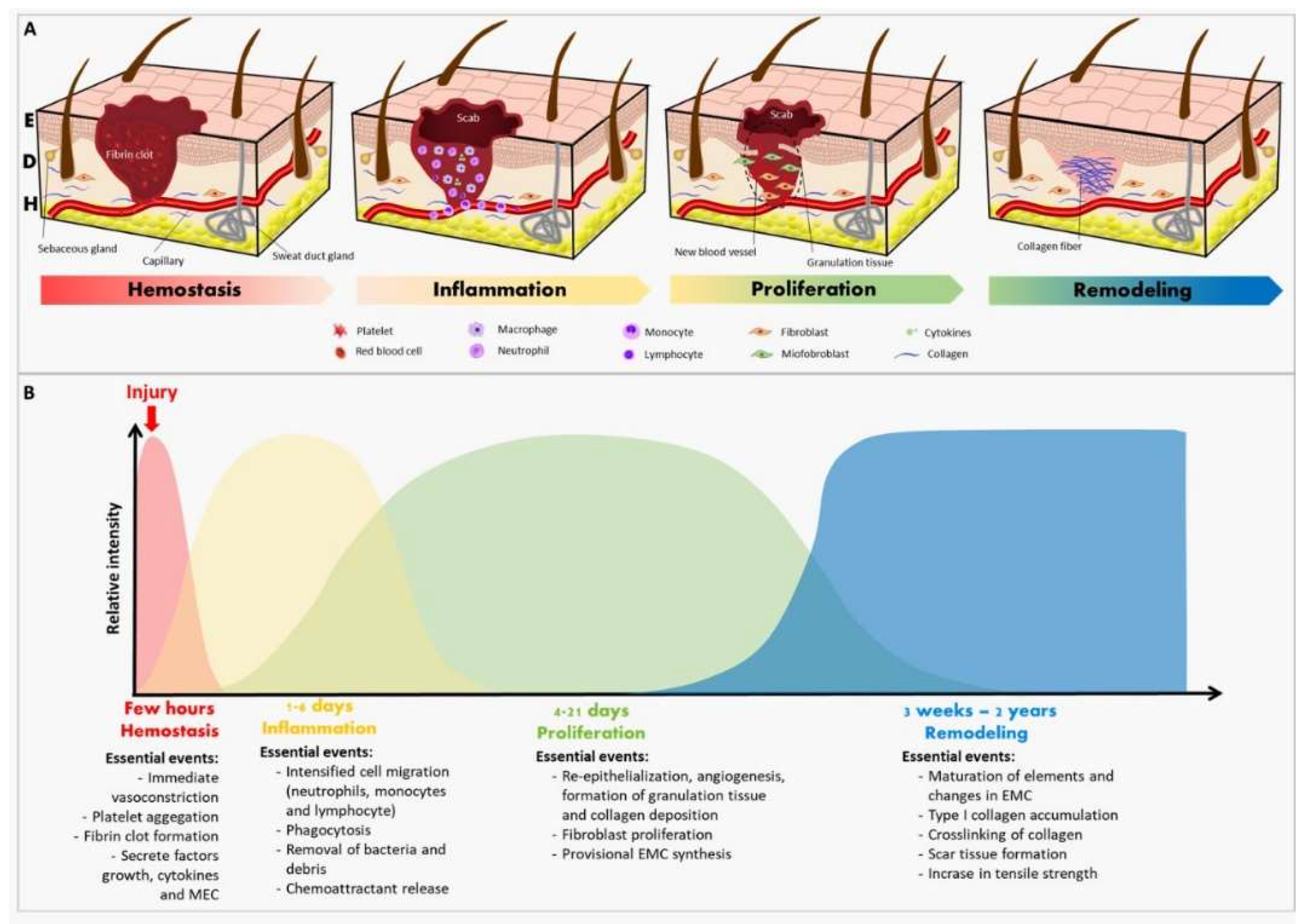

Table 1.
Effects on the healing process of wounds treated with different metal nanoparticles synthesized from plant extracts.
Table 1.
Effects on the healing process of wounds treated with different metal nanoparticles synthesized from plant extracts.
| Metal | Plant | Experimental model | Phytochemicals | Methodology | Results | References |
|---|---|---|---|---|---|---|
| Gold | Acalypha indica | In vivo assay with BALB/c mice | Absent | A 20 mm wound was made on the animal’s back, treated with AuNPs and excised to histological evaluation. | AuNPs accelerated the inflammatory stage, thus initiating blood vessel formation and collagen matrix remodeling faster than in control animals. Skin regeneration and wound contraction of treated animals were also faster than the control ones. | [58] |
| Gold | Abelmoschus esculentus | In vivo assay with male Sprague Dawley adult rats | Absent | A 2cm full-thickness skin excision was performed on the animals´ back and treated for 12 days with a colloidal solution of AuNPs synthesized with fresh okra. | AuNPs increased the percentage of wound contraction in treated animals. | [59] |
| Gold | Chamaecostus cuspidatus | In vivo assay with male Wistar rats | Absent | Absent | Rats treated with AuNPs and plant extract presented a better wound healing when compared to control rats after 4 weeks of the surgery. | [60] |
| Zinc | Acacia modesta | In vivo assay with Sprague Dawley rats | Absent | The wounds were performed on surgical sutures and treated with zinc oxide nanoparticles (ZnO-NPs). | Treated rats demonstrated a rapid rate of epithelialization, faster wound contraction, mild inflammation, and absence of infection at the wound site. Histopathology showed increased collagen fibers, fibroblastic cells, lower inflammatory cells, and rapid angiogenesis when compared to the standard surgical treatment. | [61] |
| Zinc | Acacia nilotica | In vivo assay with rats | Absent | A 2cm skin excision was performed on the animal’s back. KPC infection was induced by inoculation of 20 μL of KPC bacterial suspension (CFU 108) on the day of surgery (day 0). On day 3, the treatment based on an ointment containing the synthesized ZnO-NPs was started. | On the 14th day after surgery, the infected and uninfected control animals presented 63% and 64% of wound contraction, respectively, while the infected animals treated with the ointment based on imipenem presented 54% of contraction. Infected animals treated with ZnO-NPs presented a higher value of 98%. | [62] |
| Titanium | Ocimum sanctum | In vivo assay with diabetic male albino Wistar rats | Alkaloid, flavonoids, saponins, tannins, terpenoids, steroids, phenols, anthraquinones, proteins, and carbohydrates | A dorsal excision was performed on animals´ back and treated with chitosan gel containing TiO2 NPs. The analysis of wound area contraction, the epithelialization time, and the wound closure time. Were also evaluated. | Animals treated with the gel containing TiO2 NPs presented a faster (p<0.05) epithelialization when compared to the animals treated with the chitosan gel. | [63] |
| Cerium |
Abelmoschus esculentus |
In vivo assay with male albino rats | Absent | Rat skin incisions were performed and treated with chitosan hydrogel membrane loaded with 1% and 5% cerium nanoparticles | Animals treated with cerium nanoparticles presented a maximum wound contraction, while the group treated with the chitosan membrane had a visibly larger wound size. | [64] |
Table 2.
Effects on the healing process of wounds treated with silver nanoparticles synthesized from plant extracts.
Table 2.
Effects on the healing process of wounds treated with silver nanoparticles synthesized from plant extracts.
| Plant | Experimental model | Phytochemicals | Methodology | Results | References |
|---|---|---|---|---|---|
| Azadirachta indica | In vivo assay with adult male albino mice | Flavonoids, phenolics, terpenoids, and terpenes. | Wounds treated with AI-AgNPs (0.3, 1, and 3 mg) immobilized in the PF127 hydrogel. | Almost complete wound closure with on day 10 of the group treated with 1.0 mg AI-AgNPs-PF127 hydrogel | [65] |
| Aloe barbadensis miller and Curcuma longa | In vitro wound healing and cytotoxicity assays using human embryonic kidney cell lines (HEK-293) | Absent | Inoculation of AgNPs in cell culture. Cellular modifications were observed by using an optical microscope, while the area covered by cells was measured using Image-J software. | AgNPs with turmeric extract presented non-toxic pattern, in addition to faster and more sustained cell growth compared to other AgNPs | [66] |
| Delonix elata | In vivo treatment of anorectal wounded area in patients weighing 64 kg | Alkaloids, saponins, theroids, tannins, carotene, phenolics, anthocyanins, glycosides, flavonoids, and others. | Diary treatment with the application of cloths coated with AgNPs. The percentage of wound closure was calculated daily. | The AgNPs synthesized by the aqueous extract of the D. elata leaf presented healing properties after anorectal surgical wounds in humans. | [67] |
| Lindera strychnifolia | Cell culture in NIH3T3 cell line | Absent | Cell scraping method in NIH3T3 cells | AgNPs presented a wound closure percentage of 64% compared to the control | [68] |
| Madhuca longifolia | In vivo wound healing bioassay in Swiss albino mice | 3-hydroxy flavones, 3,6 dihydroxyflavone, dihydroquercetin, Quercetin, Myricetin 3-O-arabinoside, Myricetin 3-O-galactoside, and dihydroxyl quercetin | A 100 mm2 excision was performed and treated with paraffin-based ointment containing 70 mg/g of AgNPs. The percentage of wound closure in each case was calculated from the reduction in the wounded area | The percentage of wound closure was 80.33% for the group of ointment containing the AgNPs | [69] |
| Catharanthus roseus | In vivo assay with male albino mice using an excision wound model | Absent | A 2x2 cm2 excision was performed on the animal’s back and treated with 2 mL of the synthesized AgNP (2 mM), once a day for 12 days | Animals of the test group (treated with AgNP) presented better wound healing activity (98% of wound closure) compared to control group (85% of wound closure) | [70] |
| Prosopis juliflora | Excision wound model performed in mice | Absent | A 10 mm skin excision was performed and treated topically with an ointment containing AgNPs and Carbopol. The treatment was regular and the reduction in the wound area was measured and photographed on the 1st, 6th, 10th, and 15th days | The percentage of wound closure in animals treated with AgNPs and Carbopol was significantly higher than in the other two groups (treated only with Carbopol or povidone-iodine) | [71] |
| Rhizophora apiculata | Cell culture in murine L929 cell line | Glycosides, saponins, terpenoids, flavonoids, and phenols. | Scratch wound migration assay | AgNPs presented better potent cell migration and wound closure than the plant extract. | [72] |
| Curcuma longa L | Cell culture in murine L929 cell line | Absent | Scratch wound migration assay | AgNPs increased cell migration in the injured area, thus indicating active proliferation and growth of fibroblastic cells | [73] |
| Ardisia solanacea | Cell culture in human fibroblasts BJ-5Ta | Alkaloids, tannins, phenolic compounds, and flavonoids | Scratch wound migration assay | Authors reported a positive effect of the wound healing activity of the synthesized AgNPs | [74] |
| Parrotiopsis jacquemontiana | Skin excision in male rats | Flavonoids, tannins, coumarins, phlobatannins, steroids, phenols, alkaloids, saponins, sterols, betacyanin, vitamin C, proteins, oils, and resins | The wound area was measured in mm every 5 days, and the contraction was calculated using the measurements | The wound closure rate increased over time for all of the groups, being the values from AgNPs higher than the positive control, and these higher from the negative control. No scar formation was observed at the end of day 15 with AgNPs. | [75] |
| Euphorbia milii | Skin excision in albino male rats | Absent | A 50 mm2 dorsal excision was performed and treated with an ointment containing 10% of the synthesized AgNPs. The wound area was analyzed from the day of excision with an interval of three days until complete epithelialization | The control group presented 77.08% of wound contraction, while group I (treated with nitrofurazone ointment) presented 82.56%, and group II (treated with the AgNPs), 91.45% | [76] |
| Scutellaria barbata | Cell culture in L929 fibroblasts cell line | Absent | Scratch wound migration assay | AgNPs induced wound healing by proliferation, differentiation, and migration of L929 fibroblast cells | [9] |
| Syzygium aromaticum | In vivo assay with male and female albino rats | Absent | A dorsal excision was performed on the animal´s back and the AgNPs formulation was applied once to the wound site for 10 consecutive days until the wound was completely healed | The wound closure was achieved in 46 days and 28 days respectively for the animals treated with AgNP 3% and AgNP 5% in base cream. On day 20, the control group presented a closure rate of 94.25%, while AgNP 5% and AgNP 3% presented 98.76% and 97.06%, respectively | [77] |
| Tridax procumbens | In vivo assay with mice | Absent | A 4.5 cm2 skin excision was performed on the animals´ back and chitosan-based gel loaded with AgNPs was applied once a day until complete healing | Animals treated with AgNPs presented significant progressive healing when compared to the groups receiving negative (placebo) and standard (1% silver sulfadiazine) treatments | [78] |
|
Catharanthus roseus and Azadirachta indica |
In vivo assay with female BALB/c mice | Absent | A 5mm excision was performed on the animals´ back and treated with the formulation containing AgNPs from C. roseus or the one containing AgNPs from A. indica. Commercially available povidone-iodine ointment was used as a positive control | The wound closure rate for the AgNPs´ treatment was significantly higher (94% ± 1% for C. roseus and 87% ± 1% for A. indica) when compared to the controls (76% ± 1% for the negative and 79 % ± 1% for the positive). Wounds treated with AgNPs did not show microbial growth, bleeding, or pus formation during the experimental period, while wounds in the negative control group showed notable irritation. | [79] |
| Pisonia Alba | Cell culture of human dermal fibroblasts (HDF) | Absent | Scratch wound migration assay and analysis of cell migration in cells treated with the AgNPs. Measurements performed at 0, 24, and 48 hours of incubation | AgNPs were reported as a stimulant for collagen production and deposition in the wound site. Wound closure rates of 23.32% ± 2.29 and 17.21% ± 1.00 were observed at a concentration of 25 μg/mL after 24 h and 48 h of incubation, respectively | [80] |
| Cotyledon orbiculata | Cell culture of HaCaT, KMST-6, and CHO | Polyphenols, flavanols, tannins, and flavonols | Scratch wound migration assay with the aqueous extract of C. orbiculata and the AgNPs | C. orbiculata aqueous extract and AgNPs presented faster closure when compared to the negative control (untreated cells) in all cell lines. In HaCaT and CHO cells, AgNPs (at 2.5 µg/mL) were more efficient than the aqueous extract and the positive control (allantoin) | [81] |
| Periploca hydaspidis | In vivo assay with Sprague-Dawley rats | Tannins, flavonoids, phenols, coumarins, alkaloids, anthocyanins, saponins, glycosides, vitamin C | A cut was performed on the animal’s back and treated with the plant extract or the AgNPs | Plant extract (20%, 35%, and 75%) and AgNPs (30%, 60%, and 100%), presented a higher wound closure rate when compared to the negative control (25%, 45%, and 85%) for specific experimental days, in this case 5, 10, and 15 days, respectively | [82] |
Disclaimer/Publisher’s Note: The statements, opinions and data contained in all publications are solely those of the individual author(s) and contributor(s) and not of MDPI and/or the editor(s). MDPI and/or the editor(s) disclaim responsibility for any injury to people or property resulting from any ideas, methods, instructions or products referred to in the content. |
© 2024 by the authors. Licensee MDPI, Basel, Switzerland. This article is an open access article distributed under the terms and conditions of the Creative Commons Attribution (CC BY) license (http://creativecommons.org/licenses/by/4.0/).
Copyright: This open access article is published under a Creative Commons CC BY 4.0 license, which permit the free download, distribution, and reuse, provided that the author and preprint are cited in any reuse.


