Submitted:
01 February 2024
Posted:
02 February 2024
You are already at the latest version
Abstract
Keywords:
1. Introduction
2. Results and Discussion
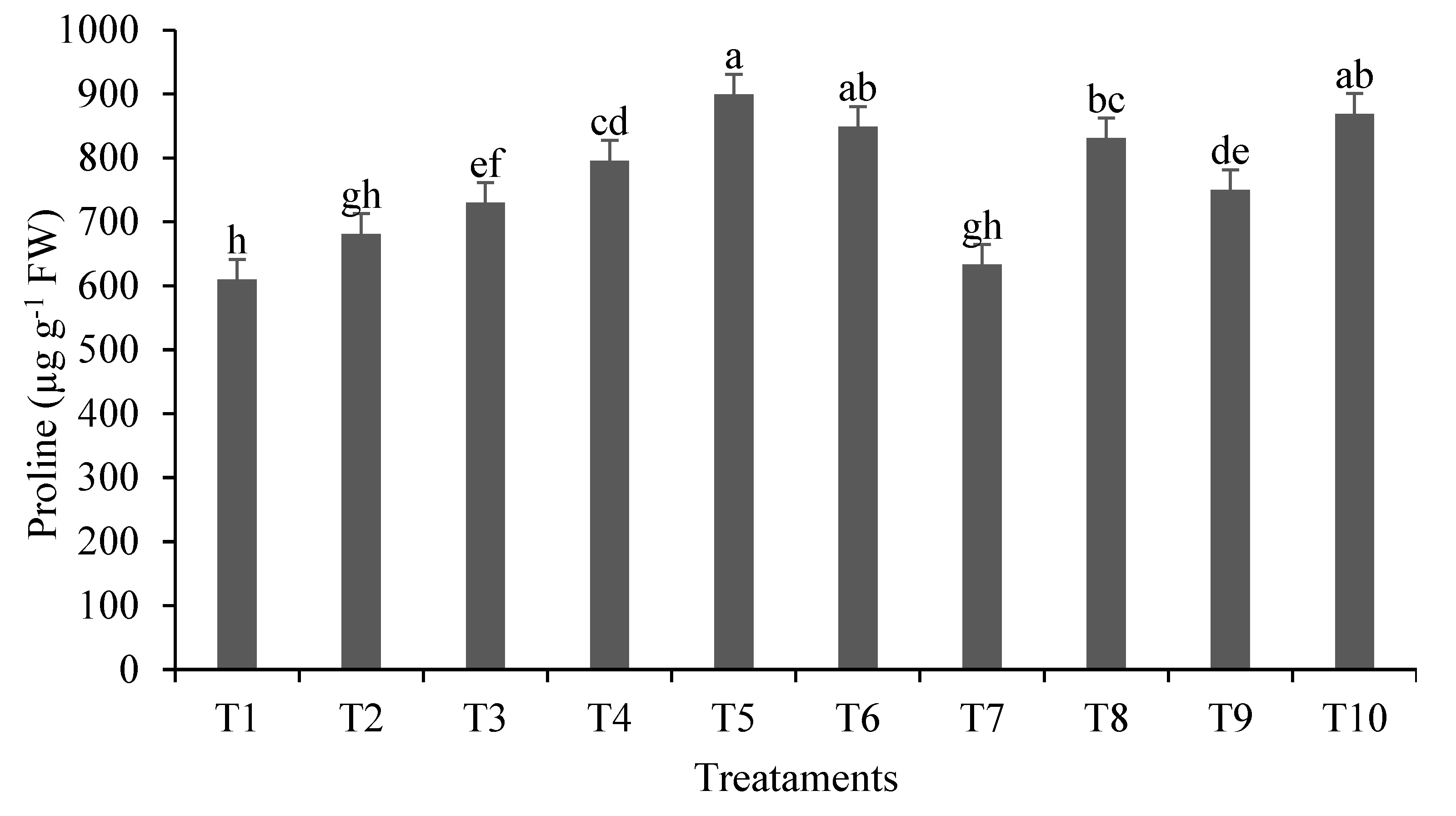
2.1. Primary and Secondary Metabolites
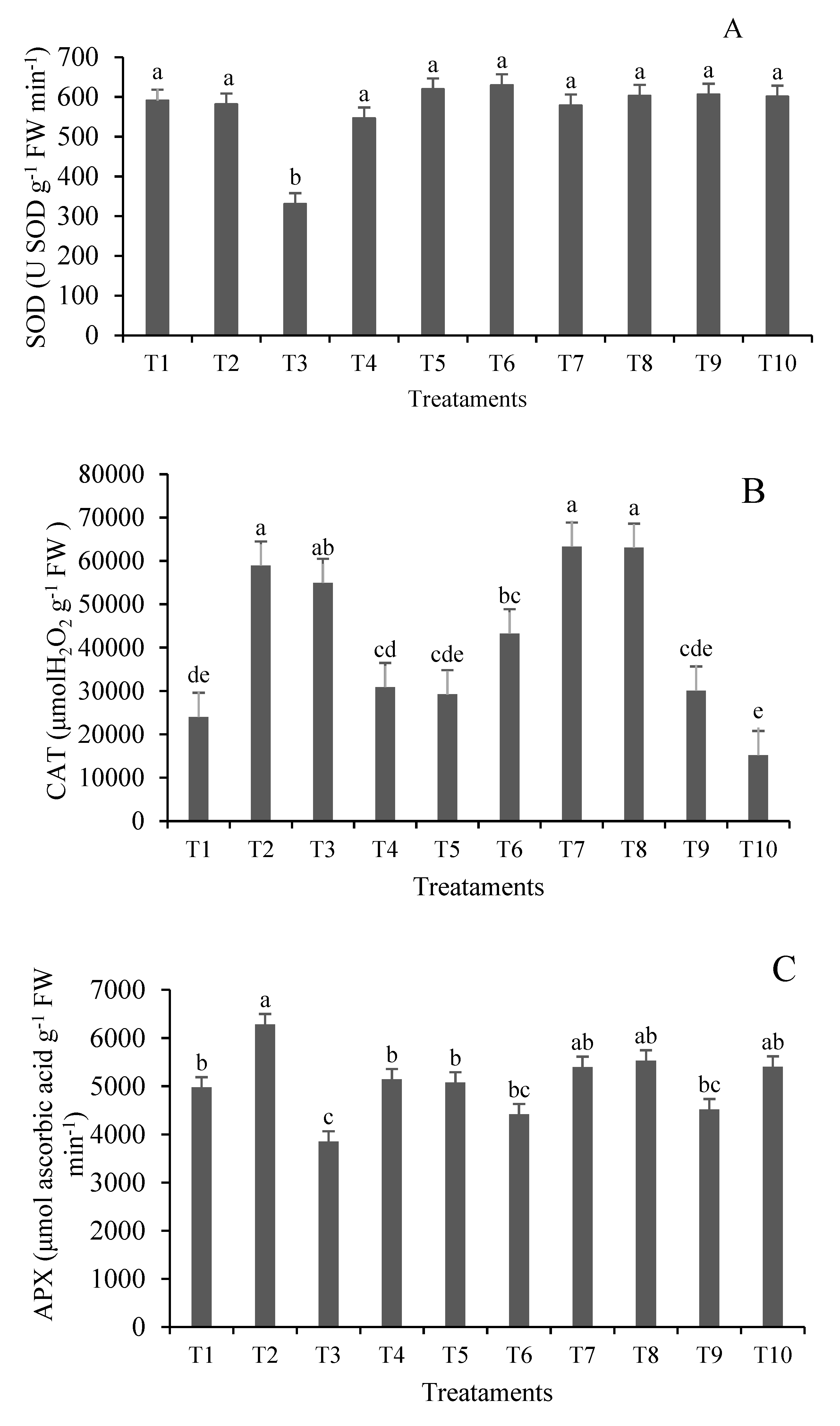
2.2. Response of Antioxidant Enzymes to Lead Stress
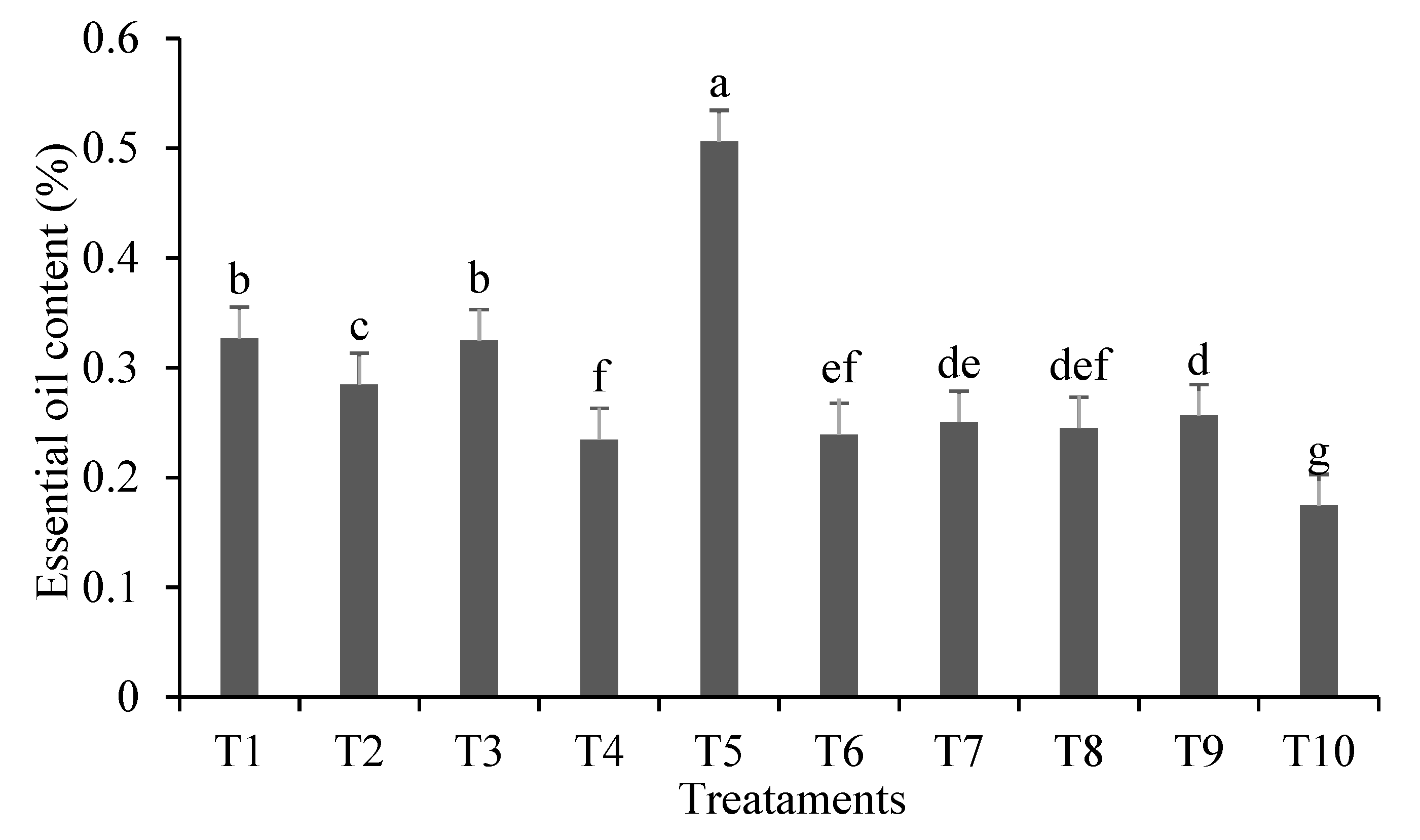
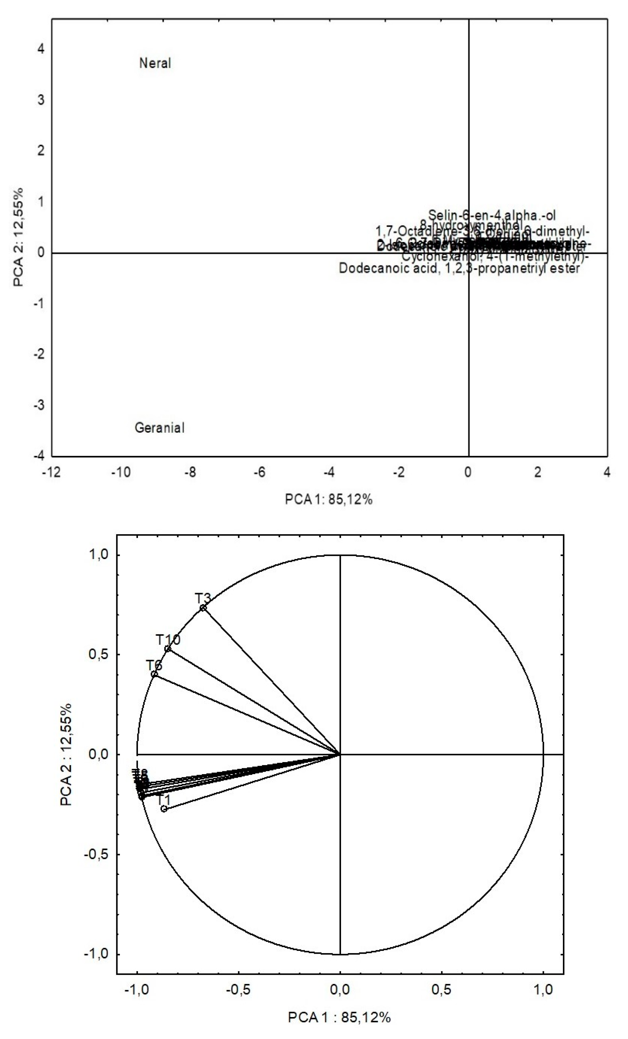
2.3. Extraction, Evaluation of EO Content and Chemical Identification by GC/MS
2.4. Figures, Tables and Schemes
| pH (CaCl2) | P | C | Al3+ | H++Al3+ | Ca2+ | Mg2+ | K+ | SB | CTC | V | |
|---|---|---|---|---|---|---|---|---|---|---|---|
| mg dm-3 | g dm-3 | -----------------------Cmolc dm-3------------------------- | % | ||||||||
| Soil | 6.07 | 1.54 | 4.09 | 0.00 | 1.89 | 1.38 | 0.75 | 0.05 | 2.18 | 4.07 | 53.52 |
| Ref1 | 3.8-6.6 | 16-24 | 0.8-15.9 | - | 0.6-5.0 | 0.3-7.2 | 0.3-3.3 | 0.1-0.7 | - | 2.2-12.5 | - |
| Treatment | TSS | TSR | RSS | RSR |
|---|---|---|---|---|
| T1 | 184.56 ± 7.75d | 3849.19 ± 423.70b | 2337.80 ± 43.32cd | 2419.50 ± 45.82b |
| T2 | 108.66 ± 1.11e | 2676.01 ± 123.40c | 2780.62 ± 65.93a | 2178.07 ± 101.15c |
| T3 | 119.52 ± 3.24e | 4714.73 ± 329.25a | 2639.45 ± 69.21ab | 2183.84 ± 40.01c |
| T4 | 472.14 ± 11.63b | 1254.81 ± 35.89d | 2595.69 ± 106.51ab | 2368.12 ± 62.52bc |
| T5 | 117.85 ± 0.33e | 561.02 ± 68.17e | 2497.59 ± 20.95bc | 2389.64 ± 46.96bc |
| T6 | 788.39 ± 51.84a | 238.93 ± 1.99e | 2524.99 ± 121.51bc | 2317.78 ± 44.04bc |
| T7 | 125.56 ± 0.97e | 115.91 ± 1.11e | 2439.73 ± 86.55bc | 2415.42 ± 53.76b |
| T8 | 243.07 ± 1.59c | 200.51 ± 4.41e | 2216.46 ± 55.18d | 2361.74 ± 97.46bc |
| T9 | 106.13 ± 0.68e | 129.63 ± 0.82e | 2569.70 ± 70.05abc | 2764.81 ± 88.66a |
| T10 | 216.50 ± 3.25cd | 105.30 ± 0.60e | 2474.95 ± 33.65bc | 2492.77 ± 43.43b |
| Treatment | FlavS | FlavR | PhenS | PhenR | DPPH |
|---|---|---|---|---|---|
| T1 | 152.15 ± 5.81f | 32.34 ± 2.45f | 1207.59 ± 20.61e | 262.64 ± 4.97j | 36.91 ± 0.74a |
| T2 | 225.12 ± 1.16e | 40.12 ± 1.52ef | 1325.29 ± 34.80d | 309.28 ± 11.57i | 34.69 ± 0.07a |
| T3 | 224.81 ± 1.53e | 25.12 ± 4.38f | 1958.21 ± 7.87c | 348.88 ± 11.26h | 20.13 ± 0.28c |
| T4 | 236.78 ± 12.54e | 44.96 ± 2.51ef | 2479.73 ± 43.84b | 385.89 ± 3.56g | 12.70 ± 0.12d |
| T5 | 382.69 ± 9.29d | 68.30 ± 10.16e | 2771.76 ± 50.72a | 511.74 ± 1.23f | 12.64 ± 0.22d |
| T6 | 132.69 ± 1.34g | 434.81 ± 11.33d | 1284.57 ± 2.72de | 617.23 ± 2.39e | 35.50 ± 1.59a |
| T7 | 234.51 ± 2.71e | 468.45 ± 27.29c | 1330.47 ± 41.42d | 722.12 ± 22.95d | 34.53 ± 1.38a |
| T8 | 438.75 ± 2.29c | 584.72 ± 1.48b | 1351.20 ± 19.47d | 875.95 ± 13.61c | 15.07 ± 1.22d |
| T9 | 492.54 ± 2.55b | 604.36 ± 1.29b | 1893.07 ± 33.93c | 1081.00 ± 11.06b | 13.07 ± 0.10d |
| T10 | 674.06 ± 2.87a | 767.54 ± 1.24a | 1374.15 ± 28.82d | 2175.48 ± 2.96a | 28.21 ± 0.31b |
| Peak | 1RI | Component | T1 | T2 | T3 | T4 | T5 | T6 | T7 | T8 | T9 | T10 |
|---|---|---|---|---|---|---|---|---|---|---|---|---|
| 1 | 8.342 | β Myrcene | 2.46 | 3.79 | 8.04 | 4.73 | 6.63 | 3.66 | 7.31 | 8.05 | 4.87 | 1.79 |
| 2 | 14.568 | Neral (Citral Z) | 22.79 | 33.02 | 63.18 | 33.83 | 34.54 | 33.31 | 33.69 | 33.58 | 33.59 | 66.79 |
| 3 | 15.265 | Geranial (Citral E) | 35.7 | 48.02 | t | 50.44 | 47.03 | 49.31 | 50.79 | 44.74 | 51.01 | 16.95 |
| 4 | 15.550 | Epoxy-linalooloxide | 2.66 | t | t | t | t | t | t | t | t | t |
| 5 | 16.856 | Dodecanoic acid. 2-hexen-1-yl ester | 3.86 | t | 2.9 | t | t | t | 2.43 | t | t | t |
| 6 | 17.664 | Cyclohexanol. 4-(1-methylethyl)- | 5.97 | t | t | t | t | t | t | t | t | t |
| 7 | 42.681 | Dodecanoic acid. 1.2.3-propanetriyl ester | 19.87 | t | t | t | t | t | t | t | t | t |
| 8 | 8.280 | methyl heptenone | t | 0.34 | t | t | t | t | t | t | t | t |
| 9 | 10.908 | cis-Myroxide | t | 0.39 | 0.75 | 0.5 | 0.64 | 0.47 | 0.85 | 0.96 | 0.54 | t |
| 10 | 11.179 | Linalool | t | 0.53 | 0.98 | 0.51 | 0.6 | t | t | 0.56 | t | 0.51 |
| 11 | 12.202 | 6-Octenal. 7-methyl-3-methylene- | t | 0.39 | 0.75 | 0.4 | 0.42 | 0.39 | t | 0.43 | t | 0.38 |
| 12 | 12.689 | Isoneral | t | 0.7 | 2.34 | 0.76 | 0.82 | 0.7 | t | 0.8 | 0.62 | 0.58 |
| 13 | 13.125 | Isogeranial | t | 0.89 | 2.68 | 1 | 1.12 | 0.92 | 0.85 | 1.07 | 0.91 | 0.85 |
| 14 | 15.802 | 2-Undecanone | t | 0.3 | 0.52 | 0.3 | t | 0.38 | t | 0.36 | 0.28 | |
| 15 | 16.947 | 2.7-Dimethyl-2.7-octanediol | t | 2.41 | 2.34 | 2.3 | 2.96 | t | 3.19 | 2.5 | 2.16 | |
| 16 | 17.783 | 8-hydroxymenthol | t | 4.31 | 6.94 | 4.21 | 3.72 | 5.13 | 2.79 | 5.13 | 4.32 | 3.85 |
| 17 | 22.920 | Selin-6-en-4.alpha.-ol | t | 3.05 | 7.02 | 0.97 | 1.56 | 1.59 | t | 0.51 | 1.08 | 3.28 |
| 18 | 23.580 | α Cadinol | t | 1.16 | 3.64 | t | 0.6 | 0.59 | t | t | t | 1.31 |
| 19 | 15.602 | 1-Undecyne | t | t | t | t | t | 0.27 | t | 0.24 | t | t |
| 20 | 15.675 | 2-Isopropenyl-5-methylhex-4-enal | t | t | t | t | t | 0.31 | t | 0.35 | t | t |
| 21 | 20.070 | 2-Tridecanone | t | t | t | t | t | t | t | t | 0.43 | 0.56 |
| Total | 93.31 | 99.3 | 99.74 | 99.99 | 99.98 | 99.99 | 98.71 | 99.97 | 99.87 | 99.29 |
3. Materials and Methods
3.1. Experimental Design
3.2. Determination of Total and Reducing Sugars
3.3. Determination of Total Phenolic Content, Flavonoids and DPPH Antioxidant Activity
3.4. Antioxidant Enzymes
3.4.1. Superoxide Dismutase (SOD, EC 1.15.1.1)
3.4.2. Catalase (CAT, EC 1.11.1.6)
3.4.3. Ascorbate Peroxidase (APX, EC 1.11.1.11)
3.5. Proline
3.6. Essential Oil Extraction and Yield Evaluation
3.7. Chemical Identification of Essential Oil by GC/MS
3.8. Statistical Analysis
5. Conclusions
6. Patents
Author Contributions
Data Availability Statement
Acknowledgments
Conflicts of Interest
References
- Bharagava, R.N.; Saxena, G.; Mulla, S.I. Introduction to Industrial Wastes Containing Organic and Inorganic Pollutants and Bioremediation Approaches for Environmental Management. In Bioremediation of Industrial Waste for Environmental Safety; Springer Singapore: Singapore, 2020; pp. 1–18. [Google Scholar]
- Wei, B.; Yang, L. A Review of Heavy Metal Contaminations in Urban Soils, Urban Road Dusts and Agricultural Soils from China. Microchem. J. 2010, 94, 99–107. [Google Scholar] [CrossRef]
- Agwu, K.K.; Okoye, C.M.I.; Okeji, M.C.; Clifford, E.O. Potential Health Impacts of Heavy Metal Concentrations in Fresh and Marine Water Fishes Consumed in Southeast, Nigeria. Pakistan J. Nutr. 2018, 17, 647–653. [Google Scholar] [CrossRef]
- Manoj, M.C.; Kawsar, M. Metal Contamination Assessment in a Sediment Core from Vagamon Lake, Southwest India: Natural/Anthropogenic Impact. Environ. Nanotechnology, Monit. Manag. 2020, 14, 100362. [Google Scholar] [CrossRef]
- Lermen, C.; Morelli, F.; Gazim, Z.C.; Silva, A.P. da; Gonçalves, J.E.; Dragunski, D.C.; Alberton, O. Essential Oil Content and Chemical Composition of Cymbopogon Citratus Inoculated with Arbuscular Mycorrhizal Fungi under Different Levels of Lead. Ind. Crops Prod. 2015, 76, 734–738. [Google Scholar] [CrossRef]
- Sharma, P.; Dubey, R.S. Lead Toxicity Im Plants. Brazilian J. plant Physiol. 2005, 17, 35–52. [Google Scholar] [CrossRef]
- Kou, M.; Xiong, J.; Li, M.; Wang, M.; Tan, W. Interactive Effects of Cd and Pb on the Photosynthesis Efficiency and Antioxidant Defense System of Capsicum Annuum L. Bull. Environ. Contam. Toxicol. 2022. [Google Scholar] [CrossRef] [PubMed]
- Huihui, Z.; Xin, L.; Zisong, X.; Yue, W.; Zhiyuan, T.; Meijun, A.; Yuehui, Z.; Wenxu, Z.; Nan, X.; Guangyu, S. Toxic Effects of Heavy Metals Pb and Cd on Mulberry (Morus Alba L.) Seedling Leaves: Photosynthetic Function and Reactive Oxygen Species (ROS) Metabolism Responses. Ecotoxicol. Environ. Saf. 2020, 195, 110469. [Google Scholar] [CrossRef]
- Ali, M.; Nas, F.S. The Effect of Lead on Plants in Terms of Growing and Biochemical Parameters: A Review. MOJ Ecol. Environ. Sci. 2018, 3, 265–268. [Google Scholar] [CrossRef]
- Al-Khatib, I.A.; Anayah, F.M.; Al-Sari, M.I.; Al-Madbouh, S.; Salahat, J.I.; Jararaa, B.Y.A. Assessing Physiochemical Characteristics of Agricultural Waste and Ready Compost at Wadi Al-Far’a Watershed of Palestine. J. Environ. Public Health 2023, 2023, 1–13. [Google Scholar] [CrossRef]
- Zago, V.C.P.; das Dores, N.C.; Watts, B.A. Strategy for Phytomanagement in an Area Affected by Iron Ore Dam Rupture: A Study Case in Minas Gerais State, Brazil. Environ. Pollut. 2019, 249, 1029–1037. [Google Scholar] [CrossRef]
- Zakka Israila, Y. The Effect of Application of EDTA on the Phytoextraction of Heavy Metals by Vetivera Zizanioides, Cymbopogon Citratus and Helianthus Annuls. Int. J. Environ. Monit. Anal. 2015, 3, 38. [Google Scholar] [CrossRef]
- Oladeji, O.S.; Adelowo, F.E.; Ayodele, D.T.; Odelade, K.A. Phytochemistry and Pharmacological Activities of Cymbopogon Citratus: A Review. Sci. African 2019, 6, e00137. [Google Scholar] [CrossRef]
- Ajayi, E.O.; Sadimenko, A.P.; Afolayan, A.J. GC–MS Evaluation of Cymbopogon Citratus (DC) Stapf Oil Obtained Using Modified Hydrodistillation and Microwave Extraction Methods. Food Chem. 2016, 209, 262–266. [Google Scholar] [CrossRef]
- Balakrishnan, B.; Paramasivam, S.; Arulkumar, A. Evaluation of the Lemongrass Plant (Cymbopogon Citratus) Extracted in Different Solvents for Antioxidant and Antibacterial Activity against Human Pathogens. Asian Pacific J. Trop. Dis. 2014, 4, S134–S139. [Google Scholar] [CrossRef]
- Olorunnisola, S.K.; Asiyanbi, H.T.; Hammed, A.M.; Simsek, S. Biological Properties of Lemongrass: An Overview. Int. Food Res. J. 2014, 21, 455–462. [Google Scholar]
- Galanakis, C.M. The Food Systems in the Era of the Coronavirus (COVID-19) Pandemic Crisis. Foods 2020, 9, 523. [Google Scholar] [CrossRef] [PubMed]
- Kanyinda, J.-N.M. Preparation of Papers for European Journal of Medical and Health Sciences (EJMED). Eur. J. Med. Heal. Sci. 2020, 2, 3–6. [Google Scholar]
- Bashan, Y.; De-Bashan, L.E. How the Plant Growth-Promoting Bacterium Azospirillum Promotes Plant Growth—A Critical Assessment. In; 2010; pp. 77–136.
- Vejan, P.; Abdullah, R.; Khadiran, T.; Ismail, S.; Nasrulhaq Boyce, A. Role of Plant Growth Promoting Rhizobacteria in Agricultural Sustainability—A Review. Molecules 2016, 21, 573. [Google Scholar] [CrossRef]
- Zeffa, D.M.; Perini, L.J.; Silva, M.B.; de Sousa, N.V.; Scapim, C.A.; Oliveira, A.L.M. de; Amaral Júnior, A.T. do; Azeredo Gonçalves, L.S. Azospirillum Brasilense Promotes Increases in Growth and Nitrogen Use Efficiency of Maize Genotypes. PLoS One 2019, 14, e0215332. [Google Scholar] [CrossRef]
- Fukami, J.; Cerezini, P.; Hungria, M. Azospirillum: Benefits That Go Far beyond Biological Nitrogen Fixation. AMB Express 2018, 8, 73. [Google Scholar] [CrossRef]
- Fukami, J.; Ollero, F.J.; Megías, M.; Hungria, M. Phytohormones and Induction of Plant-Stress Tolerance and Defense Genes by Seed and Foliar Inoculation with Azospirillum Brasilense Cells and Metabolites Promote Maize Growth. AMB Express 2017, 7, 153. [Google Scholar] [CrossRef] [PubMed]
- da Cruz, R.M.S.; Alberton, O.; da Silva Lorencete, M.; da Cruz, G.L.S.; Gasparotto-Junior, A.; Cardozo-Filho, L.; de Souza, S.G.H. Phytochemistry of Cymbopogon Citratus (D.C.) Stapf Inoculated with Arbuscular Mycorrhizal Fungi and Plant Growth Promoting Bacteria. Ind. Crops Prod. 2020, 149, 112340. [Google Scholar] [CrossRef]
- Li, X.; Zhou, Y.; Yang, Y.; Yang, S.; Sun, X.; Yang, Y. Physiological and Proteomics Analyses Reveal the Mechanism of Eichhornia Crassipes Tolerance to High-Concentration Cadmium Stress Compared with Pistia Stratiotes. PLoS One 2015, 10, e0124304. [Google Scholar] [CrossRef] [PubMed]
- Basu, S.; Roychoudhury, A.; Saha, P.P.; Sengupta, D.N. Differential Antioxidative Responses of Indica Rice Cultivars to Drought Stress. Plant Growth Regul. 2010, 60, 51–59. [Google Scholar] [CrossRef]
- Chaoui, A.; Mazhoudi, S.; Ghorbal, M.H.; El Ferjani, E. Cadmium and Zinc Induction of Lipid Peroxidation and Effects on Antioxidant Enzyme Activities in Bean (Phaseolus Vulgaris L.). Plant Sci. 1997, 127, 139–147. [Google Scholar] [CrossRef]
- Yang, J.; Ye, Z. Antioxidant Enzymes and Proteins of Wetland Plants: Their Relation to Pb Tolerance and Accumulation. Environ. Sci. Pollut. Res. 2015, 22, 1931–1939. [Google Scholar] [CrossRef] [PubMed]
- Keser, G.; Saygideger, S. Effects of Lead on the Activities of Antioxidant Enzymes in Watercress, Nasturtium Officinale R. Br. Biol. Trace Elem. Res. 2010, 137, 235–243. [Google Scholar] [CrossRef]
- Guo, Z.; Gao, Y.; Cao, X.; Jiang, W.; Liu, X.; Liu, Q.; Chen, Z.; Zhou, W.; Cui, J.; Wang, Q. Phytoremediation of Cd and Pb Interactive Polluted Soils by Switchgrass (Panicum Virgatum L.). Int. J. Phytoremediation 2019, 21, 1486–1496. [Google Scholar] [CrossRef] [PubMed]
- Yang, Y.; Han, X.; Liang, Y.; Ghosh, A.; Chen, J.; Tang, M. The Combined Effects of Arbuscular Mycorrhizal Fungi (AMF) and Lead (Pb) Stress on Pb Accumulation, Plant Growth Parameters, Photosynthesis, and Antioxidant Enzymes in Robinia Pseudoacacia L. PLoS One 2015, 10, e0145726. [Google Scholar] [CrossRef]
- Etesami, H. Bacterial Mediated Alleviation of Heavy Metal Stress and Decreased Accumulation of Metals in Plant Tissues: Mechanisms and Future Prospects. Ecotoxicol. Environ. Saf. 2018, 147, 175–191. [Google Scholar] [CrossRef]
- Gibson, S.I. Control of Plant Development and Gene Expression by Sugar Signaling. Curr. Opin. Plant Biol. 2005, 8, 93–102. [Google Scholar] [CrossRef] [PubMed]
- Lastdrager, J.; Hanson, J.; Smeekens, S. Sugar Signals and the Control of Plant Growth and Development. J. Exp. Bot. 2014, 65, 799–807. [Google Scholar] [CrossRef] [PubMed]
- Nelson, D.L.; Cox, M.M. Princípios de Bioquímica de Lehninger; Artmed, 2018. [Google Scholar]
- Kunz, S.; Pesquet, E.; Kleczkowski, L.A. Functional Dissection of Sugar Signals Affecting Gene Expression in Arabidopsis Thaliana. PLoS One 2014, 9, e100312. [Google Scholar] [CrossRef] [PubMed]
- Sakr, S.; Wang, M.; Dédaldéchamp, F.; Perez-Garcia, M.-D.; Ogé, L.; Hamama, L.; Atanassova, R. The Sugar-Signaling Hub: Overview of Regulators and Interaction with the Hormonal and Metabolic Network. Int. J. Mol. Sci. 2018, 19, 2506. [Google Scholar] [CrossRef] [PubMed]
- Schubert, K.R.; Evans, H.J. Hydrogen Evolution: A Major Factor Affecting the Efficiency of Nitrogen Fixation in Nodulated Symbionts. Proc. Natl. Acad. Sci. 1976, 73, 1207–1211. [Google Scholar] [CrossRef] [PubMed]
- Taiz, L.; Zeiger, E. Fisiologia e Desenvolvimento Vegetal; 6th ed.; 2017.
- Rolland, F.; Moore, B.; Sheen, J. Sugar Sensing and Signaling in Plants. Plant Cell 2002, 14, S185–S205. [Google Scholar] [CrossRef] [PubMed]
- Sami, F.; Yusuf, M.; Faizan, M.; Faraz, A.; Hayat, S. Role of Sugars under Abiotic Stress. Plant Physiol. Biochem. 2016, 109, 54–61. [Google Scholar] [CrossRef] [PubMed]
- Singh, S.; Srivastava, P.K.; Kumar, D.; Tripathi, D.K.; Chauhan, D.K.; Prasad, S.M. Morpho-Anatomical and Biochemical Adapting Strategies of Maize (Zea Mays L.) Seedlings against Lead and Chromium Stresses. Biocatal. Agric. Biotechnol. 2015, 4, 286–295. [Google Scholar] [CrossRef]
- Keunen, E.; Peshev, D.; Vangronsveld, J.; Van Den Ende, W.; Cuypers, A. Plant Sugars Are Crucial Players in the Oxidative Challenge during Abiotic Stress: Extending the Traditional Concept. Plant. Cell Environ. 2013, 36, 1242–1255. [Google Scholar] [CrossRef]
- Hu, M.; Shi, Z.; Zhang, Z.; Zhang, Y.; Li, H. Effects of Exogenous Glucose on Seed Germination and Antioxidant Capacity in Wheat Seedlings under Salt Stress. Plant Growth Regul. 2012, 68, 177–188. [Google Scholar] [CrossRef]
- Shimoi, K.; Masuda, S.; Shen, B.; Furugori, M.; Kinae, N. Radioprotective Effects of Antioxidative Plant Flavonoids in Mice. Mutat. Res. Mol. Mech. Mutagen. 1996, 350, 153–161. [Google Scholar] [CrossRef] [PubMed]
- Franco; Navarro; Martínez-Pinilla Hormetic and Mitochondria-Related Mechanisms of Antioxidant Action of Phytochemicals. Antioxidants 2019, 8, 373. [CrossRef] [PubMed]
- Hatier, J.-H.B.; Gould, K.S. Foliar Anthocyanins as Modulators of Stress Signals. J. Theor. Biol. 2008, 253, 625–627. [Google Scholar] [CrossRef] [PubMed]
- Di Ferdinando, M.; Brunetti, C.; Fini, A.; Tattini, M. Flavonoids as Antioxidants in Plants Under Abiotic Stresses. In Abiotic Stress Responses in Plants; Springer New York: New York, NY, 2012; pp. 159–179. [Google Scholar]
- D’Amelia, V.; Aversano, R.; Chiaiese, P.; Carputo, D. The Antioxidant Properties of Plant Flavonoids: Their Exploitation by Molecular Plant Breeding. Phytochem. Rev. 2018, 17, 611–625. [Google Scholar] [CrossRef]
- Tattini, M.; Galardi, C.; Pinelli, P.; Massai, R.; Remorini, D.; Agati, G. Differential Accumulation of Flavonoids and Hydroxycinnamates in Leaves of Ligustrum Vulgare under Excess Light and Drought Stress. New Phytol. 2004, 163, 547–561. [Google Scholar] [CrossRef] [PubMed]
- Agati, G.; Azzarello, E.; Pollastri, S.; Tattini, M. Flavonoids as Antioxidants in Plants: Location and Functional Significance. Plant Sci. 2012, 196, 67–76. [Google Scholar] [CrossRef]
- Brand-Williams, W.; Cuvelier, M.E.; Berset, C. Use of a Free Radical Method to Evaluate Antioxidant Activity. LWT - Food Sci. Technol. 1995, 28, 25–30. [Google Scholar] [CrossRef]
- Soobrattee, M.A.; Neergheen, V.S.; Luximon-Ramma, A.; Aruoma, O.I.; Bahorun, T. Phenolics as Potential Antioxidant Therapeutic Agents: Mechanism and Actions. Mutat. Res. Mol. Mech. Mutagen. 2005, 579, 200–213. [Google Scholar] [CrossRef]
- Schutzendubel, A.; Polle, A. Plant Responses to Abiotic Stresses: Heavy Metal-Induced Oxidative Stress and Protection by Mycorrhization. J. Exp. Bot. 2002, 53, 1351–1365. [Google Scholar] [CrossRef]
- Sharma, P. Efficiency of Bacteria and Bacterial Assisted Phytoremediation of Heavy Metals: An Update. Bioresour. Technol. 2021, 328, 124835. [Google Scholar] [CrossRef]
- Wang, Q.; Xiong, D.; Zhao, P.; Yu, X.; Tu, B.; Wang, G. Effect of Applying an Arsenic-Resistant and Plant Growth-Promoting Rhizobacterium to Enhance Soil Arsenic Phytoremediation by Populus Deltoides LH05-17. J. Appl. Microbiol. 2011, 111, 1065–1074. [Google Scholar] [CrossRef]
- Gupta, D.K.; Huang, H.G.; Corpas, F.J. Lead Tolerance in Plants: Strategies for Phytoremediation. Environ. Sci. Pollut. Res. 2013, 20, 2150–2161. [Google Scholar] [CrossRef]
- Bacilio, M.; Vazquez, P.; Bashan, Y. Alleviation of Noxious Effects of Cattle Ranch Composts on Wheat Seed Germination by Inoculation with Azospirillum Spp. Biol. Fertil. Soils 2003, 38, 261–266. [Google Scholar] [CrossRef]
- Lewinsohn, E. Histochemical Localization of Citral Accumulation in Lemongrass Leaves (Cymbopogon Citratus(DC.) Stapf., Poaceae). Ann. Bot. 1998, 81, 35–39. [Google Scholar] [CrossRef]
- de Souza, B.C.; da Cruz, R.M.S.; Lourenço, E.L.B.; Pinc, M.M.; Dalmagro, M.; da Silva, C.; Nunes, M.G.I.F.; de Souza, S.G.H.; Alberton, O. Inoculation of Lemongrass with Arbuscular Mycorrhizal Fungi and Rhizobacteria Alters Plant Growth and Essential Oil Production. Rhizosphere 2022, 22, 100514. [Google Scholar] [CrossRef]
- Ben Taarit, M.; Msaada, K.; Hosni, K.; Hammami, M.; Kchouk, M.E.; Marzouk, B. Plant Growth, Essential Oil Yield and Composition of Sage (Salvia Officinalis L.) Fruits Cultivated under Salt Stress Conditions. Ind. Crops Prod. 2009, 30, 333–337. [Google Scholar] [CrossRef]
- Abdelmajeed, N.A.; Danial, E.N.; Ayad, H.S. The Effect of Environmental Stress on Qualitative and Quantitative Essential Oil of Aromatic and Medicinal Plants. Arch. Des Sci. 2013, 66, 100. [Google Scholar]
- Bernstein, N.; Chaimovitch, D.; Dudai, N. Effect of Irrigation with Secondary Treated Effluent on Essential Oil, Antioxidant Activity, and Phenolic Compounds in Oregano and Rosemary. Agron. J. 2009, 101, 1–10. [Google Scholar] [CrossRef]
- Shahi, A.K.; Kaul, M.K.; Gupta, R.; Dutt, P.; Chandra, S.; Qazi, G.N. Determination of Essential Oil Quality Index by Using Energy Summation Indices in an Elite Strain of Cymbopogon Citratus (DC) Stapf [RRL(J)CCA12]. Flavour Fragr. J. 2005, 20, 118–121. [Google Scholar] [CrossRef]
- Sambatti, J.A.; Souza Junior, I.G.; Costa, A.C.S.; Tormena, C.A. Estimativa Da Acidez Potencial Pelo Método Do PH SMP Em Solos Da Formação Caiuá: Noroeste Do Estado Do Paraná. Rev. Bras. Ciência do Solo 2003, 27, 257–264. [Google Scholar] [CrossRef]
- Hoagland, D.R.; Arnon, D.I. The Water-Culture Method for Growing Plants without Soil. Circ. Calif. Agric. Exp. Stn. 1950, 347. [Google Scholar]
- Miller, G.L. Use of Dinitrosalicylic and for Determination of Reducing Sugar. Anal. Chem. 1959, 426–428. [Google Scholar] [CrossRef]
- Santos, A.A. dos; Deoti, J.R.; Müller, G.; Dário, M.G.; Stambuk, B.U.; Alves Junior, S.L. Dosagem de Açúcares Redutores Com o Reativo DNS Em Microplaca. Brazilian J. Food Technol. 2017, 20. [Google Scholar] [CrossRef]
- McDonald, S.; Prenzler, P.D.; Antolovich, M.; Robards, K. Phenolic Content and Antioxidant Activity of Olive Extracts. Food Chem. 2001, 73, 73–84. [Google Scholar] [CrossRef]
- Zhishen, J.; Mengcheng, T.; Jianming, W. The Determination of Flavonoid Contents in Mulberry and Their Scavenging Effects on Superoxide Radicals. Food Chem. 1999, 64, 555–559. [Google Scholar] [CrossRef]
- Koleva, I.I.; van Beek, T.A.; Linssen, J.P.H.; Groot, A. de; Evstatieva, L.N. Screening of Plant Extracts for Antioxidant Activity: A Comparative Study on Three Testing Methods. Phytochem. Anal. 2002, 13, 8–17. [Google Scholar] [CrossRef]
- Bonacina, C.; da Cruz, R.M.S.; Nascimento, A.B.; Barbosa, L.N.; Gonçalves, J.E.; Gazim, Z.C.; Magalhães, H.M.; de Souza, S.G.H. Salinity Modulates Growth, Oxidative Metabolism, and Essential Oil Profile in Curcuma Longa L. (Zingiberaceae) Rhizomes. South African J. Bot. 2022, 146, 1–11. [Google Scholar] [CrossRef]
- Giannopolitis, C.N.; Ries, S.K. Superoxide Dismutases. Plant Physiol. 1977, 59, 309–314. [Google Scholar] [CrossRef] [PubMed]
- Havir, E.A.; McHale, N.A. Biochemical and Developmental Characterization of Multiple Forms of Catalase in Tobacco Leaves. Plant Physiol. 1987, 84, 450–455. [Google Scholar] [CrossRef]
- Anderson, M.D.; Prasad, T.K.; Stewart, C.R. Changes in Isozyme Profiles of Catalase, Peroxidase, and Glutathione Reductase during Acclimation to Chilling in Mesocotyls of Maize Seedlings. Plant Physiol. 1995, 109, 1247–1257. [Google Scholar] [CrossRef]
- Nakano, Y.; Asada, K. Hydrogen Peroxide Is Scavenged by Ascorbate-Specific Peroxidase in Spinach Chloroplasts. Plant Cell Physiol. 1981. [Google Scholar] [CrossRef]
- Bates, L.S.; Waldren, R.P.; Teare, I.D. Rapid Determination of Free Proline for Water-Stress Studies. 1973, 207, 205–207. [Google Scholar] [CrossRef]
- Statsoft Statsoft. Eletronic Statistics Textbook. Tulsa: StatSoft 2017.


Disclaimer/Publisher’s Note: The statements, opinions and data contained in all publications are solely those of the individual author(s) and contributor(s) and not of MDPI and/or the editor(s). MDPI and/or the editor(s) disclaim responsibility for any injury to people or property resulting from any ideas, methods, instructions or products referred to in the content. |
© 2024 by the authors. Licensee MDPI, Basel, Switzerland. This article is an open access article distributed under the terms and conditions of the Creative Commons Attribution (CC BY) license (http://creativecommons.org/licenses/by/4.0/).


