
Submitted:
01 February 2024
Posted:
02 February 2024
You are already at the latest version
Abstract
This study investigated the presence, distribution, and antimicrobial resistance profiles of extend-ed-spectrum beta-lactamase (ESBL)-producing Enterobacteriaceae in a dairy herd located in Northern Italy. A questionnaire was administered, and the feces of clinically healthy pre-weaned dairy calves, their mothers, and the cows treated for mastitis, waste milk, water, and environmental samples, were collected and inoculated on Chromagar ESBL™ medium. The questionnaire identi-fied several risk factors including administration of waste milk to male calves, lack of routine cleaning of calf feeding and housing equipment, and blanket dry cow therapy. Escherichia coli was identified by MALDI-TOF MS in the feces of 28 of 37 (75.67%) calves, 2 of 3 (66.67%) treated cows, 8 of 14 (57.15%) environmental samples, and the waste milk. Six of 14 (42.85%) environmental samples were also positive for ESBL Acinetobacter baumannii, while all fecal samples were negative. ESBL production was confirmed for all E. coli isolates based on the double disk susceptibility test (DDST), while none were carbapenemase producers. Upon minimal inhibitory concentration (MIC) analysis, all of them showed multiple resistances and were categorized as multi-drug resistant (MDR). This study confirms the widespread presence of MDR, ESBL-E. coli in dairy calves and highlights their diffusion in the farm environment.
Keywords:
antimicrobial resistance
; biosecurity
; dairy calves
; ESBL E. coli
; multi-drug resistant
; waste milk
1. Introduction
Antimicrobial resistance (AMR) is a problem of global concern [1]. The spread of antibiotic-resistant bacteria, as stated by the World Health Organization (WHO), can render many drugs previously essential for treating infections in humans and animals ineffective [2]. About 50-80% of the total antibiotic use in developed countries has been attributed to livestock [3], although significant efforts are being made to rationalize antibiotic use. According to a recent report published by the European Food Safety Authority [4], in Europe antibiotic use has decreased to become lower in food-producing animals than in humans. Nevertheless, the animal industry still plays a crucial role in the occurrence and transmission of AMR [5].
In recent years, the prevalence of bacterial strains producing extended-spectrum beta-lactamases (ESBLs), i.e., enzymes that confer resistance to beta-lactams, has increased worldwide [6,7]. Beta-lactam drugs represent the most widely used group of antimicrobial agents and comprise penicillins and cephalosporins. Resistance to these drugs occurs through several mechanisms, among which the most widely used by Gram-negative bacteria is the production of β-lactamases. These enzymes hydrolyze a specific site in the β-lactam ring structure, causing it to open: the drugs are thus unable to bind to target proteins present on the bacterial wall [8].
The presence of ESBL-producing Escherichia coli is a common occurrence in dairy cattle, with the highest incidence observed in calves [9]. Food-producing animals acquire AMR microorganisms due to several factors such as antibiotic use, forage, soil, water, and interaction with wildlife. Resistant bacteria colonizing the animal gastrointestinal tract are then shed in feces, thus favoring intra-farm spread and maintenance as well as environmental contamination via the farm waste products, including untreated wastewater, sewage sludge, and organic fertilizers such as manure [3,10,11]. Adding to the control of pathogens from outside and inside the farm, biosecurity measures can therefore be also crucial for avoiding the selection, maintenance, and spread of AMR microbes within and outside the farm. Accordingly, although a rational use of antibiotics is key for reducing AMR, adequate farm management practices also play a fundamental role in containing the AMR burden [4].
With these premises, we investigated the distribution of ESBL-producing E. coli in a medium-large herd in Northern Italy. A detailed questionnaire was administered to the farmer to assess farming practices, and calf feces and cow feces were assessed together with samples from the farm and animal environment to gain a better picture and further information on their prevalence, distribution, and antimicrobial resistance traits.
2. Results
2.1. Bacteriological Culture Results
Bacteriological results are summarized in Table 1. ESBL E. coli was isolated from the feces of 15 of 18 (83.3%) female calves, 13 of 19 (68.4%) male calves, 2 of 3 (66.7%) cows treated for IMI, and waste milk. The feces of the dams of the enrolled calves were all negative. ESBL E. coli were also isolated from 8 of 14 (57.14%) environmental samples, including male and female calf pens, the cow feeding rack, the alley floors, the calf drinking water, and the calf feeding buckets. The remaining 6 environmental samples were negative for ESBL E. coli but were positive for ESBL Acinetobacter baumannii (42.85%).
2.2. Antimicrobial Susceptibility Testing of ESBL E. coli
All E. coli isolates were phenotypically positive for the double disk synergy test (DDST), confirming the production of ESBL. However, all of them were susceptible to the carbapenem class. Based on the MIC results (Figure 1) the highest level of resistance was observed for β-lactams, with all isolates being resistant to ampicillin, cefazolin, and cefotaxime (100%), while only 2.6% of isolates were resistant to amoxicillin/clavulanic acid. Concerning aminoglycosides, 97.4% of isolates were resistant to kanamycin, 97.4% to aminosidine, and 15.4% to gentamicin. Concerning fluoroquinolones, 12.8% were resistant to enrofloxacin and 17.9% to flumequine. Resistance to florfenicol was 46.2%. Concerning sulfonamides, 84.6% were resistant to sulfisoxazole, and 48.7% to trimethoprim/sulfamethoxazole. For the tetracycline class, 89.7% of isolates were resistant. All isolates were susceptible to colistin (100%), which resulted in the lowest resistance rate. Notably, all ESBL E. coli isolates were MDR, being resistant to at least 3 classes of antibiotics. The MIC results are detailed in Supplementary Table S1.
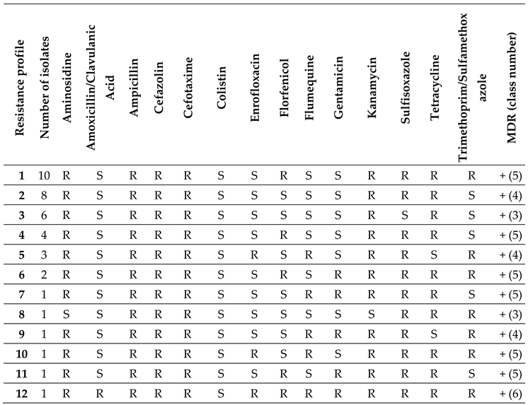
Table 2 reports the 12 different ESBL E. coli resistance profiles observed in this study. Profile 1 was the most frequent (10 out of 39) and was found in 9 calves (3 male and 6 female calves) and the calf feeding bucket. Profile 2 (8 of 39) was found in 7 calves (4 male and 3 female calves) and waste milk. Profile 3 (6 of 39) was found in 2 out of 3 cows treated for IMI, 3 alley floor samples, and a male calf. Profile 4 (4 of 39) was found in 3 female and 1 male calf. Profile 5 (3 of 39) was found in 2 female calves and a female calf pen. Profile 6 (2 of 39) was found in a male pen and the cow feeding rack. Profiles from 7 to 12 were found only once and in 4 male calves, the calf drinking water, and a female calf, respectively.
2.3. Hierarchical Clustering of E. coli Isolates Based on the MIC Results
Hierarchical clustering of ESBL E. coli isolates based on the MIC test results was carried out [12]. Results are illustrated in Figure 2. Two main branches were observed. One included the isolates from 12 calves, waste milk, treated cows, and alley floors. Within this branch, all the isolates from cows and alley floors and one calf isolate were separated from the waste milk isolate and 11 closely related calf isolates. Another branch included 16 calf isolates and the isolates from calf pens, water and feeding buckets, and the cow feeding rack. Within this branch, one subgroup included the isolates from 4 calves, male pens, and the cow feeding rack, separated from the isolates from 9 calves and the water and feeding buckets, while another subgroup included three calves and the female pen.
Based on the threshold Euclidean distance of 7.5, several statistically significant clusters grouped more than one sample. The largest one included the waste milk and 11 calf isolates, suggesting a relevant role for this risk factor in ESBL E. coli diffusion. The second largest cluster included the isolates from 9 calves, closely related to the common feeding bucket and the calf drinking water. The third largest cluster grouped the isolates from one calf, treated cows, and the primiparous and fresh cow alleys, indicating fecal shedding from treated cows as another diffusion route. The fourth one included two female calf isolates and the female calf pens. The other 4 calf isolates, the male pens, and the cow feeding rack were also closely related.
2.4. Results of the Biosecurity Questionnaire
The questionnaire highlighted notable issues concerning the risk factors for the occurrence of ESBL E. coli: 1. Feeding of waste milk to male calves; 2. No pasteurizer cleaning; 3. Daily cleaning of the nipple buckets with water without detergent or disinfectant; 4. Common buckets between female and male calves; 5. Mixed use of calf pens; 6. Occasional cleaning of single calf pens during the winter season; 7. Occasional cleaning of cattle water troughs; 8. Antibiotic treatment of cases of environmental mastitis; 9. Blanket dry cow therapy (Supplementary File S2).
3. Discussion
This study aimed to assess the presence, distribution, and antimicrobial resistance profiles of ESBL-producing E. coli in a medium-sized dairy herd in Northern Italy, hosting nearly 1,000 animals, including calves, heifers, and lactating and dry cows. We collected calf and cow feces, waste milk, environmental samples, and water, and we administered a questionnaire to assess the associated risk factors. As a result, most pre-weaned calves, including males and females, carried ESBL E. coli in their intestines, and ESBL E. coli were also diffusely present in the environment and farm equipment in contact with them. This result is in agreement with a previous paper [13] showing that the shedding of AMR E. coli increased with herd size. The herd management interview enabled the gathering of information on potential risk factors for the distribution of ESBL microorganisms on the farm. Not much is known about the diffusion and transmission of ESBL E. coli among calves and cows, and how they are affected by environmental factors [14]. The hierarchical clustering of ESBL E. coli isolates based on the MIC results suggested that isolates with different resistance characteristics are circulating in the farm and that different sources and routes could be involved in their dissemination and maintenance, facilitated by incorrect or inadequate management, biosecurity, and hygiene practices.
The farmer used waste milk for feeding male calves. According to hierarchical clustering based on the isolate MIC profiles, the largest statistically significant cluster included about 40% of all calves’ fecal isolates and the waste milk isolate. ESBL E. coli were also detected in the feces of two out of three cows treated for mastitis that contributed to the waste milk. Among the risk factors associated with the spread of ESBL-producing bacteria on cattle farms, the use of waste milk containing antibiotic residues as calf feed appears to play a crucial role [3,15]. However, waste milk is still used for this purpose in different countries [16], although many of them, including Italy, have recently issued guidelines discouraging or forbidding this practice.
Based on hierarchical clustering, the MIC profiles of the isolates from the calf feeding bucket and drinking water were related to the fecal isolates of over 30% of the calves. Incorrect management practices such as shared or improperly cleaned feeding equipment [3] can favor the diffusion of AMR-carrying bacteria on the farm. Notably, the feeding buckets and calf water buckets were not cleaned with detergents or disinfectants, and sometimes not even rinsed with water between feedings; furthermore, the number of buckets was not adequate for the number of animals on the farm. ESBL E. coli were isolated from all these pieces of equipment as well as from the calf drinking water. Moreover, the pasteurizer used to reduce bacterial contamination of milk was not cleaned between cycles. Poor cleaning is one of the factors favoring bacterial contamination and multiplication, leading to higher microbial loads [17]. Indeed, we isolated ESBL E. coli also from the farm’s pasteurized waste milk.
Shared calf pens and their poor hygiene may also play a role in promoting the diffusion of AMR bacteria. We isolated ESBL E. coli from both male and female calf pens, and we observed a relationship between the MIC profiles of these isolates and those from calf feces based on hierarchical clustering. As highlighted in an EFSA scientific opinion paper on calf welfare, the level of cleanliness of the areas used for housing calves is a major determinant of their health [18]. Inaccurate cleaning procedures of the single pens or calf hutches may not adequately remove fecal contamination from the walls, leading them to serve as a reservoir [14].
The cows underwent blanket dry cow therapy (BDCT) with a β-lactam, specifically amoxicillin/clavulanic acid. Although this practice is not allowed in Italy, some farms are still using it. BDCT has been reported to be linked to a significant increase of ESBL E. coli in calf feces during the colostral phase [9].
The ESBL E. coli isolated from the feces of cows treated for mastitis clustered with the isolates from the cows’ alley floors, suggesting fecal shedding. E. coli ESBL shedding can vary greatly among individuals [19], and antibiotic treatment for mastitis could play a role in increasing animal colonization, shedding, and subsequent environmental contamination by AMR-carrying bacteria [20]. The farm evaluated in this study did not have a sick pen, and this represents a lack of biosecurity.
ESBL E. coli often carry multiple resistance genes that confer resistance to other antimicrobial drugs than β-lactams, leading to MDR [21]. All the isolates obtained in our study, both from the calves, their equipment, and the farm environment, were MDR. On the other hand, resistance to colistin, an antibiotic of last resort for humans, was not detected. Many developed countries have prohibited its usage in food-producing animals, and the Antimicrobial Advice Ad Hoc Expert Group (AMEG) [22] has placed antibiotics in this category as very important in human medicine. All ESBL E. coli isolates were also carbapenem-sensitive. This is also a positive finding, as carbapenemase-producing Enterobacteriaceae cause serious human infections. The study by Waade et al. conducted in Germany in 2021 reported similar results since the ESBL-producing isolates were 92.9% E. coli; 60.6% of ESBL-producing isolates were resistant to one or more classes of antibiotics including penicillins and cephalosporins but were sensitive to carbapenems [23].
As a final consideration, although these bacteria were never isolated from fecal samples, 42.85% of environmental samples were positive for ESBL-producing A. baumannii. A. baumannii is reported as a relevant cause of nosocomial infections in humans, and MDR strains can pose significant risks for human health [24]. MDR A. baumannii has also been associated with inadequate cleaning of dairy equipment and the dairy cattle environment [25]. Therefore, improving farm management practices can help control also this relevant AMR microorganism.
4. Materials and Methods
2.1. Farm Description and Ethics Statement
The farm was located in Northern Italy, and it was characterized by a high prevalence of ESBL-producing Enterobacteriaceae in the feces of calves according to previous information available to our laboratory. The herd consisted of 1,000 animals, out of them 450 lactating Italian Friesian cows, and is accredited free from infectious bovine rhinotracheitis (IBR), vaccinated for neonatal diarrhea agents, and type-1 and type-2 bovine viral diarrhea virus (BVDV). The farm does not use an in-house colostrum bank, but the colostrum is taken by the calves directly from the dam. Waste milk with antibiotics is pasteurized and used for feeding male calves.
2.2. Questionnaire
A questionnaire was completed together with the herd manager (Supplementary File S2). The questionnaire follows the Biocheck.UGent checklist [26] and was used to assess different aspects of herd management, the use of antibiotics, and farm biosecurity. The questionnaire was also integrated with further aspects based on previous studies on ESBL E. coli risk factors [15,20,27]. The form was divided into several sections: I. General questions about farm organization: how many animals are present in the different categories, who works with the animals; II. Health management in the sick pen and management of outbreaks; III. Reproduction management; IV. Calving pen management and hygiene questions; V. Calf rearing: colostrum feeding management, milk feeding management, calf housing, vaccinations, and treatments; VI. Health management of the herd; VII. Milking management.
2.3. Animals and Sample Collection
During our visit, the feces of all dairy calves between 7 and 21 days of age, balanced for sex, not treated with antibiotics, and free from diarrhea, were collected for a total of 37 samples. According to the calf manager, the males (19) had been fed with waste milk, while the females (18) with a commercial milk replacer. We sampled the feces of the 26 dams present on the farm (the 9 missing dams had been sold or sent to the slaughterhouse) and of 3 cows treated for intramammary infection (IMI) with amoxicillin/clavulanic acid that contributed to the waste milk. All the fecal samples were collected from the rectal ampoule using gloves, stored refrigerated in the laboratory, and frozen at -20°C until analysis. Waste milk was collected directly from the pasteurizer and kept refrigerated until arrival at the laboratory. Three calf pens were sampled by rubbing sterile gauzes against the inner wall of the pens over an area of about 150 X 30 cm2 at the height of the calves’ noses avoiding obvious fecal smears, then stored in sterile 50-mL Falcon® tubes. Two disposable swabs were collected by rubbing the bottom and inner wall of a calf feeding bucket, and the inner part of the nipple, respectively. Two water samples of 150 mL were collected into sterile containers from the calf watering buckets and one from the cow watering trough, respectively. Two environmental samples were also taken with gauzes from the cow feeding rack and one from the cow’s berth tube. Disposable fabric socks were used to collect three samples from the barn floors by walking down the alleys one time and then inserted in sterile plastic bags: one from the cow alley, one from the primiparous cow alley, and one from the fresh cow alley, respectively. All environmental and water samples were stored at refrigerated temperature until arrival at the laboratory.
2.4. ESBL E. coli Isolation and Characterization
Environmental swabs and feces (0.1 g) were enriched in 5 mL of Müeller Hinton broth (Microbiol, Cagliari, Italy) and incubated at 37° C under aerobic conditions for 18-24 h. For environmental samples, 30 mL of Müeller Hinton broth was added to the Falcon tubes and plastic bags containing the samples and incubated at 37° C for 18-24 h. One hundred mL of water was added to an equal amount of double-strength enrichment broth and incubated at 37°C for 48 h. All the feces and environmental samples were cultured on CHROMagar™ ESBL agar plates (CHROMagar, Paris, France) and MacConkey agar as a control medium (Oxoid Ltd., Basingstoke, United Kingdom), and incubated at 37°C for 18-24 h. Pasteurized waste milk was seeded on blood agar plates (Microbiol, Cagliari, Italy) and CHROMagar™ ESBL agar plates in amounts of 100 μL and incubated at 37°C for 24 hours. Colonies indicating ESBL bacteria grown on CHROMagar™ ESBL agar plates were picked and submitted to species identification with the MBT Microflex LT/SH MALDI-TOF mass spectrometer (Bruker Daltonik GmbH, Germany) as described previously [28]. After species identification, the colonies recovered from CHROMagar™ ESBL agar plates were sub-cultured on blood agar plates (Microbiol, Cagliari, Italy) and subjected to ESBL phenotyping assessment using the double-disc synergy test (DDST) and to assess carbapenemase production according to the EUCAST guidelines [29].
2.5. Antimicrobial Susceptibility Testing
A SensititreTM ITISVE1 plate (Thermo Fisher Scientific®, Waltham, MA, USA) was used to determine the MIC of the antimicrobials commonly used in dairy herds against the ESBL-E. coli isolates. The plate contained the following antibiotics: flumequine (range 1 - 16 μg/ml); amoxicillin/clavulanic acid (0,25 - 32 μg/ml); ampicillin (0,25 - 32 μg/ml); cefazolin (0,5 - 8 μg/ml); cefotaxime (0,5 - 4 μg/ml); sulfisoxazole (128 - 512 μg/ml); colistin (0,03125 - 8 μg/ml); enrofloxacin (0,015625 - 32 μg/ml); florfenicol (1 - 64 μg/ml); gentamicin (0,25 - 32 μg/ml); tetracycline (0,5 - 16 μg/ml); trimethoprim/sulfamethoxazole (0,0625 - 16 μg/ml); aminosidine (1 - 32 μg/ml); kanamycin (2 - 32 μg/ml). Quality control for Sensititre plates was performed using E. coli strain ATCC25922 and Sensititre™ SWIN™ Software System (SensititreTM, Thermo Fisher Scientific®, Waltham, MA, USA). The MIC results were interpreted according to the manufacturer’s instructions using CLSI VET08 4th edition [30] (V= Vet), CLSI VET06 1st edition [31] (V= Vet), CLSI M100 29th edition (H= Human) [32], EUCAST v.11.0 [33], CASFM 2019 [34].
2.6. Hierarchical Clustering
Non-supervised hierarchical cluster analysis using Ward’s method was performed based on the MIC values [12,35]. A total of 21 parameters were obtained by assigning to each MIC value ranging from >512 µg/mL to ≤ 0.015625 a number from 1 to 21 according to decreasing antibiotic concentrations. The profiles obtained for each sample after the conversion were used to construct a dendrogram. This technique was chosen for its effectiveness in minimizing variance within the clusters, allowing us to identify groups of isolates with similar resistance patterns. The resulting dendrogram provides a visual representation of the progressive merging of the clusters based on the Euclidean distance. The dendrogram was cut (maximum distance for clustering) at a height of 7.5. This cut-off point was chosen based on statistical significance, ensuring that each cluster represented a distinctive group of isolates with similar characteristics. The analysis was conducted using the SciPy library (version 1.11.4, https://scipy.org/) within the Python environment (version 3.10.12, https://www.python.org/).
5. Conclusions
This study confirms the widespread diffusion of ESBL E. coli in dairy farms and highlights the relevant presence and circulation of MDR strains. Rational antibiotic use remains the most relevant driver enabling the reduction and control of AMR bacteria. Nevertheless, adherence to good internal and external biosecurity practices, hygiene of facilities and equipment, correct feeding procedures, and correct animal management might also significantly contribute to reducing and controlling the spread of AMR bacteria within the farm.
Supplementary Materials
The following supporting information can be downloaded at: Preprints.org, Supplementary File S1: Questionnaire of the study multi-drug resistant extended-spectrum beta-lactamase (ESBL)-producing Escherichia coli in a dairy herd: distribution and antimicrobial resistance profiles. Supplementary Table S2: MIC values expressed in µg/mL for the ESBL E. coli isolates assessed in this study with the plate assay.
Author Contributions
Conceptualization: M.P., R.P, M.F.A.; methodology, M.P., R.P, M.F.A.; farm activities and sample collection: M.P, L.F.P.; formal analysis: M.P., L.F.P., L.M.; resources: R.P., M.F.A.; data curation: M.P., A.G., M.F.A.; writing, original draft: M.P., M.F.A.; writing, review and editing, all authors; supervision: R.P., M.F.A. All authors have read and agreed to the published version of the manuscript.
Funding
This research received no external funding. All funds were from the University of Milan.
Institutional Review Board Statement
The study was conducted in accordance with the Declaration of Helsinki and approved by the Institutional Committee for Animal Welfare of the University of Milan (protocol number 99_2023).
Informed Consent Statement
Written informed consent has been obtained from the farmer to publish this paper.
Data Availability Statement
All pertinent data generated in this study are provided in the supplementary materials.
Acknowledgments
Fernando Ulloa was supported by a fellowship from the Universidad Austral de Chile.
Conflicts of Interest
The authors declare no conflicts of interest.
References
- European Centre for Disease Prevention and Control. Available online: https://www.ecdc.europa.eu/en/publications-data/antimicrobial-resistance-surveillance-europe-2022-2020-data (accessed on 29 January 2024).
- World Health Organization. Available online: https://www.who.int/publications-detail-redirect/9789240062702 (accessed on 29 January 2024).
- Ma, Z.; Lee, S.; Jeong, K.C. Mitigating Antibiotic Resistance at the Livestock-Environment Interface: A Review. J Microbiol Biotechnol 2019, 29, 1683–1692. [Google Scholar] [CrossRef] [PubMed]
- European Centre for Disease Prevention and Control (ECDC); European Food Safety Authority (EFSA); European Medicine Agency (EMA). Third Joint Inter-Agency Report on Integrated Analysis of Consumption of Antimicrobial Agents and Occurrence of Antimicrobial Resistance in Bacteria from Humans and Food-Producing Animals in the EU/EEA. EFSA Journal 2021, 19, e06712. [Google Scholar] [CrossRef]
- Cuong, N.V.; Padungtod, P.; Thwaites, G.; Carrique-Mas, J.J. Antimicrobial Usage in Animal Production: A Review of the Literature with a Focus on Low- and Middle-Income Countries. Antibiotics (Basel) 2018, 7, 75. [Google Scholar] [CrossRef] [PubMed]
- Bush, K.; Fisher, J.F. Epidemiological Expansion, Structural Studies, and Clinical Challenges of New β-Lactamases from Gram-Negative Bacteria. Annu Rev Microbiol 2011, 65, 455–478. [Google Scholar] [CrossRef] [PubMed]
- Shaikh, S.; Fatima, J.; Shakil, S.; Rizvi, S.M.D.; Kamal, M.A. Antibiotic Resistance and Extended Spectrum Beta-Lactamases: Types, Epidemiology and Treatment. Saudi J Biol Sci 2015, 22, 90–101. [Google Scholar] [CrossRef] [PubMed]
- Reygaert, W.C. An Overview of the Antimicrobial Resistance Mechanisms of Bacteria. AIMS Microbiol 2018, 4, 482–501. [Google Scholar] [CrossRef] [PubMed]
- Tetens, J.L.; Billerbeck, S.; Schwenker, J.A.; Hölzel, C.S. Short Communication: Selection of Extended-Spectrum β-Lactamase-Producing Escherichia Coli in Dairy Calves Associated with Antibiotic Dry Cow Therapy-A Cohort Study. J Dairy Sci 2019, 102, 11449–11452. [Google Scholar] [CrossRef] [PubMed]
- Massé, J.; Lardé, H.; Fairbrother, J.M.; Roy, J.-P.; Francoz, D.; Dufour, S.; Archambault, M. Prevalence of Antimicrobial Resistance and Characteristics of Escherichia Coli Isolates From Fecal and Manure Pit Samples on Dairy Farms in the Province of Québec, Canada. Front Vet Sci 2021, 8, 654125. [Google Scholar] [CrossRef]
- Cho, S.; Jackson, C.R.; Frye, J.G. Freshwater Environment as a Reservoir of Extended-Spectrum β-Lactamase-Producing Enterobacteriaceae. J Appl Microbiol 2023, 134, lxad034. [Google Scholar] [CrossRef]
- Berrazeg, M.; Drissi, M.; Medjahed, L.; Rolain, J.M. Hierarchical Clustering as a Rapid Tool for Surveillance of Emerging Antibiotic-Resistance Phenotypes in Klebsiella Pneumoniae Strains. J Med Microbiol 2013, 62, 864–874. [Google Scholar] [CrossRef]
- Duse, A.; Waller, K.P.; Emanuelson, U.; Unnerstad, H.E.; Persson, Y.; Bengtsson, B. Risk Factors for Antimicrobial Resistance in Fecal Escherichia Coli from Preweaned Dairy Calves. J Dairy Sci 2015, 98, 500–516. [Google Scholar] [CrossRef] [PubMed]
- Homeier-Bachmann, T.; Kleist, J.F.; Schütz, A.K.; Bachmann, L. Distribution of ESBL/AmpC-Escherichia Coli on a Dairy Farm. Antibiotics (Basel) 2022, 11, 940. [Google Scholar] [CrossRef] [PubMed]
- Weber, L.P.; Dreyer, S.; Heppelmann, M.; Schaufler, K.; Homeier-Bachmann, T.; Bachmann, L. Prevalence and Risk Factors for ESBL/AmpC-E. Coli in Pre-Weaned Dairy Calves on Dairy Farms in Germany. Microorganisms 2021, 9, 2135. [Google Scholar] [CrossRef] [PubMed]
- European Food Safety Authority Panel on Biological Hazards (BIOHAZ); Ricci, A.; Allende, A.; Bolton, D.; Chemaly, M.; Davies, R.; Fernández Escámez, P.S.; Girones, R.; Koutsoumanis, K.; Lindqvist, R.; et al. Risk for the Development of Antimicrobial Resistance (AMR) Due to Feeding of Calves with Milk Containing Residues of Antibiotics. EFSA Journal 2017, 15, e04665. [CrossRef]
- Elizondo-Salazar, J.; Jones, C.; Heinrichs, A. Evaluation of Calf Milk Pasteurization Systems on 6 Pennsylvania Dairy Farms. Journal of dairy science 2010, 93, 5509–5513. [Google Scholar] [CrossRef] [PubMed]
- European Food Safety Authority Panel on Biological Hazards (BIOHAZ); Koutsoumanis, K.; Allende, A.; Álvarez-Ordóñez, A.; Bolton, D.; Bover-Cid, S.; Chemaly, M.; de Cesare, A.; Hilbert, F.; Lindqvist, R.; et al. Update of the List of Qualified Presumption of Safety (QPS) Recommended Microorganisms Intentionally Added to Food or Feed as Notified to EFSA. EFSA Journal 2023, 21, e07747. [CrossRef]
- Munns, K.D.; Selinger, L.B.; Stanford, K.; Guan, L.; Callaway, T.R.; McAllister, T.A. Perspectives on Super-Shedding of Escherichia Coli O157:H7 by Cattle. Foodborne Pathog Dis 2015, 12, 89–103. [Google Scholar] [CrossRef]
- Gonggrijp, M.A.; Santman-Berends, I.M.G.A.; Heuvelink, A.E.; Buter, G.J.; van Schaik, G.; Hage, J.J.; Lam, T.J.G.M. Prevalence and Risk Factors for Extended-Spectrum β-Lactamase- and AmpC-Producing Escherichia Coli in Dairy Farms. J Dairy Sci 2016, 99, 9001–9013. [Google Scholar] [CrossRef]
- Ibrahim, D.R.; Dodd, C.E.R.; Stekel, D.J.; Meshioye, R.T.; Diggle, M.; Lister, M.; Hobman, J.L. Multidrug-Resistant ESBL-Producing E. coli in Clinical Samples from the UK. Antibiotics 2023, 12, 169. [Google Scholar] [CrossRef]
- European Medicines Agency (EMA). Available online: https://www.ema.europa.eu/en/news/categorisation-antibiotics-used-animals-promotes-responsible-use-protect-public-and-animal-health (accessed on 29 January 2024).
- Waade, J.; Seibt, U.; Honscha, W.; Rachidi, F.; Starke, A.; Speck, S.; Truyen, U. Multidrug-Resistant Enterobacteria in Newborn Dairy Calves in Germany. PLOS ONE 2021, 16, e0248291. [Google Scholar] [CrossRef]
- Mohamed, H.M.A.; Abd-Elhafeez, H.H.; Al-Jabr, O.A.; El-Zamkan, M.A. Characterization of Acinetobacter Baumannii Isolated from Raw Milk. Biology (Basel) 2022, 11, 1845. [Google Scholar] [CrossRef]
- Zucali, M.; Bava, L.; Tamburini, A.; Brasca, M.; Vanoni, L.; Sandrucci, A. Effects of Season, Milking Routine and Cow Cleanliness on Bacterial and Somatic Cell Counts of Bulk Tank Milk. J Dairy Res 2011, 78, 436–441. [Google Scholar] [CrossRef]
- Biocheck.UGent. Available online: https://biocheckgent.com/en (accessed on 29 January 2024).
- Santman-Berends, I.M.G.A.; Gonggrijp, M.A.; Hage, J.J.; Heuvelink, A.E.; Velthuis, A.; Lam, T.J.G.M.; van Schaik, G. Prevalence and Risk Factors for Extended-Spectrum β-Lactamase or AmpC-Producing Escherichia Coli in Organic Dairy Herds in the Netherlands. J Dairy Sci 2017, 100, 562–571. [Google Scholar] [CrossRef]
- Rosa, N.M.; Penati, M.; Fusar-Poli, S.; Addis, M.F.; Tola, S. Species Identification by MALDI-TOF MS and Gap PCR–RFLP of Non-Aureus Staphylococcus, Mammaliicoccus, and Streptococcus Spp. Associated with Sheep and Goat Mastitis. Veterinary Research 2022, 53, 84. [Google Scholar] [CrossRef]
- Clinical & Laboratory Standards Institute (CLSI). Available online: https://clsi.org/ (accessed on 29 January 2024).
- Clinical and Laboratory Standards Institute (CLSI); Performance Standards for Antimicrobial Disk and Diluition Susceptibility Tests fro Bacteria Isolated from Animals, 4th ed.; Publisher: Wayne, PA, USA, 2018.
- Clinical and Laboratory Standards Institute (CLSI); Methods for Antimicrobial Susceptibility Testing of Infrequently Isolated or Fastidious Bacteria Isolated From Animals, 1st ed.; Publisher: Wayne, PA, USA, 2017.
- Clinical and Laboratory Standards Institute (CLSI); Performance Standards for Antimicrobial Susceptibility Testing, 29th ed.; Publisher: Wayne, PA, USA, 2019.
- Clinical Breakpoints and Dosing of Antibiotics (EUCAST). Available online: https://www.eucast.org/clinical_breakpoints (accessed on 29 January 2024).
- Comité de l’antibiogramme de la Société Francaise de Microbiologie (CASFM). Available online: https://www.sfm-microbiologie.org/wp-content/uploads/2019/01/CASFM_VET2018.pdf (accessed on 29 January 2024).
- Ward Jr., J. H. Hierarchical Grouping to Optimize an Objective Function. Journal of the American Statistical Association 1963, 58, 236–244. [Google Scholar] [CrossRef]
Figure 1.
Distribution of resistance and susceptibility of the ESBL E. coli isolates to the different antimicrobials according to the plate MIC test.
Figure 1.
Distribution of resistance and susceptibility of the ESBL E. coli isolates to the different antimicrobials according to the plate MIC test.
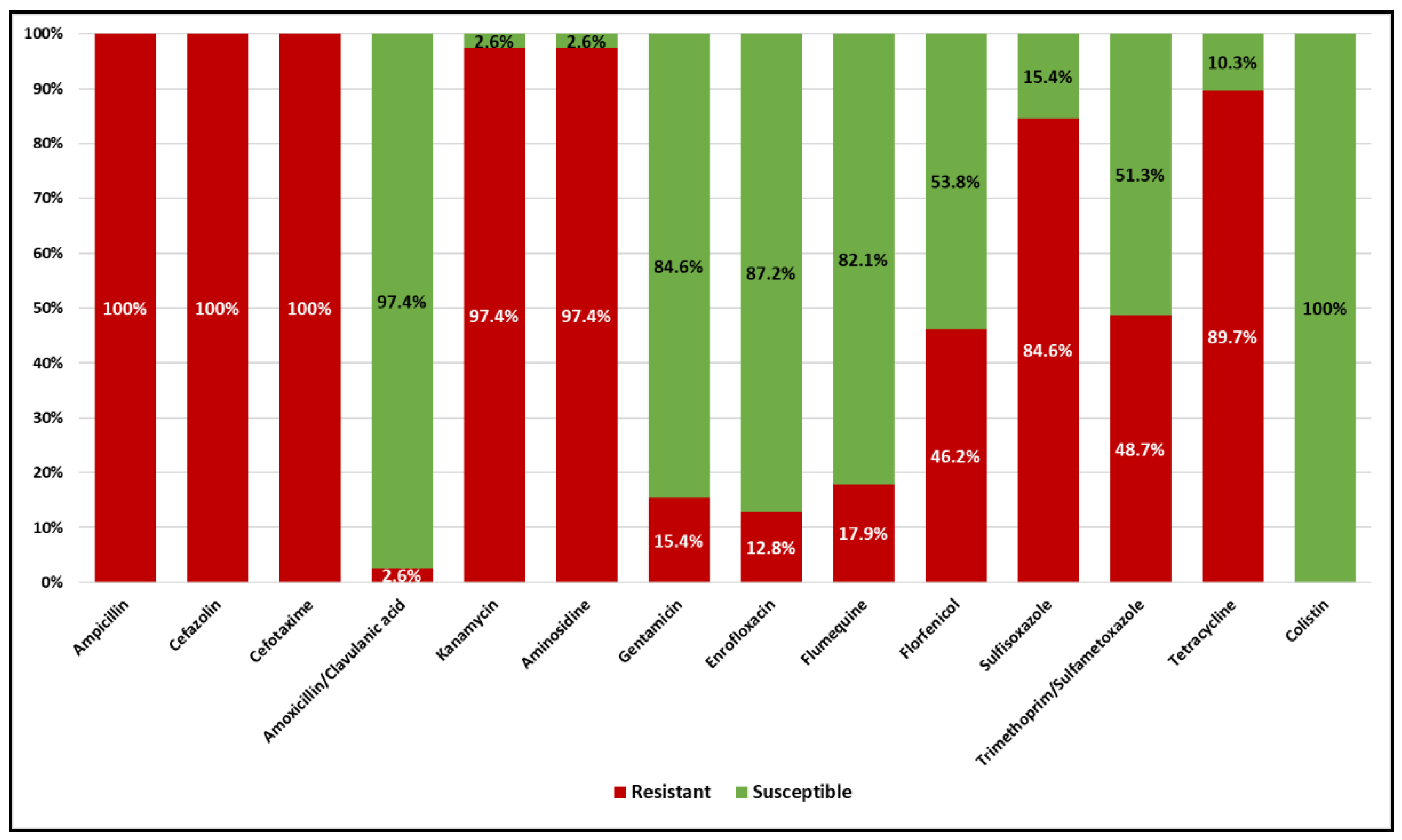
Figure 2.
Hierarchical clustering of the ESBL E. coli isolates’ MIC profiles based on Ward’s method. Female calves (F) are illustrated in red. Male calves (M) are illustrated in blue. Cows, environment, and water samples are illustrated in gold. The dotted line indicates the significant Euclidean distance cut-off point at 7.5.
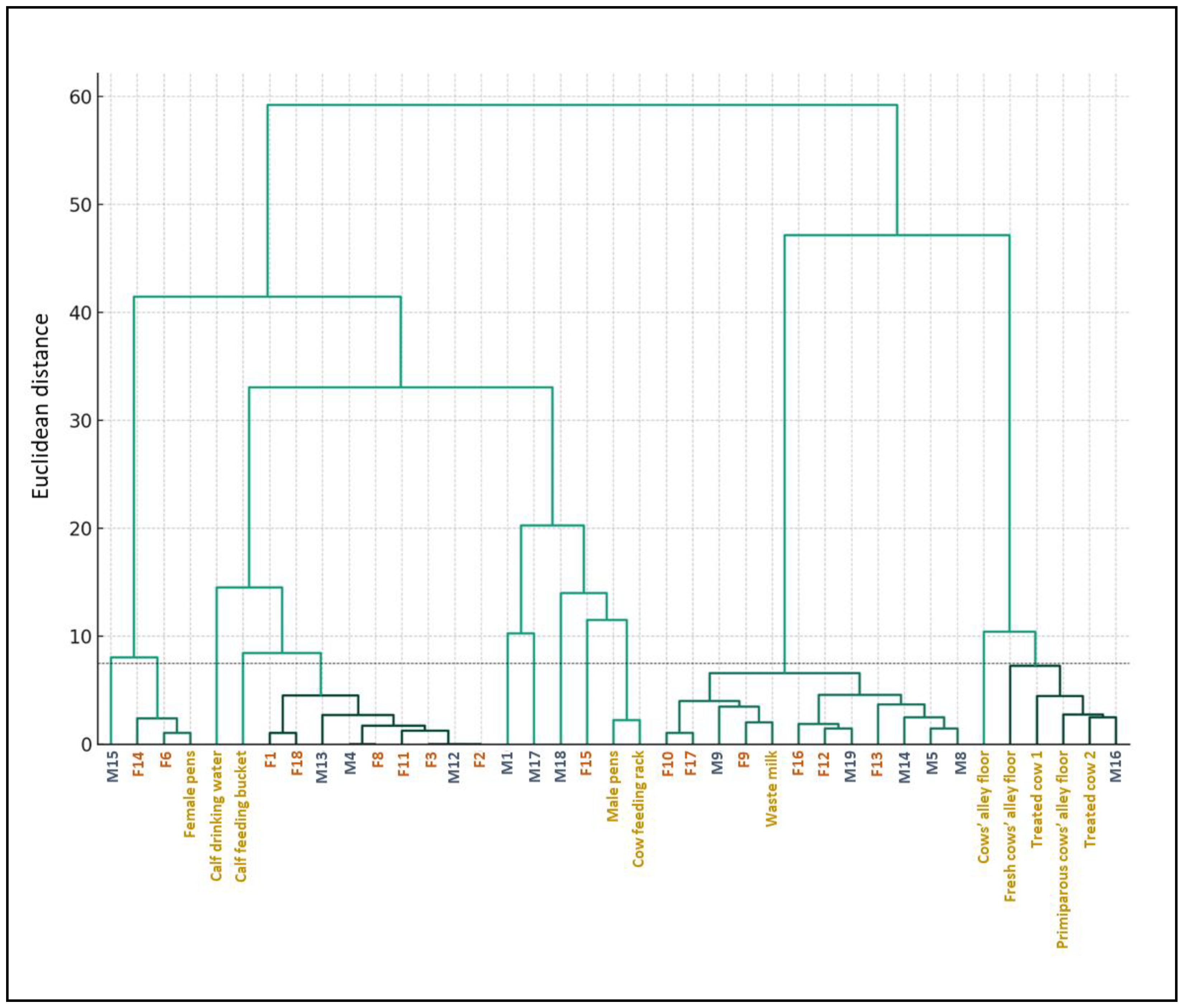

Table 1.
Summary of analyzed samples and respective bacteriological results.
| Sample type | N | ESBL E. coli (%) | Negative (%) | ESBL A. baumannii (%) |
|---|---|---|---|---|
| Female calf feces | 18 | 15 (83.3%) | 3 (16.7%) | 0 |
| Male calf feces | 19 | 13 (68.4%) | 6 (31.6%) | 0 |
| Treated cow feces | 3 | 2 (66%) | 1 (33%) | 0 |
| Dam feces | 26 | 0 | 26 (100%) | 0 |
| Waste milk | 1 | 1 (100%) | 0 | 0 |
| Male calf pens | 1 | 1 (100%) | 0 | 0 |
| Female calf pens | 1 | 1 (100%) | 0 | 0 |
| Mixed-use calf pens | 1 | 0 | 0 | 1 (100%) |
| Calf feeding bucket | 2 | 1 (50%) | 0 | 1 (50%) |
| Calf drinking water | 2 | 1 (50%) | 0 | 1 (50%) |
| Cow alleys | 3 | 3 (100%) | 0 | 0 |
| Cow’s berth tube | 1 | 0 | 0 | 1 (100%) |
| Cow water trough | 1 | 0 | 0 | 1 (100%) |
| Cow feeding rack | 2 | 1 (50%) | 0 | 1 (50%) |
Table 2.
Antimicrobial resistance profiles of the ESBL E. coli isolates. R, resistant; S, sensitive; MDR, multi-drug resistant. The number of antimicrobial classes is reported in parentheses.
Table 2.
Antimicrobial resistance profiles of the ESBL E. coli isolates. R, resistant; S, sensitive; MDR, multi-drug resistant. The number of antimicrobial classes is reported in parentheses.
Disclaimer/Publisher’s Note: The statements, opinions and data contained in all publications are solely those of the individual author(s) and contributor(s) and not of MDPI and/or the editor(s). MDPI and/or the editor(s) disclaim responsibility for any injury to people or property resulting from any ideas, methods, instructions or products referred to in the content. |
© 2024 by the authors. Licensee MDPI, Basel, Switzerland. This article is an open access article distributed under the terms and conditions of the Creative Commons Attribution (CC BY) license (http://creativecommons.org/licenses/by/4.0/).
Copyright: This open access article is published under a Creative Commons CC BY 4.0 license, which permit the free download, distribution, and reuse, provided that the author and preprint are cited in any reuse.


