Preprint
Article

Phytochemical Characterization and Evaluation of the Biological Activities of Opuntia ficus indica L

Altmetrics

Downloads

242

Views

200

Comments

0

Submitted:

05 February 2024

Posted:

06 February 2024

You are already at the latest version

Alerts
Abstract
In the context of sustainable and eco-friendly agriculture, current scientific researches are focusing on plant-based bioproducts to remedy the toxicity caused by chemical fertilizers and pesticides. This study was conducted on cladodes of Opuntia ficus-indicato screen the bioactive molecules and to evaluate the activity againstfungal agentsisolated from tomato fruits. Quantitative analysis of the methanolic extractrevealed its richeness in total polyphenols (86.6 mg GAE/100 g FW), flavonoids (13.4 mg QE/100 g FW), condensed tannins (08.9 mg TAE/100 g FW), and carotenoids(0.9 mg β-CE/100 g FW). DPPH test revealed that the cladodes extract had a high antioxidant potential (0.6 mg/ml). On the other hand, 7 fungal agents were isolated from infected tomato fruits and identifyed asbelonging to the following genera: Rhizoctonia (EC2), Fusarium (EC1 and EC3), Alternaria (EC4), Mucor (EC5), Aspergillus (EC6) and Penicillium (EC7). At a concentration 0.02% of cladodehydro-ethanolic extract, results of the antifungal activity showed aninhibition of mycelial growth up to70% for the isolates ofAlternariaspEC4 (70.91%), Rhizoctonia solaniEC2 (58.49%), Fusarium oxysporiumEC3 (57.63%) and Fusarium solaniEC1 (53.13%). While,inhibitory activities lower than 50% were obtained for fungal isolates MucorspEC6 (31.08%), Aspergillus sp EC5 (35.14%) and Penicillium sp EC7 (28.38%).At 0.04%all thecladode hydro-ethanolic extractwere able to inhibit mycelium growth in more than 50%.Similarly, maximum spore inhibition development were obtained with 0.04 cladode hydro-ethanolicreaching more than 60%. Inhibition percentages of 83.02%, 85.96%, 87.76% and 90.20% were obtained for fungal isolates Fusarium oxysporium EC3, Fusarium solani EC1, Rhizoctonia solani EC2 and Alternaria sp EC4, respectively. All together, the results show that Opuntia ficus-indica extract has potential tobe used effectively as a biopesticide against fungal agents of tomato fruits.
Keywords: 
Subject: Biology and Life Sciences  -   Biology and Biotechnology

1. Introduction

Tomato was ranked as the third most relevant vegetablein the world because it is grown in almost in more than 130 major countries over an estimated area of 2.5 million hectares [2]. Tomato is a short cycle crop, usually with high yields. In Algeria, tomatoes account for 1% of world production, 30% of which was produced in the Mediterranean [1]. According to FAO (2022), total world tomato production for both processing and fresh tomatoin 2021 amounted to over 189.1 million metric tonnes, with an increment of 2% per from 2018 to 2020[3].
However, tomato crops are at risck of being attacked by almost an handred of well known biological pests, as well as by new diseases emerging at an alarming rate [3]. These numerousdeseasedcan affect roots, crown, stem, leaves and even tomato fruits [4]. They are generally the result of unfavourable weather conditions or nutrient deficiencies [5]. Recent studies based on surveys and statistics in various countries have revealed that losses of fruit and vegetables due to fungal infections are estimated at an average of 60 million tonnes per year at consumer level. This represents 16% of the food that reaches consumers. Of this amount, 80% of food could be avoided or potentially avoided by improving home storage conditions, a saving of 47 million tonnes per year [6].Tomatoes with a short shelf life, are considered to be one of the most easily spoiled products [5]. Several fungi belonging to the genera Aspergillus, Penicillium and Fusarium are known to contaminate agricultural products and/or to produce toxic secondary metabolites [7,8]. These fungican cause yield losses between 30 and 50% of the crop in the event of epidemic development [9]. Once harvested, tomatoes are also subject to various fungal infections that limit their shelf life to just a few days. Among the fungimost found in fresh tomatoes are Aspergillussp, Mucorsp, Fusarium oxysporum, Fusarium solani and Alternariasp [5]. Chemical pesticides, using fungicides and insecticides [10], mainly control these diseases. However, numerous studies indicate the emergence of fungi resistance to these chemical substances. These substances cause both toxicological and ecological problems [11]. In addition, they have negative effects on other microorganisms that are a source of soil fertilization and important components of the soilfood webs [12]. To remedy this situation, scientific research has turned to our natural heritage, in particular aromatic and medicinal plants. Plants represent an inexhaustible source of substances and natural bioactive compounds. Numerous studies have highlighted the presence of secondary metabolites with biological activities such as polyphenols, alkaloids and terpenes [13].
Nowadays, Opuntia cladodes, fruits and flowers are the subject of numerous studies due to their sought-after properties in the food, cosmetic and pharmaceutical fields [14,15,16]. The Opuntiaficus-indica fruit, theprickly pear, is essentially found in the western Mediterranean: southern Spain, Portugal and North Africa (Tunisia, Algeria and Morocco)[17,18]. The genus Opuntia belongs to the Cactaceae family and account approximately with 300 species [19]. Itoriginated in Mexico and grows in arid and semi-arid regions [20]. Despite this, the Opuntia ficus-indica species is the most widely consumed and studied. The cladodes of this species are characterised by their high mucilage production [21] mainly composed of polysaccharides, minerals, amino acids, vitamins, phenolic acids and flavonoids [22,23,24], and by their therapeutical potential, antibacterial, and antifungal function [25]. Algeria has a rich and diverse plant life. Among the medicinal plants that make up the plant cover,Opuntia genus is widely distributed, especially in arid and semi-arid regions. This plant species is used in many regions of the world, mainly for food and traditional medicine [26]. This study focused mainly on thecharacterization of the phytochemical composition of extracts obtained by assaying bioactive substances and determining the antifungal activity of extracts of Opuntia ficus indica L cladodes against certain fungal agents isolated from tomato fruits.

2. Materials and Methods

2.1. Sampling of Plant Material

2.1.1. Opuntia Ficus Indica

The cladodes of Opuntia ficus indica L plant used in this study were collected in a rural area called Douar Al-Abayatt affiliated to the commune of Sidi Kadaa, located 18 km from the department of Mascara (Figure 1). Harvesting tooked place during February 2023, at 35°20’51.3’’N 0°18’46.

2.1.2. Tomato Fruits

Seven (07) infected tomato fruit samples (E1, E2, E3, E4, E5, E6 and E7) were obtained from various vegetable marchants in Mascara department which showed visual symptoms of various tomato diseases.Characterization of tomato fungal diseases was carried based on naked eye observable symptomssuch as glueiness and the appearance of necrotic spots or lesions.

2.2. Isolation and Identification of Fungal Agents

2.2.1. Isolation

Infected tomato fruits were cleaned with sterile distilled water. They were then rinsed with alcohol to remove surface microorganisms and kept until drying [27]. For the isolation of fungal agents, PDA (Potato Dextrose Agar) medium was used. Fragments of infected tomato fruits obtained were aseptically inoculated into Petri dishes containing PDA medium at rate of one fragment per dish.The Petri dishes were then sealed with parafilm and incubated at 25°C for 7 days [28].

2.2.2. Purification

Successive transplantof fungal isolates formed was performed aseptically. Culture explants were selected from the peripheral zone of colony growth and transferred to Petri dishes containing PDA medium.Incubation was performed at 25°C for 7 days. Purification was repeated 3 to4 times [29].

2.2.3. Identification

2.2.3.1. Macroscopic Identification

Macroscopic identification was carried out by examining cultures formed on PDA medium incubated at 25°C for 7 days. The examination aimed to determine the following cultural characteristics: growth rate, texture and colour of the verso (front) side of the thallusand recto (back) coloursideof colony [30].

2.2.3.2. Microscopic Identification

Microscopic observation of fingalisolates was carried out using the scotch tape method, which involved making an imprint on colony surface of the fungal isolate using adhesive tape. Once removed from the culture, the scotch tape was placed on a slide containing methylene blue. The preparation was observed by experts in fungal identification based on morphological characters using optical microscope at magnifications x10 and x40[31]Microscopic observation aimed at: examination of the mycelium (presence or absence of partitions, and branching mode); determination of the fruiting bodies (sporulation) and their contents (shape) and study of the conidia (morphology) [30].

2.3. Screening of Bioactive Molecules

2.3.1. Preparation of the Methanolic Extract

The methanolic extract of Opuntia ficus-indicawas prepared by homogenising 10 g of cladodes FW in 10 mL of methanol 95% (vol/vol) followed bystirring at 700 rpm for 30min at room temperature. The extractwas filtered through filter paper to remove solid particles [32], the liquid was keped for subsequent analysis.

2.3.2. Determination of Total Polyphenols

The total phenolic content of Opuntia ficus-indica cladodes was determined using Folin-Ciocalteaumodified method [33]. Briefly, 100 µL of methanolic extract was mixed with 900 µL of Folin-Ciocalteau reagent (diluted 1:10 with water). After 5 min, 750 µL of sodium bicarbonate aquous solution (7%) was added to the mixture and vortexed for 30 s. The above solution was then left to incubateat room temperature for 90 min and the absorbance was measured at 765 nm. Resultswere expressed as the mean (mg Gallic Acid Equivalent±SD/100 g Fresh Weight (FW)) for 3 replicates. Total polyphenol content was expressed as Gallic Acid Equivalents (GAE) utilizing gallic acid calibration curve from 0.006 mg/mL to 0.2 mg/mL.

2.3.3. Determination of Total Flavonoids

Total flavonoids were determined using the method described by Dehpeur et al. [34]. 500 μL of methanolic extract was added to 1500 μL of methanol (95%), 100 μL of 10% (w/v) AlCl3, 100 µl of sodium acetate (1 M) and 2.8 mL of distilled water. The mixture was stirred and incubated in the dark at room temperature for 30 min. Then, absorbance was measured at 415 nm.The blank was prepared by replacing the extract with methanol (95%). Results were expressed as the mean (mg Quercetin Equivalent±SD/100 g Fresh Weight (FW)) for 3 replicates. Total flavonoid content was expressed as Quercetin Equivalent (QE) using calibration curve ofquercetin from 0.0025 mg/ml to 0.08 mg/ml.

2.3.4. Determination of Condensed Tannins

This assay was carried out using the colorimetric method ofJoslyn [35].Whichis based on the reduction of phosphomolybdic and tungstic acid in an alkaline medium. In the presence of tannins, itgives a blue colour whose intensity is measured at 760 nm. 0.5 m of methanolicextract, 2.5 mL of Folin-Ciocalteu reagent solution and 5 mL of sodium carbonate (7.5%). The mixture formed was then left to stand for 30 min at room temperature in the dark; followed by incubationfor 5 min at 55°C. The solutionwas piked in cold water for 30 min. Then, absorbance was measured using a spectrophotometer at 760 nm. Results were expressed as the mean (mg Tannic Acid Equivalent±SD/100 g Fresh Weight (FW)) for 3 replicates. Condensed tanninsconcentrationwas expressed as Tannic Acid Equivalent (TAE)utilizing tannic acid calibration curve ranging from 0.02 mg/L to 0.1 mg/mL.

2.3.5. Determination of Carotenoids

The modified method of Sass-kiss et al. [36] was adopted for the determination of carotenoid content. It consisted of homogenising 4g of cladodes with 10 mL of a mixture of solvents (hexane/acetone/ethanol) (1V:2V:2V); followed by shaking at 300-400 rpm/15 min. The solution was centrifugated at 5500 rpm/15minat 4 °C. The top layer of hexane containing the pigment was sampled and its absorbance was measured at 430 nm. A blank was prepared in 95% methanol.Results were expressed as mean (mg β-carotene equivalent± SD/100 g Fresh Weight (FW)). Total carotenoid content was expressed as β-carotene equivalent (β-CE) by extrapolation on a β-carotene calibration curve from 0.002 mg/mLto 0.01 mg/mL.

2.3.6. Determination of Antioxidant Activity Using DPPH Test

DPPH (2, 2-diphenyl-1-picryl hydrazyl) assay was performed as described by Aruwa et al. [37]. 50 μL of each dilution of the extract (1 to 5 mg/ml) with 5 ml of 0.004% (w/v) DPPH solution was vortexed for 30s and incubated in the dark for 30 min at room temperature. The absorbances of the mixtures obtained were read using a UV-visible spectrophotometer at 517 nm. The blank was prepared in methanol 80% (v/v) and DPPH in methanol was used as a negative control. Ascorbic acid was used as a positive control. DPPH is a stable violet free radical in solution,its colour disappears rapidly when it is reduced to diphenyl picryl hydrazine (yellow) by a compound with anti-radical properties, resulting in discolouration. The intensity of the colour is perversely proportional to the capacity of the antioxidants present in the medium to donate protons. It has a characteristic absorbance in the range 512 to 517 nm [38].
The percentage of DPPH inhibition was calculated using the following formula:
Preprints 98289 i001
Where:
A0: Absorbance of the negative control
A1: Absorbance of the extract/standard
The percentage of radical scavenging activity relative to the extract concentration curve was plotted and the sample concentration that was required for 50% radical scavenging activity was determined and expressed as the EC50 value. Lower EC50 values indicate high antioxidant capacity. The experiment was carried out in triplicate.

2.4. Antifungal Activity

2.4.1. Preparation of Ethanolic Extract

Hydro-ethanolic extracts were prepared according to the modified method of Ghoul et al. [32]. 300 g of Opuntia ficus-indica cladodes were homogenised in ethanol 70% (vol/vol) with stirring at XXXX rpm for xxx min, at room temperature. The mixture was filtered through a filter paper to remove solid particles. The filtrate was evaporated to dryness at 40 °C. Six g of the dry extractwere recovered. 2 g and 4 g of the dry extract were homogenized in two tubes containing 10 mL of distilled waterto obtain the concentrations of 0.2 and 0.4 g/mL. The hydroalcoholic extract of the cladodes was sterilised under UV light for 1-2 days.

2.4.2. Preparation of PDA Medium with Different Concentrations of Cladode Hydro-Ethanolic Extract

Under sterile conditions, 10 mL of hydro-ethanolic extract of Opuntia ficus-indica cladodes containing 0.2 or 0.4 g/mLwere mixed with PDA medium (10 mLof extract + 90 mLof PDA medium). Followed by, shaking until homogenization of each mixture at temperature of 50°C. The extracts were prepared at final concentrations in the culture medium of 0.02% and 0.04% [39, modified].

2.4.3. Antifungal Activity

2.4.3.1. Cladode hydro-Ethanolic Effect on Mycelial Growth

Agar discs with 6 mm diameter of each fungus culture (grown at 25°C for 7) days were aseptically placed in the center of Petri dishes containing PDA medium at different concentrations of hydro-ethanolic extract (0.02% and 0.04%) [39, modified]. The dishes were then sealed with para-film. Petri dishes wereincubated at 25 °C for 7 days. Controls were prepared by inoculating each fungus on PDA medium alone (without the hydroalcoholic extract). Results were expressed by measuring the diameters of the inhibition zones formed (in mm). The antifungal activity was determined by the percentage of inhibition using the following formula [40]:
Preprints 98289 i002
Where:
MGIP (%): Mycelial growth inhibition percentage
D0: Growth diameter of the control
D1: Growth diameter with hydro-ethanolic extract

2.4.3.2. Cladodehydro-ethanoliceffect on Sporulation

Treated dishes previously used to determine the effect of cladode extract on mycelial growth were also used to test its effect on sporulation. For each fungal isolate, an explant forming a surface area of 1 cm² was taken from a culture on PDA medium incubated at 25 °C/10 days and introduced into test tubes containing 10 ml of sterile distilled water. The spore suspension was vortexed and filtered through filter paper to remove mycelial fragments. The total number of spores was counted using a Malassez cell as follows: a drop of the spore suspension was introduced using a Pasteur pipette into the space between the slide and coverslip. The spores are counted using the Mallassez cell counting perimeters. Sporulation results were expressed as spores/ml compared to control and antifungal activity was determined by the percentage of inhibition using the following formula [41].
Preprints 98289 i003
Where:
SIP (%): Sporulation inhibition percentage.
ST: Average number of spores estimated in the control.
S1: Average number of spores estimated in the presence of treatment
Statistical Analysis
The data experiments expressing antifungal activity of cladode hydro-ethanolic effect on mycelial growth and sporulation were analysed by 5% one-way ANOVA followed by 5% Tukey-Kramer HSD, and compared by correlationtests using JMP 17 software.

3. Results

3.1. Characterisation of Tomato Fungal Diseases

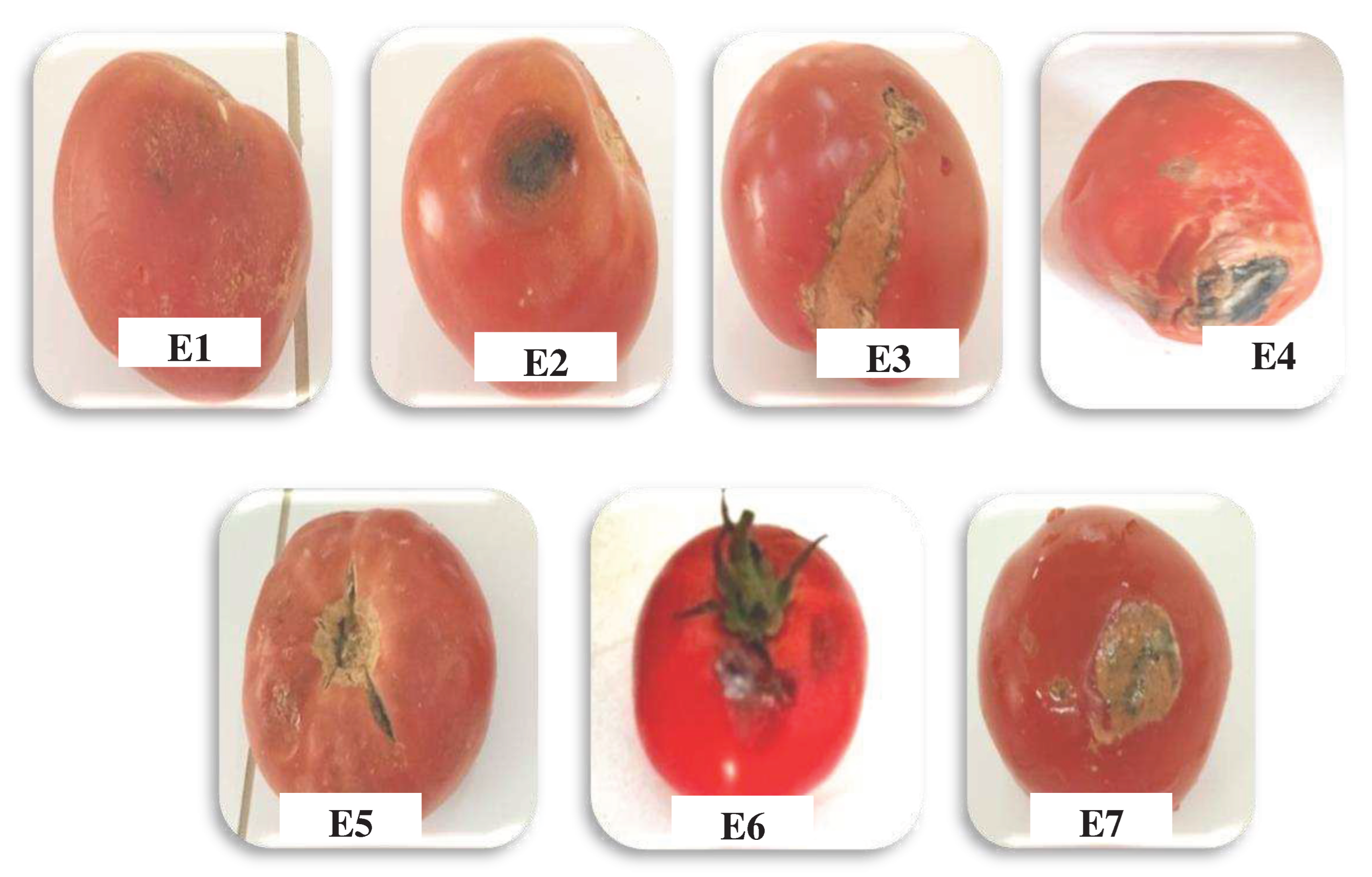
The seven (07) infected tomato fruit samples obtained (E1, E2, E3, E4, E5, E6 and E7) presented: black, white, brown or grey spots or soft, moist tissue(Figure 2).
Table 1 summarises the symptoms, fungal disease agent and diseases of the infected tomato fruit samplescolected.

3.2. Identification of Fungal Agents

3.2.1. Macroscopic Identification

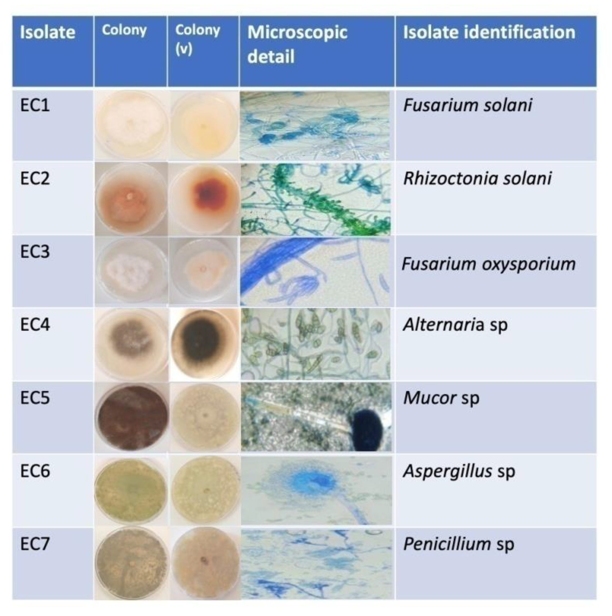
Macroscopic characterization of the 7 fungal agentsisolated from infected tomato fruits are summarised in Table S1. The 7 isolates revealed different types of colonies (Figure 3). Macroscopic observation of isolate EC1 showed a snow-white colony colour with a cottony appearance, a beige-white colour of verso and an average mycelial growth rate. Macroscopic observation of isolate EC2 revealed orange colony and a dark orange of the verso. The surface had cottony texture and mycelium growth was slow. Macroscopic observation of isolate EC3 showed a pinkish-whitecolour at the end of the colonies and a beige colour on the verso, with a cottony, dry appearance on the surface and slow mycelial growth. Macroscopic observation of isolate EC4 revealed brownish-blackcoloured colonies at the ends and a black colour on the verso. The texture was cottony and thick on the surface and the rate of mycelial growth was slow. Macroscopic observation of isolate EC5 showed black colonies with a beigecolour of the verso. The surface was granular and dry, and rapid growth rate. Macroscopic observation of isolate EC6 revealed a green coloration of the colonies and a yellow colour of the verso, with a granular and powdery appearance. Macroscopic observation of isolate EC7 showed bluish-green colonies and a beige-yellow colour of the verso, with a very powdery surface texture with the presence of droplets.

3.2.2. Microscopic Identification

Microscopic characteristics of the 7 fungal agents isolated from infected tomato fruits are grouped in (Spplemanty material: Table S2).
Microscopic identification of the 7 fungal isolates showed different aspects (Figure 3). Microscopic observation of isolate EC1 revealed the presence of septate mycelia, cylindrical microconidia, frequent chlamydospores and branched conidiophores. Microscopic observation of isolate EC2 revealed the presence of septate mycelia with elongated and often slightly swollen hyphae, branched at right angles. Microscopic observation of isolate EC3 showed septate mycelium with septate hyphae. Also, smooth or rough, globose hyaline chlamydospores, fusiform, multi-septate septate macroconidia produced by phialides onconidiophores. Microscopic observation of isolate EC4 revealed septate mycelium bearing numerous chains of conidia. The conidia are multicellular asexual spores divided by transverse and/or longitudinal partitions.Microscopic observation of isolate EC5 showed non-partitioned mycelia with non-septate thallus, and black globose unicellular conidia. Microscopic observation of isolate EC6 revealed the presence of septate mycelia, septate thallus, conidiophores terminated by an erect globose head, unicellular and globose conidia. Microscopic observation of isolate EC7 showed the presence of septate haylans hyphae, with septate mycelia, numerous isolated and branched conidiophores, and branched phialides.
Identification of the 7 fungal agents isolated from infected tomato fruits is shown in Table 2. This identification revealed that the isolates belonged to 6 genera: Rhizoctonia (EC2), Fusarium (EC1 and EC3), Alternaria (EC4), Mucor (EC5), Aspergillus (EC6) and Penicillium (EC7).

3.3. Screening of Bioactive Molecules

Table 3 shows the results of the determination of the main secondary metabolites of the specimens obtained from Opuntia ficus indica cladodes, including total polyphenols, flavonoids, condensed tannins and carotenoids.
In this study, the result of the determination of total polyphenols in the extract of Opuntia ficus-indica cladodes was estimated at a value of 86.63±0.008 mg GAE/100 g FW. Furthermore, the value determined for flavonoids was 13.4±0.01 mg QE/100g FW. The content of condensed tannins in the extract of Opuntia ficu- indica cladodes was estimated to be 08.9±0.11 mg TAE/100 g FW. For carotenoids, the assay revealed a content of 0.94±0.17mg β-CE /100 g FW.The results of the evaluation of the antiradical activity of showed that the IC50 of Opuntia ficus indica cladode extract was 0.64±0.005mg/ml compared to the control (ascorbic acid) which was 0.39±0.003mg/ml. Thus,expressing good free radical (DPPH) scavenging and antioxidant capacities.

3.4. Antifungal Activity

3.4.1. Cladode Hydro-Ethanoliceffecton Mycelial Growth

Antifungal effect of Opuntia ficus indica cladode extract on the mycelial growth of the fungi isolated is shown in (Spplemanty material: Figure S1).
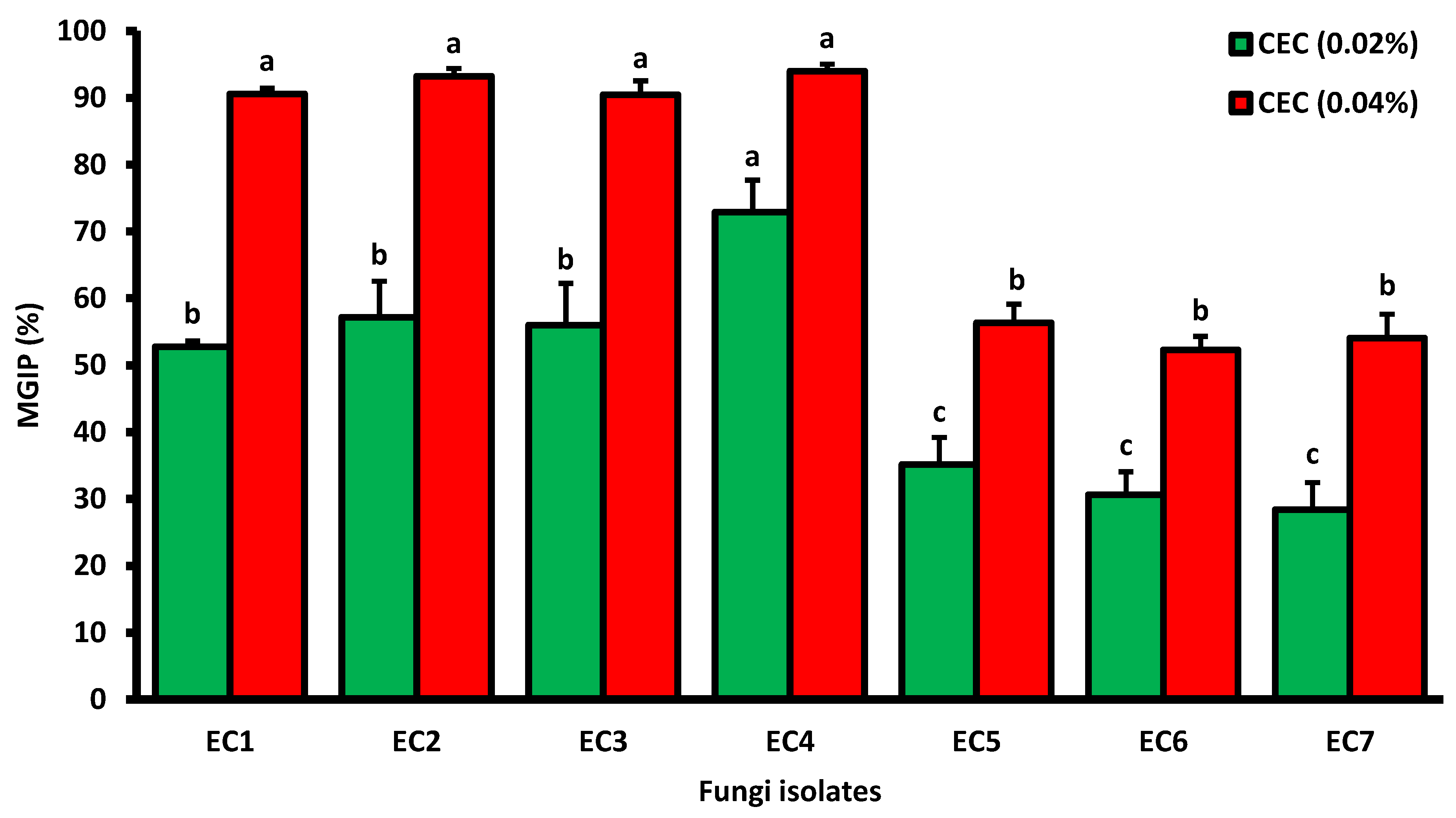
Extract of Opuntia ficus indica cladodes exhibited significant inhibitory activitiesof the mycelial growth against all the fungal agentstested (Figure 5). At a concentration 0.02% of cladode extract, the fungus EC4 showed high sensitivity characterised by an inhibition percentage of 70.91%, followed by the fungal isolates EC2, EC3 and EC1 representing inhibition percentages of of 58.49%, 57.63% and 53.13%, respectively.Fungal isolates EC5, EC6 and EC7 showed low inhibition percentages matching 35.14%, 31.08%, and 28.38%, respectively. At a concentration 0.04% of cladode extract, the results showed an increase in the inhibition percentage compared with the 0.02% concentration, reaching more than 50% and in variable levels. Maximum inhibition percentages of 90.63%, 91.53%, 92.45%, and 94.55% were obtained for fungal isolates EC1, EC3, EC2 and EC4, respectively. Followed by the inhibition percentages of fungal isolates EC6, EC7 and EC5, revealing inhibition percentages of mycelial growthcorrespending to 52.70%, 54.05% and 56.76%, respectively. Thus, it seems clearly to be a proportional relationship between the concentration of Opuntia ficus indica cladode extract and the inhibition of fungal mycelial growth.

3.4.2. Cladode Hydro-Ethanolic Effect on Sporulation

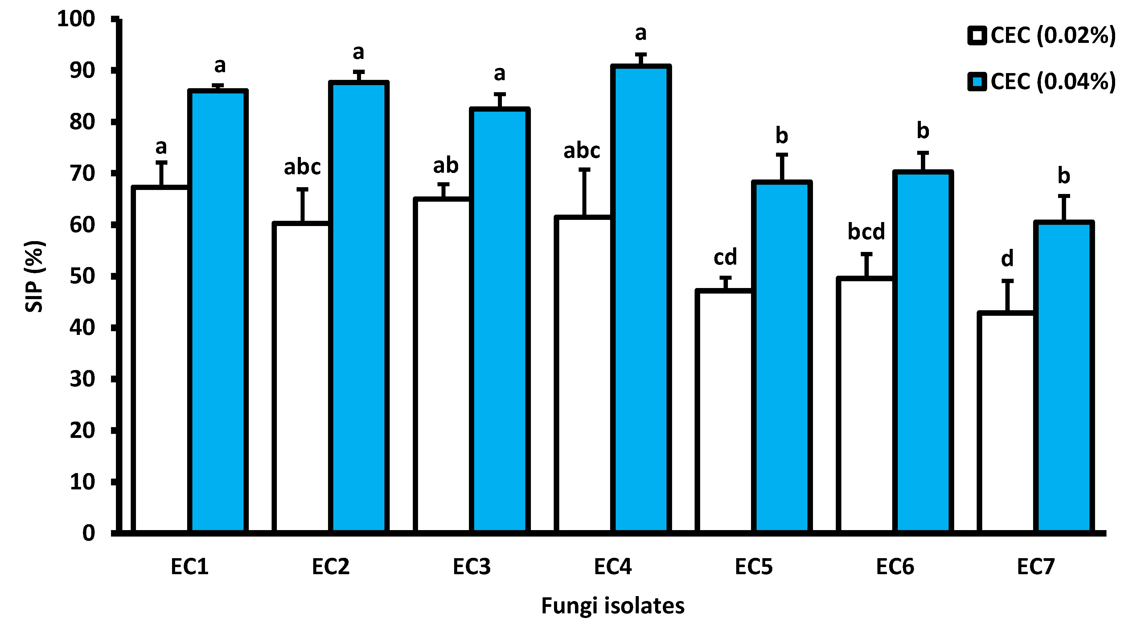
Results of the effect of Opuntia ficus indica cladode extract on sporulation expressed as a percentage of inhibition showed variable levels compared with the control (Figure 6). Opuntia ficus indica cladode extract showed sporulation inhibitory activity against all the fungal isolates tested. At a concentration 0.02% of cladode extract, the highest sporulation inhibition levels were 60.78%, 61.22%, 64.15%, and 68.42% obtained for fungal isolates EC4, EC2, EC3 and EC1, respectively. While, average activity values, less than 50%, were obtained for fungal isolates EC6, EC5 and EC7 resulting in sporulation inhibition rates of 49.38%, 47.56%, and 47.97%, respectively. At a concentration 0.04% of cladode extract, the results showed an increase in the rate of sporulation inhibition reaching more than 50% for all the fungal isolates and at varying proportions.Maximum sporulation inhibition was observed for fungal isolates EC4, EC2, EC1, and EC3 characterizied by inhibition percentages of 90.20%, 87.76%, 85.96%, and 83.02, respectively. For fungal isolates EC6, EC5, and EC7 sporulation inhibition percentages of 70.37%, 68.29%, and 60.76% were obtained, respectively.
Comparaison of the effect of Opuntia ficus indica cladode extract on fungal agents by correlation test showed that the results of mycelial growth inhibition are in perfect agreement with the results of sporulation inhibition. The correlation coeficents unregistered were 0.827 and 0.949 for CEC concentrations 0.02% and 0.04%, respectively (Figure 7).

4. Discussion

The use and characterisation of plant extracts seems to be verypromisingfor the biological control of fungal agents and an alternative to chemical pesticides protecting major crops of worldwide importance such as tomatoes. The mycelium of F. oxysporum is septate and spindle-shaped, and multi-septate macroconidia are produced [42]. Under microscope (x40 magnification), the hyphae of Alternaria sp are septate, the conidiophores are simple, smooth and sometimes branched. The conidia are divided by transverse and/or longitudinal partitions [30]. Microscopic observation of Mucor sp is characterizied also by an unpartitioned mycelium and black globular unicellular conidia [31]. Aspergillus sp show compartmentalised mycelia and globular unicellular conidia [43,44]. Penicillium sp manifest septate hyphae bearing conidiophores, and branching penicilli consisting of phialides. Fusarium solani present numerous unicellular or bicellular microconidia, and macroconidia [42]. The mycelia formed by Rhizoctonia solani were septate, showing 90° branching and a slight constriction at their base, browning as they age, with elongated and often slightly swollen hyphal cells [45].
Total polyphenols represent between 8 and 9 mg/100g FW (Fresh Weight) in Opuntia ficusindica cladodes [46]. According to the study established by [47], the value varies from 41.6 to 23.4 mg GAE/100 g, to 45.6-52.6 mg GAE/100 ml of fresh cladode juice [48]. According to another study, in two-year-old cladodes, the content is also higher, reaching 73.9 mg GAE/100 g MF [49]. Various studies have shown that external factors (geographical and climatic), genetic factors, but also the degree of maturity of the plant and storage time have an important influence on the metabolism of polyphenols [50]. Phenol content differs according to tissue type, stage of development and post-harvest handling. Chlorenchyma contains more than parenchyma, young nopals exceed old cladodes and cooked ones are less abundant in polyphenols than raw ones [51]. Phenolic compounds are secondary metabolites that constitute one of the most representative and widespread groups in the plant kingdom, with more than 8,000 phenolic combinations [52,53]. Ferulic acid, coumaric acid, hydroxybenzoic acid, caffeic acid, salicylic acid and gallic acid are among the phenolic acids detected in this plant part [46,54].
The total flavonoid content of Opuntia ficus indica cladodes measured in this study appears to be higher than the content reported by Boutakiot [48], which is 1.24±0.01 mg RE (Rutin equivalent)/100 ml juice. While, according to another study reported by Boukhalfa and Hamdi [55] the total flavonoid content of Opuntia ficus indica is 240 mg QE/g FW. Variation in flavonoid content is related to cultivar type, extraction methods, protocol, dosage and environmental conditions [56]. Flavonoids are universal plant pigments, responsible for the colouring of flowers, fruit and leaves, and are almost always water-soluble. They play a role in the defence and protection of tissues against the harmful effects of ultraviolet radiation [13]. The term flavonoids refer to a very large group of natural compounds belonging to the polyphenol family [57], which are complex phenolic compounds with a structure consisting of two aromatic rings (rings A and B) and an oxygenated heterocycle (ring C) [58,59]. Some of them also play a key role in phytoalexins, i.e., metabolites that the plant uses in large quantities to suppress infections caused by fungi or bacteria [60]. In Opuntia ficus indica cladodes, examples of these molecules include isorhamnetin, kaempferol, quercetin and iso-quercitrin, rutin, catechin and epicatechin, nicotiflorin and narcissi [46,54]. The main functions of flavonoids are to contribute to plant colour, and they can also play a crutialrole in plant protection. Flavonoids have other interesting functions in controlling plant growth and development by interacting in complex ways with various growth hormones [60].
Hadj Sadok [47], reported that condensed tannins content varying between 6.45 and 6.93 mg/100 g FW. According to another the study established by Boutakiout [48], cladode juice is richer in condensed tannins containing 18.23±0.36 mg TAE/100 ml. The level of tannins in a plant depends on two main factors: the stage of vegetative development and environmental conditions. Their concentration varies considerably between different plant species and within the same species, as it depends on the degree of maturity, the age of the leaves and flowers and the season [61]. Tannin compounds are poorly represented in cladodes and are responsible for the astringency of certain fruits and beverages [62,63]. They are widely available in the plant kingdom, since all organs can close them: root, rhizome, bark, leaf, flower, fruit, rosehip, seed and wood. Their molecular weight varies from 500 to 3,000 Daltons. They have a polyphenolic structure, are soluble in water, alcohol and acetone, and sparingly soluble in ether [13]. Tannin synthesis is a method of defence against free-living plant pathogens (bacteria, fungi and viruses) or those transmitted by nematodes, as well as predators (insects and herbivores) [64,65,66].
Somme investigation reported an increase in carotenoid content during growth ranging between 0.047±0.05 to 0.077±0.06 mg/100 g FW [47]. In terms of variables linked to plant development conditions, the polarity of the different classes of carotenoids (xanthophylls, carotenes, and carotenoid esters) has more influence than their solubility in the extraction solvent and therefore the extraction itself [67]. Carotenoids and their antioxidant activity is linked to the long-chain polyene that reacts with ROO, HO, O2 and R radicals by adding a simple electrotransferase [68]. According to Robards [69], extraction of carotenoids using organic solvents is the most widely used and cost-effective method. Hexane is the best solvent for polar electrolytes while ethanol remains the best choice in the case of polar carotenoids [70]. Carotenoids in Opuntia ficus indica are subdivided into α-cryptoxanthin (20%), β-carotene (36%) and lutein (44%), totalling 229 μg/g DM [71]. Carotenoids play several essential roles in photosynthesis, helping to collect light energy, maintain the structure and functionality of photosynthetic complexes and act as protective agents dealing with reactive oxygen species and the dissipation of excess light energy [72]. Carotenoids are also precursors of vitamin A [73].
The results of the assessment of antioxidant activity in this investigation revealed good activity. Similarly, Msaddak [74] mentioned 1.45 mg/ml which indicate significant anti-radical activity. Also, according to the study conducedby Boutakiout [48], cladode juice is characterised by its antioxidant richness of 1.78±0.03 µmol TE (Trolox Equivalent)/ml, which is considered to be a powerful antioxidant source. The polyphenols contained in cladode extract are probably responsible for the antioxidant activity. Studies show that anti-free radical activity is correlated with the level of polyphenols and flavonoids in medicinal plant extracts [75]. Cladodes are an important source of natural antioxidants in the same way as other known conventional sources [49]. The ability of plant species to resist attack from insects and micro-organisms is often linked to their phenolic compound content. These compounds have anticancer, anti-inflammatory, anti-arteriosclerotic, analgesic, anticoagulant, antimicrobial, antiviral, anticancer, antiallergic, vasodilatory and antioxidant activities [76]. The DPPH test is one of the most widely used tests for determining the anti-free radical activity of plant extracts [77].
Taken together, the effect study of Opuntiaficus indica cladode extract on fungal agents showed that the results of mycelial growth inhibition are in perfect agreement with the results of sporulation inhibition. The antifungal activity on mycelial growth and sporulation of the fungal agents tested is probably related to the high levels of secondary metabolites (flavonoids, polyphenolsand tannins) in the extract of Opuntia ficus indica cladodes. These compounds can cross cell membranes, penetrate the cell interior and interact with critical intracellular sites such as enzymes and proteins, leading to cell death [78]. Flavonoids are metabolites that plants synthesise in large quantities to combat infections caused by fungi [76]. Among the polyphenols, significant synthesis of tannins in parasitized plants corresponds to a defence reaction [79]. A number of investigations had established the high antifungal activities of Opuntia ficus-indica cladodes. The antimicrobial activity of O. ficus-indica aqueous and ethanolic extracts demonstrated distinct responses against 7 pathogenic fungi: Aspergillus niger (MT628904.1), Curvularia khuzestanica (MH688044.1), Penicillium funiculosum (JX500735.1), Talaromyces funiculosus (KX262973.1), Penicillium minioluteum (JN620402.1), Aspergillus chevalieri (MT487830.1), and Aspergillus terreus (MT558939.1). T. funiculosus exhibited the highest inhibition zone against ethanolic extract (0.40 ± 0.10 mm) among the examined fungus, while P. funiculosum displayed a smaller zone of inhibition (0.07 ± 0.06 mm). The examined fungi all displayed low levels of antibacterial activity in the aqueous extract, ranging from 0.13 to 0.17 [80]. Moreover, Hajar et al. [81] reported that two types of fungi were used to test the efficacy of the methanolic extracts: Aspergillus fumigatus and Aspergillus flavus. The research demonstrated that the investigated extracts’ effectiveness in preventing the growth of the fungi under study varied. The methanolic extracts of cladodes inhibited the growth of A. fumigatus by a ratio of 89.57% at a concentration of 1000 mg/ml, whereas they inhibited the growth of A. flavus by a ratio of 85.40% at the same concentration. In an additional investigation focused on assessing the antifungal efficacy against Aspergillus niger, the cladode methanolic extract demonstrated inhibiting ratios of 73.49% and 76.14% at the 500 mg/ml and 1000 mg/ml, respectively, compared to the fluconazole inhibiting ratio of 65.66% at the tested concentration. Conversely, the lowest effect was achieved by the aqueous extracts, which inhibited ratios to 12.53% at a concentration of 1000 mg/ml [82].
No bioproducts or biopesticides based on Opuntia ficus-indica cladode extract are currently commercialized on the market. Except a number of investigations which have described the extract of this plant’s cladodes as insecticidal [83,84] or larvicidal [85] bioproduct. This encourages the development of a new bio-fungicide against tomato pests.

5. Conclusion

Within the general framework of biofungicidefrom plants, Opuntia ficus-indica was chosen for this study. It is a succulent xerophytic plant of the CAM (Crassulacean Acid Metabolism) type belonging to cactus family. Results of phytochemical screening of the methanolic extract carried out on Opuntia ficus-indica cladodes showed that they contain a high percentage of phenolic compounds (flavonoids, polyphenols, carotenoid, and condensed tannins). On the other hand, seven species of fungi were isolated from infected tomato fruits and identified (macroscopic identification and microscopic identification) which belong to six different genera/species: Fusarium oxysporium, Fusarium solani, Rhizoctonia solani, Alternaria sp, Mucor sp, Aspergillus sp and Penicillium sp.
Antifungal activity of hydo-ethanoic extract was evaluated with two concentrations (0.02% and 0.04%) using two tests: the effect of the extract on mycelial growth and the effect of the extract on sporulation of fungal isolates. Those results showed maximum inhibition percentages at a concentration of 0.04% of the cladodes extract, compared with the 0.02% concentration, reaching more than 50% for the fungal isolates Fusarium oxysporium (91.33%), Fusarium solani (90.63%) and Rhizoctonia solani (92%), Alternariasp (94.55%). Opuntia ficus indica cladodes extract also showed significant inhibitory activity on sporulation at the 0.04% concentration against all the fungal isolates tested, reaching over 60%. These inhibitory effects (effect on mycelial growth and effect on sporulation) seem to variate according to the concentration of cladode extract and the fungal isolate tested. This study is an important preliminary step in the characterisation of Opuntia ficus indica cladodes extract for possible use as a biopesticide against fungal agents of tomato fruits, including during the biological conservation or control of diseases caused to field or greenhouse crops.

References

  1. Bekkar, K. Etude de l’effet des facteurs abiotiques et nutritionnels sur la production d’oospores chez Phytophthora infestans (Mont.) De Bary. Thèse de doctorat, Ecole Nationale Supérieure Agronomique d’El-Harrach, Alger, Algérie, 2014.
  2. Blancard, D.;Marchoux, G.; Laterrot, H.; Candresse, T. Les Maladies De La Tomate: Identifier, connaître, maîtriser. Editions Quae, Versailles, FR, 2009; p. 691.
  3. Branthôme, F.X. Worldwide (total fresh) tomato production in 2021. Available online: https://www.tomatonews.com/en/worldwide-total-fresh-tomato-production-in-2021_2_1911.html (accessed on 1 November 2023).
  4. Messiaen, C.M.; Blancard, D.; Rouxel, F.; Lafon, R. Les maladies des plantes maraichères. Ed. Institutnati. Rech. Agro., Paris, FR, 1993; p. 568.
  5. Naika, S.;Jeud, J.V.L.; Jeffau, M.; Hilmi, M.; Vandam, B. Culture de la tomate: production, transformation et commercialisation. (5eme edn). Digigrafi, Wageningen, NL, 2005; p.105.
  6. Zhang, G.; Wang, L.; Pan, J. Probing the binding of the flavonoid diosmetin to human serum albumin by multispectroscopic techniques. J. Agric. Food Chem. 2012, 60, 2721-2729. [CrossRef]
  7. Doyle, M.P.; Beuchat, L.R.; Montville, T.J. Food microbiology: Fundamentals and frontiers. ASM press, Washington DC, USA, 1998 ; pp. 598-611.
  8. Meyer, A.;Deiana, J.; Bernard, A. Cours de microbiologie générale: avec problèmes et exercices corrigés. Wolters Kluwer, Paris, FRA, 2004.
  9. Yang, Z.; Yuan, L.; Duan, Y. The investigation and prevention of tomato root knot nematode in Yunnan Yuanmou. Plant Prot Technol. 2011, 44–45;
  10. Merghid, M.;Debbache, M.; Foughali, I. Impacts des pesticides utilisés dans la plasticulture sur la santé humaine En Algérie. Etude de cas la wilaya de Constantine. Mémoire de Master, Université des Frères Mentouri Constantine, Constantine, Algérie, 2017.
  11. Alleche, N. Activité antifongique de quelques extraits d’une plante endémique sur des moisissures du blé stocké. Mémoire de Master, Université des Frères Mentouri Constantine, Constantine, Algérie, 2017.
  12. Abouthiam, A. Problématique de I’utilisation des insecticides chimiques dans la lutte anti-acridienne au Sahel. In La lutte anti-acridienne. Ed. AUPELF-UREF, Montreal, CA, 1991. pp.193-206.
  13. Bruneton, J. Pharmacognosie, phytochimie, plantes Médicinales. Éditions Tec & Doc, 3e édition, Lavoisier, Paris, FRA, 1999.
  14. Msaddak, L. Propriétés techno-fonctionnelles et substances bioactives de deux ingré-Dients alimentaires : cladodes du figuier de barbarie et feuilles de vigne. Thèse de doctorat, Université de Gabès, Tunis, Tunisie, 2018.
  15. Belmadadi, I.M.A. Etude du potentiel antioxydant d’Aloe vera et de la figue de Barbarie. Mémoire de Master, Université Mohamed El Bachir El Ibrahimi, Bordj Bou Arreridj, Algérie, 2018.
  16. Benattia, F.K. Analyse et Application des Extraits de pépains de Figues de Barbarie.Thèse de doctorat, Université Abou BekrBelkaid, Tlemcen, Algérie, 2018.
  17. Salem, H.B.; Nefzaoui, A.; Salem, L.B. Supplementing spineless cactus (Opuntia ficus-indica f. inermis) based diets with urea-treated straw or old man saltbush (Atriplex nummularia). Effects on intake, digestion and sheep growth. J. Agric. Sci. 2002, 138, 85-92. [CrossRef]
  18. Arba, M. Le cactus Opuntia, une espèce fruitière et fourragère pour une agriculture durable au Maroc. In Actes du Symposium International AGDUMED-durabilité des systèmes de culture en zone méditerranéenne et gestion des ressources en eau et en sol. Cana Print, Rabat, MAR, 2009; pp. 14-16.
  19. Yahia, E.M.; Sáenz, C. Cactus pear (Opuntia species). In book: Postharvest biology and technology of tropical and subtropical fruits. Acai to citrus Publisher: Woodhead Publishing, ENG, 2011; pp. 290-329.
  20. Felker, P.; Rodriguez, S.; del, C.; Casoliba, R.M.; Filippini, R.; Medina, D.; Zapata, R. Comparison of Opuntia ficus indica varieties of Mexican and Argentine origin for fruit yield and quality in Argentina. J. Arid Environ. 2005, 60, 405–422. [CrossRef]
  21. Di Lorenzo, F.; Silipo, A.; Molinaro, A.; Parrilli, M.; Schiraldi, C.; D’Agostino, A.; Izzo, E.; Rizza, L.; Bonina, A.; Bonina, F. The polysaccharide and low molecular weight components of Opuntia ficus indica cladodes: structure and skin repairing properties. Carbohydr. Polym. 2017, 157, 128-136. [CrossRef]
  22. Malainine, M.E.; Dufresne, A.; Dupeyre, D.; Mahrouz, M.; Vuong, R.; Vignon, M.R. Structure and morphology of cladodes and spines of Opuntia ficus-indica. Cellulose extraction and characterisation. Carbohydr. Polym. 2003, 51, 77-83. [CrossRef]
  23. Stintzing, F.C.; Herbach, K.M.; Mosshammer, M.R.; Carle, R.; Yi, W.; Sellappan, S., Akoh, C.C.; Bunch, R.; Felker, P. Color, betalain pattern, and antioxidant properties of cactus pear (Opuntia spp.) clones. J. Agric. Food Chem. 2005, 53, 442-451. [CrossRef]
  24. Aragona, M.; Lauriano, E.R.; Pergolizzi, S.; Faggio, C. Opuntia ficus-indica (L.) miller as a source of bioactivity compounds for health and nutrition. Nat. Prod. Res. 2018, 32, 2037-2049. [CrossRef]
  25. Goudjil, S.;Naceri, K.; Noura, S. Caractérisation physicochimique et effet antibactérien de la cladode d’Opuntia ficus indicainermis(L) Mill. de la région de contribuTtioianret, en vue d’explorer son potentiel thérapeutique. Thèse de doctorat, Université Ibn khaldoun, Tiaret, Algérie, 2018.
  26. Contreras-Padilla, M.; Perez-Torrero, E.; Hernández-Urbiola, M.I.; Hernández-Quevedo, G.; del Real, A.; Rivera-Muñoz, E.M.; Rodríguez-García, M.E. Evaluation of oxalates and calcium in nopal pads (Opuntia ficus-indica var. redonda) at different maturity stages. J.Food Compos. Anal. 2011, 24, 38-43. [CrossRef]
  27. Meena, M.; Swapnil, P.; Upadhyay, R.S. Isolation, characterization and toxicological potential of Alternaria-mycotoxins (TeA, AOH and AME) in different Alternaria species from various regions of India. Sci. Rep. 2017, 7, 8777. [CrossRef]
  28. Cassagne, H. Milieux de culture et leurs applications. Edition de la Tourelle, Paris, FRA, 1966;379 p.
  29. Pitt, J.I.; Hocking, A.D. Fungi and Food Spoilage, 3rd Ed. Springer Dordrecht, Heidelberg, London, New York, 2009, 524 p.
  30. Bessadat, N. Isolement, identification et caractérisation des Alternariasp. Résponsable de la détérioration des plantes maraichères par des systèmes enzymatiques et moléculaires. Thèse de doctorat, Université d’Oran es-senia, Oran, Algérie, 2014.
  31. Botton, B.; Bretton, A.; Fever, M.; Gautier, S.; Guy, P.; Larpent, J.P.; Reymond, P.; Sanglier, J.; Vayssier, Y.; Veau, P. Moisissures utiles et nuisibles. Importance industrielle, (edn) Masson, collection biotechnologie, Paris, FRA, 1990; pp. 309-512.
  32. Ghoul, M.; Minet, J.; Bernard, T.; Dupray, E.; Cornier, M. Marine macroalgae as a source for osmoprotection for Escherichia coli. Microb. Ecol.1995, 30, 171-81. [CrossRef]
  33. Singleton, V.L.; Orthofer, R.; Lamuela-Raventos, R.M. Analysis of total phenols and other oxidation substrates and antioxidants by means of Folin-Ciocalteu reagent. Meth. Enzymol. 1999, 299, 152-178. [CrossRef]
  34. Dehpeur, A.A.; Ebrahimzadeh, A.A.; Fazel, N.S.; Nabavi, S.M. Antioxidant activity of the methanol extract of Ferulaassafoetida and its essential oil composition. Grasas Aceites. 2009, 60, 405–412. [CrossRef]
  35. Joslyn, M. Tannins and related phenolics. In Methods in food analysis, 701-725. J. Cell. Biochem. 1970, 22, 188-919. [CrossRef]
  36. Sass-Kiss, A.; Kiss, J.; Milotay, P.; Kerek, M.M.; Toth-Markus, M. Differences in anthocyanin and carotenoid content of fruits and vegetables. Food Res. Int. 2005, 38, 1023-1029. [CrossRef]
  37. Aruwa, C.E.; Amoo, S.O.; Kudanga, T. Opuntia (Cactaceae) plant compounds, biological activities and prospects–A comprehensive review. Food Res. Int. 2018, 112, 328-344. [CrossRef]
  38. Sanchez-Moreno, C. Methods used to evaluate the free radical scavenging activity in foods and biological systems. Int. J. Foods Sci. Tech. 2002, 8, 121-137. [CrossRef]
  39. Serhan, A.R. Additional interaction of mint leaf extracts with fungi that have an antagonistic property on certain fungi associated with legume seeds. Arab. J. Plant Prot. 2006, 24, 118-124 (In Arabic).
  40. Kismoune, S. L’effet de l’extrait aqueux de cyprès sur la croissance de champignon Phythophthorainfestans. Mémoire de Master, Université Mouhamed SeddikBenyahia, Jijel, 2021.
  41. Leroux, P.;Credet, A. Document sur l’étude de l’activité des fongicides. INRA, Versailles, FRA, 1978, 12 p.
  42. Chabasse, D.;Bouchara, J.; De Gentile, L.; Brun, S.; Cimon, B.; Penn, P. Les moisissures d’intérêt médical. Cahier N°25 de formation de biologiemédicale. 2002; 157 p. [CrossRef]
  43. Guiraud, J.P.Microbiologie alimentaire. Dunod, Paris, FRA, 1998; pp.7-330.
  44. Jernejc, K.; Cimerman, A. Morphological characteristics, extracellular and inracellular protein and enzyme patterns of five Aspergillus species. Food Technol. Biotechnol. 2001, 39, 333-340. [CrossRef]
  45. Blancard, D. Biologie, épidémiologie. Available online: http://ephytia.inra.fr/fr/C/5217/Tomate-Biologie-epidemiologie (accessed on 1 November April 2023).
  46. Ventura-Aguilar, R.I.;Bosquez-Molina, E.; Bautista-Baños, S.; Rivera-Cabrera, F. Cactus stem (Opuntia ficus-indica Mill): anatomy, physiology and chemical composition with emphasis on its biofunctional properties. J. Sci. Food Agric. 2017, 97, 5065-5073. [CrossRef]
  47. Hadj Sadok,T.;Aid, F.;Bellal, M.; Abdul Hussain, M.S. Composition chimique des jeunes cladodes d’Opuntia ficus indicaet possibilité de valorisation alimentaire. Thèse de doctorat, Ecole NationaleSupérieurAgronomique, Alger, Algérie, 2010.
  48. Boutakiout, A. Etude physico-chimique, biochimique et stabilité d’un nouveau Produit: jus de cladode du figuier de Barbarie marocain (Opuntia ficus-indica et Opuntia megacantha). Thèse de doctorat, Université d’Angers, Angers, France, 2015.
  49. Ayadi, M.A.; Abdelmaksoud, W.; Ennouri, M.; Attia, H. Cladodes from Opuntia ficus indica as a source of dietary fiber: effect on dough characteristics and cake making. Ind Crops Prod. 2009, 30, 40-47. [CrossRef]
  50. Aganga, A.A.; Mosase, K.W. Tannins content, nutritive value and dry matter digestibility of Lonchocarpuscapassa, Ziziphus mucronata, Sclerocaryabirrea, Kirkia acuminata and Rhus lancea seeds. Anim. Feed Sci. Technol. 2003, 91, 107-113. [CrossRef]
  51. Ventura-Aguilar, R.I.; Bosquez-Molina, E.; Bautista-Baños, S.; Rivera-Cabrera, F. Cactus stem (Opuntia ficus-indica Mill): anatomy, physiology and chemical composition with emphasis on its biofunctional properties. J. Sci. Food Agric. 2017, 97, 5065-5073. [CrossRef]
  52. Bruneton, J. Pharmacognosie. 5 Édition. Phytochimie– Plantes médicinales. Tec and Doc, Lavoisier, Paris, FRA, 2015; 1504 p.
  53. Šaponjac, V.T.; Canadanovi’c-Brunet, J.; Cetkovi’c, G.; Djilas, S. Detection of Bioactive Compounds in Plants’and Food Products. In Emerging and Traditional Technologies for Safe, Healthy and Quality Food, 1st ed.; Nedovi’c, V., Raspor, P., Levi’c, J., TumbasŠaponjac, V., Barbosa-Cánovas, G.V., Eds.; Springer International Publishing: Basel, Switzerland, 2016; pp. 81–109.
  54. Guevara-Figueroa, T.; Jiménez-Islas, H.; Reyes-Escogido, M.L.; Mortensen, A.G.; Laursen, B.B.; Lin, L.W.; De Leo’n-Rodrı’guez, A.; Fomsgaard, I.S.; de la Rosa, A.P.B. Proximate composition, phenolic acids, and flavonoids characterization of commercial and wild nopal (Opuntia spp.). J. Food Compos. Anal.2010,23, 525-532. [CrossRef]
  55. Boukhalfa, S.; Hamdi, S. Évaluation phytochimique et étude des activités biologiques des extraits bruts des plantes médicinales locales: Opuntia ficus indica et Thymus lanceolatus. Mémoire de Master, Université des Frères Mentouri Constantine, Constantine, Algérie 2016.
  56. Maataoui, B.S.;Hmyene, A.; Hilali, S. Activites anti-radicalaires d’extraits de jus de fruits du figuier de barbarie (Opuntia ficus indica). Leban. Sci. J. 2006, 7, 3-8.
  57. Bennick, A. Interaction of plant polyphenols with salivary proteins. Crit. Rev. Oral Biol. Med. 2002, 13, 184-196.
  58. Middleton,Jr.E.;Kandaswami, C.;Theoharides, T.C. The effects of plant flavonoids on mammalian cells: implications for inflammation, heart disease, and cancer. Pharmacol. Rev. 2000, 52, 673-751.
  59. Bennick, A. Interaction of plant polyphenols with salivary proteins. Crit. Rev. Oral Biol. Med. 2002,13, 184-196.
  60. Youla, A.;Latrous, I.E. Contribution à l’étude phytochimique des flavonoïdes chez l’espèce (Melissa officinalis L.) et évaluation de leur pouvoir antibactérien. Mémoire de Master, Université des Frères Mentouri Constantine, Constantine, Algérie, 2017.
  61. Skadhauge, B.; Gruber, M.Y.; Thomsen, K.K.; Von Wettstein, D. Leucocyanidin reductase activity and accumulation of proanthocyanidins in developing legume tissues. Am. J. Bot. 1997, 84, 494-503. [CrossRef]
  62. Tirilly, Y.; Bourgeois, C. M. Technologie des légumes. Editions Tec & Doc, Lavoisier, FRA, 1999 ; 558 p.
  63. Scalbert, A.; Williamson, G. Dietary intake and bioavailability of polyphenols. J. Nutr. 2000, 130, 2073S-2085S. [CrossRef]
  64. Collingborn, F.M.; Gowen, S.R.; Mueller-Harvey, I. Investigations into the biochemical basis for nematode resistance in roots of three musa cultivars in response to radopholussimilis infection. J. Agric. Food Chem. 2000,48, 5297-5301. [CrossRef]
  65. Waghorn, G. Beneficial and detrimental effects of dietary condensed tannins for Sustainable sheep and goat production—Progress and challenges. Anim. Feed Sci. Technol.2008, 147, 116-139. [CrossRef]
  66. Hassanpour, S.; Maheri-Sis, N.; Eshratkhah, B.; Mehmandar, F.B. Plants and secondary metabolites (tannins): A review. Int. J. For.Soil Eros. 2011, 1, 47–53.
  67. Dias, M.G.; Camões, M.F.G Oliveira, L. Carotenoids in traditional Portuguese fruits and vegetables. Food Chem. 2009, 113, 808-815. [CrossRef]
  68. López-Lázaro, M.; Martín-Cordero, C.; Ayuso, M.J. Two new flavonol glycosides as DNA topoisomerase I poisons. Z Naturforsch C. 2000, 55, 898-902. [CrossRef]
  69. Robards, K. Strategies for the determination of bioactive phenols in plants, fruit and vegetables. J. Chromatogr. A. 2003, 1000, 657-691. [CrossRef]
  70. Amorim-Carrilho, K.T.; Cepeda, A.; Fente, C.; Regal, P. Review of methods for analysis of carotenoids. Trends Anal. Chem. 2014, 56, 49-73. [CrossRef]
  71. Jaramillo-Flores, M.E.; González-Cruz, L.; Cornejo-Mazon, M.; Dorantes-Alvarez, L.;Gutierrez-Lopez, G.F.; Hernandez-Sanchez, H. Effect of thermal treatment on the antioxidant activity and content of carotenoids and phenolic compounds of cactus pear cladodes (Opuntia ficus-indica). Food Sci Technol Int.2003, 9, 271-278. [CrossRef]
  72. Ootaki, T.; Yamazaki, Y.; Noshita, T.; Takahashi, S. Excess carotenoids disturb prospective cell-to-cell recognition system in mating responses of phycomycesblakesleeanus. Mycoscience. 1996, 37, 427-435. [CrossRef]
  73. Krinsky, N.I.; Johnson, E.J. Carotenoid actions and their relation to health and disease. Mol. Aspects Med. 2005, 26, 459-516. [CrossRef]
  74. Msaddak, L. Propriétés techno-fonctionnelles et substances bioactives de deux ingré-dients alimentaires : cladodes du figuier de barbarie et feuilles de vigne. Thèse de doctorat, Université de Gabès, Tunis, Tunisie, 2018.
  75. Mariod, A.A.; Ibrahim, R.M.; Ismail, M.; Ismail, N. Antioxidant activity and phenolic content of phenolic rich fractions obtained from black cumin (Nigella sativa) seedcake. Food Chem. 2009, 116, 306-312. [CrossRef]
  76. Benhamama, L. Contribution à l’étude phytochimique et évaluation de l’activitéAntioxydante de la plante médicinale Crataegus monogyna. Mémoire de Master, Université des Frères Mentouri Constantine, Constantine, Algérie, 2015.
  77. Laguerre, M.; Lecomte, J.; Villeneuve, P. Evaluation of the ability of antioxidants to counteract lipid oxidation: existing methods, new trends and challenges. Prog. Lipid Res. 2007, 46, 244-282. [CrossRef]
  78. Cristani, M.; D’arrigo, M.; Giuseppina, M.; Castelli, F.; Sarpietro, M.; Micieli, M.; Venuti, V.; Bisignano, G.; Saija, A.; Trombetta, D. Interaction of four monoterpenes contained in essential oils with model membranes: implications for their antibacterial activity. J Agric Food Chem. 2007, 55, 6301-6305. [CrossRef]
  79. Guignard, J.L. Abrégé de biochimie végétale, Ed. Masson, Paris, FRA;160 p.
  80. Alghamdi, A.; Alshehri, W.; Sajer, B.; Ashkan, M.; Ashy, R.; Gashgari, R.; Hakmi, H. Biological Activities and GC-MS Analysis of Aloe vera and Opuntia ficus-indica Extracts. J. Chem. 2023, 2023, 1-15. https://doi.org/10.1155/2023/6504505. [CrossRef]
  81. Hajar, N.; Nawal, A.; Amjad, D. A study to determine total phenolic content of Opuntia ficus-indica extracts and their activity against some pathogenic fungi. World J Pharm Pharm. Sci. 2018, 8, 98-109.
  82. Ali, N.; Nasser, H.; Deeb, A. Evaluating The inhibitory Efficacy Of Cactus plant Extracts (Opuntia ficus-indica) Against The Isolation Of Aspergillus niger Fungus. Tishreen University Journal-Basic Sciences Series. 2018, 40, 227-241.
  83. Paiva, P.M.; Santana, G.M.; Souza, I.F.; Albuquerque, L.P.; Agra-Neto, A.C.; Albuquerque, A.C.; Luz, L.A.; Napoleão, T.H.; Coelho, L.C.B.B. Effect of lectins from Opuntia ficus-indica cladodes and Moringa oleifera seeds on survival of Nasutitermes corniger. Int. Biodeterior. Biodegradation. 2011, 65, 982-989. [CrossRef]
  84. de Santana Souza, C.; Procópio, T.F., do Rego Belmonte, B.; Paiva, P.M.G.; de Albuquerque, L.A.; Pontual, E.V.; Napoleão, T.H. Effects of Opuntia ficus-indica lectin on feeding, survival, and gut enzymes of maize weevil, Sitophilus zeamais. Appl Biol Chem. 2018, 61, 337–343. [CrossRef]
  85. Ferrreira, E.C.B.; Nova, I.C.V.; de Almeida, W.A.; dos Santos Albuquerque, F.M.; dos Santos Cruz, G.; da Costa, H.N.; Procópio, T.F.; da Silva, W.A.V.; Ferreira, M.R.A.; Paiva, P.M.G.; Soares, L.A.L.; Teixeira, A.A.C.; Teixeira, V.W.; Napoleão, T.H.; Barros, R.; Pontual, E.V. Opuntia ficus-indica cladode extract is an embryotoxic, larvicidal, and oviposition-deterrent agent for the diamondback moth, Plutella xylostella. Crop Prot. 2021, 139, 105351. [CrossRef]
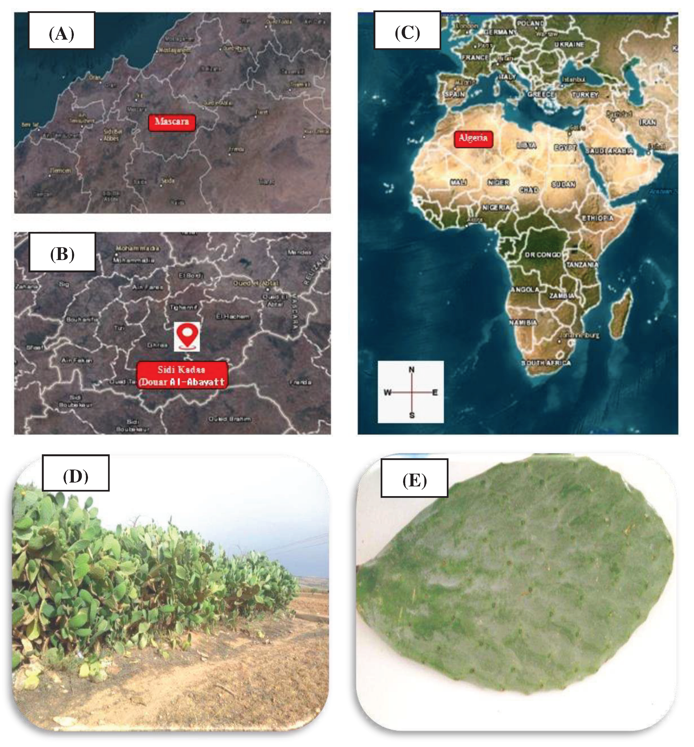
Figure 1. Map showing the localization of the sampling place of plant material extracted from https://mol.org/regions/ (A: Mascara department, B: Sidi Kada (Douar El-Abyatt), C: Algeria country) and specimen OFI-02-2023 of Opuntia ficus indica L (D: Opuntia ficus-indica plant; E: Opuntia ficus-indica cladode).
Preprints 98289 g001
Figure 2. Disease symptoms in samples of infected tomato fruits.
Preprints 98289 g002
Figure 3. Macroscopic and microscopic aspects of fungal agentsobtained from infected tomato fruits (v: verso sides of colonies). You need to introduce details on the microscopic observation (optic maicrospyobservation, xx x amplification) Photos wer obtained with .......
Preprints 98289 g003
Figure 5. Effect of Opuntia ficus indica cladode extract on mycelial growth of fungal agentsisolated from infected tomato fruits. MGIP (%): Mycelial growth inhibition percentage; CEC: Cladode extract concentration.Data are expressed as mean±s.d. (P < 0.05, same letter show no significant difference in one-way ANOVA followed by Tukey-Kramer HSD test).
Preprints 98289 g005
Figure 6. Effect of Opuntia ficus indica cladode extract on sporulation of fungal agents isolated from infected tomato fruits. SIP (%): Sporulation inhibition percentage; CEC: Cladode extract concentration.Data are expressed as mean±s.d. (P < 0.05, same letter show no significant difference in one-way ANOVA followed by Tukey-Kramer HSD test).
Preprints 98289 g006
Figure 7. Correlation test of MGIP (%) vs SIP (%) for CEC (0.02% and 0.04%).
Preprints 98289 g007
Table 1. Symptoms, fungi and diseases of the infested tomato fruit samples.
Samples Symptom Fungiisolated Disease
E1 White spots Fusarium solani Fusariosis
E2 Black spots Rhizoctonia solani Brown rot
E3 Brown spots Fusarium oxysporium Fusariosis
E4 Black spots Alternaria sp Alternariosis
E5 Soft, dampfabric Mucor sp Mucormycosis
E6 Grey stains Aspergillus sp Aspergillosis
E7 Green stains Penicillium sp Penicillosis
Table 2. Genus and species identification of fungal agents isolated from infected tomato fruits.
Isolate code Identification (Genus/species)
EC1 Fusarium solani
EC2 Rhizoctonia solani
EC3 Fusarium oxysporium
EC4 Alternaria sp
EC5 Mucor sp
EC6 Aspergillus sp
EC7 Penicillium sp
Table 3. Phytochemical screening of bioactive molecules in Opuntia ficus indica cladodes.
Component Content
Total polyphenols 86.63±0 .008 mg GAE/100g FW
Flavonoids 13.40±0.01 mg QE/100g FW
Condensed tannins 08.90±0.11mg TAE/100g FW
Carotenoids 0.94±0.02 mg β-CE /100g FW
Antioxidant activity(DPPH test) IC 50 (%)
Cladode extract Ascorbic acid
0.64±0.005 mg/ml 0.39±0.003 mg/ml
GAE: Gallic Acid Equivalent; QE: Quercetin Equivalent; TAE: Tannic Acid Equivalent; β-CE: β-carotene Equivalent.
Disclaimer/Publisher’s Note: The statements, opinions and data contained in all publications are solely those of the individual author(s) and contributor(s) and not of MDPI and/or the editor(s). MDPI and/or the editor(s) disclaim responsibility for any injury to people or property resulting from any ideas, methods, instructions or products referred to in the content.
Copyright: This open access article is published under a Creative Commons CC BY 4.0 license, which permit the free download, distribution, and reuse, provided that the author and preprint are cited in any reuse.
Prerpints.org logo

Preprints.org is a free preprint server supported by MDPI in Basel, Switzerland.

Subscribe

© 2024 MDPI (Basel, Switzerland) unless otherwise stated