Preprint
Article

Anatomical Characteristics of Tapped Styrax sumatrana Wood Grown in North Sumatra, Indonesia

Altmetrics

Downloads

98

Views

44

Comments

0

A peer-reviewed article of this preprint also exists.

Submitted:

08 February 2024

Posted:

08 February 2024

You are already at the latest version

Alerts
Abstract
This study investigated the anatomical characteristics and pulp quality of tapped Styrax sumatrana wood to facilitate its further utilization. The transverse surface of the tapped S. sumatrana was light or greyish brown in the sapwood and reddish brown in the heartwood. The resin canals of the tapped wood were formed along the growth rings at the boundaries between the heartwood and sapwood. Furthermore, microscopic analyses revealed an irregular outline and rounded epithelial cells at the edges of the intercellular traumatic resin canal (TC). Approximately 8–16 epithelial cells surrounded the resin canals. The fibers in the tapped S. sumatrana were generally thin-walled, whereas those near the TC were thick-walled. Moreover, S. sumatrana were diffuse-porous and exhibited intermediately distinct or indistinct growth ring boundaries. The vessels were mainly in radial multiples and clusters of 3–5. In addition, they showed a diagonal-to-radial pattern arrangement and a few tangential bands. Deposits were observed in some vessels in the tapped part but were absent in the untapped part. The fiber length of the tapped S. sumatrana wood was classified as moderate to extensive and categorized as 2nd grade pulp quality. Finally, the cell walls were classified as thin to moderate.
Keywords: 
Subject: Biology and Life Sciences  -   Forestry

1. Introduction

Plants can produce exudates that usually come out when plants are injured. The exudates cover the wound and harden after being exposed to air [1, 2]. Resin, latex, and gum are the common exudates used. Resin is a type of exudate consisted of terpenoids or phenolics and is secreted by internal or surface glands of plant tissues [3, 4].
One of the plant genera that produce resinous exudates is Styrax spp. The Styrax genus, which belongs to the family Styracaceae, comprises approximately 130 species [5]. It has major habitats in the tropical, subtropical, and temperate regions of Asia, Europe, and America [6]. Three Styrax species have been identified In Southeast Asia: Styrax benzoin, Styrax paralleloneurum synonymous to Styrax sumatrana, and Styrax tonkinesis.
Styrax spp. is one of the most important plants in North Sumatra with potential for development and has been utilized to produce a resin called benzoin. Benzoin has high economic value as a raw material for cosmetics and a perfume binder to keep the fragrance from fading quickly. Benzoin is also useful as a preservative and raw material for pharmaceutical drugs. It can also be used as a mixture in manufacturing ceramics to make them stronger and less likely to break [7].
S. sumatrana can reach a height of 35 m and a diameter at breast height (DBH) of 40–60 cm. This wood is suitable for light construction, furniture manufacturing, handicrafts, as well as pulp and paper [6,7,8,9]. S. sumatrana, known as the kemenyan, haminjon toba, or benzoin tree, is a commercial tree endemic to North Sumatra, Indonesia. The area of ​​Styrax plantation in North Sumatra was 23,172 ha, with a total resin production of 8,845 tons [10]. The resin produced from Sumatra benzoin contains styrene, benzoic acid, isovanillin, cinnamic acid, p-coumaryl benzoate, and p-coumaryl cinnamate, which are commonly used in incense, seasoning materials, fragrances, and medicines [6, 11].
S. sumatrana resin is harvested through mechanical treatment of the stem. A few holes with a depth of 3–5 cm are made in the stems using a sharp object and patted every four days for three months. The resin is subsequently harvested 3–4 months after treatment [12]. The productive period of this tree as a resin producer is approximately 20 years, and Sumatra residents typically use it for firewood after its productive years. Following the productive phase, the resin tapping process leaves distinctive tapping scars on the wood of the Styrax tree.
Intercellular traumatic canals (TC) may develop in response to various types of injuries such as physical damage, infections, and insect attacks. Typically, these canals consist of epithelial cells producing natural resin and are organized in tangential bands or exist as a singular canal [13]. Intercellular traumatic resin canals are found in various species, such as the Meliaceae family due to mechanical stimuli [8], Tsuga sieboldii by piercing metal pins around the stem [14], and Picea engelmannii due to the attack of Dendroctonus rufipennis [15]. In addition, Deng et al. [16] revealed that traumatic resin canals in Styrax are formed by parenchymal cells in the secondary xylem by schizolysigeny after injury.
Previous studies have demonstrated the anatomical characteristics of Styrax wood grown in Indonesia. Pasaribu et al. [17] reported that S. sumatrana generally shows distinctive anatomical characteristics with S. paralleloneurum. S. sumatrana and S. paralleloneurum showed multiseriate rays with three to six and three to four seriate rays, respectively. Moreover, S. paralleloneurum showed large intercellular traumatic resin (TC), whereas S. sumatrana had a small TC in the growth ring. However, the fiber quality of both species was categorized as 1st-grade pulp quality.
S. sumatrana grown in North Sumatra is commonly wounded by resin tapping, which may affect its anatomical characteristics. So far, the anatomical characteristics of tapped S. sumatrana wood remain understudied. Therefore, the present study investigated the qualitative anatomical characteristics and fiber quality of tapped S. sumatrana according to the IAWA list for hardwood identification to provide valuable information on wood identification and quality to facilitate further utilization of the species.

2. Materials and Methods

2.1. Materials

A S. sumatrana tree tapped by the local community for resin harvesting was used in this study. The tree was located at an altitude of approximately 900 m above sea level in Banuaji IV village in the Adiankoting sub-district, North Tapanuli regency, North Sumatra, Indonesia (2°0’10.08” N, 99°4’14.52” E). Wood discs were obtained at breast height from 10-year-old trees with a diameter of 12 cm.

2.2. Methods

2.2.1. Macroscopic and Microscopic Observation

The transverse surfaces of the wood disc underwent abrasion with coarse sandpaper (plate number 1 from Deerfos Co., Ltd. in Incheon, Korea) via a sanding machine (TW/BD-46 model, running at 2070 rpm and 450 W from Rexon Industrial Corp., Ltd. in Taichung, Taiwan). Following this initial sanding, the wood discs were manually sanded with fine sandpaper (CC-600Cw from Daesung Abrasive Co., Ltd. in Seoul, Korea). The macroscopic structures of the transverse and longitudinal surfaces were observed under stereo microscope (SMZ 745T, Nikon, Tokyo, Japan)
Here, 20 (L) × 20 (R) × 20 (T) mm3 wood blocks were extracted from the tapped and untapped parts near the bark of the wood discs for microscopic observation. The samples were soaked in a mixture of glycerin and water (50:50) and heated on a heating plate for 30–45 min. Cross, tangential, and radial sections with approximately 20 μm thickness were prepared using a sliding microtome (Nippon Optical Works Co., Ltd., Nagano, Japan). All slices were stained with 1% safranin and 1% light-green solution. Additionally, they were dehydrated using a graded series of alcohol (50%, 70%, 90%, 95%, and 99%) and xylene. Permanent slides were prepared using Canada balsam. The stained sections were observed using an optical microscope (Eclipse Si, Nikon, Tokyo, Japan) connected to i-Solution-lite software (IMT i-Solution Inc., Burnaby, BC, Canada).
For the scanning electron microscopy (SEM), 0.5 × 0.5 × 0.5 cm3 wood samples were vacuumed and coated with gold using a sputter coater (Cressington sputter coater 108; Watford, UK). The samples were observed using a scanning electron microscope (JEOL JSM-6360, 15 kV, Tokyo, Japan).

2.2.2. Cell Maceration

Matchstick-sized samples were prepared for maceration. The wood samples were macerated in a boiling solution containing 60% glacial acetic acid and 30% hydrogen peroxide at 80°C for 1–2 h [19]. The macerated samples were rinsed under running tap water until neutralization. Next, fibers were separated using a pin while washing in distilled water. The fibers were stained using 1 % safranin solution for approximately 3 h and then washed with distilled water. They were subsequently arranged on a microscope slide, dripped with glycerin, and mounted with a cover glass for observation under an optical microscope (Eclipse Si) connected to i-Solution Lite software (IMT i-Solution Inc.).

2.2.3. Measuring Dimensions of Vessels and Fibers, as Well as Their Derivative Value

The fiber length and diameter, lumen diameter, and vessel length and diameter were each measured using 25 cells. The derivative values of the fiber, such as the Runkel ratio (RR), felting power (FP), mulhsteph ratio (MR), coefficient rigidity (CR), and flexibility ratio (FR) were analyzed to elucidate the quality of the wood fiber for pulp and paper production. The fiber quality was determined as previously described [18], as shown in Table 1. The pulp quality classification is shown in Table 2. Fibers were subsequently separated using a pin while washing in distilled water.

3. Results and Discussion

3.1. Macroscopic Characteristics in the Transverse and Longitudinal Surfaces

Figure 1 shows the transverse, tangential, and radial surfaces of an S. sumatrana wood disc wounded by tapping. S. sumatrana wood showed light or greyish-brown sapwood and reddish-brown heartwood on the transverse surface. The heartwood proportion was higher than that of sapwood (3:1). Pasaribu et al. [17] reported that untapped S. sumatarana and S. paralleloneurum were light brown or slightly grayish-brownish-yellow, and that there was no color difference between heartwood and sapwood in either species. Similarly, Damayanti et al. [18] reported that S. benzoin was light brown or slightly grayish-brownish-yellow, whereas the heartwood and sapwood showed no color difference.
S. sumatrana wood showed clear TCs, forming a complete ring, and a resin pocket consisting of whitish substances in the earlywood (Figures 1A and C). The radial section showed clear dark lines representing TC in the longitudinal direction (Figure 1D). In other hardwood species, Dünisch and Baas [13] mentioned that Meliaceae species had two kinds of channels between cells: single or small groups of channels (called local intercellular canals), and the other type formed thin bands of channels along the outer edge of the wood disc. The length of the TC was extended from millimeters to a couple of meters and, in some cases, could be extended down the entire axis of the tree.

3.2. Microscopic Characteristics

3.2.1. Cross-Sections

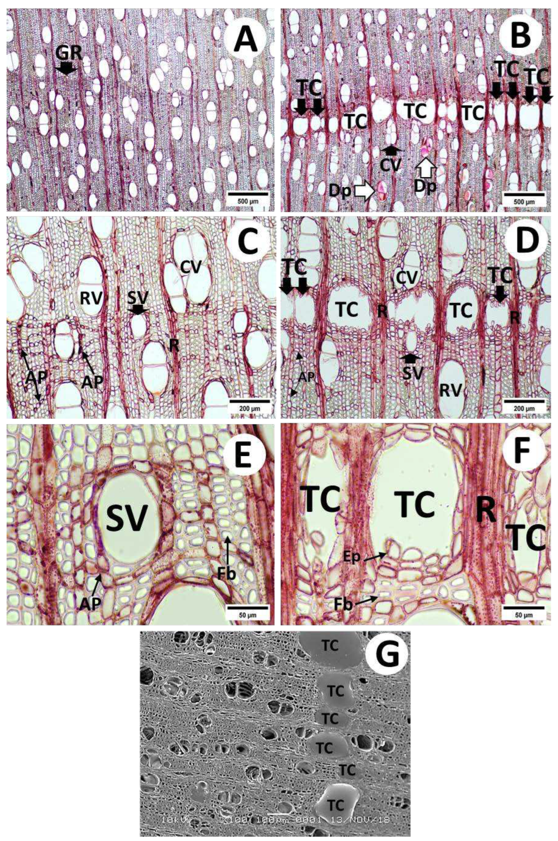
Optical micrographs of cross sections of the tapped and untapped parts of S. sumatrana are presented in Figure 2. The wood discs showed an intermediate difference between distinct and indistinct growth ring boundaries, with diffuse-porous vessels observed (Figures 2A and 2B). The vessels were mainly in radial multiples and clusters of 3–5 and partly solitary. They also showed an angular outline (Figures 2C and 2D). In addition, S. sumatrana tended to have a diagonally to radially arranged pattern and a few tangential vessel bands. Gum or deposits were observed in some vessels in the tapped part (Figure 2B), whereas they were absent in the untapped part of the wood discs (Figure 2A). The number of vessels and diameter of vessel lumina were 7–11 per mm2 and 96–190 (131 ± 28) μm, respectively.
TCs were arranged in tangential bands resembling growth rings (Figures 2B, 2D, and 2G). In addition, the TC showed an irregular outline and rounded parenchymal cells (epithelial cells) at the edges of the canal. Approximately 8–16 epithelial cells surrounded these resin canals (Figure 2F), and TCs fulfilled with resin are also shown in Figure 2G. The fibers in the tapped and untapped parts of S. sumatrana were generally thin-walled, whereas those near the TC were thick-walled (Figures 2E and 2F). The apotracheal axial parenchyma was diffuse and diffuse-in-aggregate, whereas the paratracheal axial parenchyma was vasicentric and banded, including reticulate, narrow, and marginal bands (Figures 2C, 2D, and 2E).
Tapped S. sumatrana generally showed anatomical characteristics comparable to Styrax wood [17, 21]. These studies identified mechanical treatment during resin tapping as the cause of TC in Styrax wood and other hardwoods. Pasaribu et al. [17] reported that TCs were formed at the growth ring boundary of untapped S. sumatrana and S. paralleloneurum. However, the TC formed in S. paralleloneurum was larger than that in S. sumatrana. Damayanti et al. [21] and Pasaribu et al. [17] attributed these differences in TC size to genetic differences or natural wounding during wood harvesting. Dünisch and Baas [13] observed that wounded Meliaceae species typically developed intercellular canals, which were commonly found either individually or in small clusters, often appearing as thin tangential bands. They also mentioned that there were thick-walled fibers and increased production of reddish to dark red-colored accessory compounds near the intercellular canals. Rounded parenchyma cells were frequent at the edges of intercellular canals and may extend into the secretion.

3.2.2. Radial and Tangential Sections

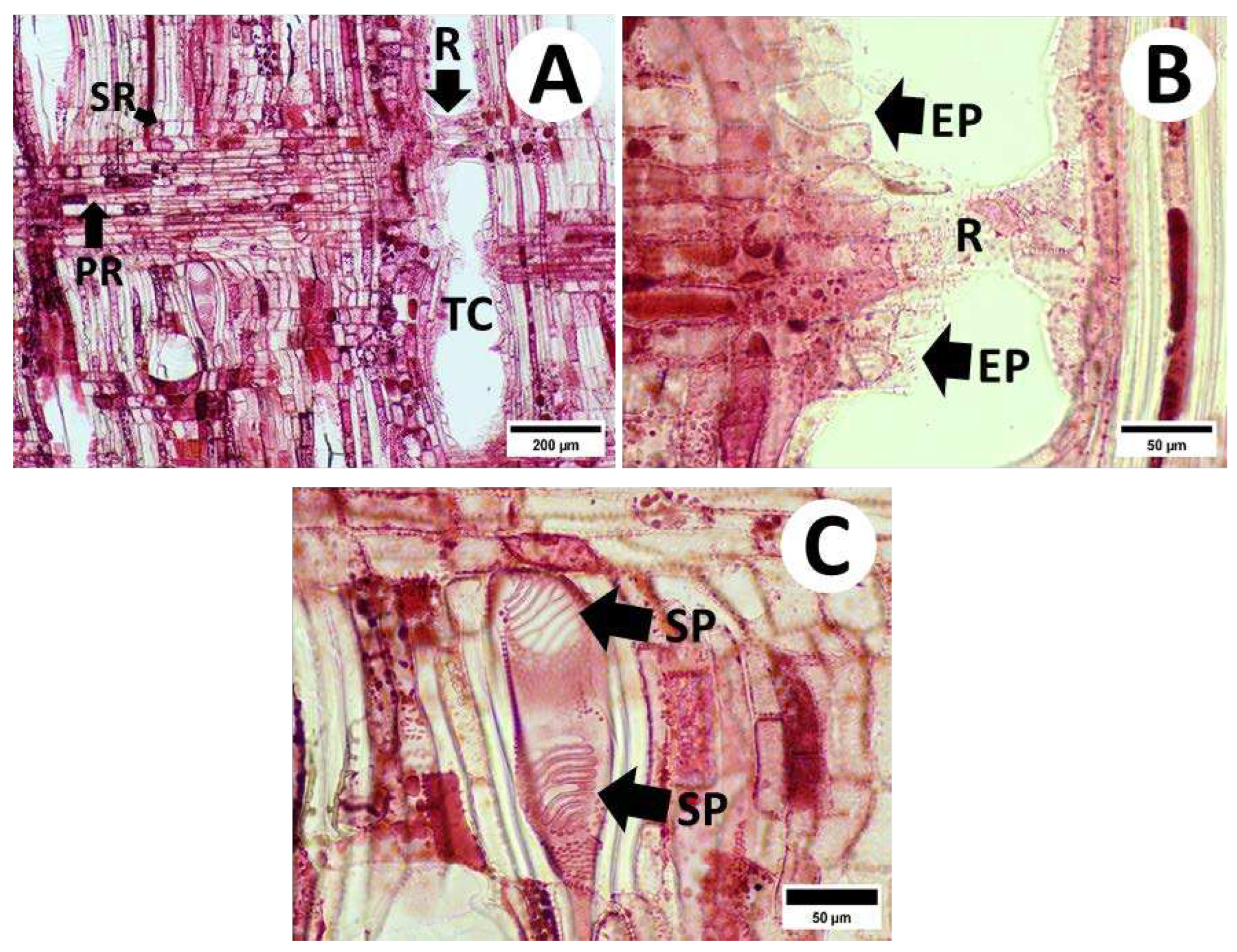
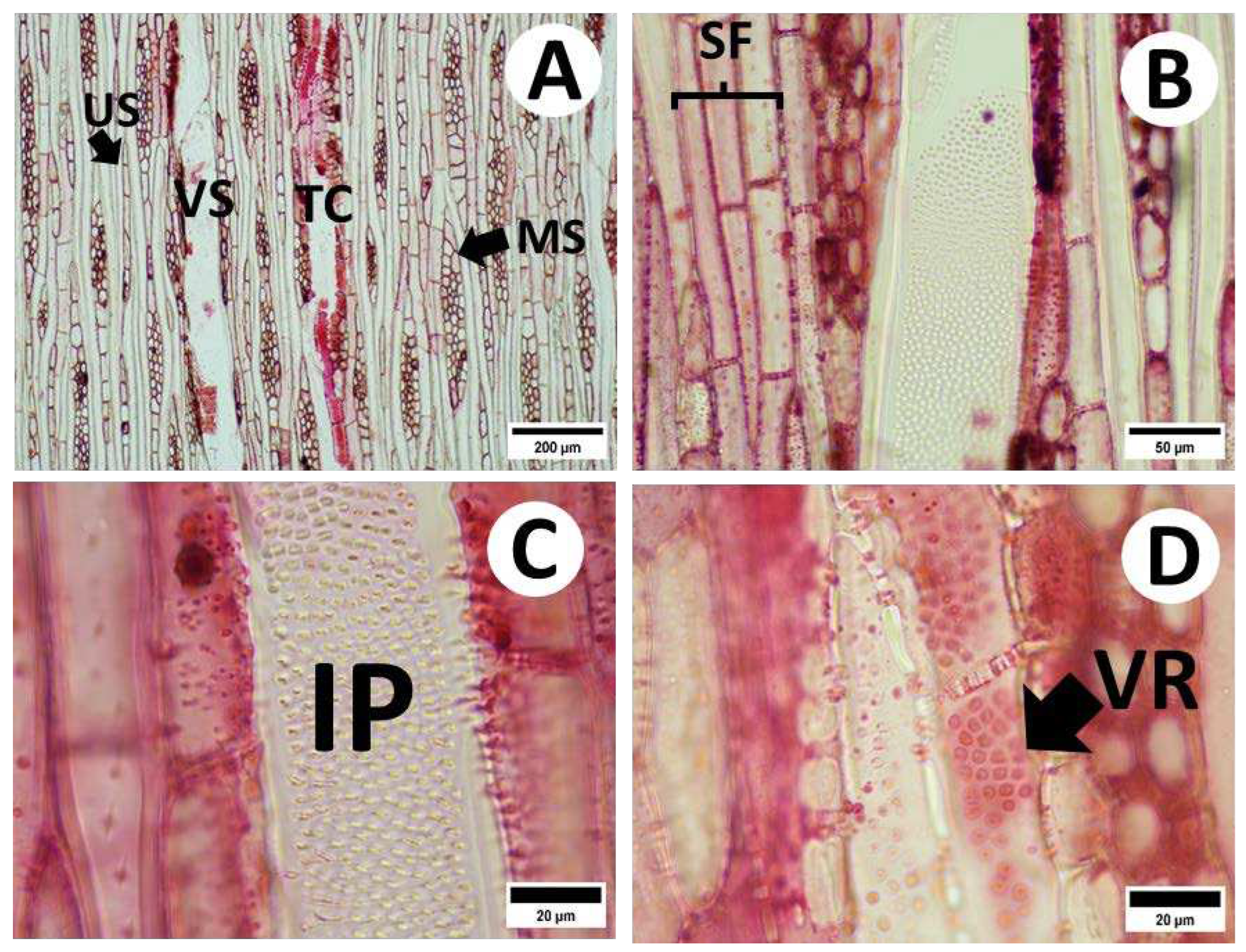
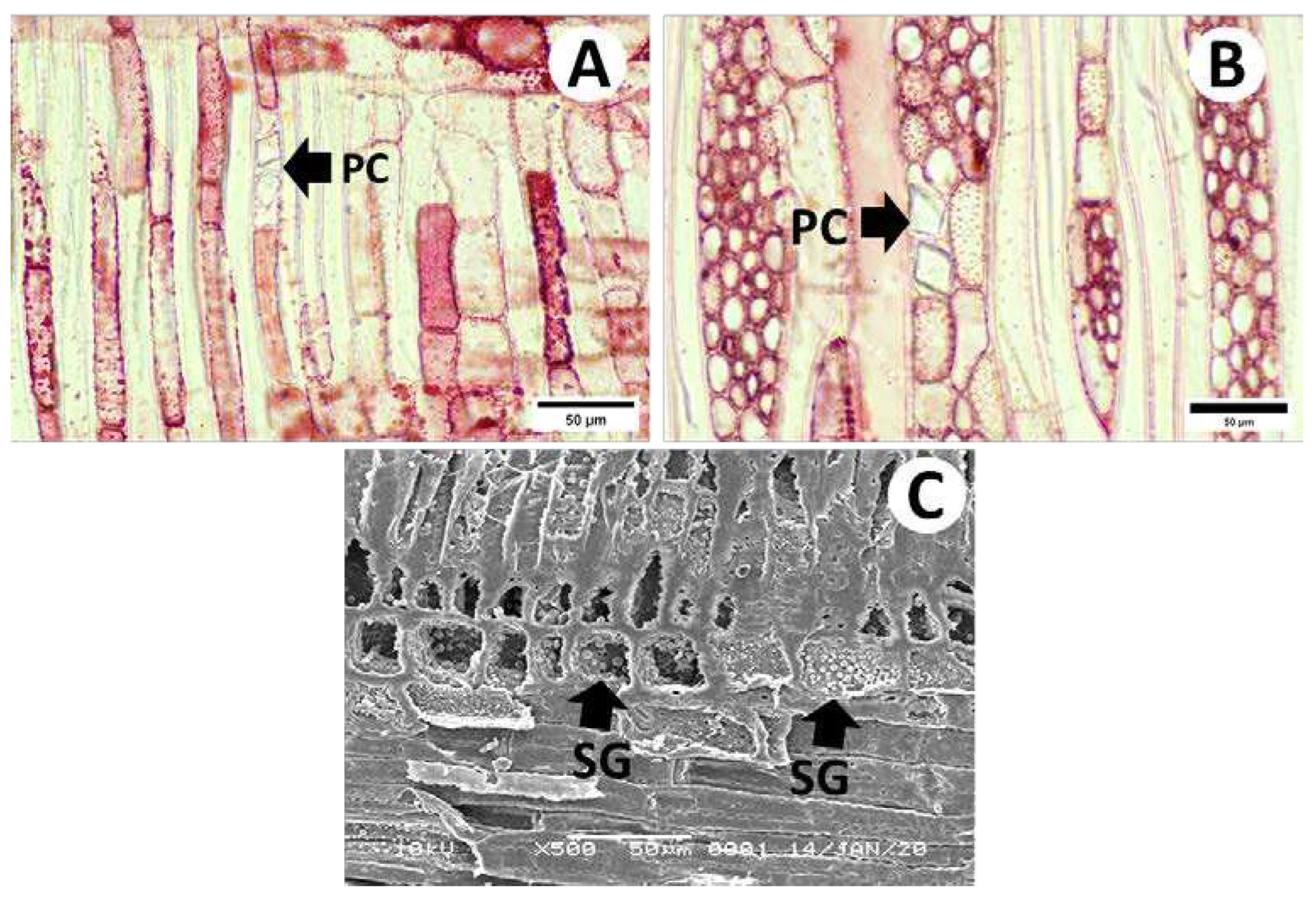
Optical micrographs of the radial and tangential sections of tapped S. sumatrana are shown in Figure 3 and Figure 4, respectively. Procumbent body ray and square marginal cells were observed in the radial section (Figure 3A). Epithelial cells were observed at the edges of the TC, whereas a rejuvenated ray was observed within the TC (Figure 3B). The vessels contained scalariform perforation plates (Figure 3C). The width of the rays was uniseriate to multiseriate (2–4 seriates) (Figure 4A), and the fibers were non-septated and septated with distinctly bordered pits (Figures 4A and B) in the tangential section. The intervessel pits were alternating (Figure 4C), and the vessel ray pits had distinct borders similar to the intervessel pits in size and shape (Figure 4D). Chambered prismatic oxalate crystals were observed in the axial (Figure 5A) and ray (Figure 5B) parenchyma. Silica grains were also observed in the ray cells (Figure 5C). Dickison and Phend [22] found crystals in the ray cells of Styrax hypargyreus, Styrax officinalis, and Styrax suberifolius. In addition, silica grains were found in Styrax argenteus, Styrax fanshawei, Styrax glabratus, Styrax guianensis, S. hypargyreus, Styrax hypochryseus, Styrax leprosus, Styrax pallidus, and Styrax tarapotensis. However, Pasaribu et al. [17] reported the absence of silica in S. sumatrana.

3.3. Vessel and Fiber Characteristics

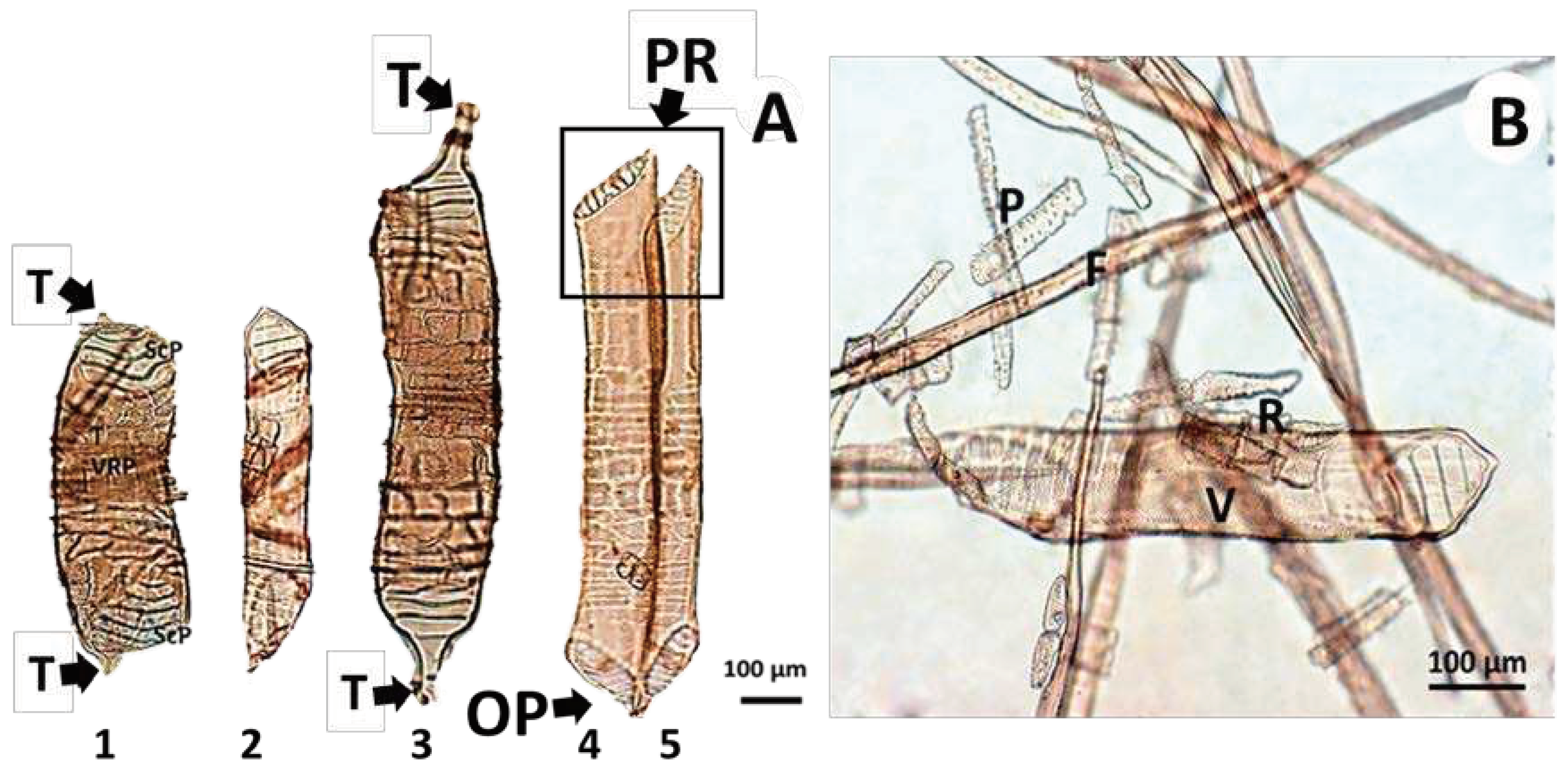
Optical micrographs of the cell components of tapped S. sumatrana are shown in Figure 6. The vessel elements were tubes with tails at both ends (Figure 6A), with a few vessels showing parallel and opposite arrangements of the perforation plates on the tips of their elements (Figures 6A: 4 and 5).
The length of the vessels ranged from 436 to 965 μm and the average was 721±171 μm. The vessel length of S. sumatrana in the present study was shorter than that of Styrax wood in previous studies [17, 21, and 24]. Pasaribu et al. [17] reported an S. sumatrana vessel length of 396–1449 μm and an average of 1031±178 μm. In addition, the vessel length of S. paralleloneurum was 671–1374 μm, and the average was 1026±187 μm. In addition, the vessel length of S. paralleloneurum was 671–1374 μm, and the average was 1026±187 μm. Damayanti et al. [21] reported S. benzoin and S. paralleloneurum vessel element lengths of 1135±168 μm and 1055±166 μm, respectively. Machado et al. [24] reported that the vessel lengths of Styrax latifolium, Styrax martii, Styrax leprosus, and Styrax camporum from Brazil were 970±197 μm, 873±197 μm, 853±157 μm, and 738±121 μm, respectively.
The vessel lumina diameter of S. sumatrana ranged from 96 to 190 with an average of 131 ± 28 μm. As reported by Pasaribu et al. [17] and Damayanti et al. [21], vessel lumina diameters of S. sumatrana, S. benzoin, and S. paralleloneurum were 168 ± 37 μm, 160±21 μm, and 140±25 μm, respectively, showing larger value than S. sumatrana in the present study. Machado et al. [24] reported that the vessel lumina diameters of Styrax latifolium, Styrax martii, Styrax leprosus, and Styrax camporum from Brazil were 91±14 μm, 97±17 μm, 69±17 μm, and 72±14 μm, respectively, which is distinctively smaller than the vessel lumina diameter of S. sumatrana in the present study.
The dimensions and derivative values of the tapped S. sumatrana wood fibers are listed in Table 3. The fiber length was 1390.8 μm, which is categorized into moderate to extensive based on the Hardwood List of the IAWA Committee [23]. The fiber length of S. sumatrana was smaller than that of Styrax wood reported in previous studies. Pasaribu et al. [17] reported that the fiber length and diameter of untapped S. sumatrana ranged from 1525 to 2290 μm and the average was 1860±163 μm. Damayanti et al. [21] reported an S. benzoin fiber length of 1930±184 μm and an S. paralleloneurum fiber length of 1870±139 μm. Machado et al. [24] reported that the fiber lengths of Styrax latifolium, Styrax martii, and Styrax leprosus were 1904±317 μm, 1798±262 μm, and 1811±294 μm, respectively.
The fiber diameter was 29.23 μm, whereas the wall thickness and fiber lumina diameter were 5.62 μm and 17.99 μm, respectively. The fiber wall of S. sumatrana in the present study was classified as thin to moderate based on the Hardwood List of the IAWA Committee [23]. Damayanti et al. [21] mentioned that the fiber wall thickness and lumen diameter of tapped S. benzoin were 2.3±0.4 and 32.5±3.1, respectively. The fiber walls of untapped S. sumatrana and tapped S. benzoin in previous studies were distinctively thinner than those of tapped S. sumatrana in the present study. Pasaribu et al. [17] reported that the fiber and lumina diameter of untapped S. sumatrana ranged from 25 to 48 μm and from 20 to 43 μm, respectively, and the average was 35±3 μm in fiber diameter and 31± 3 μm in lumina diameter. In addition, the authors also mentioned that the fiber wall thickness of S. sumatrana ranged from 1 to 3 μm with an average of 2.3±0.4 μm.
RR is the double cell wall thickness ratio to the lumen diameter. A lower RR value is more favorable for producing high-quality fiber pulp, which is essential for achieving complete flatness and adequate fiber bonding in pulp sheets [25, 26]. In the present study, the average RR of S. sumatrana wood was 0.64, and the RR ranged from 0.42 to 1.00, indicating a 3rd grade pulp quality. The RR in the present study demonstrated a lower pulp grade than that of Styrax wood reported in previous studies. Pasaribu et al. [17] reported a 0.15 RR in S. sumatrana and S. paralleloneurus, demonstrating the potential production of 1st grade pulp. Damayanti et al. [21] reported that the RR of both tapped S. benzoin and untapped S. paralleloneurus was 0.14, which was another indicator of 1st-grade pulp.
The FP refers to the ratio of fiber length to diameter. A higher FP indicates a greater inter-fiber bonding and tear strength, which positively correlates with the tensile strength of paper sheets [25, 26]. The FP of S. sumatrana wood was 46.38 and ranged from 34.73 to 57.40, showing 3rd grade pulp quality. The FP of S. sumatrana and S. paralleloneurus were 52.42 and 47.70, respectively in a previous study, indicating 3rd grade pulp quality [17]. The FP of tapped S. benzoin and untapped S. paralleloneurus revealed 3rd grade pulp quality at 52.09 and 53.12, respectively, in another study [21].
The MR refers to the ratio of the surface area of the fiber wall to the total surface area of the fiber. A smaller MR value leads to greater density in pulp sheets, with high mechanical strength [25, 26]. The MR of tapped S. sumatrana wood ranged from 50.73% to 74.98% and averaged at 62.13 % in this study, showing 3rd grade pulp quality. This MR demonstrated a lower pulp quality grade than that of Styrax wood reported in previous studies. Pasaribu et al. [17] reported an S. sumatrana and S. paralleloneurus MR of 24.35 and 23.72, respectively, demonstrating 1st grade pulp quality. Similarly, the MR of tapped S. benzoin and untapped S. paralleloneurus showed 1st grade pulp quality at 23.26 and 22.94, respectively [21].
The coefficient of rigidity (CR) is the ratio of the cell wall thickness to the fiber diameter, and a higher CR of a fiber reduces tensile strength [25, 26]. The CR of tapped S. sumatrana ranged from 0.15 to 0.25, with an average value of 0.19 in the present study, showing 3rd grade pulp quality. However, this quality class was considerably lower than that of other Styrax woods reported in previous studies. The CR of untapped S. sumatrana, tapped S. benzoin, and untapped S. paralleloneurus were approximately 0.06, showing 1st grade pulp quality [17, 21]
The FR is the ratio of the lumen to fiber diameter. Here, the FR of tapped S. sumatrana ranged from 0.50 to 0.70, with an average of 0.61. However, it was lower than that of other Styrax wood reported in previous studies. The FR of untapped S. sumatrana, tapped S. benzoin, and untapped S. paralleloneurus was approximately 0.88, showing 1st grade pulp quality [17, 21]
The total score of tapped S. sumatrana wood fibers in the present study demonstrated a 2nd grade pulp quality. In contrast, the total scores of untapped S. sumatrana, tapped S. benzoin, and untapped S. paralleloneurus showed 1st grade pulp quality in previous studies [12, 13].

5. Conclusions

The qualitative anatomical characteristics of tapped wood in the present study showed general characteristics of Styrax woods. TC formation was observed along the growth rings in the tapped part of the wood disc. The TC showed an irregular outline with 8–16 epithelial cells at the edges of the canal. Tapped S. sumatrana generally exhibited thin-walled fibers, whereas thick-walled fibers were observed near the TC. Furthermore, deposits were observed in some vessels in the tapped part but not in the untapped part.
Chambered prismatic oxalate crystals were observed in both axial and ray parenchyma, whereas silica grains were observed in ray cells. The fiber length and cell wall of tapped S. sumatrana were classified as moderate to extensive and thin to moderate, respectively. Moreover, the derivative value of the tapped S. sumatrana wood fibers indicated 2nd grade pulp quality.
In conclusion, the formation of TCs aligned tangentially was the most distinct characteristic of tapped S. sumatrana wood. The fiber quality of the tapped wood was categorized as 2nd grade, which is acceptable for industry applications.

Author Contributions

Conceptualization, A.D., A.H.I, R.D., and A.S.; methodology, A.D. and B.D.P.; software, A.D. and B.D.P.; validation, A.H.I. and N.H.K.; formal analysis, A.D. and B.D.P.; investigation, A.D. and B.D.P.; resources, A.H.I. and N.H.K; data curation, A.D. and B.D.P.; writing—original draft preparation, A.D. and B.D.P.; writing—review and editing, A.D., B.D.P. A.H.I, and N.H.K.; visualization, A.D. and B.D.P.; supervision, A.H.I. and N.H.K.; project administration, A.H.I. and N.H.K.; funding acquisition, A.H.I. and N.H.K.

Funding

This research was funded by the Deputy for Strengthening Research and Development, Ministry of Research and Technology/National Research and Innovation Agency, Republic of Indonesia through the Basic Research of Higher Education Excellence (PDUPT) Grant (Grant no. 11/AMD/E1/KP.PTNBH/2020, date: May 11, 2020). This study was also supported by the Science and Technology Support Program through the National Research Foundation of Korea (NRF) funded by the Ministry of Science and ICT (MSIT) (Grant no. 2022R1A2C1006470), the Basic Science Research Program through the NRF funded by the Ministry of Education (Grant no. 2018R1A6A1A03025582), and the R&D Program for Forest Science Technology (Project No. 2021350C10-2323-AC03) provided by the Korea Forest Service (Korea Forestry Promotion Institute).

Data Availability Statement

The datasets generated and analyzed in the current study are not publicly available but are available from the corresponding author upon reasonable request.

Conflicts of Interest

The authors declare no conflict of interest.

References

  1. Nussinovitch, A. Plant Gum Exudates of the World: Sources, Distribution, Properties, and Applications: CRC Press, Boca Raton, 2010. [CrossRef]
  2. Boer, E.; Ella, A.B. Introduction. In: Boer E, Ella AB. (eds). Plant Resources of South-East Asia No 18: Plants Producing Exudates: Prosea, Bogor, Indonesia, 2001.
  3. Langenheim, J.H. Plant resins. American Scientist. 1990, 78, 16-24.
  4. Langenheim, J.H. Plant resins: chemistry, evolution, ecology, and ethnobotany: Timber Press, Portland, 2003. [CrossRef]
  5. Fritsch, P. W; Morton, C. M.; Chen, T.; Meldrum, C. Phylogeny and biogeography of the Styracaceae. International Journal of Plant Sciences. 2001, 162(S6), pp.S95-S116. [CrossRef]
  6. Hoesen, D.S.H. Styrax L. In Plant Resources of South-East Asia: Plants Producing Exudates, Boer, E., Ella, A.B., Eds; Prosea: Bogor, Indonesia. 2001, Volume 18, pp. 112–119.
  7. Simamora, S.W.; Muhdi; Batubara, R. Modifikasi Teknik Pemanenan Dengan Perlakuan Fisik Dalam Upaya Peningkatan Produktivitas Getah Kemenyan Toba (Styrax Sumatrana). Peronema Forestry Science Journal 2015, 4, 159-165.
  8. Iswanto, A.H.; Susilowati, A.; Azhar, I.; Riswan, R.; Supriyanto, S.; Tarigan, J.E.; Fatriasari, W. Physical and mechanical properties of local Styrax woods from North Tapanuli in Indonesia. J. Korean Wood Sci. Technol. 2016, 44(4), 539–550. [CrossRef]
  9. Iswanto, A.H.; Siregar, Y.S.; Susilowati, A.; Darwis, A.; Hartono, R.; Wirjosentono, B.; Rachmat, H.H.; Hidayat, A.; Fatriasari, W. Variations in chemical constituent of Styrax sumatrana wood growing at different cultivation sites in North Sumatra, Indonesia. BIODIVERSITAS 2019, 20 (2), 448–452. [CrossRef]
  10. Badan Pusat Statistik Propinsi Sumatera Utara. Indikator Pertanian Propinsi Sumatera Utara 2023: Badan Pusat Statistik Propinsi Sumatera Utara, Medan, Indonesia, 2023.
  11. Hovaneissian M., Archier P., Mathe C., Culioli G., Vieillescazes C. Analytical investigation of Styrax and benzoin balsams by HPLC-PAD-fluorimetry and GC-MS. Phytochemical Analysis. 2008, 19(4):301-310. [CrossRef]
  12. Jayusman. Recognising Incense Trees (Styrax spp.): Species with Broad Utilization Spectrums for Optimalization. IPB Press: Bogor, Indonesia, 2014.
  13. Dünisch O., Bass P. On the origin of intercellular canals in the secondary xylem of selected Meliaceae species. IAWA J 2006, 27:281–297. [CrossRef]
  14. Kuroda K, Shimaji K. Traumatic resin canal formation as a marker of xylem growth. Forest Sci 1983, 29(3):653-659. [CrossRef]
  15. DeRose RJ, Bekker MF, Long JN. Traumatic resin ducts as indicators of bark beetle outbreaks. Can. J. For. Res. 2017, 47:1168–1174. [CrossRef]
  16. Deng, X.Q.; Cheng, S.P.; Pan, N.X.; Chen, J.L. The effects of ethrel upon benzoin production and balsamic ducts of Styrax hypoglauca Perk. J. Integr. Plant Biol. 1978, 20, 26–29.
  17. Pasaribu, G.; Jasni, J; Damayanti, R.; Wibowo, S. Anatomical, physical, mechanical properties of Kemenyan Toba (Styrax sumatrana) and Kemenyan Bulu (Styrax paralleloneurus). J. For. Prod. Res. 2013, 31(2), 161–169.
  18. Franklin, G.L. Preparation of thin sections of synthetic resins and wood resin composites, and a new macerating method for wood. Nature 1945, 155(3924), 51–59. [CrossRef]
  19. Nurachman, R.; Siagian, M. Dimensi serat jenis kayu Indonesia. Laporan No. 75. Lembaga Penelitian: Bogor, Indonesia, 1976.
  20. Adi, D. S.; Wahyuni, I.; Risanto, L.; Rulliaty, S.; Hermiati, E.; Dwianto, W.; Watanabe, T. Central kalimantan’s fast growing species: Suitability for pulp and paper. Indonesian J. For. Res. 2015, 2(1), 21–29. [CrossRef]
  21. Dickison, W.C.; Phend, K.D. Wood anatomy of the Styracaceae: evolutionary and ecological considerations. IAWA Bulletin n.s., 1985, 6(1), 3–22.
  22. IAWA Committee. IAWA List of Microscopic Features for Hardwood Identification with an Appendix on Non-anatomical Information; National Herbarium of the Netherlands: Leiden, the Netherlands, 1989; pp. 219–332. [CrossRef]
  23. Damayanti, R.; Mandang, Y.I.; Waluyo, T.K. Anatomical properties and fiber quality of Styrax stem from North Sumatra. J. For. Prod. Res. 2007, 25(3), 273–290.
  24. Machado, S. R., Rodella, R. A., Angyalossy, V., Marcati, C. R. Structural variations in root and stem wood of Styrax (Styracaceae) from Brazilian forest and cerrado. IAWA Journal 2007, 28(2), 173-188. [CrossRef]
  25. Wistara, N.J.; Carolina, A.; Pulungan, W.S.; Emil, N.; Lee, S.H.; Kim, N.H. Effect of tree age and active alkali on kraft pulping of white jabon. J. Korean Wood Sci. Technol. 2015, 43, 566–577. [CrossRef]
  26. Darwis, A.; Karliati, T.; Sutrisno, Alamsyah, E.M.; Rumidatul, A.; Melani, L.; Kim, H.J.; Iswanto, A.H.; Fatriasari, W. Chemical properties, crystallinity, and fiber biometry of Jabon (Anthocephalus cadamba) wood for pulp raw material: the effect of age and position. Nord. Pulp Pap. Res. J. 2023, 1–11. [CrossRef]
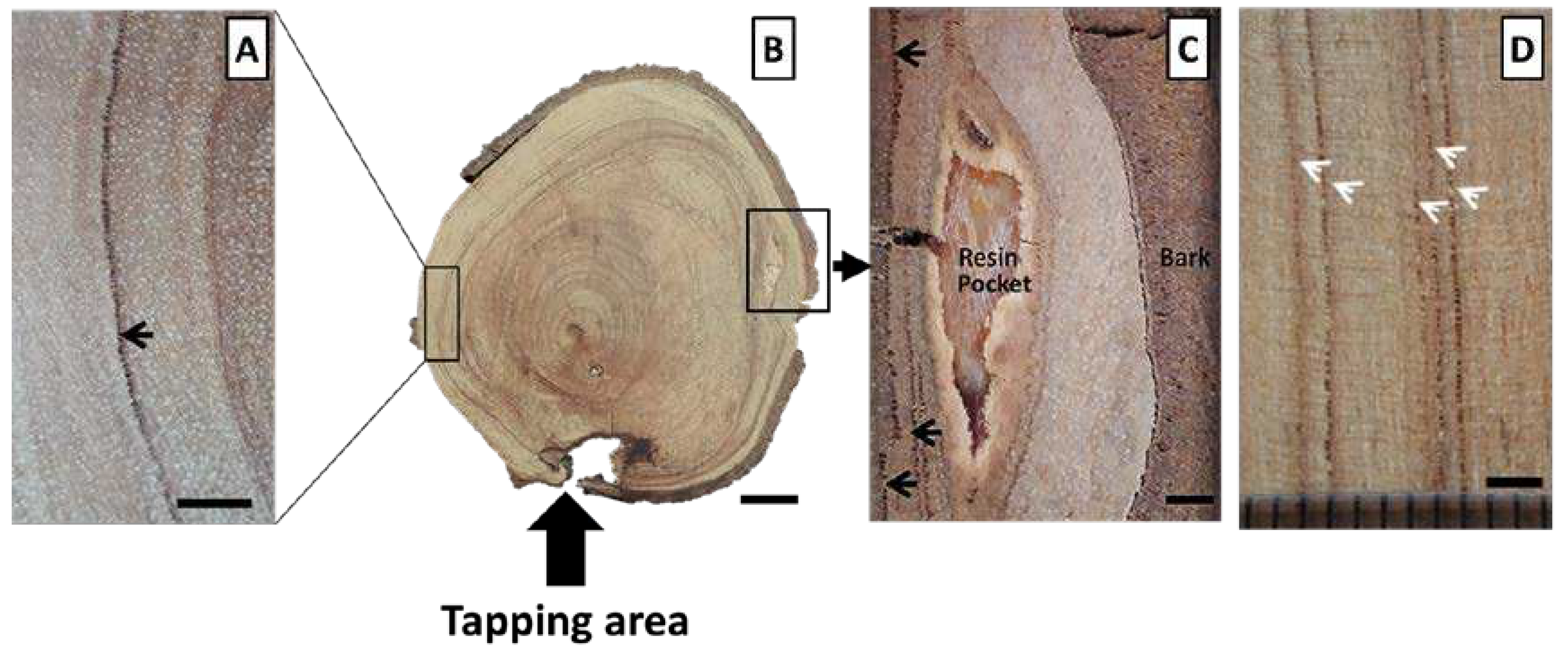
Figure 1. Transverse (A, B, and C) and radial surfaces (D) of a tapped Styrax sumatrana wood disc. The resin pocket can be seen in Figure 1C. The dark lines (arrows) in the transverse surface indicate traumatic intercellular canal bands in the tapped S. sumatrana wood. Scale bars: 2 mm (A, C, and D) and 30 mm (B).
Preprints 98494 g001
Figure 2. The optical (A to F) and scanning electron (G) micrographs of cross-sections of the untapped (A, C, E) and tapped (B, D, F, G) parts in tapped S. sumatrana. Growth ring (GR), traumatic resin canals (TC), clustered vessels (CV), radially arranged vessels (RV), solitary vessels (SV), deposits in vessels (DP), rays (R), axial parenchyma (AP), fibers (Fb), and epithelial cells (Ep). Scale bars: 500 µm (A and B), 200 µm (C and D), 50 µm (E and F), and 100 µm (G).
Preprints 98494 g002
Figure 3. Optical micrographs of the radial section in tapped S. sumatrana. Square body ray (SR), procumbent body ray (PR), traumatic intercellular canal (TC), epithelial cells (EP), rejuvenated ray (R), and scalariform perforation plate (SP). Scale bars: 200 µm (A), 50 µm (B and C).
Preprints 98494 g003
Figure 4. Optical micrographs of the tangential section in tapped S. sumatrana. Multiseriate (MS) and uniseriate rays (US), septate fibers with bordered pits (SF), intervessel pits (IP), and vessel-ray pits (VR). Scale bars: 200 µm (A), 50 µm (B), and 20 µm (C and D).
Preprints 98494 g004
Figure 5. Oxalate prismatic crystals (PC) in axial (A) and ray (B) parenchyma, as well as silica grain (SG) in ray parenchyma (C) of tapped S. sumatrana wood. Scale bars: 50 µm.
Preprints 98494 g005
Figure 6. The compositions of cells in tapped S. sumatrana. Vessel elements (1–5), tails of vessel element (T), parallel (PR) and opposite arrangement of perforation plates (OP) (A: 4 and 5), fibers (F), parenchyma cell (P), and ray (R). Scale bars: 100 μm.
Preprints 98494 g006
Table 1. Fiber derivative values and their formula equation.
The derivative values of fiber Equation
Runkel ratio (RR) 2 W l d
Felting power (FP) L D
Mulhsteph ratio (MR) D 2 l d 2 D 2 x 100 %
Coefficient rigidity (CR) W D
Flexibility ratio (FR) l d D
Table 2. Pulp quality classification [19, 20].
Parameter Class I Class II Class III Class IV
Value Score Value Score Value Score Value Score
Fiber length
(µm)
2200 100 1600–2200 75 900–1600 50 900 50
Runkel ratio 0.25 100 0.2–0.5 75 0.5–0.1 50 1 50
Felting power 90 100 70–90 75 40–70 50 40 50
Flexibility ratio 0.8 100 0.6–0.8 75 0.4–0.6 50 0.6 50
Coefficient of
rigidity
0.1 100 0.1–0.15 75 0.15–0.2 50 0.2 50
Mulhsteph ratio 30% 100 30–60% 75 60–80% 50 80% 50
Total score 451–600 301–450 151–300 150
Table 3. Dimension and derivative value of tapped S. sumatrana wood fiber.
Min Max Average SD Pulp quality
Dimension of fibers (μm)
Length 1113.25 1774.02 1390.77 180.27 (50) III
Diameter 24.04 34.72 29.23 3.07 -
Lumina Diameter 14.69 24.37 17.99 3.08 -
Wall thickness 4.675 7.51 5.62 0.78 -
Biometrics
Runkel ratio 0.42 1.00 0.64 0.15 (50) III
Felting power 34.73 57.40 46.38 7.52 (50) III
Mulhsteph ratio (%) 50.73 74.98 62.13 6.53 (50) III
Coefficient of rigidity 0.15 0.25 0.19 0.03 (50) III
Flexibility ratio 0.50 0.70 0.61 0.05 (75) II
Total score (320) II
Disclaimer/Publisher’s Note: The statements, opinions and data contained in all publications are solely those of the individual author(s) and contributor(s) and not of MDPI and/or the editor(s). MDPI and/or the editor(s) disclaim responsibility for any injury to people or property resulting from any ideas, methods, instructions or products referred to in the content.
Copyright: This open access article is published under a Creative Commons CC BY 4.0 license, which permit the free download, distribution, and reuse, provided that the author and preprint are cited in any reuse.
Prerpints.org logo

Preprints.org is a free preprint server supported by MDPI in Basel, Switzerland.

Subscribe

© 2024 MDPI (Basel, Switzerland) unless otherwise stated