Preprint
Article

Design, Synthesis and Molecular Modeling of Benzofuran-8-Hyroxyquinoline Hybrids as Multi-Target Inhibitors and Potential Anti-Alzheimer’s Disease

Altmetrics

Downloads

142

Views

63

Comments

0

Submitted:

12 February 2024

Posted:

13 February 2024

You are already at the latest version

Alerts
Abstract
Cholinesterase (ChE) and secretase (BACE) inhibitors, and fibril- and β-amyloid-suppressing medicines are used to treat Alzheimer's disease (AD) symptomatically. The prevalence and complex nature of AD have increased the urgent need for multi-targeted directed ligands (MTDLs). This is because MTDLs can prevent potential drug-drug interactions during poly-therapy and have a better therapeutic profile than single targeted agents. Using piperazine linker or spacer and two primary scaffolds, benzofuran and 8-hydroxyquinoline, a unique class of multi-targeted medicines was recently reported by our group. These compounds showed re-markable effectiveness in inhibiting Aβ1-42 aggregation and acting as iron chelators.The results prompted us to synthesize an additional series of compounds as multimodal anti-AD agents and investigate its inhibitor activities to ChE (AChE/BChE) and BACE1 enzymes. The resulting in-hibitory effects suggested that the compounds under study might be used to improve cognitive function. The docking analysis' findings revealed that the compounds bind to AChE and BChE by forming H-bond interactions with amino acid residues at binding sites and µ-stacking inter-actions with aromatic residues, whereas the binding to BACE1 only revealed H-bond interac-tions with amino acid residues at binding sites.
Keywords: 
Subject: Chemistry and Materials Science  -   Medicinal Chemistry

1. Introduction

A serious neurodegenerative brain condition, Alzheimer’s disease (AD) is characterised by a decline in cognitive performance and ultimately, memory loss. The condition deteriorates with age. Ever since AD was formally diagnosed in 1910, it has been a persistent problem and a difficult sickness for medical experts worldwide to comprehend [1]. Over 55 million people worldwide suffer with dementia, and over 60% of them reside in low- or middle-income countries. According to data from https://www.who.int/news-room/fact-sheets/detail/dementia (accessed October 9, 2022), the proportion of older people in the population is rising in almost every nation. By 2030, it is expected that this number will rise to 78 million, and by 2050, it will reach 139 million. We still don’t fully comprehend the pathophysiological pathways behind AD. Nonetheless, despite its complexity, four hypotheses, among others, can be used to identify a number of neurodegenerative pathways. 1. The aspartyl protease β-site APP cleaving enzyme-1 (BACE1) and γ-secretase progressively degrade amyloid precursor protein (APP) to produce insoluble amyloid beta (Aβ) plaques (Figure 1). 2. Tau protein hyperphosphorylation leads to intracellular accumulations of neurofibrillary tangles (NFTs) in neuronal and glial cells. 3. The pathology of AD is related to the biometals’ malfunction that results in detrimental excess level of heavy metals such as copper and iron. 4. Oxidative stress causes brain neuronal cells to die and synapses to disappear [2]. High level in the inflammatory mediators and low level in the neurotransmitter acetylcholine (ACh) have a distinct role in AD pathology [3,4,5].
When acetylcholinesterase (AChE) is suppressed in the early stages of AD, ACh levels increase and cognitive function improves. As AD progresses and AChE levels are thought to be declining, AChE inhibitors (AChEIs) appears to be ineffective [6]. On the other hand, butyrylcholinesterase (BuChE) levels either remain constant or even increase [7] and BuChEIs are effective in the late stages of AD since BChEIs can hydrolyze ChE to compensate for the drop in AChE activity [8]. The intracellular buildup of hyperphosphorylated neurofibrillary tangles of tau protein, oxidative and inflammatory stresses are additional hypotheses that support the multifactorial character of AD illness [9,10]. The five subsites that make up the active sites of ChE enzymes are the catalytic active site (CAS), peripheral anionic site (PAS), acyl binding pocket, oxyanion hole, and anionic subsite [11]. One of the distinctions between the structure of both AChE and BuChE is the size of the acyl pocket. More specifically, the acyl pocket of BuChE has smaller residues like Leu286 and Val288 that enable a larger site, whereas the acyl pocket of AChE contains larger and more aromatic Phe295 and Phe297 residues that enable a smaller site. The aforementioned structural differences have an impact on selective inhibitor design [12]. The inhibition of Aβ peptide accumulation in the brain can be considered as a treatment strategy in delaying the start of AD [3]. Reduced plaque aggregation has been demonstrated to be a benefit of heavy metals (Cupper, Iron) chelators [13]. A common course of treatment for AD involves acetylcholinesterase inhibitors such donepezil (E2020, Aricept®), galantamine, and rivastigmine, which improve cholinergic neurotransmission [14]. Butyl choline esterase has been found to be a co-regulator of acetylcholine activity, which has recently attracted the attention of chemists and biologists who are concerned in the role of these enzymes in AD [15]. Given the complexity of AD, one gene change might not be sufficient to deliver the necessary treatment. Multi-targeted directed ligands (MTDLs) are now more important than ever to prevent interaction between multi-therapeutics drugs and to have a better therapeutic treatment than single-targeted therapy. This is due to the prevalence of Alzheimer disease (AD) in addition to the growing understanding of its multifactorial nature. In light of these results, the exciting notion of developing multi-target molecules may lead to a more effective therapeutic strategy [16]. Based on this new paradigm, several researchers created a number of hybrid compounds, including SKF-64346 and derivatives of acridine (Figure 2). The alkylamine moiety of these hybrid medicines inhibits cholinesterase (AChE and BuChE), while the benzofuran or acridine motif inhibits Aβ-aggregation [17,18]. Many compounds (Figure 2) were developed to exhibit antiaggregant and anticholinesterase activities [19]. HLA-20 (8-hydroxyquinoline derivatives) and M30’s capacities to chelate iron, scavenge free radicals, and prevent iron-catalyzed lipid membrane peroxidation have all been proven [20,21].
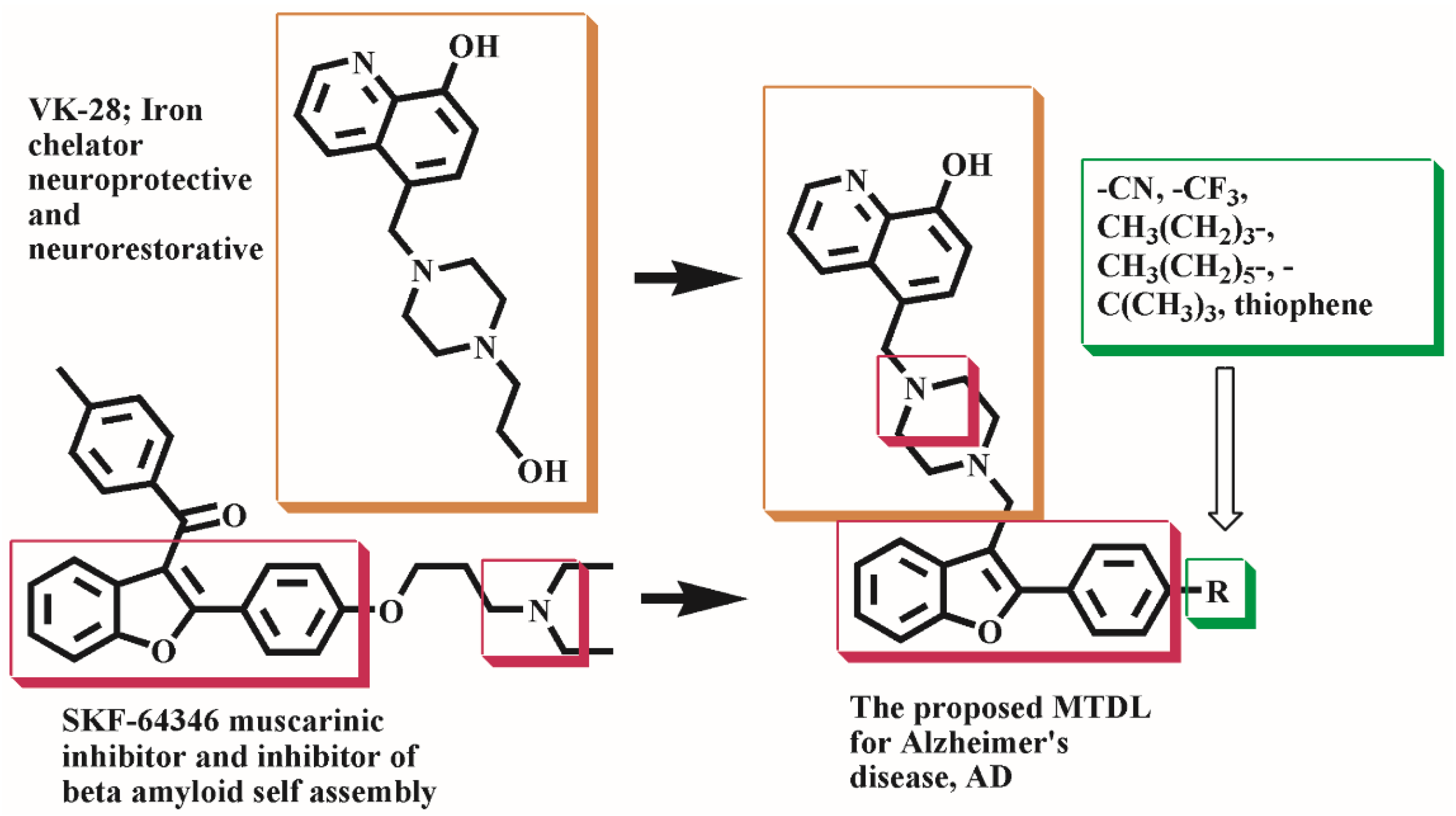
The lead compound SKF-64346, an AChE/BChE and γ-secretase inhibitor has motivated us to develop a number of hybrid molecules through linking the benzofuran and 8-hydroxyquinoline moieties by piperazine spacer that also facilitates BBB crossing. To assess the effect of the substitutes on the electrical configuration of the whole structure and their impact on the compounds’ biological activity, a range of substitutions were introduced to position 2 of the benzofuran scaffold (Figure 3). Promising results obtained in our published work where the hybrid compounds explored significant inhibition of Aβ1-42 self-aggregation and chelated bio-metals such as Fe3+ [24]. These findings prompted us continue our work for the synthesis of new derivatives and perform biological evaluation against more AD-target enzymes ChE ‘including AChE/BChE’ and BAC1.

2. Results and Discussions

2.1. Chemistry
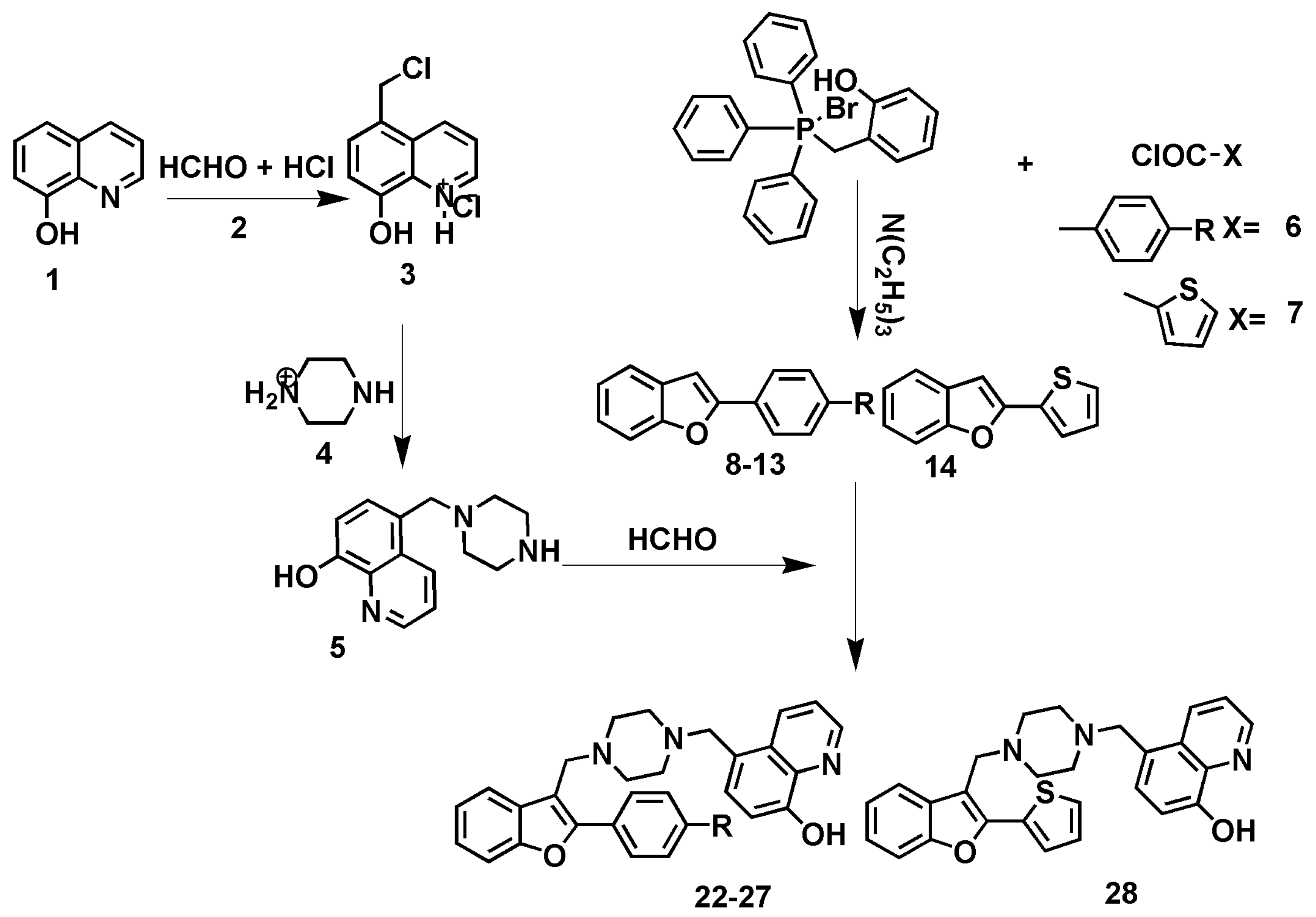
The synthetic route for the intermediates and final compounds is depicted in scheme 1. The starting materials 5-chloromethyl-8-hydroxyquinoline hydrochloride 3 and 5-((piperazin-1-yl)methyl)quinolin-8-ol 5 [22,23] were prepared according to reported procedures. Wittig reaction was utilized to prepare 2-Aryl[b]benzofurans intermediate compounds 8-14 through the reaction of 2-hydroxybenzyltriphenyl phosphonium bromide with substituted-benzoic acid chloride in aprotic solvent, toluene, and trimethylamine. Mannich reaction was utilized for aminoalkylation of compounds 8-14 with 5-((piperazin-1-yl)methyl)quinolin-8-ol revealing 5-((4-((2-(4-substitutedphenyl)benzofuran-3-yl)methyl)piperazin-1-yl)methyl)quinolin-8-ol final compounds 22-28. Structure elucidations of compounds (8-14, 22-28) was done using IR, 1H NMR, 13C NMR, mass spectral data and CHN analysis. Two distinctive bands were visible in the IR spectra of compounds 8-14 at 3095-2975 cm-1 due to aromatic CH stretching and at 868-725 cm-1 due to aromatic CH out-of-plane bending. Conversion of 8-14 into 22-28 leads to the development of new bands at 3378-3315 cm-1 related to phenolic OH stretching of the 8-hydroxyquinoline fragment. 1H NMR spectra of 8-14 revealed signals at δ 6.66–8.68 ppm characteristic of 2-phenylbenzofuran. Transformation of compounds 8-14 into compounds 22-28 resulted in emergence of new NMR signals due to aliphatic protons of both piperazine ring appeared as two signals at δ 2.55-291 and 2.68-2.98 ppm and two N-methylene protons at δ 3.51-3.81 and at 3.69-4.29 ppm. In additions, compounds 22-28 showed signals at chemical shift range δ 6.89-8.87 ppm. Moreover, the butyl-, pentyl- and hexyl substituents showed characteristic four-signals pattern at δ 0.85-0.93, 1.22-1.42, 1.56-1.67, and 2.58-2.66 ppm for the intermediate compounds 10, 12, 13 and the final compounds 24, 26, 27. Compounds 11, 25 showed characteristic t-butyl group as strong singlet signal at δ 1.32-1.39 ppm. 13C NMR spectra of compounds 8-14 revealed distinguishing signals at δ 100.69-102.25 ppm because of benzofuran-C3 that disappeared upon C3-aminoalkylation into compounds 22-28.
Scheme 1. The synthesis pathway. Reagents and conditions: (i) Et3N, toluene, reflux 10 hrs; (ii) Formaline 37%, DMF, stir 3 hr at r.t. R = CN,8,22; CF3, 9,23; n-butyl, 10,24; t-butyl, 11,25; n-pentyl, 12,26; n-hexyl, 13,27.
Preprints 98786 sch001

2.2. Inhibition of AChE, BChE and BACE1 Activities

Table 1 displays moderate to strong in vitro inhibitory effects of compounds 15-28 against AChE and BChE. Compounds 15-28 explored an inhibition range of 33.4% - 81.3% against AChE while it showed an inhibition range of 54.58%-94.47% against BChE and an inhibition range of 20.9% - 97.9% against BACE1.

2.3. Docking Study of Compounds 15-28 against AChE and BChE Proteins Structures

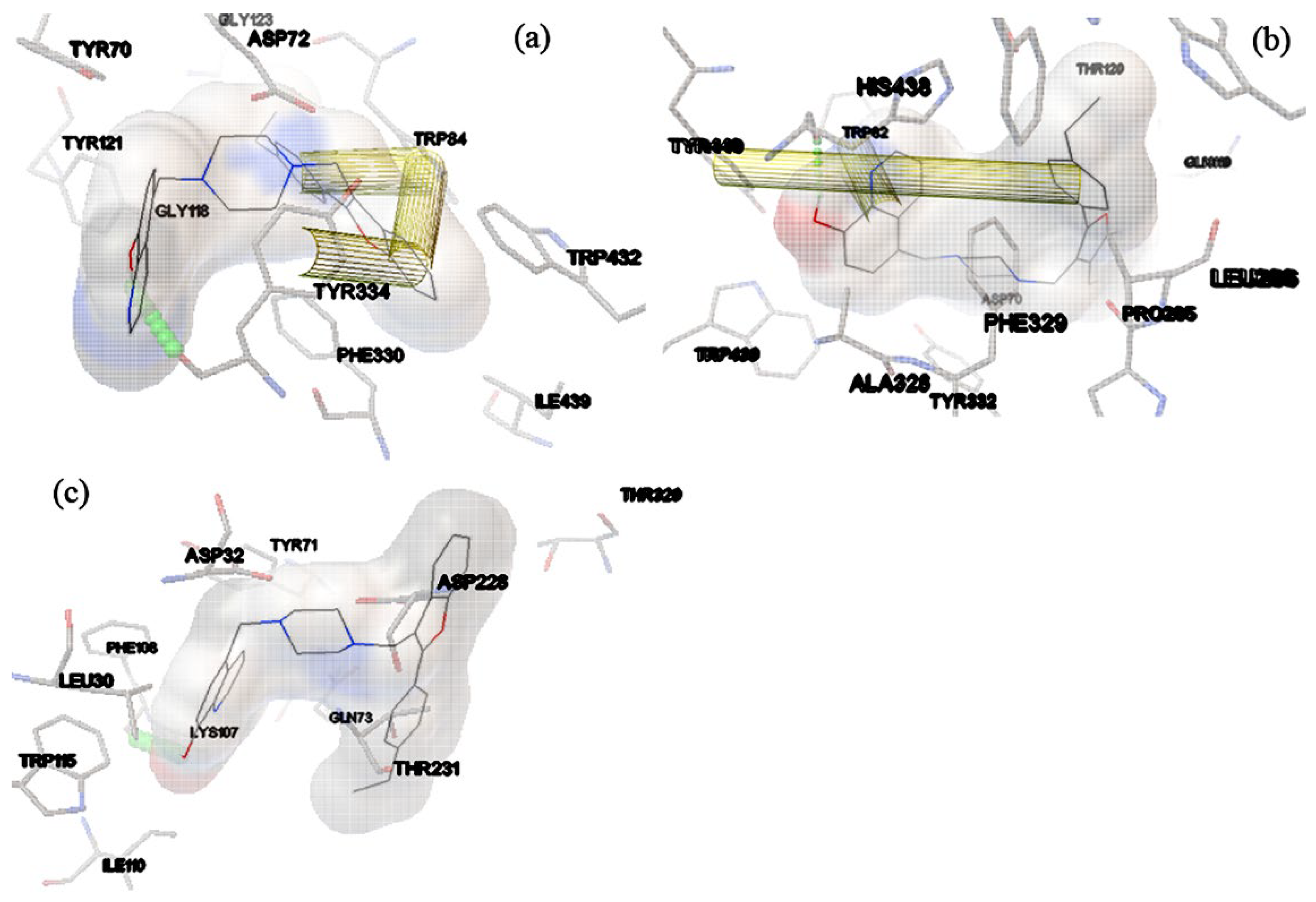
Using Autodock4 [25], we were able to ascertain the molecular interactions and binding of the ligands 15–28 to AChE/BChE. The crystal structures of Torpedo California AChE (5nnu) [26] and human BChE(6i0b) [27] have been obtained through the protein database PDB. Before being docked to the macromolecule structure, the synthesized compounds 15–28 were sketched, optimized, and uploaded to pdbqt format. The experimental AChE/BChE-inhibition percent was in accordance with the docking scores (Table 1). For the complexes of compounds 15-28 with the protein structure, docking study has estimated the energy of binding in the range from -15.63 to -9.92 kcal mol-1 for AChE and -13.79 to -10.93 kcal mol-1 to BChE (Table 1) and revealed major contribution of hydrogen bonds and hydrophobic interactions (Table 2). Figure 4 shows the binders’ docked poses inside the binding site and reveals the hydrogen-bonding residues to the ligands and the hydrophobic amino acids that are essential for ligand-target interactions. The interactions of compounds 15-28 with AChE showed hydrogen bonds to amino acid residues Tyr70, Tyr121, Ser122, Tyr334, His440 while the amino acid residues Tyr70, Val71, Asp72, Gly117, Gly118, Tyr121, Ser122, Gly123, Phe330, Leu333, Tyr334, Trp432, Met436, Ile439, His440, Tyr442 were engaged in van der Waals interactions, and the amino acid residues Tyr70, Trp84, Phe330, Phe331, Tyr334, Trp432 and His440 were involved in π–π stacking interactions (Table 2; Figure 4). Further, with hBChE, compounds 15-28 formed hydrogen bonds to the amino acids Tyr82, Tyr128, Tyr332, Tyr449, His438 and involved in van der Waals interactions to residues side chains of Asp70, Trp82, Gly116, Gly117, Gly119, Tyr120, Gly121, Ser196, Glu197, Ser198, Trp231, Leu286, Ser287, Ala328, Phe329, Tyr332, Trp430 while the amino acid residues Trp82, Tyr128, Ala328, Phe329, Tyr332, Phe398, Trp430, His438, Tyr440 were engaged in Pi–Pi interactions (Table 7; Figure 4a,b).
Table 2. Experimental and predicted BACE1 inhibition % and docking results of (10 µmol) compounds 15-28.
No. Binding Energy Kcalmol-1 Inhibition constant (Ki nmol) Exp.
inhibition %
Binding residues Hydrogen bonds (distance, Å)
15 -12.54 0.638 83.4 Leu30, Asp32, Tyr71, Gln73, Lys107, Ile110, Trp115, Asp228, Thr231, Thr320 Phe108
16 -13.16 0.227 87.0 Gln12, Gly13, Leu30, Asp32, Tyr71, Gln73, Lys107, Phe108, Asp228,Gly230, Thr231, Thr282 Thr72
17 -12.86 0.376 63.1 Gln12, Gly13, Leu30, Asp32, Tyr71, Gln73, Lys107, Phe108, Asp228,Gly230, Arg235, Thr282 Thr72
18 -13.65 0.099 74.5 Gln12, Gly13, Leu30, Asp32, Tyr71, Gln73, Lys107, Phe108, Asp228,Gly230, Thr231, Thr282 Thr72
19 -13.62 0.103 72.1 Gln12, Gly13, Leu30, Asp32, Tyr71, Gln73, Lys107, Phe108, Asp228,Gly230, Thr231, Thr282 Thr72
20 -13.19 0.213 62.4 Gly13, Asp32, Tyr71, Gln73, Gly74, Lys107, Phe108, Asp228,Gly230, Thr231, Thr282 Thr72
21 -11.32 5.02 60.5 Gln12, Gly13, Tyr71, Gln73, Gly230, Thr231, Thr282 Thr72
22 -12.05 1.47 61.9 Leu30, Tyr71,Thr72, Gln73, Lys107, Ile110, Trp115, Gly230, Thr231, Arg235 Phe108
23 -11.33 4.9 79.7 Gln12, Gly13, Leu30, Tyr71, Gln73, Asp228, Gly230, Thr231, Arg235, Val232 Thr72
24 -10.12 38.3 89.2 Gln12, Gly13, Tyr71, Gln73, Lys107, Phe108, Asp228,Gly230, Thr231, Thr282 Thr72
25 -11.82 2.16 83.1 Leu30, Tyr71,Thr72, Gln73, Ile110, Ile118, Asp228, Thr229, Gly230, Thr231, Thr232 Phe108
26 -12.51 0.681 91.0 Gln12, Gly13, Leu30, Tyr71, Thr72, Gln73, Phe108, Ile110, Trp115, Asp228,Gly230, Thr231, Arg235 Lys107
27 -12.59 0.593 81.3 Gly13, Asp32, Tyr71, Gln73, Phe108, Ile110, Ile226, Asp228,Gly230, Thr231,Thr232,Arg235, Val332, Thr329 Lys107
28 -9.98 56.95 78.6 Tyr71, Lys107, Phe108, Ile110, Asp228,Thr229, Thr231, Arg235, Val332 ----
Xray -11.45 4.0 nd Leu30, Asp32, Tyr71, Lys107, Phe108, Ile110, Trp115,Asp228,Thr229, Gly230, Arg235 Gln73, Phe108, Asp228
Table 3. Docking study results of compounds 15-28 to AChE and BChE.
No Binding residues Hydrogen bonds (distance, Å) Pi-pi interactions
AChE BChE AChE BChE AChE BChE
15 Tyr70,Asp72,Gly118, Tyr121, Gly123, Phe330, Tyr334, Trp432, Ile439 Asp70,Trp82,Gln119, Tyr120,Pro285,Leu286, Ala328,Phe329,Tyr332, Trp429 Tyr334 His438 Trp84, Tyr334 Tyr440
16 Tyr70,Asp72,Gly118, Tyr121, Ser122, Phe330, Leu333, Trp432, Met436 Asp70,Gly116, Gln119, Ser196,Pro285, Ser287, Asn289, Ala328, Phe329, Tyr332 Tyr334 His438 Trp84 Trp82, Tyr440
17 Asp72, Gly118, Tyr121, Ser122, Gly123, Phe330 Gly115,Gly117,Gln119, Gly121,Glu197,Ser198, Trp231, Pro285,Leu286, Ser287,Phe329, Phe398 Tyr334 His438 Tyr70, Trp84, Trp432 Trp82, Phe329, Phe398
18 Tyr70, Asp72, Gly118, Tyr121, Ser122, Trp432, Met436 Tpr82, Gly115,Gly117, Gln119,Ser198, Pro285, Leu286,Ser287, Ala328 Tyr334 His438 Trp84, Phe330 Tyr332
19 Tyr70,Asp72,Gly118, Tyr121, Ser122, Phe330, Trp432, Met436 Asp70,Gly117, Leu286, Ala328,Phe329,Tyr332, Trp430, Tyr440 Tyr334 His438 Trp84 Trp82, His438
20 Tyr70,Asp72,Gly117, Gly118, Phe330, Trp432, Met436, Ile439, His440, Tyr442 Gly115, Gly117, Gln119, Pro285, Ser287, Ala328 Tyr332, Trp430, Tyr440 Tyr334 His438 Trp84 Trp82, Tyr440
21 Tyr70,Asp72, Asn85, Pro86,Gln89,Tyr121, Tyr334, Phe330, Tyr442, Ile439 Gly116, Thr120, Glu197, Pro285, Ala328, Phe329, Tyr332, Trp430 His440 His438 Trp84, Phe330, His440 Trp82, Tyr128, Tyr332
22 Asp72, Gly118, Tyr121, Phe290, Tyr334, Trp432, Ile439 Gly117, Thr120, Trp231, Leu286, Phe398 His440 His438 Trp84, Phe330, His440 Trp82, Ala328
23 Tyr70, Val71, Asp72, Asn85, Tyr121, Ser122, Trp279, Phe331, Tyr334, His440, Gly441 Asp70, Trp82, Gly115, Gly116, THR120, Tyr128, Pro285, Leu286, Ala328, Tyr332, Trp430 Tyr334 His438 Trp84 Tyr440
24 Asp72, Gly118, Tyr121, Ser122, Leu127, Trp279, Trp432, Ile439 Gly115, Gly116, Gly117, Gln119, Tyr128, Ser198, Pro285, Ala328, Phe329, Tyr332, Trp430, Tyr440 Tyr334 His438 Trp84, Phe330 Trp82, Trp430, Tyr440
25 Asp72, Ser81, Trp84, Gly118, Gly119, Tyr121, Phe290, Phe330, Phe331, Trp432 Gly117, Thr120, Trp198, Leu286, Ala328, Tyr332, Phe398, Trp430, Tyr440 Tyr334 His438 Phe330 Trp82, His438, Tyr440
26 Val71, Asp72, Gly118, Gly119, Tyr121, Trp279, Phe290, Phe330,Tyr334, His440 Asp70, Trp82, Gly116, THR120, Glu197, Ser198, Pro285, Ala328,Tyr332, Trp430 Ser122 His438 Tyr70 His438
27 Asp72, Trp84, Gly118, Tyr121, Ser122, Trp279, Phe290, Phe330,Phe331,Tyr334, Gly335, His440 Gly115, Gly116, THR120, Glu197, Ser198, Ser287, Ala328, Phe329, His438 Tyr70 Trp82 Phe331 Tyr332, Tyr440
28 Tyr70, Asp72, Trp84, Asn85,Gly118,Tyr121, Ser122, Trp279, Tyr334 Gly116, Ser198, Thr284, Pro285, Ala328, Tyr332, Tyr440 His440 His438 Phe330 Trp82, Tyr440
Xray Tyr70, Asp72, Tyr121, Trp279, Phe330, Tyr334, Gly335, Trp432, Met436, Ile439, His440, Gly441, Asp70, Gly115,Gln119, Ser196, Glu197 Ser198, Pro285,Leu286,Ala328, Phe329, Trp430, Met437, Tyr440 Tyr121 His438 Trp84 Ala328,Tyr440

2.4. Docking study of compounds 15-28 against BAC1 enzyme structures

Using Autodock4.0, we were able to ascertain the molecular interactions and binding affinities between compounds 15–28 and BACE1. The crystal structure of BACE1 (4ivt) [28] was first obtained from the protein database PDB. Before being docked to the macromolecule structure, the compounds 15–28 were sketched, optimized, and uploaded to pdbqt format. The results of the docking investigation were consistent with the experimental BACE1 inhibition values. The early assessment of binding energies based on the docking data revealed the efficiency of the complex system (Table 2), binding locations, and interacting residues with the binders 15–28 (Table 2, Figure 4c). For the complexes of compounds 15-28 with BACE1, docking study has estimated the energy of binding in the energy-value range -13.65 to -9.98 kcal mol-1 and revealed the major contributions of the hydrogen bonds and hydrophobic interactions. The docked poses inside the binding site showed the hydrogen-bonding to Thr72, Phe108, Lys107 and van der Waal interactions to hydrophobic side chains of the residues Gln12, Gly13, Leu30, Leu30, Asp32, Tyr71,Thr72, Gln73, Lys107, Phe108, Ile110, Trp115, Gly230, Thr231, Arg235 that is (Table 2, Figure 4c).

2.5. Molecular Dynamics Simulations

The simulation approach allowed the observation of how binders’ conformations changed within the macromolecule’s binding region. By comparing the RMSD values of the resultant conformations to the starting structure, the dynamic stabilities of the complex system was investigated.
Using Amber tools22 [29] compounds 22, 18 and 26 structural alterations were discovered during the simulation period of their complexes with 5nuu, 6i0b and 4ivt respectively. The difference between the final and the initial protein-ligand coordinates was calculated using Root Mean Square Deviation (RMSD). The stability of the system was measured by the divergence from the original conformation that took place during the simulation. Less variations result from more stable structures.
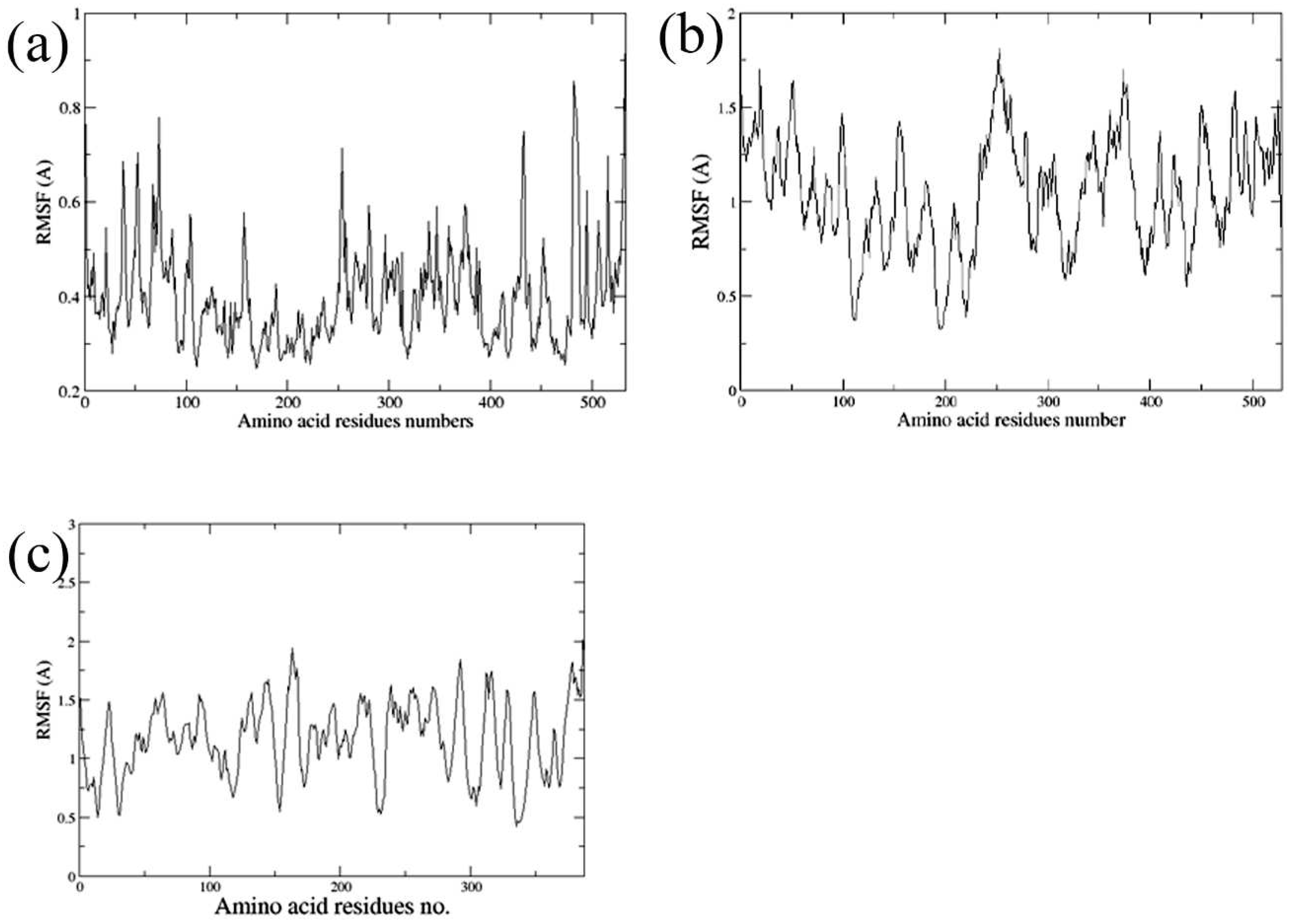
A root mean square fluctuation (RMSF) study can be used to determine which parts of the protein-ligand complex are more flexible. In Figure 5, the RMSF was shown through the final 4 ns. We looked at how flexibility evolved over MD simulations in order to pinpoint some significant trends for the protein-binder complex system. For the average flexibility of each residue in the macromolecule peptides, 5nuu-compound 22, 6i0b-compound 18, and 4ivt-compound 26, the RMSF at the C-α of each amino acid residue was taken into account and the areas with the greatest fluctuations during the simulation are shown by peaks.
The amino acids that has low values of rmsf are less mobile and stabilized by the hydrogen bond or hydrophobic interaction with the binder ligand. For tcAChE the residues are Tyr76, Val68, Asp69, Ser78, Trp81, Asn82, Tyr118, Ser119, Glu196, Trp276, Ile284, Phe287, Phe327, Phe328, Tyr331, Trp429, Ile436, His437, Tyr439. For hBChE the residues are Leu107, Ile108, Trp109, Ile110, Tyr111, Phe115, Gln116, Thr117, Glu194, Ser195, Ala196, Ala199, Tyr393, Asn394, Phe395, Ile396, Met435, His435, Tyr437, Glu438, Ile439. For BACE1 the residues are Gln12, Leu30, Tyr71, Thr72, Gln73, Lys107, Phe108, Ile110, Trp115, Thr231, Thr232, Asn233, Ser325, Gln326, Ser327, Thr329, Ile386,
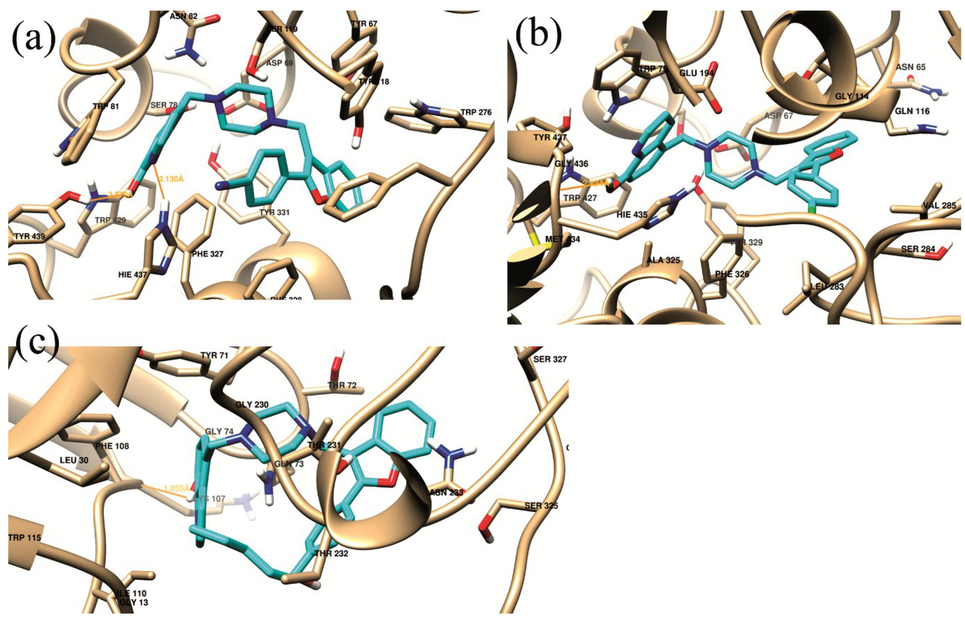
Figure 6. shows the average structure for the 5nnu-22, 6i0b-18 and 4ivt-26 generated from the last 4 ns trajectory data of the MD simulations process. The van der Waals interaction energy has a major contributions to the total binding more than the electrostatic contributions. This suggest the crucial role of the hydrophobic residues delineating the binding sites of tcAChE, hBChE and BACE1 protein structure.
Table 4 shows the MM/GBSA total binding energy of 5nuu-compoud 22 (-57.0944 kcal mol–1), 6i0b-compound 18 (-51.7731 kcal mol–1) and 4ivt-compound 26 (c) (-42.8924 kcal mol–1). In the three complex systems, 5nnu -22, 6i0b-18 and 4ivt-26, the ligands are anchored inside the binding site through the hydrogen bond of the ligands to Hie437, Hie435 and Lys107 respectively (Table 4). At the same time, the van der Waals interactions of the ligands with the hydrophobic side chains of the surrounding amino acid residues support the bound conformations as explored by MM/PBSA pairwise decomposition analysis (Table 5). The residues Hie437, Trp79 and Tyr71 have major contributions to the ligand protein interaction energies of 5nnu, 6i0b and 4ivt respectively.

3. Experiment and procedures

3.1. Chemistry

The starting materials and reagents were purchased from Sigma-Aldrich (St. Louis, MO, USA). Without any adjustments, the melting points were calculated using a Stuart Scientific electrothermal melting point equipment from Stone, Staffordshire, UK. Thin layer chromatography (TLC) was performed using precoated silica gel plates (0.25 mm, 60G F254, Merck, Darmstadt, Germany) and a chloroform/methanol (8:2) developing solution. The chemical structure spectrum study was finished at the research center of King Saud University College of Pharmacy. An FTIR spectrophotometer was used to capture IR spectra on KBr plates. Shelton, Connecticut, USA: Perkin Elmer. An NMR spectrophotometer (Bruker, Flawil, Switzerland) that operates at 500 MHz for 1H and 125.76 MHz for 13C was used to produce nuclear magnetic resonance (NMR) spectra. Mass spectra were collected using a model 320 MS spectrometer. USA (Varian, Lexington, KY). A Perkin Elmer model 2400 elemental analyser was used to examine the elements. The intermediate compounds 5-chloromethyl-8-hydroxyquinoline hydrochloride 3, 5-chloromethyl-8-hydroxyquinoline hydrochloride 5 and the final compounds 5-((4-((2-(4-substitutedphenyl)benzofuran-3-yl)methyl)piperazin-1-yl)methyl)quinolin-8-ol derivatives 15-21 were prepare as reported [22, 24].
5-((4-((2-(4-Ethylphenyl)benzofuran-3-yl)methyl)piperazin-1-yl)methyl)quinolin-8-ol (15): White solid, yield 85%, mp 100–101 °C [24].
5-((4-((2-(4-Methoxyphenyl)benzofuran-3-yl)methyl)piperazin-1-yl)methyl)quinolin-8-ol (16): White solid, yield 80%, mp 141–142 °C [24].
5-((4-((2-(4-Fluorophenyl)benzofuran-3-yl)methyl)piperazin-1-yl)methyl)quinolin-8-ol (17): White solid, yield 75%, mp 118–119 °C [24].
5-((4-((2-(4-Chlorophenyl)benzofuran-3-yl)methyl)piperazin-1-yl)methyl)quinolin-8-ol (18): White solid, yield 90%, mp 134–135 °C [24].
5-((4-((2-(4-Bromophenyl)benzofuran-3-yl)methyl)piperazin-1-yl)methyl)quinolin-8-ol (19): Beige solid, yield 70%, mp 115–116 °C [24].
5-((4-((2-p-Tolylbenzofuran-3-yl)methyl)piperazin-1-yl)methyl)quinolin-8-ol (20): White solid, yield 78%, mp 116–117 °C [24].
5-((4-((2-Biphenylbenzofuran-3-yl)methyl)piperazin-1-yl)methyl)quinolin-8-ol (21): White solid, yield 65%, mp 206–207 °C [24].

3.1.1. General synthesis of 2-(4-substitutedphenyl)benzofuran derivatives (8-14)

2-hydroxybenzyltriphenyl phosphonium bromide (12 g, 27 mmol), 4-substitutedbenzoylchloride (5 g, 29 mmol) in presence of triethylamine (11.1 mL, 80 mmol) were combined and refluxed for 10 hours in toluene (125 mL). The reaction mixture was concentrated under reduced pressure into greasy residue that received 120 mL of ethanol before being chilled overnight. The resulting solid underwent filtering, drying, and recrystallization from ethanol.
4-(Benzofuran-2-yl)benzonitrile (8); White solid, mp 205-6 °C 3048 (arom CH str), 2220 (CN str), 881-691 (arom CH bend). 1H NMR (700 MHz, CDCl3): δ = 7.19-8.21 (9H, m, ArH). 13CNMR (100 MHz, CDCl3): δ = 101.54, 111.43, 112.85, 115.97, 120.92, 122.95, 123.32, 124.76, 127.81, 132.84, 133.91, 154.87, 156.02. Ms m/z calcd: 219.07 [M]+, found: 219.48 [M]+. Anal. calcd. for C15H9NO: C, 82.18; H, 4.14; N, 6.39. Found: C, 82.32; H, 4.21; N, 6.26.
2-(4-(triFluoromethyl)phenyl)benzofuran (9); White solid, mp 156-7 °C 3044 (arom CH str), 799-706 (arom CH bend). 1H NMR (700 MHz, CDCl3): δ = 7.16-8.00 (9H, m, ArH). 13CNMR (100 MHz, CDCl3): δ = 102.05, 111.22, 118.11, 122.45, 123.92, 123.95, 124.55, 125.15, 128.12, 130.72, 134.05, 154.83, 156.65. Ms m/z calcd: 262.06 [M]+, found: 262.38 [M]+. Anal. calcd. for C15H9F3O: C, 68.70; H, 3.46. Found: C, 68.55; H, 3.62.
2-(4-Butylphenyl)benzofuran (10); White solid, mp 133-4 °C 3055 (arom CH str), 847-696 (arom CH bend). 1H NMR (700 MHz, CDCl3): δ = 0.93 (3H, t, CH3), 1.25-1.38 (2H, m, CH2), 1.60-1.65 (2H, m, CH2), 2.66 (2H, t, CH2), 6.72-7.69 (9H, m, ArH). 13CNMR (100 MHz, CDCl3): δ = 14.35, 21.32, 31.89, 36.47, 101.83, 110.46, 119.79, 122.95, 123.47, 123.95, 126.55, 127.15, 128.12, 137.22, 153.94, 155.38. Ms m/z calcd: 250.14 [M]+, found: 250.62 [M]+. Anal. calcd. for C18H18O: C, 86.36; H, 7.25. Found: C, 86.48; H, 7.16.
2-(4-tert-Butylphenyl)benzofuran (11); White solid, mp 118-9 °C 3042 (arom CH str), 799-706 (arom CH bend). 1H NMR (700 MHz, CDCl3): δ = 1.32 (9H, t, three CH3), 6.71-7.86 (9H, m, ArH). 13CNMR (100 MHz, CDCl3): δ = 31.68, 34.78, 100.69, 111.13, 120.77, 122.68, 123.47, 123.34, 124.56, 125.75, 127.74, 151.81, 154.83, 156.37. Ms m/z calcd: 250.14 [M]+, found: 250.39 [M]+. Anal. calcd. for C18H18O: C, 86.36; H, 7.25. Found: C, 86.24; H, 7.39.
2-(4-Pentylphenyl)benzofuran (12); White solid, mp 116-7 °C 3045 (arom CH str), 829-706 (arom CH bend). 1H NMR (700 MHz, CDCl3): δ = 0.85 (3H, t, CH3), 1.22-1.30 (4H, m, two CH2), 1.60-1.65 (2H, m, CH2), 2.66 (2H, t, CH2), 6.92-7.81 (9H, m, ArH). 13CNMR (100 MHz, CDCl3): δ = 14.38, 22.40, 30.72, 31.34, 35.52, 102.10, 111.47, 121.44, 122.12, 123.02, 125.13, 126.15, 127.15, 128.02, 138.04, 154.36, 156.65. Ms m/z calcd: 264.15 [M]+, found: 264.47 [M]+. Anal. calcd. for C19H20O: C, 86.32; H, 7.63. Found: C, 86.46; H, 7.52.
2-(4-Hexylphenyl)benzofuran (13); White solid, mp 114-5 C 3044 (arom CH str), 799-706 (arom CH bend). 1H NMR (700 MHz, CDCl3): δ = 0.89 (3H, t, CH3), 1.24-1.42 (6H, m, three CH2), 1.56-1.67 (2H, m, CH2), 2.58 (2H, t, CH2), 6.68-7.82 (9H, m, ArH). 13CNMR (100 MHz, CDCl3): δ = 14.23, 22.71, 28.83, 30.88, 31.12, 35.84, 102.25, 111.61, 121.01, 123.05, 123.48, 124.11, 127.04, 127.72, 128.11, 138.33, 155.01, 156.48 Ms m/z calcd: 278.17 [M]+, found: 278.59 [M]+. Anal. calcd. for C20H22O: C, 86.29; H, 7.97. Found: C, 86.15; H, 8.13.
2-(Thiophen-2-yl)benzofuran (14); White solid, mp 186-7 °C 3041 (arom CH str), 829-707 (arom CH bend). 1H NMR (700 MHz, CDCl3): δ = 6.72-7.70 (9H, m, ArH). 13CNMR (100 MHz, CDCl3): δ = 102.08, 111.22, 121.36, 123.11, 123.76, 124.05, 125.04, 126.13, 127.15, 132.73, 155.25, 156.11 Ms m/z calcd: 200.03 [M]+, found: 200.46 [M]+. Anal. calcd. for C12H8OS: C, 71.97; H, 4.03; S, 16.01. Found: C, 71.82; H, 3.91; S, 16.25.

3.1.2. General synthesis of 5-((4-((2-(4-substitutedphenyl)benzofuran-3-yl)methyl)piperazin-1-yl)methyl)quinolin-8-ol derivatives (22-28)

To a DMF (10 mL) solution of 2-(4-Substitutedphenyl)benzofuran (8-14) (10 mmol), formaline (37%, 1.55 mL) and 5-((piperazin-1-yl)methyl)quinolin-8-ol (10 mmol) were added and stirred at r.t. for 3 h. Excess water was added to the reaction mixture and allowed to 24 h in cold. The purified precipitate was obtained after filtering, water washing, and recrystallization from ethanol.
4-(3-((4-((8-Hydroxyquinolin-5-yl)methyl)piperazin-1-yl)methyl)benzofuran-2-yl)benzonitrile (22) White solid, yield 70%, mp 177-8 °C. IR (λmax, cm-1): 3403 (OH str), 3050 (arom CH str), 2930 (aliph CH str), 2222 (CN str), 887-710 (arom CH bend). 1H NMR (700 MHz, CDCl3): δ = 2.9 (4H, s, 2CH2), 2.97 (4H, s, 2CH2), 3.75 (2H, s, CH2), 3.76 (2H, s, CH2), 7.02-8.63 (13H, m, ArH). 13CNMR (100 MHz, DMSO-d6): δ = 41.05, 51.83, 52.01, 57.54, 110.74, 112.08, 113.11, 114.56, 117.39, 120.72, 121.33, 123.22, 123.88, 124.05, 125.91, 126.24, 128.12, 128.52, 132.02, 132.35, 133.95, 136.73, 148.56, 151.40, 151.64, 155.52. Ms m/z calcd: 474.21 [M]+, found 474.58 [M]+. Anal. calcd. for C30H26N4O2: C, 75.93; H, 5.52; N, 11.81. Found: C, 75.78; H, 5.41; N, 11.97.
5-((4-((2-(4-(triFluoromethyl)phenyl)benzofuran-3-yl)methyl)piperazin-1-yl)methyl)quinolin-8-ol (23) White solid, yield 65%, mp 133-4 °C. IR (λmax, cm-1): 3403 (OH str), 3048 (arom CH str), 2955 (aliph CH str), 837-660 (arom CH bend). 1H NMR (700 MHz, CDCl3): δ = 2.91 (4H, s, 2CH2), 2.98 (4H, s, 2CH2), 3.76 (2H, s, CH2), 3.78 (2H, s, CH2), 6.99-8.54 (13H, m, ArH). 13CNMR (100 MHz, DMSO-d6): δ = 41.72, 52.04, 52.42, 58.33, 111.12, 112.63, 113.05, 117.11, 120.81, 121.14, 123.45, 123.05, 124.66, 125.12, 126.01, 126.78, 127.34, 127.95, 130.38, 131.25, 132.73, 138.16, 149.41, 150.64, 151.48, 156.33. Ms m/z calcd: 517.2 [M]+, found 517.58 [M]+. Anal. calcd. for C30H26F3N3O2. C, 69.62; H, 5.06; N, 8.12. Found: C, 69.73; H, 4.91; N, 8.05.
5-((4-((2-(4-Butylphenyl)benzofuran-3-yl)methyl)piperazin-1-yl)methyl)quinolin-8-ol (24) White solid, yield 65%, mp 171-2 °C. IR (λmax, cm-1): 3358 (OH str), 3042 (arom CH str), 2917 (aliph CH str), 796-739 (arom CH bend). 1H NMR (700 MHz, CDCl3): δ = 0.98 (3H, t, CH3), 1.39-1.45 (2H, m, CH2), 1.63-1.69 (2H, m, CH2), 2.67-2.70 (10H,m, four piperazine-CH2 and -CH2-), 3.76 (2H, s, CH2), 3.79 (2H, s, CH2), 7.0-8.48 (13H, m, ArH). 13CNMR (100 MHz, DMSO-d6): δ = 14.21, 22.08, 32.78, 36.11, 41.33, 51.82, 52.62, 57.87, 110.59, 112.77, 117.05, 120.81, 121.53, 123.14, 124.03, 124.26, 125.66, 126.11, 127.01, 127.22, 128.34, 128.47, 130.92, 137.55, 138.48, 148.74, 151.41, 151.29, 154.08. Ms m/z calcd: 505.27 [M]+, found 505.78 [M]+. Anal. calcd. for C33H35N3O2: C, 78.38; H, 6.98; N, 8.31. Found: C, 78.22; H, 6.81; N, 8.45.
5-((4-((2-(4-tert-Butylphenyl)benzofuran-3-yl)methyl)piperazin-1-yl)methyl)quinolin-8-ol (25) White solid, yield 65%, mp 220-C. IR (λmax, cm-1): 3344 (OH str), 3094 (arom CH str), 2912 (aliph CH str), 794-602 (arom CH bend). 1H NMR (700 MHz, CDCl3): δ = 1.39 (9H, s, three CH3), 2.9 (4H, t, two CH2), 2.97 (4H, t, two CH2), 3.72 (2H, s, CH2), 3.81 (2H, s, CH2), 7.01-8.61 (13H, m, ArH). 13CNMR (100 MHz, DMSO-d6): δ = 31.15, 34.78, 36.50, 51.81, 53.57, 57.06, 111.13, 112.9, 117.3, 120.77, 121.16, 123.34, 124.0, 124.56, 125.32, 125.57, 125.89, 126.08, 127.74, 129.37, 131.32, 137.05, 138.48, 151.81, 154.83, 156.17, 156.37, 162.58. Ms m/z calcd: 505.27 [M]+, found 505.78 [M]+. Anal. calcd. for C33H35N3O2: C, 78.38; H, 6.98; N, 8.31. Found: C, 78.41; H, 7.05; N, 8.24.
5-((4-((2-(4-pentylphenyl)benzofuran-3-yl)methyl)piperazin-1-yl)methyl)quinolin-8-ol (26) White solid, yield 65%, mp 198-9 °C. IR (λmax, cm-1): 3361 (OH str), 3045 (arom CH str), 2933 (aliph CH str), 798-609 (arom CH bend). 1H NMR (700 MHz, DMSO-d6): δ = 0.85 (3H, t, CH3), 1.22-1.30 (4H, m, two CH2), 1.52-1.60 (3H, m, CH2), 2.55-2.68 (10H, m, four piperazine-CH2 and pentyl –CH2), 3.83 (2H, s, CH2), 4.29 (2H, s, CH2), 7.11-8.81 (13H, m, ArH). 13CNMR (100 MHz, DMSO-d6): δ = 19.01, 25.93, 31.0, 31.15, 37.0, 36.50, 49.05, 56.04, 57.06, 62.49, 110.71, 114.12, 114.91, 120.60, 121.47, 122.16, 127.21, 128.75, 130.03, 133.30, 134.89, 145.24, 153.57, 153.67, 162.01. Ms m/z calcd: 519.29 [M]+, found 519.72 [M]+. Anal. calcd. for C34H37N3O2: C, 78.58; H, 7.18; N, 8.09. Found: C, 78.42; H, 7.05; N, 8.16.
5-((4-((2-(4-hexylphenyl)benzofuran-3-yl)methyl)piperazin-1-yl)methyl)quinolin-8-ol (27) White solid, yield 65%, mp 186-7 °C. IR (λmax, cm-1): 3358 (OH str), 3077 (arom CH str), 2927 (aliph CH str), 823-745 (arom CH bend). 1H NMR (700 MHz, CDCl3): δ = 0.81 (3H, t, CH3), 1.24-1.29 (6H, m, three CH2), 1.53-1.59 (3H, m, CH2), 2.55-2.59 (2H, t, CH2), 2.80 (4H, t, two CH2), 2.88 (4H, t, two CH2), 3.66 (2H, s, CH2), 3.69 (2H, s, CH2), 6.90-8.78 (13H, m, ArH). 13CNMR (100 MHz, CDCl3): δ = 15.11, 21.45, 29.23, 31.5, 31.56, 36.61, 38.88, 51.05, 52.34, 58.04, 111.08, 113.27, 117.05, 120.84, 121.16, 122.78, 124.25, 124.95, 125.05, 126.44, 127.01, 127.15, 127.46, 128.11, 128.43, 130.85, 137.12, 138.55, 150.05, 151.12, 151.35, 154.28. Ms m/z calcd: 533.3 [M]+, found 533.69 [M]+. Anal. calcd. for C35H39N3O2: C, 78.77; H, 7.37; N, 7.87. Found: C, 78.64; H, 7.21; N, 7.93.
5-((4-((2-(Thiophen-2-yl)benzofuran-3-yl)methyl)piperazin-1-yl)methyl)quinolin-8-ol (28) White solid, yield 65%, mp 225-6 °C. IR (λmax, cm-1): 3361 (OH str), 3055 (arom CH str), 2928 (aliph CH str), 819-709 (arom CH bend). 1H NMR (700 MHz, CDCl3): δ = 2.9 (4H, t, two CH2), 2.97 (4H, t, two CH2), 3.51 (2H, s, CH2), 3.75 (2H, s, CH2), 6.89-8.87 (13H, m, ArH). 13CNMR (100 MHz, DMSO-d6): δ = 51.15, 52.38, 40.18, 57.77, 111.22, 112.56, 117.68, 121.22, 121.73, 123.16, 124.05, 124.71, 125.11, 126.57, 126.89, 127.08, 127.45, 129.15, 131.42, 134.05, 138.17, 149.81, 150.82, 151.22, 154.69. Ms m/z calcd: 455.17 [M]+, found 455.78 [M]+. Anal. calcd. for C27H25N3O2S: C, 71.18; H, 5.53; N, 9.22; S, 7.04. Found: C, 71.04; H, 5.42; N, 9.11; S, 7.18.

3.2. AChE/BChE screening experiment.

The screening kits for human acety/butyrylcholinesterase inhibitors (ab283363 and ab241010) were bought from abcam in the UK. Using a modified-Ellman’s approach, the inhibitory activity of compounds 15–28 against AChE and BuChE were assessed. Stock solutions of the compounds 15–28 (10 µM) were made in ethanol and then diluted further using 0.1 M KH2PO4/K2HPO4 buffer (pH 8.0), resulting in a final concentration of (0.01-10 µM). The enzyme powder was dissolved in distilled water to create enzyme stock solutions. The assay combination contained 100 mL of various doses of compounds 15–28 along with 1 mL of phosphate buffer (0.1 M, KH2PO4/K2HPO4), 25 mL of AChE (0.22 U/mL, E.C. 3.1.1.7), or 25 mL of BuChE (0.06 U/mL, E.C. 3.1.1.8). In brief, after 30 minutes of incubation at 37 °C, 20 μL of 10 mmol (acetylthiocholine/butylthiocholine), 10 μL of various chemical concentrations, 10 μL of 0.22 U AChE/BChE, 40 μL of 0.02 mol/phosphate buffer (pH 7.4), and 20 μL of 4% SDS were added to a 96-well microplate to halt the reaction. To create the colour, 100 μL of DTNB was then added.
As a positive control, donepzil (the reference medication) was applied in the same concentration range. 20 mL of the 0.075 M substrate solution (acetylthiocholine/butyrylthiocholine) were added to start the reaction. At 25 °C and 412 nm, the absorbance was measured 2 minutes after the substrate was introduced. In enzyme-free assay techniques, the non-enzymatic hydrolysis of acetylthiocholine/butyrylthiocholine iodide was also considered, and the outcome was utilized as a blank [27]. Then, after subtracting the corresponding background, the absorbances for AChE in the presence and absence of the inhibitors, respectively, were calculated using the formula (1 − Ai / Ac) 100. Each experiment was run in triplicate, and the findings are shown as the mean standard deviation. Whereas Ac denotes absorbance brought on by the intact cholinesterase solution in the absence of AChE inhibitors, Ai denotes absorbance supplied by the cholinesterase solution in the presence of AChE inhibitors.

3.3. BACE-1 Screening Experiment

The BACE1 Inhibitor Screening Kit (Fluorometric) Ab283408 was bought from abcam in the UK. In order to make buffers and standard solutions, purified water was used. Using 96-well plates, spectrofluorometric studies was carried out. A buffer solution of 50 mM sodium acetate and dimethyl sulfoxide was used to dissolve the compounds 15-28 to final concentrations of (0.01-10 µM). The test compounds were treated with 20 µL of BACE-1 (25 nM) for 60 minutes. To the well, was added 20 µL of substrate (0.25 M), and then incubate for 1 hour at 37 °C. The reaction is then slowed down by adding 20 µL of sodium acetate 2.5 M to each well. Then, using a blank devoid of BACE-1, a spectrofluorometric test at 590 nm will be conducted.

3.4. Molecular Docking

The Torpedo California AChE-tacrine complex (TcAChE) (PDB code: 5nnu.pdb), the human BChE complex with ligand (hBChE) (PDB code: 6i0b.pdb), and the complex of BACE1 with the ligand inhibitor (4ivt.pdb) were all X-ray crystallized structures. After removing all water molecules and unnecessary groups. Compound 15 with significant experimental inhibition activity against AChE, BChE and BACE1 enzymes was chosen for molecular docking investigations. The docking studies were performed by Autodock 4.2 software using the Lamarckian genetic method [25]. The nonpolar hydrogen atoms were combined using AutoDock tools and saved in pdbqt format and the polar hydrogen atoms and Kollman charges of the ligands and macromolecule structures were determined. During the docking procedure, the macromolecule structures in the grid box were kept stiff while the ligands were kept flexible. Grid points, 32 x, 32 y, and 32 z, with a spacing of 0.375 Å were defined based on the user-specified number of grid points. The top-ranked ten conformations were produced.

3.5. Molecular Dynamics Simulations

AmberTools22 program was utilized in performing the molecular dynamics simulations using the docked-compounds 22, 18 and 26 to TcAChE (PDB code: 5nnu.pdb), hBChE (PDB code: 6i0b.pdb), and BACE1 (4ivt.pdb) structures, respectively. The CPPTRAJ module was utilised in analysis of the MD simulations trajectories while the MM/GBSA and MM/PBSA-pairwise decomposition protocols were used to estimate binding free energy and the interaction energy of the macromolecule-residues to the ligands.

4. Conclusions

The work included the synthesis of hybrids of benzofuran and 8-hydroxyquinoline moieties that are linked through piperazine linker. Compounds 15-28 were suggested as potential multimodal anti-AD agents aimed to their superior privilege by their dual ChE (AChE/BChE) and BAC1 inhibitor activities suggesting its potential assignment in cognitive improvement. Compounds 15-28 showed inhibition values 33.4%-81.3% (ACHE), 54.58% - 94.47% (BChE) and 20.9% - 97.9% (BACE1). The experimental results were in accordance with the in silico modeling results and revealed the favorable characteristics of the compounds as promising candidates multi-target inhibitors that could be suggested for further development and studies as potential anti-AD agents. The simulation process declared that Hie437, Hie435 and Lys107 residues are crucial for anchoring and hydrogen bonding to the ligands inside the binding site of tcAChE (5nnu -22), hBChE (6i0b-18) and BACE1 (4ivt-26) respectively. Compounds 22, 18 and 26 are suggested as lead-compounds for further optimization and studies as inhibitors of tcAChE, hBChE and BACE1 inhibitors respectively.

Author Contributions

Conceptualization: Awwad Radwan, Fars Alanazi. Data curation: Awwad Radwan, Fars Alanazi. Formal analysis: Awwad Radwan, Fars Alanazi. Software: Awwad Radwan, Fars Alanazi. Project administration: Awwad Radwan, Fars Alanazi. Resources: Awwad Radwan. Fars Alanazi. Enzyme Kits assay and analysis of the resulting data, Anas M. Abdel Rahman. Writing –original draft, review & editing: Awwad Radwan, Fars Alanazi.

Funding

This research was funded by the Deanship of Scientific Research, King Saud University, for funding through Vice Deanship of Scientific Research Chairs, Kayyali Chair for Pharmaceutical Industry, Department of Pharmaceutics, College of Pharmacy, for funding the work through Grant Number AW-2023

Data Availability Statement

All the research data are included in the manuscript.

Acknowledgments

The authors extend their appreciation to the Deanship of Scientific Research, King Saud University, for funding through Vice Deanship of Scientific Research Chairs, Kayyali Chair for Pharmaceutical Industry, Department of Pharmaceutics, College of Pharmacy, for funding the work through Grant Number AW-2023.

Conflicts of Interest

The authors declare no conflict of interest.

References

  1. Yang, H.D.; Kim, D.H.; Lee, S.B.; Young, L.D. History of Alzheimer’s disease. Dement. Neurocogn. Disord. 2016, 15, 115–121. [Google Scholar] [CrossRef]
  2. Maramai, S.; Benchekroun, M.; Gabr, M.T.; Yahiaoui, S. Multitarget therapeutic strategies for Alzheimer’s disease: Review on emerging target combinations. BioMed. Res. Int. 2020, 5120230. [Google Scholar] [CrossRef]
  3. Iraji, A.; Khoshneviszadeh, M.; Firuzi, O.; Khoshneviszadeh, M.; Edraki, N. Novel small molecule therapeutic agents for Alzheimer disease: focusing on BACE1 and multi-target directed ligands. Bioorg. Chem. 2020, 97, 103649. [Google Scholar] [CrossRef]
  4. Najafi, Z.; Mahdavi, M.; Saeedi, M.; Karimpour-Razkenari, E.; Asatouri, R.; Vafadarnejad, F.; Moghadam, F.H.; Khanavi, M.; Sharifzadeh, M.; Akbarzadeh, T. Novel tacrine-1,2,3-triazole hybrids: in vitro, in vivo biological evaluation and docking study of cholinesterase inhibitors. Eur. J. Med. Chem. 2017, 125, 1200–1212. [Google Scholar] [CrossRef]
  5. Breijyeh, Z.; Karaman, R. Comprehensive Review on Alzheimer’s Disease: Causes and Treatment. Molecules 2020, 25, 5789–5817. [Google Scholar] [CrossRef]
  6. Vafadarnejad, F.; Karimpour-Razkenari, E.; Sameem, B.; Saeedi, M.; Firuzi, O.; Edraki, N.; Mahdavi, M.; Akbarzadeh, T. Novel N-benzylpyridinium moiety linked to arylisoxazole derivatives as selective butyrylcholinesterase inhibitors: synthesis, biological evaluation, and docking study. Bioorg. Chem. 2019, 92, 103192. [Google Scholar] [CrossRef]
  7. Campanari, M.-L.; Navarrete, F.; Ginsberg, S.D.; Manzanares, J.; Sáez-Valero, J. , García-Ayllón, M.-S. Increased expression of readthrough acetylcholinesterase variants in the brains of Alzheimer’s disease patients. J. Alzheimers Dis 2016, 53, 831–841. [Google Scholar] [CrossRef]
  8. Darvesh, S.; Hopkins, D.A.; Geula, C. Neurobiology of butyrylcholinesterase. Nat. Rev. Neurosci. 2003, 4, 131–138. [Google Scholar] [CrossRef]
  9. Perry, G.; Cash, A.D.; Smith, M.A. Alzheimer Disease and Oxidative Stress. J. Biomed. Biotechnol. 2002, 2, 2,120–123. [Google Scholar] [CrossRef]
  10. Tolnay, M.; Probst, A. REVIEW: tau protein pathology in Alzheimer’s disease and related disorders. Neuropathol. Appl. Neurobiol. 1999, 25, 171–187. [Google Scholar] [CrossRef]
  11. Tripathi, M. K.; Sharma, P.; Tripathi, A.; Tripathi, P.N.; Srivastava, P.; Seth, A.; Shrivastava, S. K. Computational exploration and experimental validation to identify a dual inhibitor of cholinesterase and amyloid-beta for the treatment of Alzheimer’s disease. J. Comput. Aided. Mol. Des. 2020, 34, 983–1002. [Google Scholar] [CrossRef] [PubMed]
  12. Sharma, P.; Srivastava, P.; Seth, A.; Tripathi, P. N.; Banerjee, A. G.; Shrivastava, S. K. Comprehensive review of mechanisms of pathogenesis involved in Alzheimer’s disease and potential therapeutic strategies. Prog. Neurobiol. 2019, 174, 53–89. [Google Scholar] [CrossRef] [PubMed]
  13. Zatta, P.; Drago, D.; Bolognin, S.; Sensi, S. L. Alzheimer’s disease, metal ions and metal homeostatic therapy. Trends Pharmacol. Sci. 2009, 30, 346–355. [Google Scholar] [CrossRef] [PubMed]
  14. Birks, J. Cholinesterase inhibitors for Alzheimer’s disease. Cochrane Database Syst. Rev. 2006, 25, CD005593. [Google Scholar] [CrossRef] [PubMed]
  15. Geula, C.; Darvesh, S. Butyrylcholinesterase, cholinergic neurotransmission and the pathology of Alzheimer’s disease. Drugs Today (Barc) 2004, 40, 711–721. [Google Scholar] [CrossRef] [PubMed]
  16. Ibrahim, M. M.; Gabr, M. T. Multitarget therapeutic strategies for Alzheimer’s disease. Neural Regen. Res. 2019, 14, 437–440. [Google Scholar] [CrossRef] [PubMed]
  17. Belluti, F.; Rampa, A.; Piazzi, L.; Bisi, A.; Gobbi, S.; Bartolini, M.; Andrisano, V.; Cavalli, A.; Recanatini, M.; Valenti, P. Cholinesterase inhibitors: xanthostigmine derivatives blocking the acetylcholinesterase-induced beta-amyloid aggregation. J. med. Chem. 2005, 48, 4444–4456. [Google Scholar] [CrossRef] [PubMed]
  18. Rizzo, S.; Cavalli, A.; Ceccarini, L.; Bartolini, M.; Belluti, F.; Bisi, A.; Andrisano, V.; Recanatini, M.; Rampa, A. Structure-activity relationships and binding mode in the human acetylcholinesterase active site of pseudo-irreversible inhibitors related to xanthostigmine. ChemMedChem 2009, 4, 670–679. [Google Scholar] [CrossRef] [PubMed]
  19. Kozurkova, M. ; Hamulakova, S, Gazova Z, Paulikova H, Kristian P. Neuroactive Multifunctional Tacrine Congeners with Cholinesterase, Anti-Amyloid aggregation and neuroprotective properties. Pharmaceuticals (Basel). [CrossRef]
  20. Youdim, M.B. Multi target neuroprotective and neurorestorative anti-Parkinson and anti-Alzheimer drugs ladostigil and m30 derived from rasagiline. Exp Neurobiol. 2013, 22, 1–10. [Google Scholar] [CrossRef]
  21. 21. Amit, T; Avramovich-Tirosh, Y.; Youdim, M.B.; Mandel, S. Targeting multiple Alzheimer’s disease etiologies with multimodal neuroprotective and neurorestorative iron chelators. FASEB J. 2008, 22, 1296–1305. [CrossRef]
  22. Saadeh, H.A.; Sweidan, K.A.; Mubarak, M.S. Recent advances in the synthesis and biological activity of 8-hydroxyquinolines. Molecules 2020, 25, 4321. [Google Scholar] [CrossRef] [PubMed]
  23. Saadeh, H.A.; Sweidan, K.A.; Mubarak, M.S. Recent advances in the synthesis and biological activity of 8-hydroxyquinolines. Molecules 2020, 25, 4321. [Google Scholar] [CrossRef] [PubMed]
  24. Radwan, A.A.; Alanazi, F.K.; Raish, M. Design and synthesis of multi-functional smallmolecule based inhibitors of amyloid-β aggregation: Molecular modeling and in vitro evaluation. PLoS ONE 2023, 18, e0286195. [Google Scholar] [CrossRef]
  25. Battisti, A.; Piccionello, A.P.; Sgarbossa, A.; Vilasi, S.; Ricci, C.; Ghetti, F, et al. Curcumin-like compounds designed to modify amyloid beta peptide aggregation patterns. RSC Advances 2017; 7:31714–31724. [CrossRef]
  26. Earl, C.; Bagneris, C.; Zeman, K.; Cole, A.; Barrett, T.; Savva, R. Nucleic Acids Res. 2018, 46, 4286-4300. [CrossRef]
  27. Chalupova, K.; Korabecny, J.; Bartolini, M.; Monti, B.; Lamba, D.; Caliandro, R.; et al. Novel tacrine-tryptophan hybrids: Multi-target directed ligands as potential treatment for Alzheimer’s disease. Eur. J. Med. Chem, 2019. [Google Scholar] [CrossRef]
  28. Zou, Y.; Li, L.; Chen, W.Y.; Chen, T.T.; Ma, L.; Wang, X.; Xiong, B.; Xu, Y.C.; Shen, J. Virtual screening and structure-based discovery of indole acylguanidines as potent beta-secretase (BACE1) inhibitors. Molecules 2013, 18, 5706–5722. [Google Scholar] [CrossRef]
  29. Case, D.A.; Aktulga, H.M.; Belfon, K.; Ben-Shalom, I.Y.; Berryman, J.T., Brozell, S.R., et al., Amber 2023, University of California, San Francisco.
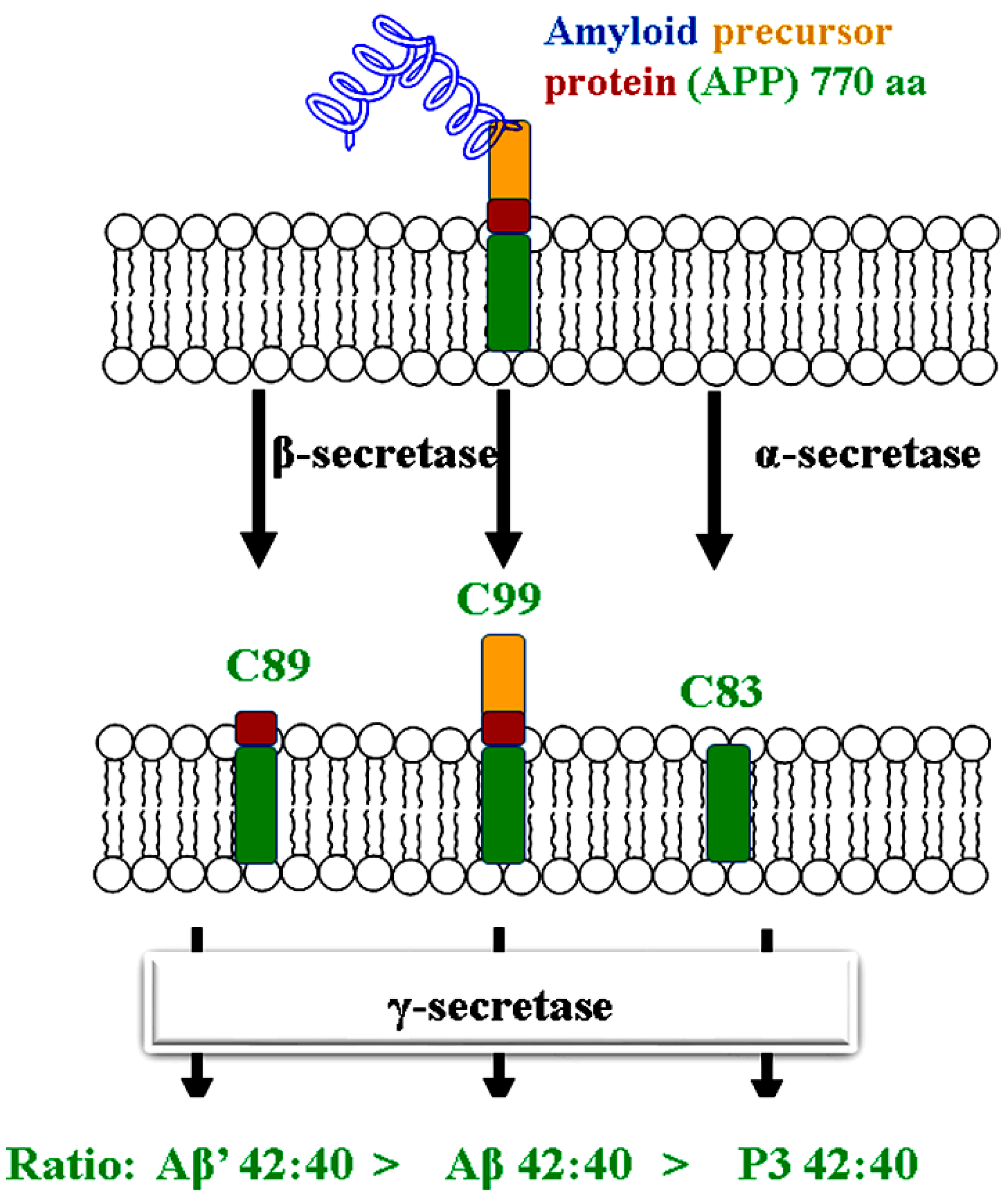
Figure 1. The biological development of β-amyloid protein AB1-42.
Preprints 98786 g001
Figure 2. Representative compounds as multi-target anti-AD agents.
Preprints 98786 g002
Figure 3. Schematic representation of the designed compounds.
Preprints 98786 g003
Figure 4. Compound 15 to the binding site of tcAChE (5nnu.pdb) (a), hBChE structure (6i0b.pdb) (b) and binding site of BACE1 structure (4ivt.pdb) (c).
Preprints 98786 g004
Figure 5. The RMSF chart of amino acid residues of 5nuu-compoud 22 (a), 6i0b-compound 18 (b) and 4ivt-compound 26 (c) complexes during simulation process.
Preprints 98786 g005
Figure 6. The averaged 5nuu-compoud 22 (a), 6i0b-compound 18 (b) and 4ivt-compound 26 (c)-structures of tcAChE, hBChE and BACE1 complexes. The hydrogen bonds (orange solid lines); Amino acids (beige-colored sticks); The ligand (a cyan-colored stick).
Preprints 98786 g006
Table 1. Experimental and predicted AChE/BChE inhibition percent and docking scores of (10 µmol) compounds 15-28.
No. Binding Energy Kcalmol-1 Inhibition constant (Ki nmol) Experimental
inhibition %
AChE hBChE AChE hBChE AChE BChE
15 -14.83 -12.36 0.013 0.87 64.9 75.69
16 -15.32 -10.92 0.006 9.89 71.3 65.88
17 -14.37 -12.36 0.03 0. 87 58.2 91.53
18 -15.63 -12.97 0.004 0.31 79.9 94.47
19 -14.85 -12.64 0.013 0.54 64.1 86.65
20 -15.16 -11.82 0.008 2.17 68.5 74.35
21 -10.42 -11.61 23.1 3.07 50.8 72.7
22 -11.9 -11.03 1.89 8.22 81.3 84.06
23 -11.98 -10.91 1.66 10.03 69.6 54.58
24 -14.42 -11.88 0.027 1.96 74.4 80.77
25 -11.24 -13.79 5.78 0.08 74.4 78.73
26 -11.4 -12.23 4.38 1.09 47.8 74.95
27 -9.92 -11.78 53.13 2.34 60.8 79.18
28 -10.97 -12.75 9.07 0.45 33.8 67.57
Xray -14.22 -11.38 0.038 4.55 -------------- -------------
Table 4. The MM/GBSA binding free energy (kcal mol–1) of 5nuu-compoud 22 (a), 6i0b-compound 18 (b) and 4ivt-compound 26 (c) of tcAChE, hBChE and BACE1 complexes.
Complex Hb-residue (Frac %) ΔGvdw ΔGelec ΔGpolara ΔGSurf b ΔGMM/GBSA
(a) HIE_437@HE2(16.25%)
SER_197@HG (11.5%)
-67.9357 -18.8145 36.9524 -7.2966 -57.0944
(b) HIE_435@O (42.25%) -60.6058 -8.3630 23.8192 -6.6235 -51.7731
(c) THR_231@HB (10.25%)
THR_232@H (9.5%)
LYS_107@O (33.75%)
-49.274 -6.0429 18.5027 -6.0780 -42.8924
a ΔGelectrostatic + ΔGpolar are the whole electorstatic contribution. b ΔGnp = ΔGvdw + ΔGsurf are the whole non polar contribution.
Table 5. The results of MM/PBSA-pairwise-residue decomposition analysis of 5nuu-compoud 22 (a), 6i0b-compound 18 (b) and 4ivt-compound 26 (c) complexes.
Complex Residue Number
Residue interaction energy kcalmol-1
(a) Asp69
-2.425
Ser78
-2.294
Trp81
-3.357
Asn82
-1.732
Tyr118
-2.301
Phe327
-2.227
Phe328
-2.342
Tyr331
-3.312
Hie437
-4.539
Tyr439
-1.749
(b) Asp67
-1.703
Trp79
-3.81
Gln116
-2.185
Thr117
-1.58
Glu194
-1.538
Ala325
-1.947
Phe326
-1.613
Tyr329
-1.768
Met434
-1.21
Hie435
-2.594
(c) Gln12
-1.046
Tyr71
-3.218
Thr72
-1.382
Gln73
-5.179
Lys107
-1.412
Phe108
-2.287
Ile110
-0.864
Gly230
-1.344
Thr231
-2.098
Thr232
-1.408
Disclaimer/Publisher’s Note: The statements, opinions and data contained in all publications are solely those of the individual author(s) and contributor(s) and not of MDPI and/or the editor(s). MDPI and/or the editor(s) disclaim responsibility for any injury to people or property resulting from any ideas, methods, instructions or products referred to in the content.
Copyright: This open access article is published under a Creative Commons CC BY 4.0 license, which permit the free download, distribution, and reuse, provided that the author and preprint are cited in any reuse.
Prerpints.org logo

Preprints.org is a free preprint server supported by MDPI in Basel, Switzerland.

Subscribe

© 2024 MDPI (Basel, Switzerland) unless otherwise stated