Preprint
Article

Pseudo-Polymorphism in 2-Pyridylmethoxy Cone Derivatives of p-tert-butylcalix[4]arene and P-Tert-Butylhomooxacalix[n]arenes

Altmetrics

Downloads

82

Views

34

Comments

0

Submitted:

15 February 2024

Posted:

16 February 2024

You are already at the latest version

Alerts
Abstract
This paper investigates pseudo-polymorphism in 2-pyridylmethoxy derivatives of p-tert-butylcalix[4]arene (PyC4), p-tert-butyldihomooxa-calix[4]arenes (PyHOC4), and p-tert-butylhexahomotrioxacalix[3]arenes (PyHO3C3), presenting 11 crystal structures with 15 crystallographically independent molecules. The macrocycle of PyC4 is smaller and less flexible with respect to those of PyHOC4 and PyHO3C3 and in solution, the cone conformation of these three molecules exhibit different point symmetries: C4, Cs and C3, respectively. A correlation is observed between the macrocycle's structural rigidity and the number of pseudo-polymorphs formed. The more rigid PyC4 displays a higher number (6) of pseudo-polymorphs compared to PyHOC4 and PyHO3C3, which exhibit a smaller number of crystalline forms (3 and 2, respectively). The X-ray structures obtained show that the conformation of the macrorings is primarily influenced by the presence of an acetonitrile guest molecule within the cavity, with limited impact from crystal packing and intermolecular cocrystallized solvent molecules. Notably, both calix[4]arene derivatives produce host-guest complex with acetonitrile, while the most flexible and less aromatic PyHO3C3 do not give crystals with acetonitrile as guest. Intertwined 1D and 2D solvent channel networks were observed in the PyHOC4-hexane and in the PyHO3C3-H2O-MeOH crystal structures, respectively, while the other pseudopolymorphs of PyHOC4 and PyHO3C3 and all PyC4 crystal forms exhibit a closely packed crystal structures without open channels.
Keywords: 
Subject: Chemistry and Materials Science  -   Materials Science and Technology

1. Introduction

In the last three decades, the field of calixarenes-based host-guest chemistry has garnered significant scientific interest due to its diverse applications in the domain of supramolecular chemistry, including sensor chemistry, ion receptors and coordination chemistry [1,2,3,4,5,6,7]. Research efforts have particularly focused on the interaction between calixarenes, especially those with oxygen donor atoms on the lower rim, and metal cations, predominantly alkali and alkaline earths [4,8,9]. Moreover, calixarenes functionalized with both N-donor and O-donor atoms have been investigated for their effectiveness in complexing transition metals [10,11], heavy metals [11,12] and lanthanide cations [8,13]. Further enhancements to calixarene properties have been explored through modifications involving the incorporation of 2-pyridyl-methyl pendant groups on the lower rim. This exploration has resulted in the synthesis of p-tert-butyl-tetrakis(2-pyridylmethyl)oxycalix[4]arene (PyC4) [14] (Figure 1). The cone conformation of this calixarene shows complexation properties towards Na+ [11,15], Ag+ [11,16] and lantanide ions [17].
Additionally, homooxacalixarenes [18], calixarene analogues in which one or more CH2 bridges are replaced by CH2OCH2 groups, were investigated. Notable among these derivatives, dihomooxacalix[4]arene with one CH2OCH2 bridge (PyHOC4) and hexahomotrioxacalix[3]arene with three CH2OCH2 bridges (PyHO3C3) (Figure 1), are attracting attention for their higher conformational flexibility compared to calix[4]arene analogues [17,19]. In fact, the comparison shows that the C4 macrocycle is a 16 membered ring (8 aromatic and 8 single bonds), the HOC4 macrocycle is an 18 membered ring (8 aromatic and 10 single bonds) and the HO3C3 macrocycle is also an 18 membered ring which includes more single bonds (6 aromatic and 12 single bonds). However, despite possessing slightly larger cavity sizes than calix[4]arenes, both HOC4 and HO3C3 can assume blocked cone conformations when functionalized with bulky groups on the lower rim, such as in PyHOC4 and in PyHO3C3. Furthermore, in the cone conformation, these three molecules exhibit different point symmetries of the macrocycle: PyC4 has a C4 symmetry, PyHOC4 has Cs symmetry, while PyHO3C3 has C3 symmetry (as shown in Figure 1).
In order to further investigate the conformational and binding properties of these macrocycles towards rare earth ions [17], we conducted a systematic structural study by single crystal x-ray diffraction on crystals obtained under various crystallization conditions for all three calixarenes. In particular, during this study, we obtained six pseudo-polymorphs of PyC4, three pseudo-polymorphs of PyHOC4 and two pseudo-polymorphs of PyHO3C3, using analogous crystallization conditions for all three. In the present paper we describe and compare the accurate crystal structures of these macrocycles obtained using state-of-the-art x-ray diffraction, employing cryo-techniques and synchrotron radiation.

2. EXPERIMENTAL

The calixarenes studied in this work were synthesized according to the literature: PyC4 [14], PyHOC4 [20], PyHO3C3 [21,22].
As a general procedure, single crystals of calixarenes were obtained through the slow evaporation of solvents. Near-saturated solutions of the macrocycles were prepared in suitable solvents. The vials, covered with perforated caps, were placed in a crystallization room at 18 °C to evaporate. The rate of evaporation is an experimental variable that depends, to some extent, on the volatility of the solvent. Crystals can grow within hours to days by selecting an appropriate solvent or mixture of solvents, depending on the boiling point, temperature, and, consequently, the evaporation rate.
The single crystal X-ray diffraction data collection was conducted at the XRD1 beamline of the Elettra synchrotron in Trieste, Italy. The rotating-crystal method was employed, utilizing a Dectris Pilatus 2M area detector and monochromatic radiation with a wavelength of 0.700 Å. Single crystals were dipped in paratone cryoprotectant, mounted on a nylon loop and flash-frozen under a nitrogen stream at 100 K. Diffraction data were indexed and integrated using the XDS package [23], while scaling was carried out with XSCALE [24]. Structures were solved using the SHELXT program [25] and the refinement was performed with SHELXL [26] by full-matrix least-squares (FMLS) method on F2.
For the refinement, non-hydrogen atoms were anisotropically refined with exception of some disordered groups having a low occupancy factor, which were refined isotropically. Hydrogen atoms, located on the difference Fourier maps, were added at the calculated positions and refined using the riding model.
Crystal Data for PyC4-MeOH-α: C68H76O4N4·0.2CH3OH (M = 1019.73 g/mol), monoclinic, space group P21/c (no. 14), a = 12.004(4) Å, b = 12.090(8) Å, c = 40.691(5) Å, β = 95.857(13) °, V = 5875(2) Å3, Z = 4, μ = 0.068 mm-1, Dcalc = 1.153 g/cm3, 92155 reflections measured (2.0° ≤ 2Θ ≤ 59.2°), 16083 unique (Rint = 0.0292, Rsigma = 0.0475) which were used in all calculations. The final R1 was 0.0616 (I > 2σ(I)) and wR2 was 0.1664 (all data).
Crystal Data for PyC4-H2O-α: C68H76O4N4·0.4H2O (M = 1020.53 g/mol), monoclinic, space group P21/c (no. 14), a = 12.005(5) Å, b = 12.121(1) Å, c = 40.565(2) Å, β = 95.98(2) °, V = 5871(2) Å3, Z = 4, μ = 0.069 mm-1, Dcalc = 1.155 g/cm3, 108841 reflections measured (3.4° ≤ 2Θ ≤ 59.2°), 15795 unique (Rint = 0.0232, Rsigma = 0.0424) which were used in all calculations. The final R1 was 0.0444 (I > 2σ(I)) and wR2 was 0.1238 (all data).
Crystal Data for PyC4-MeOH-β: C68H76O4N4·0.75CH3OH (M = 1037.35 g/mol), triclinic, space group P-1(no. 2), a = 10.410(4) Å, b = 23.014(9) Å, c = 24.874(10) Å, α= 95.813(8) °, β= 90.17(3) °, γ= 97.56(3) °, V = 5876(4) Å3, Z = 4, μ = 0.070 mm-1, Dcalc = 1.173 g/cm3, 26719 reflections measured (2.2° ≤ 2Θ ≤ 43.2°), 14199 unique (Rint = 0.0920) which were used in all calculations. The final R1 was 0.0966 (I > 2σ(I)) and wR2 was 0.2930 (all data).
Crystal Data for PyC4⸦MeCN-MeOH: C68H76O4N4·CH3CN·CH3OH (M = 1086.42 g/mol), monoclinic, space group P21 (no. 4), a = 13.936(2) Å, b = 15.260(1) Å, c = 14.125(2) Å, β = 92.794(19) °, V = 3000.3(6) Å3, Z = 2, μ = 0.072 mm-1, Dcalc = 1.203 g/cm3, 53801 reflections measured (2.8° ≤ 2Θ ≤ 59.2°), 16697 unique (Rint = 0.0392, Rsigma = 0.0418) which were used in all calculations. The final R1 was 0.0407 (I > 2σ(I)) and wR2 was 0.1131 (all data).
Crystal Data for PyC4⸦MeCN-H2O: C68H76O4N4·CH3CN·0.2H2O (M = 1057.98 g/mol), triclinic, space group P-1 (no. 2), a = 13.239(2) Å, b = 14.358(1) Å, c = 18.297(2) Å, α = 95.419(8) °, β = 100.656(11) °, γ = 117.079(9) °, V = 2979.3(6) Å3, Z = 2, μ = 0.070 mm-1, Dcalc = 1.179 g/cm3, 56137 reflections measured (2.2° ≤ 2Θ ≤ 59.2°), 15954 unique (Rint = 0.0197, Rsigma = 0.0233) which were used in all calculations. The final R1 was 0.0479 (I > 2σ(I)) and wR2 was 0.1333 (all data).
Crystal Data for PyC4: C68H76O4N4 (M = 1013.32 g/mol), monoclinic, space group P21/n (no. 14), a = 15.167(1) Å, b = 19.994(6) Å, c = 20.267(6) Å, β = 108.878(12) °, V = 5815(4) Å3, Z = 4, μ = 0.068 mm-1, Dcalc = 1.157 g/cm3, 106301 reflections measured (3.0° ≤ 2Θ ≤ 59.2°), 16910 unique (Rint = 0.0271, Rsigma = 0.0466) which were used in all calculations. The final R1 was 0.0585 (I > 2σ(I)) and wR2 was 0.1666 (all data).
Crystal Data for PyHOC4-DMSO: C69H78O5N4·0.8625C2H6SO (M = 1109.22 g/mol), triclinic, space group P-1 (no. 2), a = 20.98(2) Å, b = 22.476(18) Å, c = 28.52(3) Å, α = 72.25(2) °, β = 81.425(15) °, γ = 89.765(16) °, V = 12655(22) Å3, Z = 8, μ = 0.098 mm-1, Dcalc = 1.164 g/cm3, 101611 reflections measured (2.0° ≤ 2Θ ≤ 43.2°), 29067 unique (Rint = 0.0838, Rsigma = 0.0985) which were used in all calculations. The final R1 was 0.1659 (I > 2σ(I)) and wR2 was 0.5018 (all data).
Crystal Data for PyHOC4⸦MeCN-MeOH: C69H78O5N4·CH3CN·0.8CH3OH (M = 1110.04 g/mol), triclinic, space group P-1 (no. 2), a = 13.969(11) Å, b = 14.823(8) Å, c = 15.292(5) Å, α = 84.06(3) °, β = 83.01(4) °, γ = 87.134(16) °, V = 3124(3) Å3, Z = 2, μ = 0.071 mm-1, Dcalc = 1.180 g/cm3, 20567 reflections measured (2.6° ≤ 2Θ ≤ 41.0°), 6399 unique (Rint = 0.0543, Rsigma = 0.0605) which were used in all calculations. The final R1 was 0.1326 (I > 2σ(I)) and wR2 was 0.4251 (all data).
Crystal Data for PyHOC4-Hexane: C69H78O5N4·1.5C6H14 (M = 1172.61 g/mol), triclinic, space group P-1 (no. 2), a = 11.686(5) Å, b = 16.604(2) Å, c = 17.957(3) Å, α = 88.990(3) °, β = 86.252(5) °, γ = 77.253(5) °, V = 3391(2) Å3, Z = 2, μ = 0.068 mm-1, Dcalc = 1.148 g/cm3, 123862 reflections measured (2.2° ≤ 2Θ ≤ 59.6°), 19724 unique (Rint =0.0260, Rsigma = 0.0448) which were used in all calculations. The final R1 was 0.0643 (I > 2σ(I)) and wR2 was 0.1891 (all data).
Crystal Data for PyHO3C3: C54H63O6N3 (M = 850.07 g/mol), monoclinic, space group P21/c (no. 14), a = 24.022(3) Å, b = 10.7210(14) Å, c = 19.415(5) Å, β = 111.667(7) °, V = 4647(1) Å3, Z = 4, μ = 0.075 mm-1, Dcalc = 1.215 g/cm3, 85480 reflections measured (1.8° ≤ 2Θ ≤ 59.2°), 13548 unique (Rint = 0.0359, Rsigma = 0.0585) which were used in all calculations. The final R1 was 0.0883 (I > 2σ(I)) and wR2 was 0.2571 (all data).
Crystal Data for PyHO3C3-H2O-MeOH: C54H63O6N3·4H2O·CH3OH (M = 954.18 g/mol), monoclinic, space group P21/c (no. 14), a = 19.430(19) Å, b = 14.376(3) Å, c = 19.605(8) Å, β = 106.84(5) °, V = 5241(6) Å3, Z = 4, μ = 0.080 mm-1, Dcalc = 1.209 g/cm3, 96321 reflections measured (2.1° ≤ 2Θ ≤ 59.2°), 15397 unique (Rint = 0.0421, Rsigma = 0.0710) which were used in all calculations. The final R1 was 0.0567 (I > 2σ(I)) and wR2 was 0.1605 (all data).

3. RESULTS and DISCUSSION

3.1. PyC4 – Calix[4]arene

Three different crystal structures of PyC4 have already been reported in the literature. The first structure was documented by Pappalardo et al [14]. The diffraction data of this monoclinic crystal form, obtained from a methanol solution, was collected at 21°C. The structure reveals a co-crystalized solvent molecule with 0.6 occupancy, hydrogen-bonded to a pyridyl N atom orientated away from the cavity of the calixarene, which assumes a pinched cone conformation [14]. The other two crystal structures are two distinct tetragonal forms (P4cc and P4/n space groups) of PyC4 complexes with Na+ [15] and Ag+ [16] ions, respectively, both reported by Danil de Namor et al. In these structures, the calixarene is situated on a fourfold symmetry axis, with the hydrophobic cavity hosting an acetonitrile molecule. At the lower rim, the hydrophilic cavity encapsulates the Ag+ and Na+ ions through the ether oxygen and pyridyl nitrogen atoms. A distorted Archimedean square antiprism coordination of the cations is observed for both complexes. The role of metal ion complexation on the lower rim in preorganization of the calix cup, thereby allowing the complexation of acetonitrile guest in calix[4]arene-tetrol derivatives, has been recently reported. This results in formation of crystals with molecular cavities. On the other hand, different solvents, such as acetonitrile or water, can be used to design Supramolecular Organic Frameworks (SOF) with large hydrophobic or hydrophilic channels, respectively [27]. The different solubility of PyC4 in various solvents [15] plays an important role in the crystallization process and in the formation of different polymorphs or pseudo-polymorphic solvate crystals containing different co-crystalized solvent molecules.
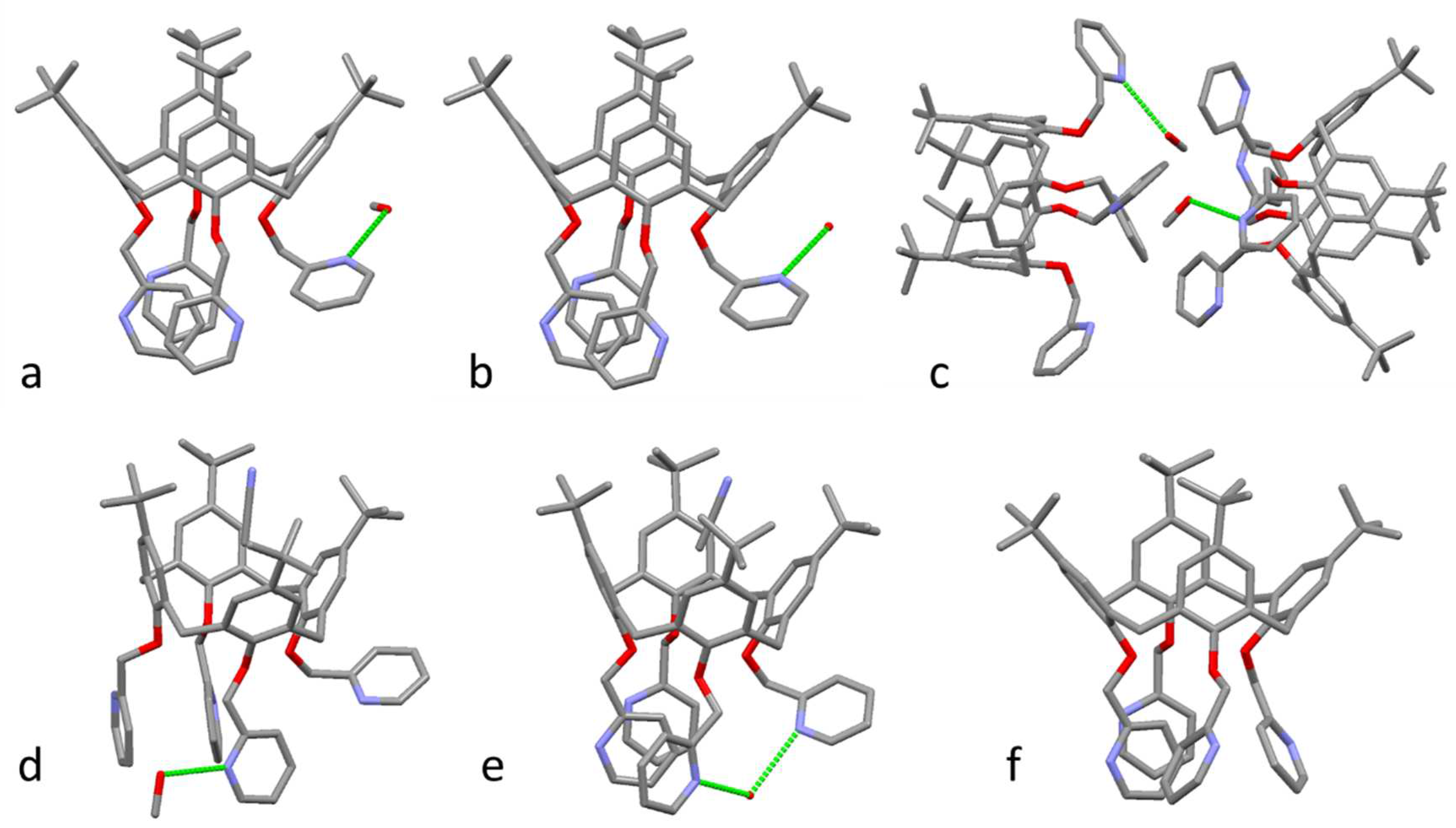
A summary of the crystallization conditions used in the various trials conducted is shown in Table S1. The asymmetric units of the six pseudo-polymorphic structures of PyC4, including the co-crystallized solvent molecules, are shown in Figure 2. The crystal data and the structure refinements of the six pseudo-polymorphs, four monoclinic and two triclinic, of PyC4 are summarized in Table S2.
The asymmetric unit of the monoclinic crystals of PyC4-MeOH-α consist of one molecule of calixarene and co-crystallized methanol molecules disordered in two positions each with 0.1 occupancy. The solvent molecules are involved in H-bonds with a pyridyl N-acceptor of the arm oriented away from the cavity of the calixarene (Figure 2a). The other three pyridyl groups are almost parallel and stack together (Figure 2a). This structure is analogous to the structure previously reported from room temperature data collection [14]. The high quality of diffraction data collected at low temperature has permitted us to observe a disorder in the outward oriented pyridyl group involved in the H-bond with the methanol molecule with partial occupancy. The presence in the Fourier difference maps of electron density attributable to half hydrogen atoms attached to both orto positions (with respect to the methylene group) and the thermal factors of the corresponding N/C atoms, indicate two overlapped orientations of the pyridyl group rotated by 180°. A second difference in comparison with the published structure is related to the disorder of the co-crystalized methanol molecules and their slightly difference in orientation. The D-A H-bond distances are 2.97 and 2.92 Å for our low temperature structure, and 2.81 Å for the room temperature structure. The expected and observed decrease in the cell volume at low temperature (3.8%) is largely due to the decrease of the b axis (3.6%).
The second crystal of PyC4 is an isomorphic solvate of PyC4-MeOH-α which differs only in the nature of the co-crystalized solvent molecules (Figure 2b). In this PyC4-H2O-α structure, the methanol site is occupied by a water molecule, split in two positions with occupancy of 0.1 and 0.3. The origin of the water molecules can be attributed to the hydrate salt used in the crystallization trials (see SI). These co-crystallized water molecules form H-bonds with the nitrogen atoms of the outward orientated pyridyl rings (D-A distances of 2.96 and 2.86 Å, Figure 2b). The pinched cone conformation and the disorder of the pyridyl arm (0.5/0.5) is very similar to that observed in PyC4-MeOH-α.
The third structure, PyC4-MeOH-β, was obtained from a triclinic non-merohedral twin crystal and corresponds to a second crystal form of PyC4-MeOH. The asymmetric unit contains two calix[4]arene molecules, each of which forms an H-bond with co-crystallized MeOH molecules (Figure 2c). These two crystallographic independent solvent molecules have occupancy factors of 0.7 and 0.8, with H-bond D-A distances of 2.97 and 2.85 Å, respectively. The two PyC4 molecules exhibit very similar conformations, with regard to both the pinched calix[4]arene cones and the orientation of the pyridyl arms (see SI and Figure 2c). All four pyridyl arms are outwards oriented and related by the pseudo-twofold axis of the calix[4]arene, which is a very different conformation with respect to the aromatic stack organization of three pyridyl moieties observed in the α form. In the β form, the arms attached to the pinched aromatic rings, one of which is involved in the H-bond interaction with the methanol molecule (Figure 3c), are further away from the central axis with respect to the other two pyridyl groups.
The crystals obtained in presence of acetonitrile are characterized by the formation of the host-guest complex between PyC4 and MeCN (PyC4⸦MeCN), with the solvent molecule hosted in the calix[4]arene cup. Due to the presence of the guest, which forms the standard C-H π interactions with the aromatic walls of PyC4, the calix[4]arene macrocycle assumes a more regular cone conformation in the solid state with respect to that observed in the absence of a guest molecule. Two pseudo-polymorphic structures of the PyC4⸦MeCN complex were obtained, a monoclinic form with co-crystallized methanol solvent molecules, PyC4⸦MeCN-MeOH (Figure 2d), and a hydrated triclinic form, PyC4⸦MeCN-H2O (Figure 2e). In the hydrated triclinic form, the conformation of the pyridyl arms is similar to that found for the α form (Figure 2a,b) with the aromatic stacking of three pyridyl groups, despite the differences in the cone pseudo-symmetry. However, in contrast to the situation observed in the α form, in the PyC4⸦MeCN-H2O structure, the N-atom of the fourth pyridyl, forming the H-bond with the co-crystallized water molecule, points towards the center of the calix[4]arene. The water molecule, with occupancy factor of 0.2, is hosted more internally, and completes a H-bond bridge with one of the stacked pyridyl, which position is slightly adjusted to accommodate the H2O molecule (Figure 2e). The monoclinic form, PyC4⸦MeCN-MeOH, shows a different disposition of the pyridyl arms. One pyridyl involved in a H-bond with a methanol molecule, which shows a two position disorder of the methyl group (0.8 and 0.2 occupancy factors), is almost parallel to the attached aromatic ring. The opposite pyridyl is almost orthogonal to its attached aromatic ring and stacks with an adjacent pyridyl, while the fourth one is outward oriented (Figure 2d). The comparison with the α and β forms evidences that the two stacked pyridyl arms in the β form are rotated by about 90° with respect to their orientations in the α form, whereas the orientations of the planes of the pyridyl groups involved in H-bond interaction do not change significantly (Figure 2d). This PyC4⸦MeCN-MeOH crystal form is the only structure characterized by a non-centrosymmetric polar space group (P21).
The sixth structure of PyC4 was obtained from an unsolvated crystal. This structure is characterized by a pinched cone conformation of the cup, with interior dihedral angles between the aromatic rings slightly more closed than in the other pseudo-polymorphs (Table 1). The conformation of the pyridyl arms is characterized by parallel π-stacking of two aromatic rings, with one orthogonal oriented to these planes. The fourth pyridyl arm is less outwardly oriented in comparison to the solvated structures (Figure 2f).

3.2. PyHOC4 – Dihomooxacalix[4]arene

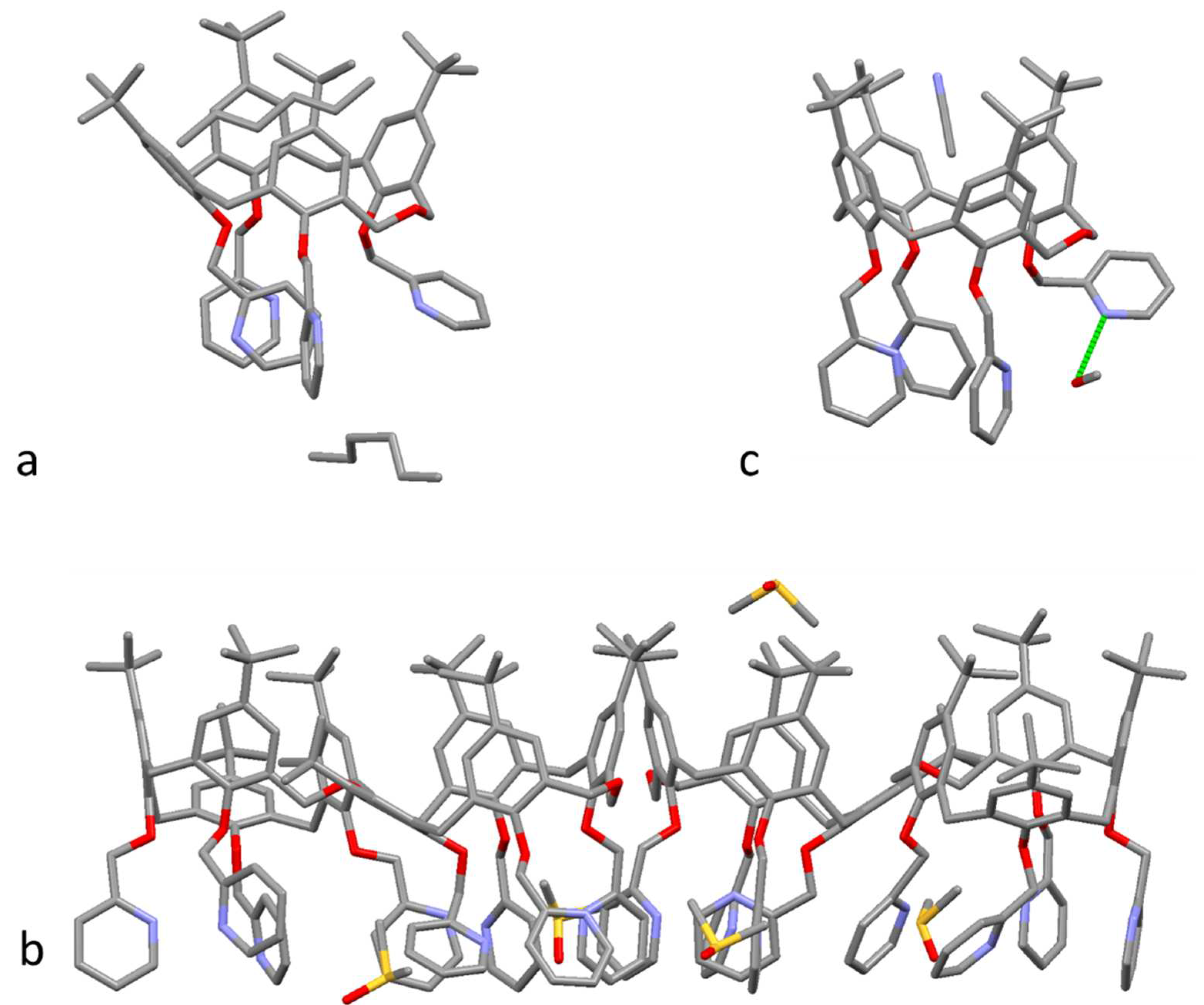
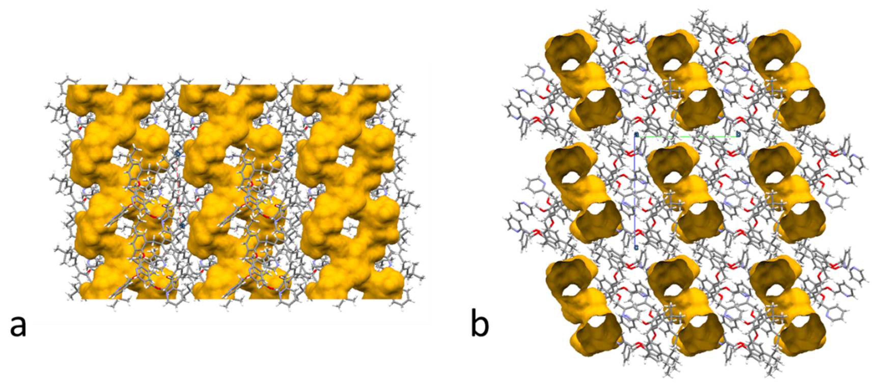
Three pseudo-polymorphic structures of dihomooxacalix[4]arene were obtained (Table S3). The first one corresponds to a triclinic crystal with one molecule of PyHOC4 and 1.5 molecules of n-hexane in the asymmetric unit (Figure 3a). The two independent solvent molecules are located in the interstitial spaces between symmetry related PyHOC4 molecules. One of these assumes an extended zig-zag conformation and it lies on an inversion centre. The cone conformation of the cup is similar to that previously observed for other dihomooxacalix[4]arene derivatives with four substituents on the lower rim, without a guest [28]. The presence of the oxa bridge results in the loss of the pseudo-C2 symmetry of the macrocycle. One of the two planes of the phenyl rings connected to the dihomooxa bridge is oriented outwards from the calixarene cone, while the other is inward oriented, both by about 22° (Table 2). The phenyl ring opposite to the outwards oriented ring is even more outwards oriented (about 47°), while the fourth phenyl ring is almost parallel to the inward oriented phenyl group and therefore it is outwards oriented (Table 2). Despite the differences in the cone conformation, the organization observed for the four pyridyl groups (Figure 3a) is analogous to the one observed for the PyC4 structure without H-bond interactions (Figure 2f). The crystal packing analysis shows that the hexane solvent molecules are present in an unusual intertwined 1D channel network developed along the crystallographic a axis (Figure 4).
A second pseudo-polymorph was obtained in the presence of DMSO solvent. This triclinic form has a large asymmetric unit composed of four PyHOC4 molecules and 3.45 DMSO molecules distributed over six sites. The cone conformation of all four crystallographic independent molecules is similar to that observed for PyHOC4-hexane (Table 2). On the other hand, a high degree of variability is observed for the orientations of the pyridyl groups across the four independent molecules. This variability is also evidenced by the observation of two-position disorder for three entire pyridyl groups. Two of the independent molecules exhibit an interesting edge-face-edge stacking of three pyridyl moieties, with the central pyridyl group located at the centre of the calixarene on the molecular axis (Figure 3b).
A third crystalline form was obtained in presence of acetonitrile and methanol. The asymmetric unit contains one host-guest complex between PyHOC4 and acetonitrile molecule (PyHOC4⸦MeCN) and a methanol molecule H-bonded to a N-atom of a pyridyl arm. As observed in the PyC4 host-guest complexes described above and in analogue dihomooxacalix[4]arenes [28,29], in the presence of the acetonitrile guest, which forms C-H π interactions with the aromatic walls of PyHOC4, the macrocycle assumes a more open cone conformation (Figure 3c) with respect to that observed in the absence of a guest molecule (Figure 3a, b). In this case, the inward oriented phenyl group is pushed externally and all four aromatic rings show an outwards orientation (Table 2). The methanol molecule found in the crystal structure forms a H-bond with a D-A distance of 2.89 Å. With the exception of the pyridyl involved in the H-bond, which shows a conformation similar to that found in all other pyridyl groups involved in H-bonds of the PyC4 structures, the other three pyridyl groups of PyHOC4⸦MeCN-MeOH exhibit a two-position disorder (0.5/0.5, 0.5/0.5 and 0.65/0.35 occupancy factors).

3.3. PyHO3C3 - Hexahomotrioxacalix[3]arene

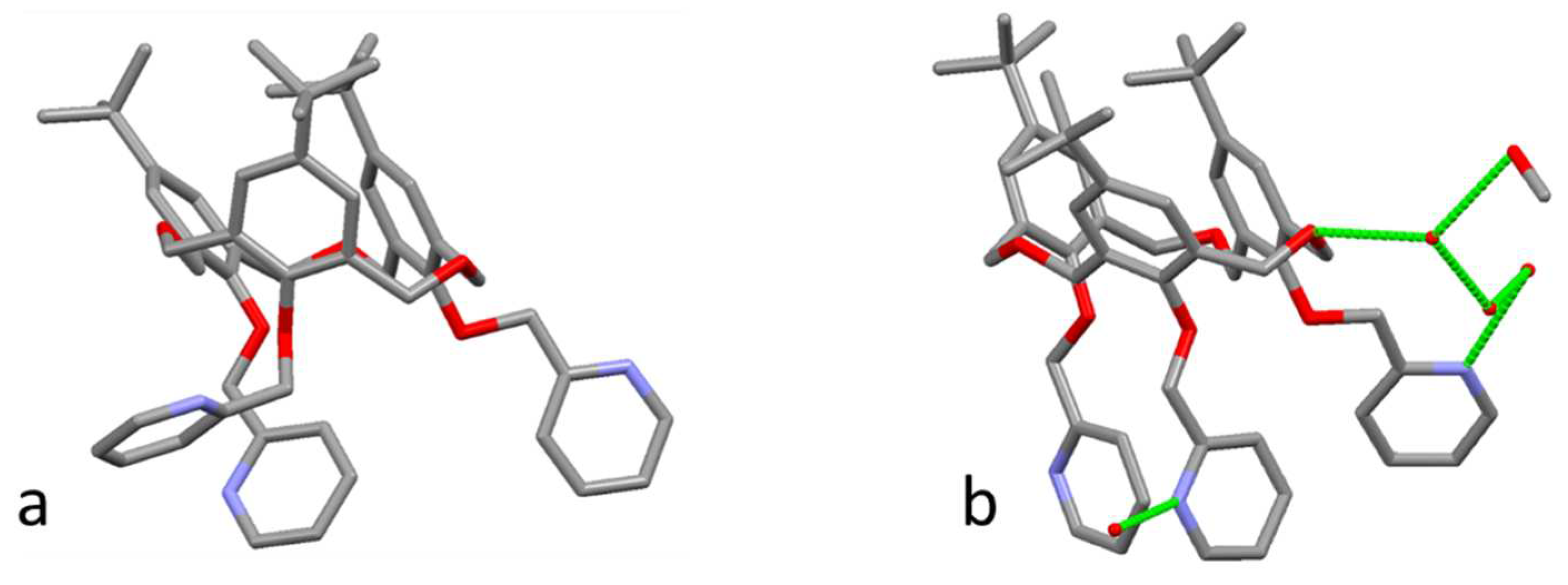
Two monoclinic crystal forms were characterized for PyHO3C3, one anhydrous and one highly hydrated crystal form (Table S4). In the anhydrous form, a single crystallographically independent molecule of PyHO3C3 is present (Figure 5a), while in the hydrate form the asymmetric unit contains one PyHO3C3 molecule, four co-crystallized water molecules and one co-crystallized methanol molecule, all with full occupancy (Figure 5b). As described above, the thrice repeated CH2-O-CH2 bridge confers higher flexibility to the PyHO3C3 macrocycle with respect to PyC4 and PyHOC4. The cone conformation of PyHO3C3 is significantly different in the two crystal forms. Both forms have one phenyl ring which leans inwards, and two which lean outwards, one of which has a very large canting angle of about 135°. However, the inwards phenyl ring of the anhydrous form is about 10° more closed than that of the hydrated form, while its outwards ring is about 10° more open than the corresponding ring in the hydrated form (Table 2). The cone conformation observed for the hydrated form is very close to the almost identical conformations observed for all seven independent molecules found in four solvate pseudo-polymorphs of hexahomotrioxacalix[3]arene functionalized with three phenylurea arms on the lower rim [30]. In the anhydrous form, the different conformation of the cone is accompanied by two position disorder (0.5/0.5) of all three pyridyl arms. In the hydrated form, the two pyridyl groups attached to the outwards orientated phenyl rings are in edge-to-face π- π interaction, while the pyridyl group attached to the inwards orientated phenyl ring is more external. This more external group and the pyridyl which is the face of the π- π interaction are involved as acceptors of H-bonds with water molecules, as is the O-atom of their interconnecting CH2-O-CH2 bridge. In fact, all the hydrogen atoms of the co-crystallized water and methanol atoms are involved in H-bonds, either with each other or with PyHO3C3 molecules. This create a 2D H-bond network and an interesting SOF with interconnected open channels filled with the co-crystallized solvent molecules (Figure 6).

4. CONCLUSIONS

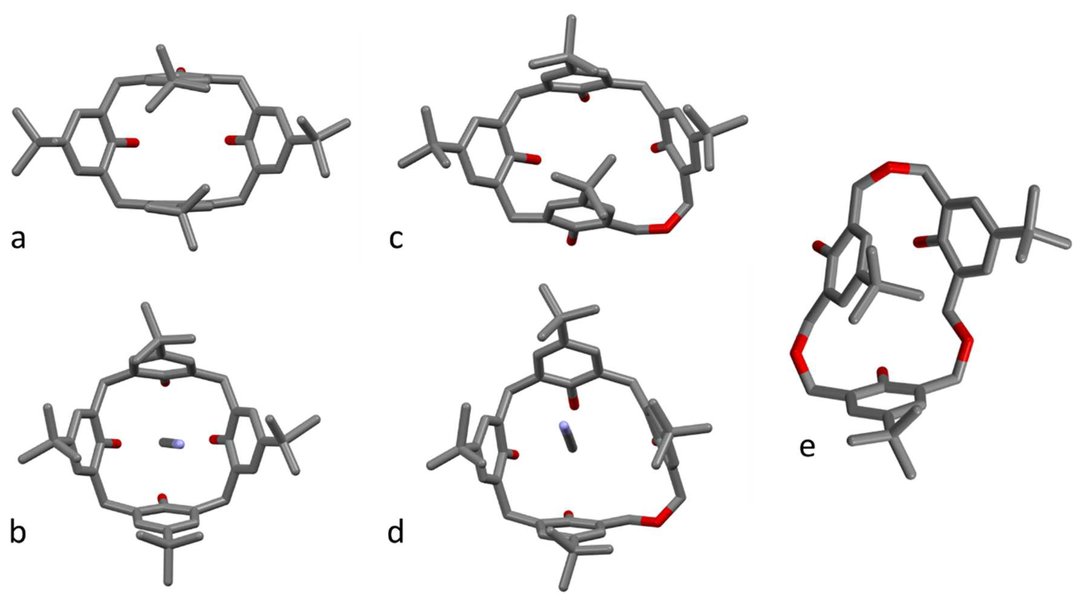
In conclusion, our investigation into the pseudo-polymorphism of 2-pyridylmethoxy cone derivatives of PyC4, PyHOC4, and PyHO3C3 has provided valuable insights. The examination of 11 crystal structures, with 15 crystallographically independent molecules, reveals that the conformation of the macrocycle is predominantly influenced by the presence of a guest molecule within the cavity, while the crystal packing and external solvent molecules play a limited role in shaping the solid-state cone conformation of these macrocycles. Despite the numerous crystallization attempts, the acetonitrile solvent forms host-guest complexes only with PyC4 and PyHOC4, while this is not the case for PyHO3C3, indicating a lower affinity of this guest molecule for the most flexible and less aromatic macrocycle although the PyHO3C3 cup and the acetronitrile molecule have the same C3 symmetry. The acetonitrile guest molecule induces a notable conformational change, resulting in the opening of the cup in PyC4 and PyHOC4 (Figure 7). This alteration also affects the orientation of the 2-pyridylmethoxy arms attached on the lower rim of the macrocycles. The diverse conformations assumed by the 2-pyridylmethoxy groups involve face-to-face and edge-to-face π-π interactions between the terminal aromatic rings.
While all the pseudo-polymorphs of PyC4 exhibit a closely packed crystal structure, PyHOC4-hexane and PyHO3C3-H2O-MeOH present intriguing intertwined 1D and 2D solvent channel networks, respectively.
Notably, a correlation between the structural rigidity of the macrocycle and the number of pseudo-polymorphs formed has been observed. The more rigid macrocycle of PyC4 displays a higher number (6) of pseudo-polymorphs, in contrast to the macrocycles with higher conformational flexibility, namely PyHOC4 and PyHO3C3, which exhibit a minor number (3 and 2, respectively) of pseudo-polymorphs.
While caution is necessary in speculating about these findings due to the potential influence of specific crystallization conditions on polymorph formation, our results suggest a trend wherein more rigid molecules tend to form a diverse array of crystal packing. This observation adds depth to our understanding of the intricate relationship between molecular flexibility and the polymorphic behaviour of calixarene derivatives.
This study not only underscores the complexity of crystal engineering in these systems but also opens avenues for further exploration into the specific conditions governing the formation of different polymorphic forms.

Supplementary Materials

Supplementary data for this article can be accessed online.

Funding

This work has received funding from MUR through the PRIN project 20227YNHEB.

Data Availability Statement

The X-ray crystallographic coordinates of the structures reported in this study were deposited at the Cambridge Crystallographic Data Centre (CCDC) under the deposition numbers CCDC 2331929-2331939. These data can be obtained free of charge from the CCDC at www.ccdc.cam.ac.uk/data_request/cif.

Acknowledgments

We thank the Elettra Synchrotron (Trieste, Italy) and the staff of the XRD1 beamline for their technical assistance.

Disclosure statement

No potential conflict of interest was reported by the authors.

References

  1. Reinhoudt, D.N. Introduction and History. In Calixarenes and Beyond; Neri, P., Sessler, J.L., Wang, M.-X., Eds.; Springer International Publishing: Cham, 2016; pp. 1–11 ISBN 9783319318677.
  2. Nimse, S.B.; Kim, T. Biological Applications of Functionalized Calixarenes. Chem. Soc. Rev. 2013, 42, 366–386. [CrossRef]
  3. Kumar, R.; Sharma, A.; Singh, H.; Suating, P.; Kim, H.S.; Sunwoo, K.; Shim, I.; Gibb, B.C.; Kim, J.S. Revisiting Fluorescent Calixarenes: From Molecular Sensors to Smart Materials. Chem. Rev. 2019, 119, 9657–9721. [CrossRef]
  4. Arora, V.; Chawla, H.M.; Singh, S.P. Calixarenes as Sensor Materials for Recognition and Separation of Metal Ions. Arkivoc 2007, 2007, 172–200. [CrossRef]
  5. Pan, Y.C.; Hu, X.Y.; Guo, D.S. Biomedical Applications of Calixarenes: State of the Art and Perspectives. Angew. Chemie - Int. Ed. 2021, 60, 2768–2794. [CrossRef]
  6. Giuliani, M.; Morbioli, I.; Sansone, F.; Casnati, A. Moulding Calixarenes for Biomacromolecule Targeting. Chem. Commun. 2015, 51, 14140–14159. [CrossRef]
  7. Ludwig, R. Calixarenes for Biochemical Recognition and Separation. Microchim. Acta 2005, 152, 1–19. [CrossRef]
  8. Sliwa, W.; Girek, T. Calixarene Complexes with Metal Ions. J. Incl. Phenom. Macrocycl. Chem. 2010, 66, 15–41. [CrossRef]
  9. Homden, D.M.; Redshaw, C. The Use of Calixarenes in Metal-Based Catalysis. Chem. Rev. 2008, 108, 5086–5130. [CrossRef]
  10. Creaven, B.S.; Donlon, D.F.; McGinley, J. Coordination Chemistry of Calix[4]arene Derivatives with Lower Rim Functionalisation and Their Applications. Coord. Chem. Rev. 2009, 253, 893–962. [CrossRef]
  11. Marcos, P.M.; Mellah, B.; Ascenso, J.R.; Michel, S.; Hubscher-Bruder, V.; Arnaud-Neu, F. Binding Properties of P-Tert-Butyldihomooxacalix[4]arene Tetra(2-Pyridylmethoxy) Derivative towards Alkali, Alkaline Earth, Transition and Heavy Metal Cations. New J. Chem. 2006, 30, 1655–1661. [CrossRef]
  12. Jin Mei, C.; Ainliah Alang Ahmad, S. A Review on the Determination Heavy Metals Ions Using Calixarene-Based Electrochemical Sensors. Arab. J. Chem. 2021, 14, 103303. [CrossRef]
  13. Wilson, L.R.B.; Coletta, M.; Evangelisiti, M.; Piligkos, S.; Dalgarno, S.J.; Brechin, E.K. The Coordination Chemistry of P-Tert-Butylcalix[4]arene with Paramagnetic Transition and Lanthanide Metal Ions: An Edinburgh Perspective. Dalt. Trans. 2022, 51, 4213–4226. [CrossRef]
  14. Pappalardo, S.; Giunta, L.; Foti, M.; Ferguson, G.; Gallagher, J.F.; Kaitner, B. Functionalization of Calix[4]arenes by Alkylation with 2-(Chloromethyl)Pyridine Hydrochloride. J. Org. Chem. 1992, 57, 2611–2624. [CrossRef]
  15. Danil de Namor, A.F.; Castellano, E.E.; Pulcha Salazar, L.E.; Piro, O.E.; Jafou, O. Thermodynamics of Cation (Alkali-Metal) Complexation by 5,11,17,23-Tetra-tert-Butyl[25,26,27,28-Tetrakis(2-Pyridylmethyl)oxy]- Calix(4)arene and the Crystal Structure–Superstructure of Its 1:1 Complex with Sodium and Acetonitrile. Phys. Chem. Chem. Phys. 1999, 1, 285–293. [CrossRef]
  16. Danil De Namor, A.F.; Piro, O.E.; Pulcha Salazar, L.E.; Aguilar-Cornejo, A.F.; Al-Rawi, N.; Castellano, E.E.; Sueros Velarde, F.J. Solution Thermodynamics of Geometrical Isomers of Pyridino Calix(4)arenes and Their Interaction with the Silver Cation. The X-Ray Structure of a 1: 1 Complex of Silver Perchlorate and Acetonitrile with 5,11,17,23-Tetra-tert-Butyl-[25,26,27,28-Tetrakis(2-Pyridylmethyl)oxy]-Calix(4)arene. J. Chem. Soc. - Faraday Trans. 1998, 94, 3097–3104. [CrossRef]
  17. Marcos, P.M.; Teixeira, F.A.; Segurado, M.A.P.; Ascenso, J.R.; Bernardino, R.J.; Cragg, P.J.; Michel, S.; Hubscher-Bruder, V.; Arnaud-Neu, F. Complexation and DFT Studies of Lanthanide Ions by (2-Pyridylmethoxy)Homooxacalixarene Derivatives. Supramol. Chem. 2013, 25, 522–532. [CrossRef]
  18. Marcos, P.M. Functionalization and Properties of Homooxacalixarenes. In Calixarenes and Beyond; Neri, P., Sessler, J.L., Wang, M.-X., Eds.; Springer International Publishing: Cham, 2016; pp. 445–466 ISBN 978-3-319-31867-7.
  19. Yamato, T.; Haraguchi, M.; Nishikawa, J.I.; Ide, S.; Tsuzuki, H. Synthesis, Conformational Studies, and Inclusion Properties of Tris[(2- Pyridylmethyl)Oxy]Hexahomotrioxacalix[3]arenes. Can. J. Chem. 1998, 76, 989–996. [CrossRef]
  20. Marcos, P.M.; Ascenso, J.R.; Pereira, J.L.C.C. Synthesis and NMR Conformational Studies of P-Tert-Butyldihomooxacalix[4]arene Derivatives Bearing Pyridyl Pendant Groups at the Lower Rim. European J. Org. Chem. 2002, 2002, 3034–3041. [CrossRef]
  21. Cragg, P.J.; Drew, M.G.B.; Steed, J.W. Conformational Preferences of O-Substituted Oxacalix[3]arenes. Supramol. Chem. 1999, 11, 5–15. [CrossRef]
  22. Marcos, P.M.; Teixeira, F.A.; Segurado, M.A.P.; Ascenso, J.R.; Bernardino, R.J.; Cragg, P.J.; Michel, S.; Hubscher-Bruder, V.; Arnaud-Neu, F. Complexation and DFT Studies of Lower Rim Hexahomotrioxacalix[3]arene Derivatives Bearing Pyridyl Groups with Transition and Heavy Metal Cations. Cone versus Partial Cone Conformation. J. Phys. Org. Chem. 2013, 26, 295–305. [CrossRef]
  23. Kabsch, W. XDS. Acta Crystallogr. Sect. D Biol. Crystallogr. 2010, 66, 125–132. [CrossRef]
  24. Kabsch, W. Integration, Scaling, Space-Group Assignment and Post-Refinement. Acta Crystallogr. Sect. D Biol. Crystallogr. 2010, 66, 133–144. [CrossRef]
  25. Sheldrick, G.M. SHELXT - Integrated Space-Group and Crystal-Structure Determination. Acta Crystallogr. Sect. A Found. Crystallogr. 2015, 71, 3–8. [CrossRef]
  26. Sheldrick, G.M. Crystal Structure Refinement with SHELXL. Acta Crystallogr. Sect. C 2015, 71, 3–8. [CrossRef]
  27. Hickey, N.; Iuliano, V.; Talotta, C.; De Rosa, M.; Soriente, A.; Gaeta, C.; Neri, P.; Geremia, S. Solvent and Guest-Driven Supramolecular Organic Frameworks Based on a Calix[4]arene-Tetrol: Channels vs Molecular Cavities. Cryst. Growth Des. 2021, 21, 6357–6363. [CrossRef]
  28. Miranda, A.S.; Marcos, P.M.; Ascenso, J.R.; Berberan-Santos, M.N.; Schurhammer, R.; Hickey, N.; Geremia, S. Dihomooxacalix[4]arene-Based Fluorescent Receptors for Anion and Organic Ion Pair Recognition. Molecules 2020, 25. [CrossRef]
  29. Miranda, A.S..; Marcos, P.M.; Ascenso, J.R.; Robalo, M.P.; Bonifácio, V.D.B.; Berberan-Santos, M.N.; Hickey, N.; Geremia, S. Conventional vs. Microwave- or Mechanically-Assisted Synthesis of Dihomooxacalix[4]arene Phthalimides: NMR, X-Ray and Photophysical Analysis. Molecules 2021, 26, 1503. [CrossRef]
  30. Teixeira, F.A.; Ascenso, J.R.; Cragg, P.J.; Hickey, N.; Geremia, S.; Marcos, P.M. Recognition of Anions, Monoamine Neurotransmitter and Trace Amine Hydrochlorides by Ureido-Hexahomotrioxacalix[3]arene Ditopic Receptors. European J. Org. Chem. 2020, 2020, 1930–1940. [CrossRef]
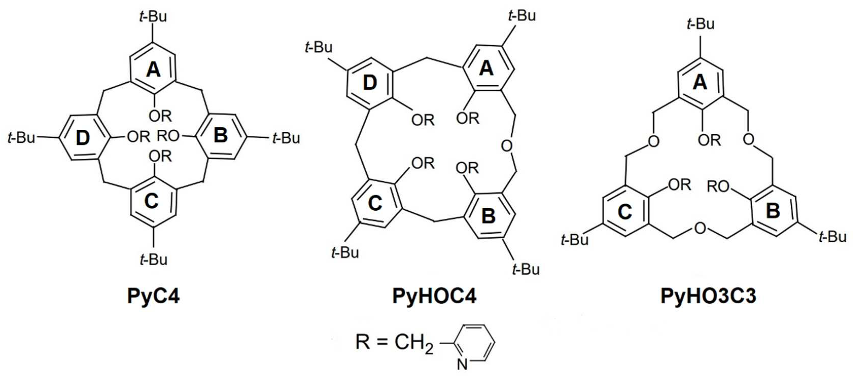
Figure 1. Chemical structures of the three macrocycles.
Preprints 98975 g001
Figure 2. Six asymmetric units of various PyC4 structures obtained in different solvents: a) PyC4-MeOH-α; b) PyC4-H2O-α; c) PyC4-MeOH-β; d) PyC4⸦MeCN-MeOH; e) PyC4⸦MeCN-H2O and f) PyC4. H-bonds between calixarene and co-crystallized solvent molecules are represented by green dashed lines. Only one position of the disorder groups is shown for better clarity.
Preprints 98975 g002
Figure 3. Asymmetric units of three different PyHOC4 structures obtained in different solvents: a) PyHOC4-DMSO; b)PyHOC4⸦MeCN-MeOH and c) PyHOC4-Hexane. Only one position of the disorder groups is shown for better clarity.
Preprints 98975 g003
Figure 4. Crystal packing of PyHOC4-hexane a) viewed along the crystallographic b axis and b) viewed along the crystallographic a axis. The surface of the solvent accessible volume calculated with a 1.2 Å probe evidences the intertwined 1D channel network developed along the crystallographic a axis.
Preprints 98975 g004
Figure 5. Asymmetric unit of two different PyHO3C3 structures: (a) the anhydrous form PyHO3C3 and (b) the hydrate form PyHO3C3-H2O-MeOH. H-bonds between calixarene and co-crystallized solvent molecules are represented by green dashed lines. Only one position of the disorder groups is shown for better clarity.
Preprints 98975 g005
Figure 6. Crystal packing of PyHO3C3-H2O-MeOH a) viewed along the crystallographic a axis and b) viewed along the crystallographic a axis. The surface of the solvent accessible volume calculated with a 1.2 Å probe evidences the 2D channel network developed in the (100) crystallographic plane.
Preprints 98975 g006
Figure 7. Effect of acetonitrile guest complexation on the macrocycle conformation. The caps are viewed perpendicular to the mean planes of the bridging methylene carbon atoms: a) PyC4, b) PyC4⸦MeCN, c) PyHOC4, d) PyHOC4⸦MeCN, e) PyHO3C3.
Preprints 98975 g007
Table 1. Interior dihedral angles between the aryl-aryl planes observed in the crystal structures. See Figure 1 for the labelling scheme of the arene moieties.
Crystal A-B B-C C-D D-A
PyC4-MeOH-  α 96 94 92 91
PyC4-H2O-  α 96 94 92 91
PyC4-MeOH-β
*
96
96
92
93
94
95
93
93
PyC4⸦MeCN-MeOH 100 102 95 100
PyC4⸦MeCN-H2O 99 100 97 101
PyC4 86 87 92 93
PyHOC4-DMSO
*
*
*
59
58
62
56
90
95
91
91
103
97
102
99
97
98
95
102
PyHOC4  ⸦MeCN-MeOH 69 112 105 96
PyHOC4  -Hexane 61 77 101 113
C-A
PyHO3C3 48 102 36
PyHO3C3-H2O-MeOH 62 98 31
* Dihedral angles of crystallographic independent molecules.
Table 2. Dihedral canting angles between the aryl planes and the mean planes of the bridging methylene carbon atoms for various calixarenes. Dihedral angles greater than 90° correspond to outward orientations of the plane with respect to the cup. See Figure 1 for the labelling scheme of the arene moieties.
Crystal A B C D
PyC4-MeOH-  α 128 98 128 91
PyC4-H2O-  α 128 98 128 91
PyC4-MeOH-β
*
136
135
96
93
130
131
89
91
PyC4⸦MeCN-MeOH 118 112 117 108
PyC4⸦MeCN-H2O 121 110 118 109
PyC4 135 85 130 87
PyHOC4-DMSO
*
*
*
117
119
116
119
72
76
74
70
135
136
134
136
102
98
100
103
PyHOC4  ⸦MeCN-MeOH 105 97 128 112
PyHOC4  -Hexane 112 68 137 106
PyHO3C3 57 119 134
PyHO3C3  -H2O-MeOH 66 110 133
* Dihedral angles of crystallographic independent molecules.
Disclaimer/Publisher’s Note: The statements, opinions and data contained in all publications are solely those of the individual author(s) and contributor(s) and not of MDPI and/or the editor(s). MDPI and/or the editor(s) disclaim responsibility for any injury to people or property resulting from any ideas, methods, instructions or products referred to in the content.
Copyright: This open access article is published under a Creative Commons CC BY 4.0 license, which permit the free download, distribution, and reuse, provided that the author and preprint are cited in any reuse.
Prerpints.org logo

Preprints.org is a free preprint server supported by MDPI in Basel, Switzerland.

Subscribe

© 2024 MDPI (Basel, Switzerland) unless otherwise stated