Preprint
Review

A Review on the Synthesis and Fabrication of Gold Nanoparticles and Their Application in Dyes Degradation

Altmetrics

Downloads

251

Views

160

Comments

0

Submitted:

21 February 2024

Posted:

21 February 2024

You are already at the latest version

Alerts
Abstract
In this review, we covered the recent advances in synthesis of gold nanoparticles and their uses in degradation of dyes. This study provides framework to develop a low cost, ecofriendly and highly efficient synthesis of gold nanoparticles. These synthetic methods do not produce any toxic byproduct. The present study is focus on removal of dyes by Au-NPs because gold nanoparticles act as good absorbent for dyes in short time. Synthesis of Au-NPs from plant extract e.g. marine alga, Scutellarin Barbata, A.nigra, Fruit peels, Bacillus marisflavi from raw silk cocoons, amylopectin and poly acrylic acid, L.asparagine, Grephene oxide, IPEI coated Au-NPs. The synthesizes Au-NPs were used further for removal of dyes like methylene blue (MB), Rhodamine B (RB) Degradation, methylene orange, acid red degradation, Congo red.
Keywords: 
Subject: Chemistry and Materials Science  -   Nanotechnology

Introduction:

The dying agents in the industry is widely used and the most fundamental agents in industries around the globe. However, their wastewater released to the environment is causing harmful effects and source of pollution for living bodies due to the presence of mutagenic and carcinogenic substances. Some of adverse effects of dyes on human being involve issues with the central nervous system, brain, liver, reproductive system, and Renal System. So, it's important to get dyes out of our surroundings. To remove these colours from the water streams, inexpensive techniques are needed. Around 100 tons of dyes and pigments are discharged annually into the aquatic environment by the textile sector. [1].
Table 1. Toxicity of some dyes [1].
Serial No Name LD50 (mg/Kg rat)
1 Malachite Green 275
2 Acid Orange 165 60
3 Basic Blue 7 100
4 Basic Blue 81 205
5 Basic Violet 16 90
6 Basic Yellow 21 171
7 Direct orange 62 150
Dyes are generally organic substances that get attached to the fabric surface and give color, also known as coloring agents. They are soluble in water, produces bright colors in aqueous medium giving it acidic properties. They are commonly used in several industries which include paper industry, food industry, printing industry, textile industry, cosmetics and many others. Researchers are trying to formulate new dye compounds that give fabrics permanent bright color but, these chemicals impart highly toxic and carcinogenic properties upon breakdown. In addition, they show resistance to aerobic biodegradation, thus possessing a challenge to the environmental researchers. They can persist for a longer period in the environment and therefore causing environmental hazards and also reduce the aesthetic value of water bodies [2].
Table 2. Classification of Dyes based on their nature and application [1,3].
Class Application Examples

Acid dyes
Nylon, wool, silk, modified acrylics, paper, leather, food, inkjet printing and cosmetics.
Acid red 88, Acid red 18


Cationic (Basic) Dyes
Poly acrylonitrile, paper, modified polyesters, modified nylons, cationic dye able polyethylene terephthalate, wool, silk, tannin mordant cotton and medicine.

Crystal Violet, Methylene Blue, Safranin, Basic fuschin

Disperse Dyes
Nylon, polyester, cellulose, acrylic fibers and cellulose acetate. Disperse Red 1, Disperse Orange 37
Direct Dyes Rayon and cotton,
Leather, paper and nylon.
Congo Red, Brilliant Blue, Copper blue 2R
Reactive Dyes Wool, nylon, cotton and other cellulosic. Reactive Black 5, Reactive Orange 16
Solvent Dyes Gasoline, plastics, oils, lubricants and waxes. Solvent Red 1, Solvent Red 49, Solvent Red 24, Solvent Red 111
Sulfur Dyes Cotton and rayon, paper, leather, silk and wood. Sulfur Brilliant Green, Sulfur black 1
Vat Dyes Cotton, rayon and wool. Vat red 10, Vat violet 13 and Vat orange 1
Azoic dyes These dyes are utilised in pigments and printing inks. Methyl Orange,
Methyl Red,
Rhodamine B


Leather dyes
By applying high temperatures to the leather, the fat-soluble components of these colours have an attraction to migrate upward into the layers, giving the leather surface a smooth finish.

Fiebing’s Leather Dye

Properties:

A dye is regarded as a good dye if it possesses certain characteristics, including the ability to impart colour, bond itself to fabrics, be lightfast, and demonstrate resistance to the action of water, diluted acids, alkalis, and certain organic solvents used in dry cleaning. The following characteristics of dyes are based on the following criteria. [3].
  • Brightness or Dullness
  • Dyeing Properties
  • Environmental Issues
  • Ease of Application
  • Low-economy cost
Table 3. Different types and properties of dyes used in industries [3].
Dye Name Type Color Molecular formula Molecular weight (g/mol) Absorption maxima (nm)
Methylene Blue Aniline Dark green C16H18N3SCl 319.85 664
Methyl Orange Azo Orange-yellow C14H14N3NaO3S
327.33 460
Methyl Red Azo Dark Red C15H15N3O2
269.30 540
Rhodamine B Azo Reddish-violet C28H31ClN2O3
479.02 554
Gentian Violet Triaryl-methane Violet C25N3H30Cl
407.98 536
Indigo Carmine Indigotine Indigo to dark blue C16H8N2Na2O8S2
466.35 610
Congo Red Azo Red C32H22N6Na2O6S2
696.66 510
Reactive Blue 4 Anthraquinone Dark blue C23H12Cl2N6Na2O8S2
681.39 596
Basic Violet 3 Triaryl-methane Bright blue purple C25H30ClN3
407.98 590
Acid Red 114 Azo Dark red powder C37H28N4Na2O10S3
830.81 514
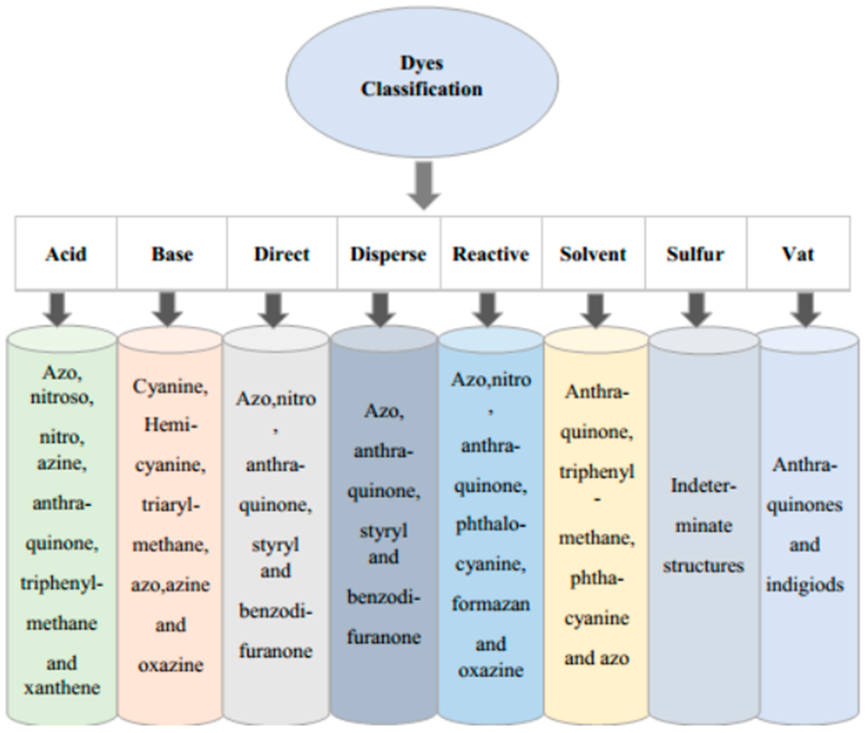
Figure 2. Classification of Dyes.
Preprints 99484 g002
Table 4. Examples of some organic dyes and their chemical formulae.
Class Chromophores Examples

Azo dyes

Preprints 99484 i001

Reactive Black 5


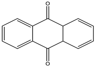
Anthraquinone dyes

Preprints 99484 i002


Reactive Black 4

Nitroso dyes

Preprints 99484 i003

Acid Green 1


Nitro dyes

Preprints 99484 i004


Acid Yellow 24


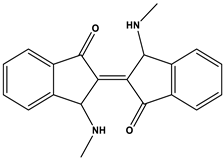
Indigoid dyes

Preprints 99484 i005


Acid Blue 71


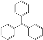
Triarylmethane dyes

Preprints 99484 i006


Malachite Green
There are different type of dyes such as reactive, disperse, direct, vat, sulphur, cationic, acid, and solvent dyes [5]. On the basis of chromophores, there are 20 to 30 diverse groups which includes azo, phthalocyanine and triarylmethane dyes. On the basis of weight dyes constitute almost 70% of world population yearly. The first pollutant that we have found in waste water was color. Even small quantity of dyes preset in water can be seen easily and effect the transparency of water as well aesthetically. Beside this the phenomenon of absorption of light and reflection affects aquatic life in water bodies. Mostly, dyes that are prepared synthetically have toxic and carcinogenic effects [6]. Industrial revolution and the usage of organic colours and aromatic compounds have caused water pollution to become a global issue today. The dyes ability to show resistance to heat, light and act as oxidizing agent and show visibility to aqueous media is major problem that has serious effect on health [7]. Industrial effluents like effluents from paper, plastic, tanning and food manufacturing, cosmetics, and pharmaceuticals has synthetic azo dyes and due to their toxic, mutagenic, and carcinogenic effect, they are not only harmful for aquatic life but also to human race. It is difficult to treat dye effluents because of its complex structure and stability [8]. Similarly, Toxic waste of many organic dyes can have harmful impacts on both human and animal health. It can be responsible for various health hazards as kidney damage, irritation of skin, liver damage, and effect on central nervous system and much more due to their carcinogenic property. They are also very dangerous for aquatic life as well because when dye pollutant present in water they eventually block sunlight for reaching to aquatic life and disturbs photosynthesis. Treatment of the toxic waste of dyes is very significant before releasing into environment [9]. Synthetic dyes can be removed by biological, chemical, and physical techniques, including ion exchange, membrane filtration, reverse osmosis, nano-filtration, and ultrafiltration. Biological techniques include adsorption by microbial biomass, algal degradation, and aerobic-anaerobic combination. Because of the adverse effect of using these methods which may include hazardous by product formation, operational difficulties, high cost, large amount of energy consumption and formation of secondary pollutants, an environmentally friendly method is required for the removal as well as degradation of dyes [10]. So, in order to remove dyes from polluted aqueous media, there are several methods that have been used like filtration, coagulation, adsorption and others. Among them, ‘‘adsorption process” is best because of less money consumption, simple and efficient method. We can use different Nano particles as an adsorbent [8].
Gold nanoparticles that are synthesized by using green chemistry can degrade cationic dye in the presence of light [8]. Degradation of dyes by using metal NPs to boost the speed of reaction is a very quick, simple, efficient and economic method. This method is most reliable one because with reducing chromophore groups it also disintegrates large molecule to smaller one, and these molecules are also eco-friendly. The process reductive degradation is rapid than photolytic one.
In photolytic oxidative degradation, we need energy consuming irradiation. Organic dyes reduction can be done by NaBH4 by transferring of electrons from BH4 to dye molecule. Due to greater potential difference between molecules, the transferring of electrons is quite tricky. Kinetically it is challenging to do degradation of other dyes but thermodynamically it is possible. When we use metal NPs as a catalyst, they reduce the kinetic energy needed for transferring of electrons [9]. Gold nanoparticles has distinctive characteristics such as thermal and electrical conductivity, their stability, best catalytic activity [11]. Au-NPs also have diverse applications in other fields as in pharmaceutics, as a catalyst, in electronics and visual sensing etc [9]. Cellulose nano crystals have been used for synthesis of Au-NPs and it plays the role of reducing agent as well as supporting one. These gold nanoparticles show brilliant properties of degradation of several organic dyes, including Congo red, Rhodamine B, Methyl orange, Methylene Blue and 4-Nitrophenol. [12].
Nanotechnology is very advance field form last decade [13]. It is defined as it process check and balance on properties of nano science that studies the nano metric scale matters, its size and characteristics [14]. Nanotechnology can be divided into two categories that are Nano devices and Nano particles/Nano materials. Nano devices are basically use for working at nano scale. Some intelligent machines and microscopic devices are included in it [15]. Nanotechnology is a source of different types of particles at nano scale level. We can define nanoparticles are those particles whose size ranges from 1nm to 100nm with only one dimension [13]. Nanoparticles exists as in form of nanotubes, dots, capsules, polymer etc [8]. Nanoparticles have a variety of properties due to difference in size and functionalities of surface area [13]. Surface area of nanoparticles is directly proportional to its catalytic activity [16]. These days’ nanoparticles become so popular due to its large surface area, selectivity, recyclability [8]. They are of different types i.e.; Polymer based nanoparticles, Solid nanoparticles (iron oxide, gold, silver), Carbon based nanoparticles, Lipid based Nano particles and Nano emulsion. These all types of nanoparticles have different applications on large scale. Different products that we use in our daily life contain nanoparticles such as different food material, cosmetic products as well as pharmaceutical products. Nanoparticles are also use for clinical purpose, diagnosis and therapies, reduction of harmful dyes, as drug carriers and drugs delivery [13]. Nowadays, due to its various applications, industries are taking lot of interest in nanotechnology. This tremendous increase of interest in nanotechnology is causing a production or synthesis of nanoparticles on large scale [14]. Different methods are used for preparation and stabilization of nanoparticles [17]. Metallic or solid nanoparticles is a new terminology that has been introduced in the field of biotechnology in recent few years. In this field, mostly gold, platinum and silver nanoparticles are synthesize due to its ecofriendly ad beneficial properties and these are termed as metallic or solid nanoparticles [17]. Here, the main focus of our review article is gold nanoparticle. We see gold is just a metal in past but later due to its physiochemical properties such as low toxicity, biocompatibility and optoelectronic property and with the advancement of nanotechnology we found out gold as a suitable material for academic research as a nanoparticle and now it is termed as gold nanoparticle [16]. It is one of the mostly studied Nobel metal because of its application in catalytic activities, optical and electronic devices, and for antimicrobial activities as well [18]. Corrosion and oxidation do not affect gold nanoparticles and are included in noble metal nanoparticle [11]. Gold nanoparticle can be synthesized by chemically and naturally both. Green Synthesis is most environment friendly for synthesizing metallic nanoparticles. There are also further different methods used for green synthesis such as hydrothermal method use for synthesizing gold nanoparticle that have pro anthocyanins functionality [18]. Additionally, thermal decomposition, solution combustion, reflux method, polyol, template, solvothermal, chemical reduction, ion-exchange, sol-gel, chemical vapors deposition, spray pyrolysis, mechanical milling, physical vapor deposition, sputtering, laser ablation, laser pyrolysis, son chemical, micro emulsion, photochemical, electrochemical, microwave, co-precipitation, and other biological, chemical, and physical methods are employed for gold nanoparticle synthesis. [19]. Adsorption is also one of the best method to synthesize gold nanoparticles. [18].
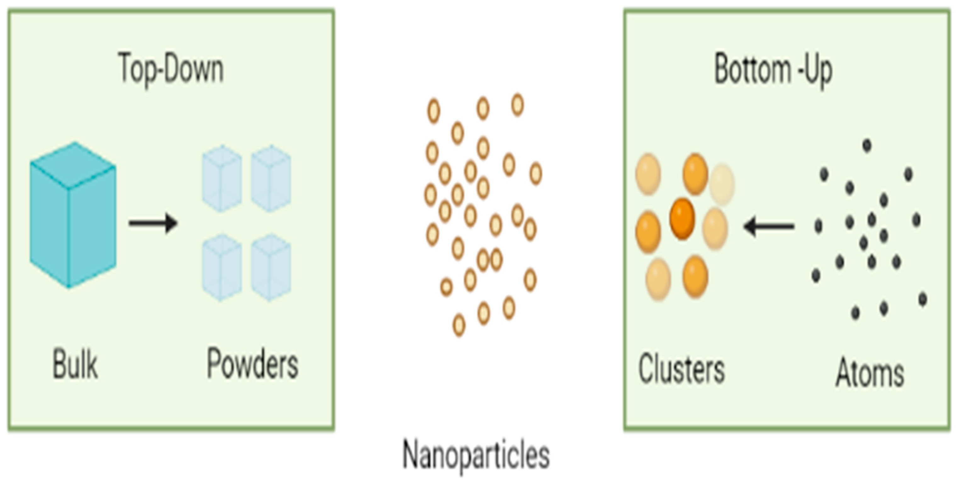
There are many methods for the synthesis of Nanoparticles which are further classified into top-up and top bottom synthesis [20]. Synthesis of gold nanoparticles can also be done by top-bottom synthesis and bottom-up Synthesis. In top-bottom synthesis, particle size should be reduced and it consumes lot of energy. Whilst in case of Bottom-up approach, it gives large amount of formation of nanoparticles in short period of time [11].
Figure 3. Top down and Bottom-up Synthesis Approach of Nanoparticles.
Preprints 99484 g003
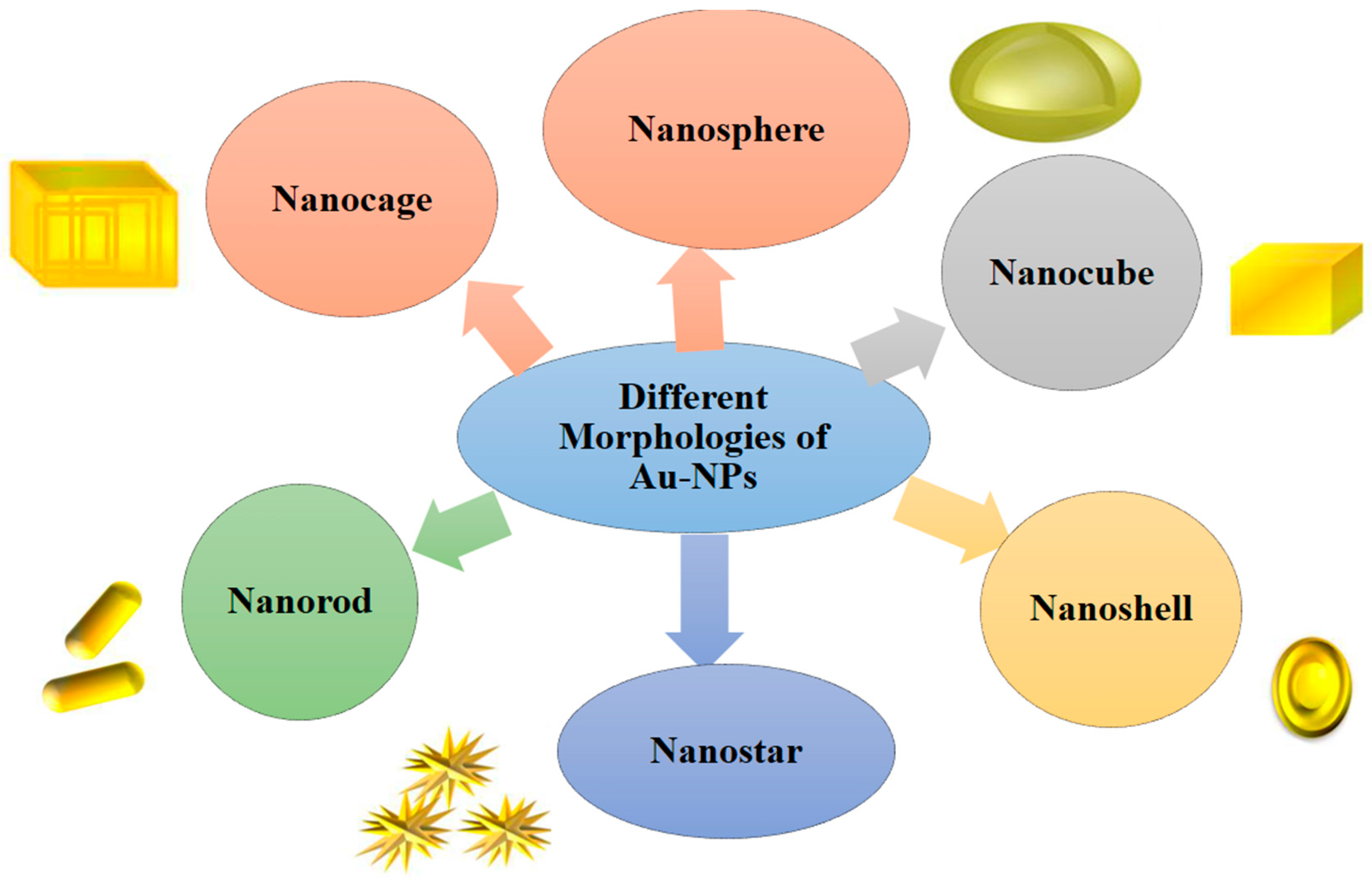
Figure 4. Different Morphologies of Au-NPs.
Preprints 99484 g004
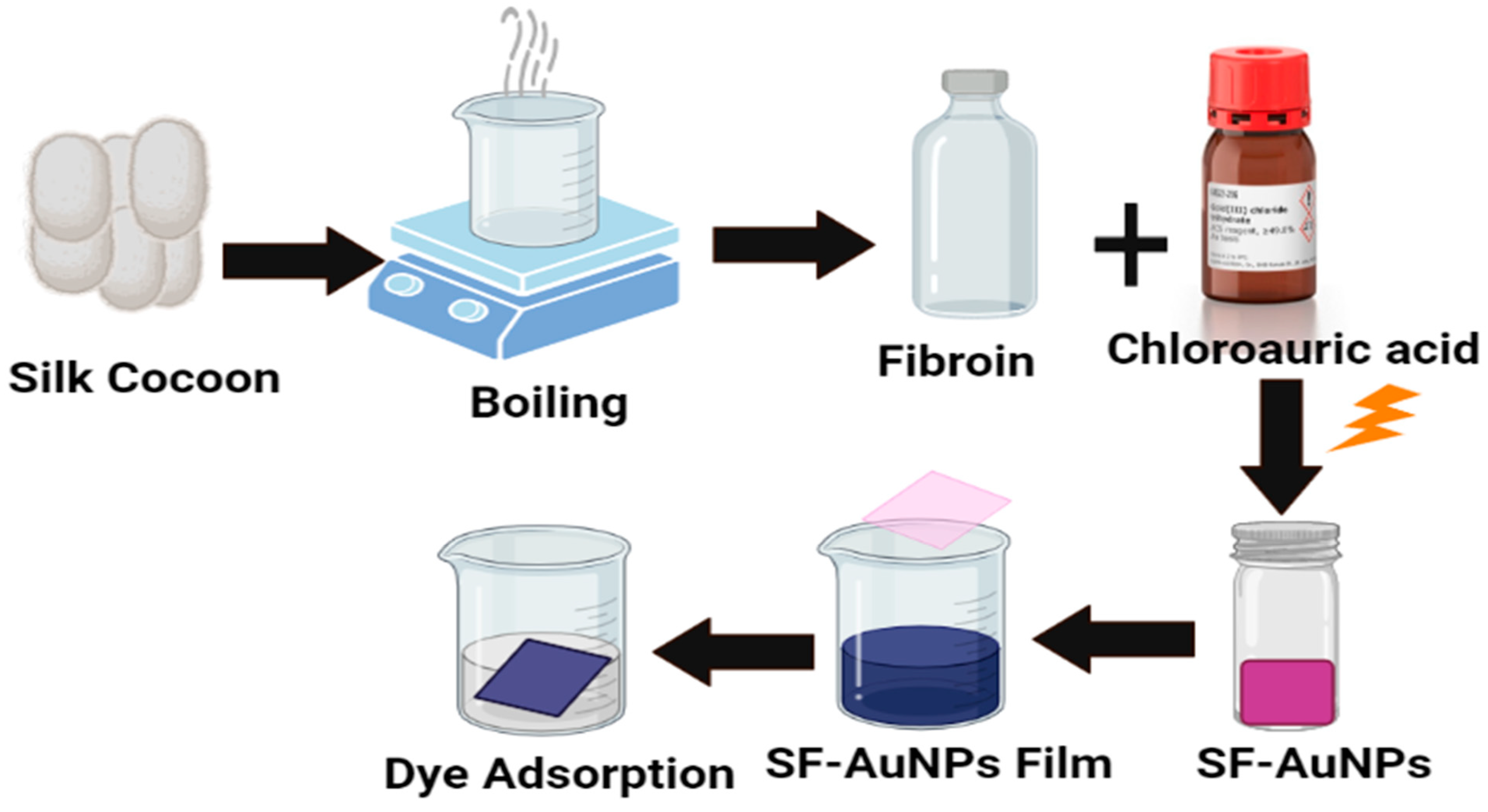
Au-NPs have different morphologies, including nanospheres, nanocubes, nanoshells, nanoflowers, nanorods etc. In medical chemistry, gold nanoparticles have a wide range of uses, including the treatment of cancer, germs, and HIV. For the removal of dyes, Excitation spectra of UV-Visible spectrometers in the wavelength range of 350-800 nm can be used to measure the size of biosynthesized gold nanoparticles, while X-ray diffraction (XRD) analysis can be used to determine the crystallographic information of gold nanoparticles. Dynamic light scattering (DLS) technique was used to measure the particle size. [22]. For example: the silk fibroin (SF) and silk fibroin-gold nanoparticles (SF-AuNPs) Nano composites for removal of methylene blue (MB) dye were reported. The AuNPs in SF were visible in the absorption band at λ= 540 nm. The crystalline character of the AuNPs was revealed by the X-ray diffraction (XRD) and transmission electron microscopy (TEM) studies. The TEM picture demonstrated the spherical form of the AuNPs, which have an average diameter of around 6.22 1.28 nm. The results were around 95.62 and 98.66 percent (SF). Moreover, samples may be recycled four times with a greater than 80% efficiency. The SF and SF-AuNPs films may thus be utilized as effective sorbents for the elimination of methylene blue dye from a solution. [23]
Figure 5. Dye Adsorption using Silk Fibroin-Gold Nanoparticles (SF-AuNPs).
Preprints 99484 g005
Table 5. Removal of dyes using variety of AuNPs.
Sources of (AuNPs) Dyes removed Morphology References
silk fibroin (SF) and silk fibroin-gold nanoparticles (SF-AuNPs Methylene blue Spherical [23]
Wedelia urticifolia(plant extracts ) Rhodamine-B Spherical [24]
Sargassum horneri(marine algae extract) SH-AuNPs Methylene blue rhodamine B, and methyl orange. Spherical [23]
(AuNPs-HNT) with halloysite nanotube phenothiazine dyes (azure B (AZB) and toluidine blue O (TBO), 4-(4-nitrophenyl)morpholine Free nit cell structure [25]
Dimrit raisin Methylene blue (MB) and methyl orange (MO) Triangular and hexagonal [26]
Rosa canina fruit extract Methylene blue (MB), Rhodamine B (RhB) and 4-nitrophenol (4-NP) Spherical [27]
(Graphene/Au-NPs) nanozymes Methylene blue (MB), rhodamine (RB), methyl orange Spherical,quasi spherical [28]
Pseudoalteromonas lipolytica (bacteria extract) methylene blue and Congo red dyes Spherical [29]
Enoki mushroom (Flammulina velutipes) Methylene Blue (MB) Colloidal (oval, spherical, drum-like, hexagonal, and triangular) and crystalline form [30]
Portobello mushroom (Agaricus Bosporus) Methylene Blue Oval, spherical, drum-like, hexagonal, and triangular [31]

Properties of gold nanoparticles:

Gold Nanoparticles have received a great fame owing to their enhanced optical, electrical, biological, magnetic and thermal capabilities. Furthermore, surface-enhanced Raman scattering (SERS) qualities are present in gold nanoparticles. [32].These proprieties of gold nanoparticles are very auspicious even that their enhanced photothermal properties can diagnose cancerous cells in human body and destroys them by photothermal therapy [33,34].The bio-functionalized gold nanoparticles with coating of graphene amplify polymer properties that can also remove toxic dyes [35].Gold nanoparticles doped with ZnO acts as photocatalyst that can be used for removal of toxic dyes [36]. Due to their excellent adsorption properties nanoparticles along with activated carbon removes Congo Red dye efficiently [37]. The electrical conduction properties of gold nanoparticles can give suitable ink ingredient and can be further used in ink-jet printing technology [38]. Exceedingly small size gold nanoparticles along with carbon nitride composites acts as efficient catalyst in removal of toxic chemical as 4-nitrophenol and harmful dyes [39].Chromophore functionalized gold nanocomposite can remove toxic dyes due to thermally activated electrons of gold [40]. Thermo physical properties of gold nanoparticles can produce laser fluorescent that can light up dye when it attaches to gold so removal of transparent dyes can be done effectively [41].

Sources of Nanoparticles:

Laccase (benzene-diol oxygen oxidoreductase) is a dimeric or tetrameric glycoprotein which contain copper atom in its active site, responsible for the transport of four electrons to the oxygen atom and forms water as a by-product. Due to the eco-friendly nature and high affinity of laccase for different aromatic compounds, it is used as a substrate for the detoxification of synthetic dyes and phenolic pollutants [42]. Nano particles synthesis uses chemicals which have negative impact on the environment so, researchers are working on the synthesis of Nano particles to synthesize it with the components which are actually not harmful to the environment like plants, bacteria, and fungi etc. Because plants operate as a reducing and stabilizing agent during the creation of nanoparticles, they are most frequently used in this process, these are economical and the presence of different phytochemicals such as flavanoids, terpenoids.Mussaenda glabrata, Galaxaura elongata, Plumeria alba, Alpinia nigra (biennial shrub) were utilized to synthesize of gold nanoparticles [43].
Brown algae Sargassum horneri (SH), a species that floats in the water, contain a lot of polyphenols, polysaccharides, and chromene chemicals. Because the hot water extract of SH possesses antiviral properties, it may be utilized to create nanoscale Au and Ag particles. Ultraviolet-visible spectroscopy (UV-Visible), dynamic light scattering (DLS), high-resolution transmission electron microscopy (HRTEM), selected area electron diffraction (SAED), energy-dispersive X-ray spectroscopy (EDS), X-ray powder diffraction (XRD), and Fourier transform infrared spectroscopy were the characterization technique used to characterize nanoparticles synthesis during their synthesis. [10].
Plants extract having metal accumulation and detoxification properties are the best candidates for the synthesis of Nano particles. Pomegranate peels (Punica granatum) contains significant number of polyphenols (15-20%) and proanthocyanins (1.2-9.0%) with highest antioxidant activity among the peel, pulp and seed fractions, used as a reducing agent in the green synthesis of nanoparticles [44]. Nano particles could also be synthesized using bacterial strains such as Bacillus sp., B. amyloliquefaciens, and B. clausii and Azoarcus sp [45]. Formation of gold nano-particles was done by obtaining aqueous extraction of fine powder of Terminalia bellirica which is a dry fruit pericarp. After formation, synthesis of gold nanoparticles was confirmed with the help of UV-Visible spectroscopy at 530nm. When leaves of Eclipta prostrate were used for synthesis of gold Nano-particles, then triangle, pentagon, and hexagon like shapes of nanoparticles were obtained [11].

Formation of gold nanoparticles and their use for dyes removal:

The Apiaceae family includes flowering herbaceous plants like fennel. For its distinctive sweet, anise-flavor, fennel seeds are widely farmed throughout Europe, the Middle East, and the Mediterranean regions. F. vulgare seeds have antibacterial, antioxidant, anticancer, anti-allergic, and memory-improving effects. The main phytochemicals found in seeds include flavonoids, poly-phenolic acids, tannins, and saponins [46].
The phytochemicals in the seeds of F. vulgare servesd as both stabilizing and reducing agents. The presence of a bright purple tint with a maximum wavelength of roughly 550 nm in the UV-Vis absorption spectra at room temperature served as proof that gold nanoparticles had formed. The reduction of MB and Rh-B in borohydrides was accomplished using the Au-NPs as nano-catalysts. The blue tint of the methylene blue in NaBH4 solution was eliminated by adding AuNPs which indicated the complete degradation of dye [46]. Antioxidant-rich substances found in Sargassum species lowered gold ions and subsequently stabilize nanoparticles. In Metal nanoparticles' surface-to-volume ratio was increased along with their catalytic effectiveness. The decrease of harmful organic dyes was accomplished using Au-NPs. For methylene blue, methyl orange, and methyl red, the findings were outstanding in terms of efficiency and degradation. The kinetic analysis demonstrated that intra-particle diffusion caused methylene blue to degrade. On contrary, the degradation of methyl orange and methyl red occurred in two stages: first, by a surface reaction, and then, second, by intra-particle diffusion. This result was significant because it showed that the stabilizing agent does not reduce the catalytic activity of Au-NPs, making the use of Sargassum spp for the production of gold attractive. Increased concentration of these algae was a fantastic substitute for the degradation of poisonous substances [47]. Gold nanoparticles are more biocompatible, long-lasting, and resistant to oxidation. (GNPs) are regarded as the most ideal for degradation of pollutants. Metal ions are converted to metal nanomaterial by phytochemicals, which also stabilizes the material for a longer time. It does this by capping the synthesized nanomaterial. Metal ions are converted to metal nanomaterial by phytochemicals, which also stabilizes it for a longer time. It accomplishes this by capping the created Nano-materials. The catalytic characteristics of the biologically produced nanoparticles are significantly influenced by the capping molecules. For the purpose of reducing anthropogenic pollutants, biosynthesized GNPs work as a promising catalyst [48].Using fruit extract from Sterculia acuminata (S. acuminata), gold nanoparticles (AuNPs) were synthesized. Due to their versatility, various poly-phenolic components in the extract served as both reducing and stabilizing agents. For numerous organic pollutants, including 4-nitrophenol (4-NP), methylene blue (MB), methyl orange (MO), and direct blue, the catalytic efficiency of the synthesized Au-NPs was assessed. It was found that certain pollutants, such as methyl orange and methylene blue, degrade more quickly than others. 4-NP was reduced, and numerous organic dyes including methyl orange, methylene blue, and straight blue were degraded to assess the catalytic activity of the generated Au-NPs. Without a catalyst at first, NaBH4 was added to tubes containing organic dye solutions. To monitor the catalytic reduction and degradation of organic dyes, UV-visible spectra were collected on a regular basis [49].
Noble metal nanoparticle-based catalysts are the first choice because to their high surface area, quick reactivity, recycling power, chemical, and physical efficiency. In addition to prevent nanoparticle aggregation, catalyst supports frequently have a high surface area and make it easier to handle, recover, and reuse materials. The effective catalytic properties of the Au-NPs and their direct attachment to cellulose fibers allow for the reduction of 4-NP, MO, and MB in deionized as well as tap water. Gold nanoparticles that have been stabilized by sodium rhodizonate easily attach to cellulose fibers (CF) to create stable Nano-composites with high catalytic activity [49]. A well-known medicinal herb in China, Eucommia ulmoides (E. ulmoides), was rich in bioactive ingredients, use of the medicinally significant E. ulmoides' aqueous extract, produced Au-NPs that acted as both a reducing and a capping agent. The delayed aggregation of particles in solution was seen to indicate that the AuNPs solution remained stable for several months. This was thought to be caused by a number of substances that protect the passive surface of the AuNPs in the colloids by acting as capping agents. Measurement of the zeta potential depicted the stability and surface charges of AuNPs [50]. In even more complicated systems, such as water-in-oil micro-emulsion droplets, the polymer can also act as a stabilizing and reducing agent. Gold nanoparticles were successfully created using the reverse micro-emulsion droplets of the quaternary system sodium dodecylsulfate (SDS)/ toluene-pentanol (1:1) / water. The polymer included in the droplets has reducing qualities, adheres to the nanoparticles' surfaces, and inhibits their aggregation. As a result, 8.6 nm nanoparticles were re-dispersed after the solvent has evaporated without changing in size [51]. Plasmonic AuNPs is currently being used in environmental applications. Due to their adequate Opto-electrical properties, AuNPs can be one of the viable candidates for the well-known photo-catalytic removal of waste treatment applications. In a specially created photo-catalytic reactor, it was discovered that hydrothermally created Au-loaded hydroxyapatite (Au-HAp) Nano-composites with mesoporous architectures and significant surface area were effective for the breakdown and removal of modelled methylene blue (MB) dye [52]. It is possible to gelate organic solvents in a remarkably brief amount of time (5 s). Through logical management of the reaction parameters, the gelator was also discovered to be capable of producing hydrogels. Further research was conducted on hydrogel created with the gelator as a reaction medium for the development of gold nanoparticles. The gelator worked as both a reducing agent and a capping agent during the synthesis of nanoparticles. Nanoparticles of both gold and silver were created in a fluid media by utilizing the gelator's twin abilities as a capping and reducing agent. The waterborne synthetic dye rhodamine B was removed using the organogel, which was created using toluene and a gelator [53].

Properties of dyes:

Methyl orange:

Methyl orange is an azo dye with a dark colour and a member of sulphonated azo group. It has been discovered to be hazardous to both people and the environment. Sulphonated azo dyes have a very poor fixing rate during the dying process, and as a result, there is a substantial release of these colors in the wastewater outflow [54]. MO dye exhibits a characteristic absorption spectrum at 465 nm. It does not degrade and turns into an organic contaminant with an alkaline pH. MO will take on a basic state when dissolved in water. In contrast, when the solution becomes more acidic, protonation causes the color to change from yellow (an alkaline color) to red (an acidic color). Low biodegradability and the presence of the azo group (N = N) make it a problem for environmental science [55]. Methyl orange (MO), the most hazardous of the dyes, can be used in the production of paper, textiles, food, and leather industry. The presence of nitrogen in MO's structure is responsible for the cause of hypersensitivity and allergy, and when water contaminated with MO comes into contact with a person's eye, it can seriously injure the cornea. When a trace amount of MO is present in the polluted water, symptoms like anaemia, dizziness, confusion, intense perspiration, and headache occur [56].The textile, ink, paint, leather, and paper industries all use methyl violet, a typical cationic dye, extensively. Despite not being particularly dangerous, methyl violet can have some negative effects on humans, such as an increase in heart rate, vomiting, shock, jaundice, cyanosis, quadriplegia, and tissue necrosis. This is due to the fact that methyl violet is a cationic dye that can easily interact with negatively charged cell membrane surfaces and can enter cells where it concentrates in the cytoplasm [57].

Malachite green:

Malachite green is a tri-arylmethane dye that is synthesized by condensing one part of benzaldehyde with two parts di-methylaniline in the presence of strong sulfuric acid or zinc chloride [58]. Malachite green (MG) is a water-soluble, cationic dye that has a powdery, green appearance [59]. In addition to being employed as a dye in the silk, wool, jute, leather, cotton, paper, and acrylic industries, it is also utilized as a food coloring agent, food additive, medicinal disinfectant, and anthelminthic [58].

Rhodamine dye:

One of the most extensively used artificial colours in the production of textiles and food products is the rhodamine dye. Rh-B dye is a neurotoxic and carcinogenic dye with developmental and simulation toxicity in both people and animals. It can irritate the skin, eyes, gastrointestinal tract, and respiratory tract. Rh-B is hazardous when consumed and inhaled, damages the thyroid and liver, and irritates the eyes and skin [60]. Rhodamine 3B (R3B) adsorbed on layered silicate surfaces retains much of its optical characteristics [61]. Rhodamine dyes are frequently used as laser dyes, fluorescence standards, single-molecule imaging agents, fluorescent markers in biological studies and chemo-sensors because of their excellent photo-physical properties, such as long absorption and emission wavelengths, high fluorescence quantum yield, large extinction coefficient and high photo stability [62]. Rhodamine is harmful to human health. When huge amount of rhodamine is used in food it can cause liver damage [63]. It has been scientifically proven that drinking water polluted with Rhodamine dyes can cause the extremely carcinogenic subcutaneous tissue transmitted sarcoma [64].

4-. nitrophenol:

A common chemical utilized in the dye, paper, pharmaceutical, insecticide, and petroleum industries as well as a precursor to 4-aminophenol is 4-nitrophenol (4-NP) [65]. There is no substitute for these molecules because of their importance in human life [66]. Despite its great utility, 4-NP is ranked in the top 114 contaminants by the United States Environmental Protection Agency (USEPA) with maximum permissible level of 0.43 mg for organic contaminants in drinking water. Furthermore, the removal of 4-NP from soil and contaminated wastewater is complicated by its poor biodegradability and high stability [65]. However, due to 4-NP's great stability and solubility in water, biological mechanisms are ineffective for removing it [67]. Therefore, it is advisable to reduce 4-NP to 4-AP since 4-AP is a starting material in many commercial synthesis including those for medicinal, photographic, and corrosion inhibitor products. As a result, the scientific community has recently paid close attention to the development of new catalysts for the conversion of 4-NP to 4-AP [68].According to the US Environmental Protection Agency, 4-Nitro Phenol (4-NP) is a dangerous chemical and a harmful contaminant. 4-NP can harm the kidneys and liver of both people and animals, as well as our central nervous system. It can also cause diseases like eye irritation confusion, un-consciousness and cyanosis. Therefore, getting rid of 4-NP is crucial [68]. Due to their highly polar properties, they are extensively soluble in water and are predicted to exhibit reverse osmosis and chemical coagulation so its removal is necessary [69].

Congo red:

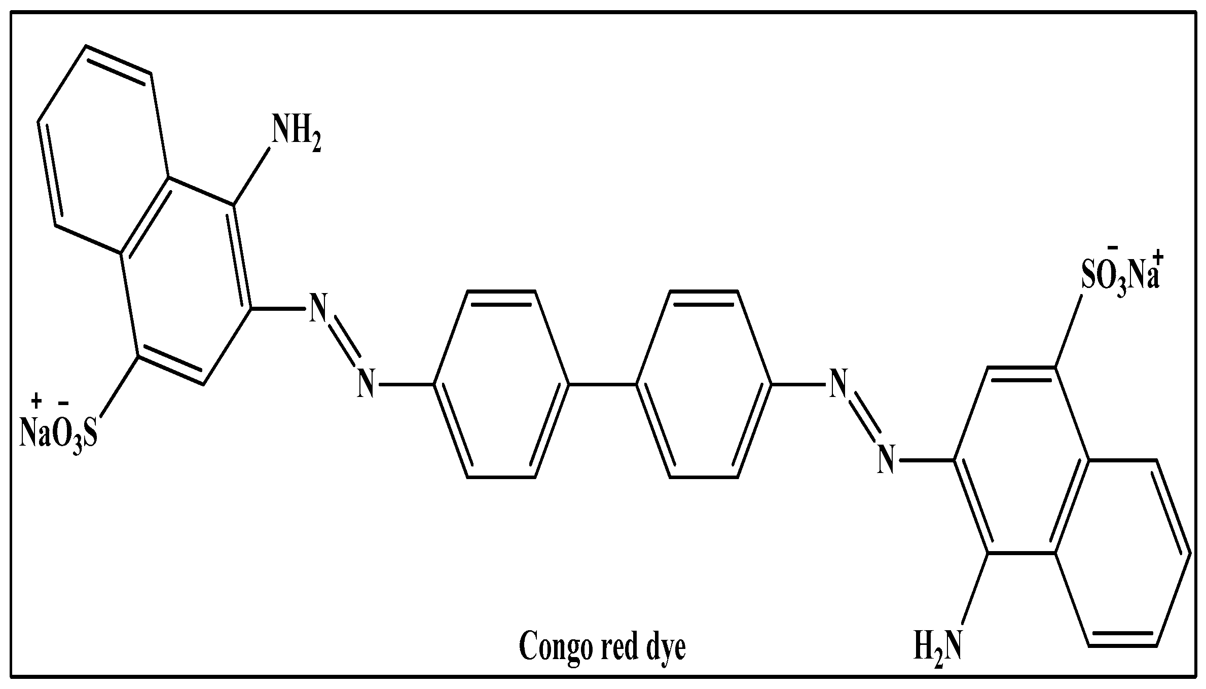
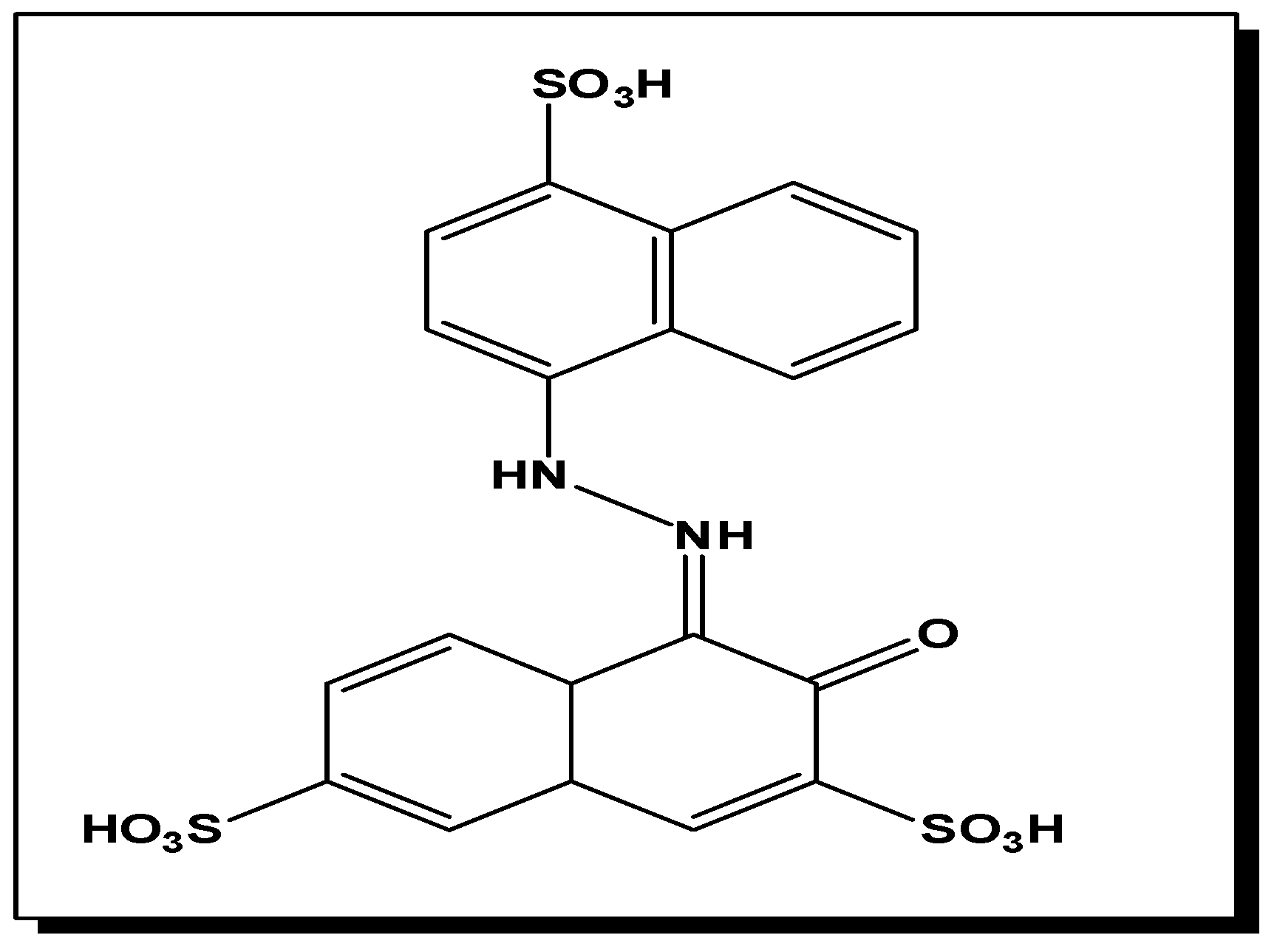
Congo red was discovered in 1884 and a significant direct dye, has a carcinogenic effect in human and animal health [70]. The big Congo red molecule has two azo linkages with 4-amino-1-naphtalenesulphonic acid groups on either end. It act as inhibitor to prevent the metal from corrosion [71]. It exists as a brownish-red crystal that is stable in air and dissolves in water at a rate of 1 g/30 mL. It is an anionic acid dye that is used in laboratories to diagnose amyloidosis, diagnose free hydrochloric acid in gastric contents, determine pH, and histologically stain for amyloid. Because of its great affinity for cellulose fibres, it is used in the textile industry and paper industry. It is a benzidine and napthoic acid derivative that breaks down into cancer-causing substances [72].
Figure 6. Structure of Congo Red Dye.
Preprints 99484 g006

Methylene blue:

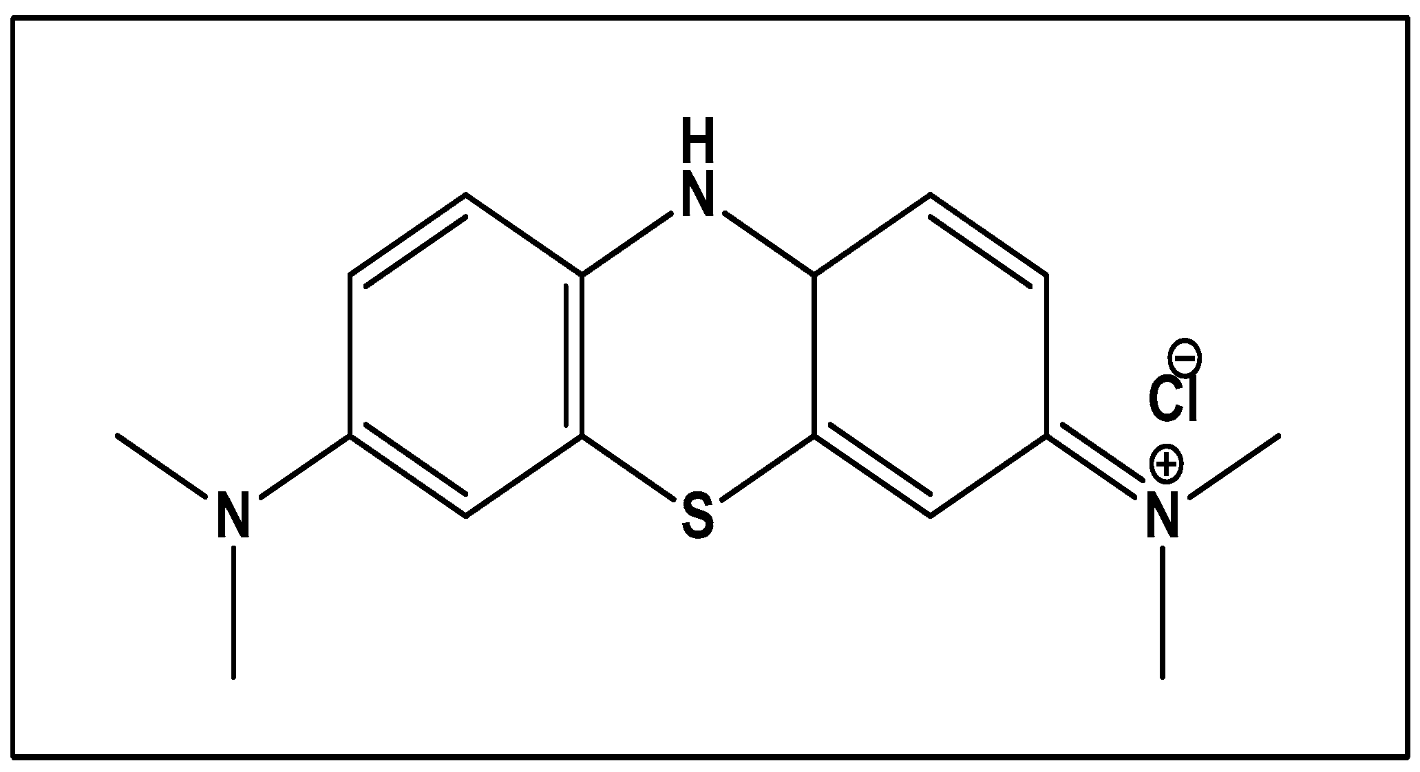
Methyl blue is crystalline, odor-free, deep green powder and when dissolve in water gives blue solution. The molecular weight of MB is 319.85 gmol‾1 and is an aromatic heterocyclic basic dye. Due to its high water solubility, it can, create a stable solution with water. MB is a positively charged chemical that belongs to the class of poly-methine dyes containing an amino autochrome unit [73]. When exposed to MB, people have the danger of developing many health problems, including methemoglobinemia, nausea, vomiting, and ocular damage. Even a small amount of dye in water could be harmful and extremely noticeable removing MB from waste effluents becomes crucial for the environment [74].
Figure 7. Structure of Methylene Blue.
Preprints 99484 g007

Degradation of organic dyes using fabricated Gold Nanoparticles

Removal of Malachite Green and Ethyl violet using Graphene oxide Gold Nanoparticles (GeONPs):
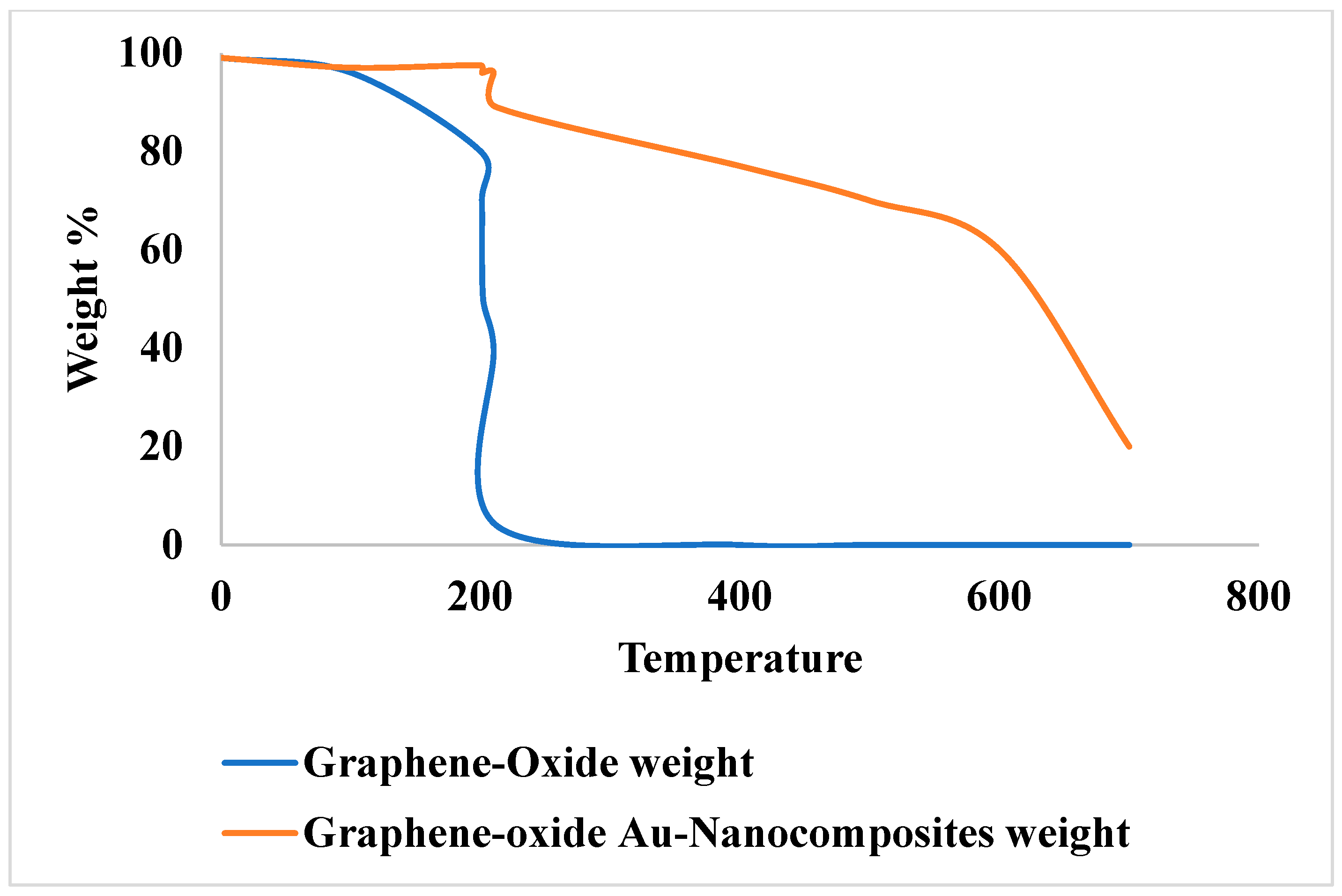
Graphene oxide gold Nano-composites was efficiently used for the removal of cationic dyes from polluted water. It was prepared by using a sodium citrate as reducing agent to reduce Au-ions and then incorporating it on Graphene oxide (G-O) sheet. Then it was characterized by different techniques like FESEM, TEM, EDX, XRD, and FT-IR AND TGA. It was observed that GeO-Au Nano-composite showed efficient adsorption of Malachite green and ethyl violet dyes [7].
Figure 8. Thermal gravimetric analysis curves for nanomaterials made of graphene oxide and graphene oxide-gold nanocomposite [7] .
Preprints 99484 g008
Graphene oxide Au-NPs was added as adsorbent in the water containing cationic dyes (Malachite green and ethyl violet) and anionic dyes (methyl orange). The aqueous solution of Methyl Orange was orange, MG was light blue and MV was dark blue. When all three dyes were mixed together giving aqueous solution of green color. After the adsorption green color turned to pale yellow. By using UV-Visible Spectrophotometer the removal of dyes was monitored from water. Two peaks were shown by UV-Visible spectrophotometer one at 600nm for cationic and other at 463nm for anionic dyes. Both peaks disappeared after 15 minutes that confirmed the removal of dyes from water [7].
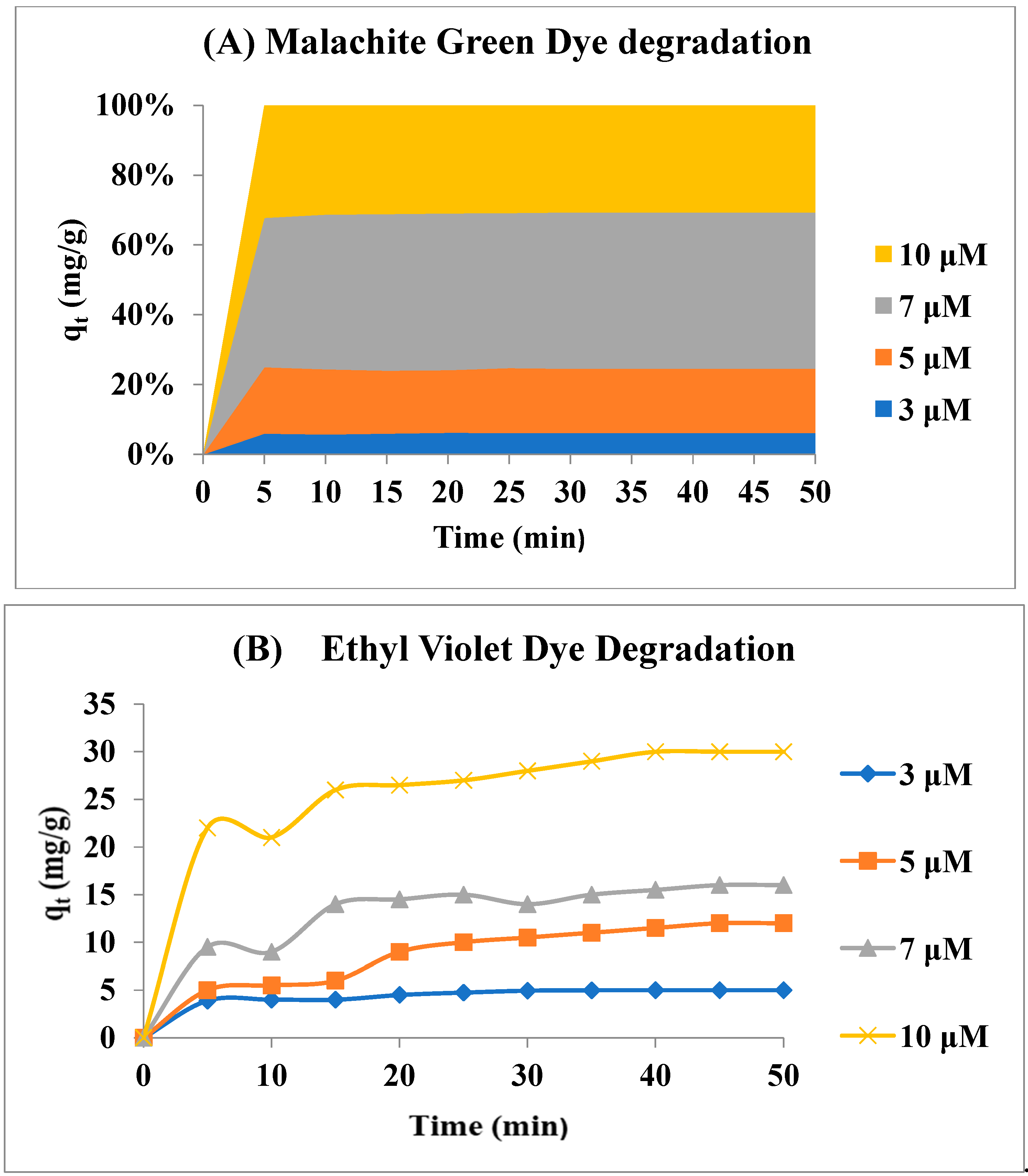
The ability of GeO and GeO-Au nanocomposite to adsorb MG and EV was investigated. For the removal of these dyes, 50ml of 10Mm solution of each dye was taken in a separate container, and GeO and GeO-Au Nanoparticles were then added as an adsorbent. It was observed that GeO-Au had a greater adsorption capacity than GeO, due to the increased adsorption of Au that was integrated on GeO sheets. The adsorption capacity of GeO-Au NPs was higher. The higher surface energy of gold nanoparticles imparted them a greater capacity for adsorption. Although this increase was not particularly significant, gold nanoparticle integration on GeO has some additional advantages, including preventing GeO sheets from aggregating upon reduction, making it simpler to separate from the adsorption medium, extending shelf life, and adding antimicrobial and catalytic properties to GeO-based composites. [7].
Figure 9. Impact of initial dye concentration on graphene oxide-gold nanocomposites' adsorption of (a) malachite green and (b) ethyl violet. Reaction Conditions: Adsorbent weight is 0.005g, and the dye solution volume is 50ml, 100rmp, and 25°C. [7].
Preprints 99484 g009
The amount of adsorbed dye was increased by enlarging the initial concentration. This increase was explained with the help of concentration gradient. By enhancing the concentration of dyes the concentration gradient was increased due to which large amount of molecule of dyes diffuse from surrounding to the surface of adsorbent. Due to the presence of large amount of adsorbate molecule in adsorption medium the total adsorption increases. By increasing the concentration gradient between the adsorbent and adsorbate and the presence of large active sites of GeO-Au Nano-composites the adsorption rate was higher at very start and then slow afterward [7].
Temperature effect: For a 50 ml, 10μM solution of each dye containing 0.005 g adsorbent and a temperature range of 25 to 45℃, the impact of temperature on the adsorption process was examined. The results Indicates that adsorption capacity was increased by enhancing temperature in case of both the dyes. The dyes molecule gain energy by interaction with active sites by raising the temperature on the adsorbent surface [7].
pH effect: pH is another factor that has an impact on the adsorption process. Different functional groups are present on the adsorbent surface. At different pH values the adsorbate surface can be protonated and deprotonated to form charged species in the medium. As a result charges on the adsorbent and the adsorbate surfaces exert electrostatic attractive or repulsive forces. The pH of dye aqueous solution was raised from 2 to 9 using 0.1 M NaOH and 0.1 M HCl aqueous solutions. It was observed that both dyes (malachite green and ethyl violet) minimal and maximum levels of adsorption occurred at pH 7 and pH 2 respectively. In the case of cationic dyes, the adsorption capability was increased by raising the pH. This was caused by growing electrostatic contact between ions and increased π-π staking between negatively charged GeO sheets and cationic dyes. As a result, the percentage of dye removal was increased [7].

Removal of Methylene Blue using AuNPs from Sesbenia grandiflora
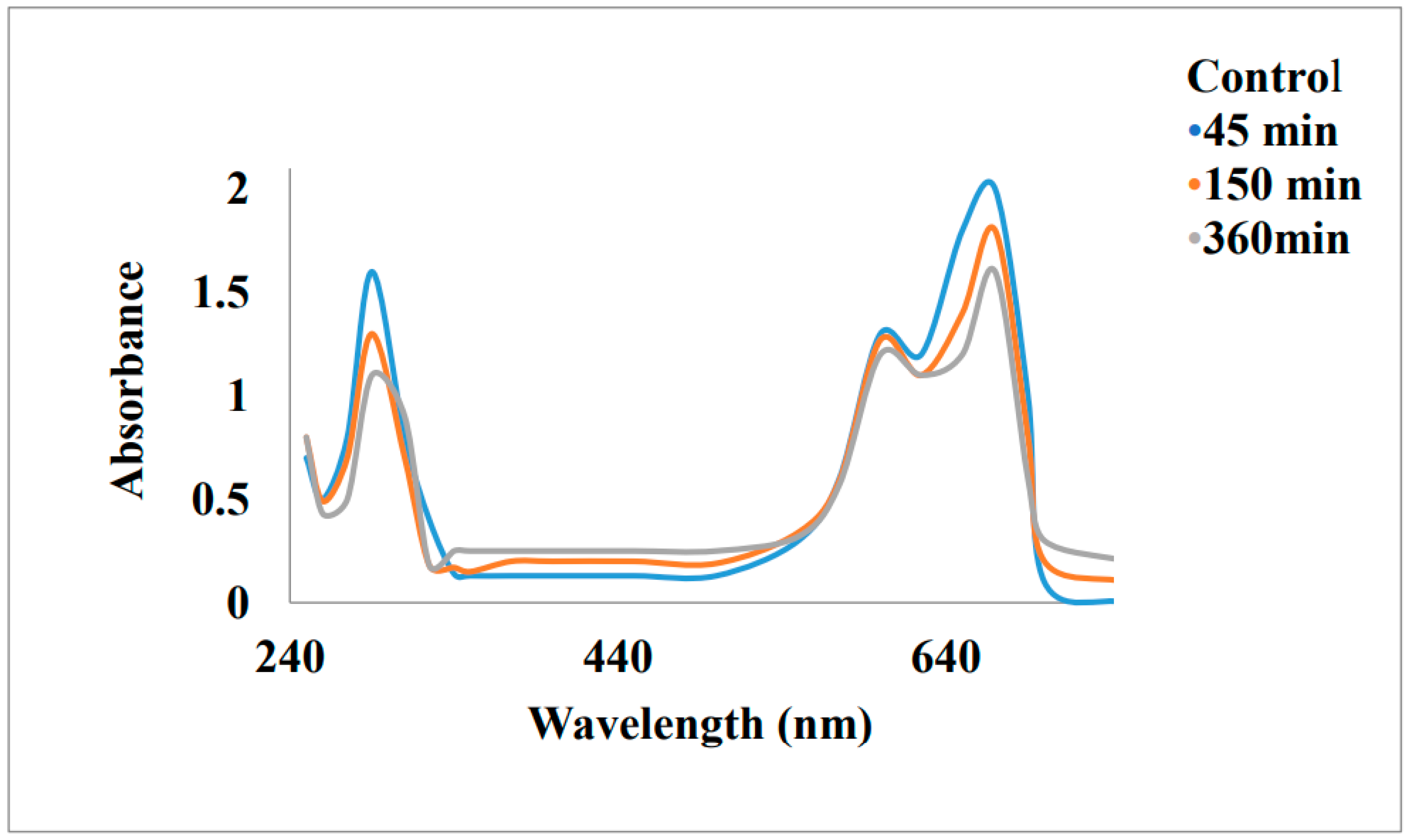
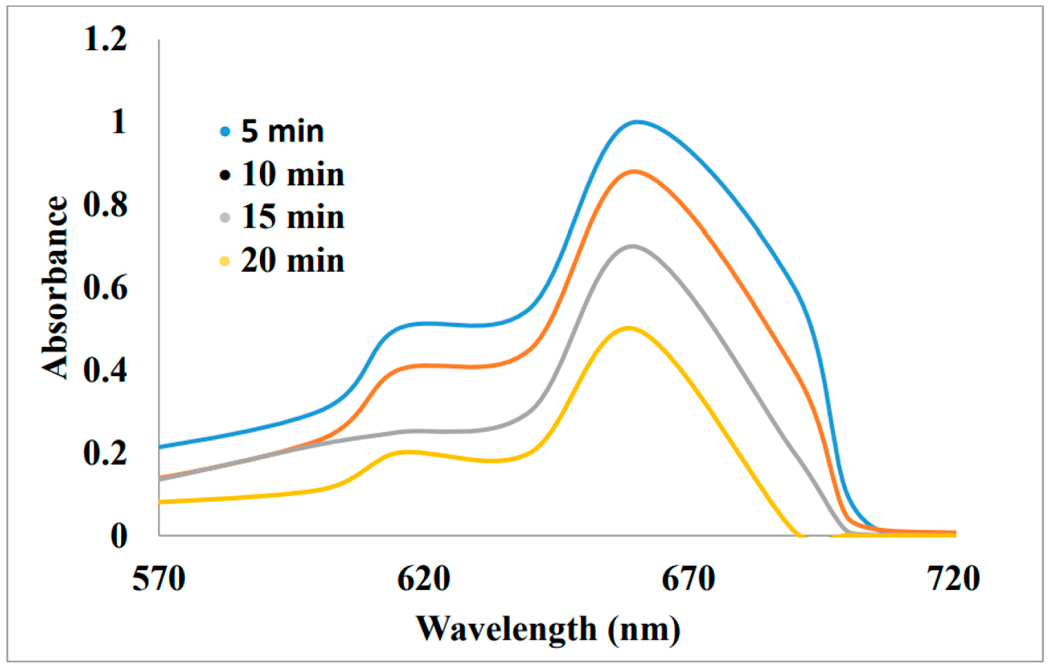
Methylene blue is used in aquatic samples to analyze trace level of sulphide ions. In aqua industry the cationic form of methylene blue dye is used as anti- Malarial agent and chemotherapeutic agent. For the reduction of MB dye biogenic gold nanoparticles were prepared from Sesbenia grandiflora extract. It was characterized by UV-Visible spectroscopy and observed that the absorbance maxima value of MB decrease with respect to time. The decrease in absorbance showed that the phyto-extract lowered the pH value. A drop in absorbance was seen at 666 and 292 nm with the addition of glass beads coated with Au-NPs, which typically implied a progressive conversion of methylene blue (MB) to leucomethylene blue (LMB). It was noticed that in the absence of catalyst the absorbance value decrease gradually in the reduction reaction. The decrease in absorbance indicated the reduction of MB. At 298k and different time interval the recorded absorbance spectra was observed for the reduction of MB [86].
Figure 10. UV-Visible spectra of methylene blue reduction by S.grandflora leaf extract [86].
Preprints 99484 g010
Figure 11. UV-Visible spectra of methylene blue reduction by S.grandflora leaf extract in the presence of glass bead coated with AuNPs [86].
Preprints 99484 g011
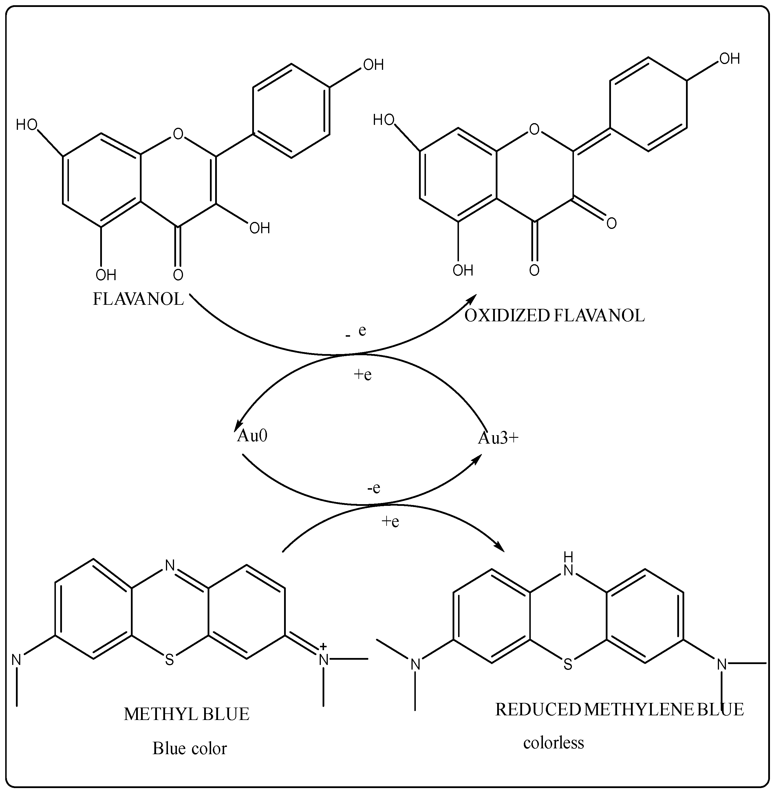
Figure 12. Reduction Mechanism of Dyes using Gold Nanoparticles.
Preprints 99484 g012

Removal of Methylene Blue using AuNPs from Cassytha Filiforms plant extract:

Methylene blue dye was removed by the synthesis of polyphenol Au-NP from Cassytha Filiforms plant extract and then characterized by using UV-visible spectroscopy, HR-TEM and XRD. These, Au-NP showed good capability in removing methylene blue by photo-catalytic degradation in the presence of sunlight. Sunlight and basicity of solution played major role in dye degradation. These gold nanoparticles have ability to degrade 87% dyes after 20minutes and also having dye loading ability of 59523mg/g. The prepared gold nanoparticles were mixed with 5ml methylene blue solution then stirred at different conditions like in sunlight, in UV-light in dark etc. For dyes degradation sample was taken from reaction mixture and then studied by UV-spectrophotometer in the presence of sunlight.

pH effect:

By increasing the PH from 7 to 9 the removal of MB was increased along with reaction rate by increasing the pH further from 11 to 13 respectively. And the removal of MB was decreased by decreasing the pH. The pH investigation showed that the phenolic OH group on the nanoparticle surface was essential for the adsorption of MB [8].
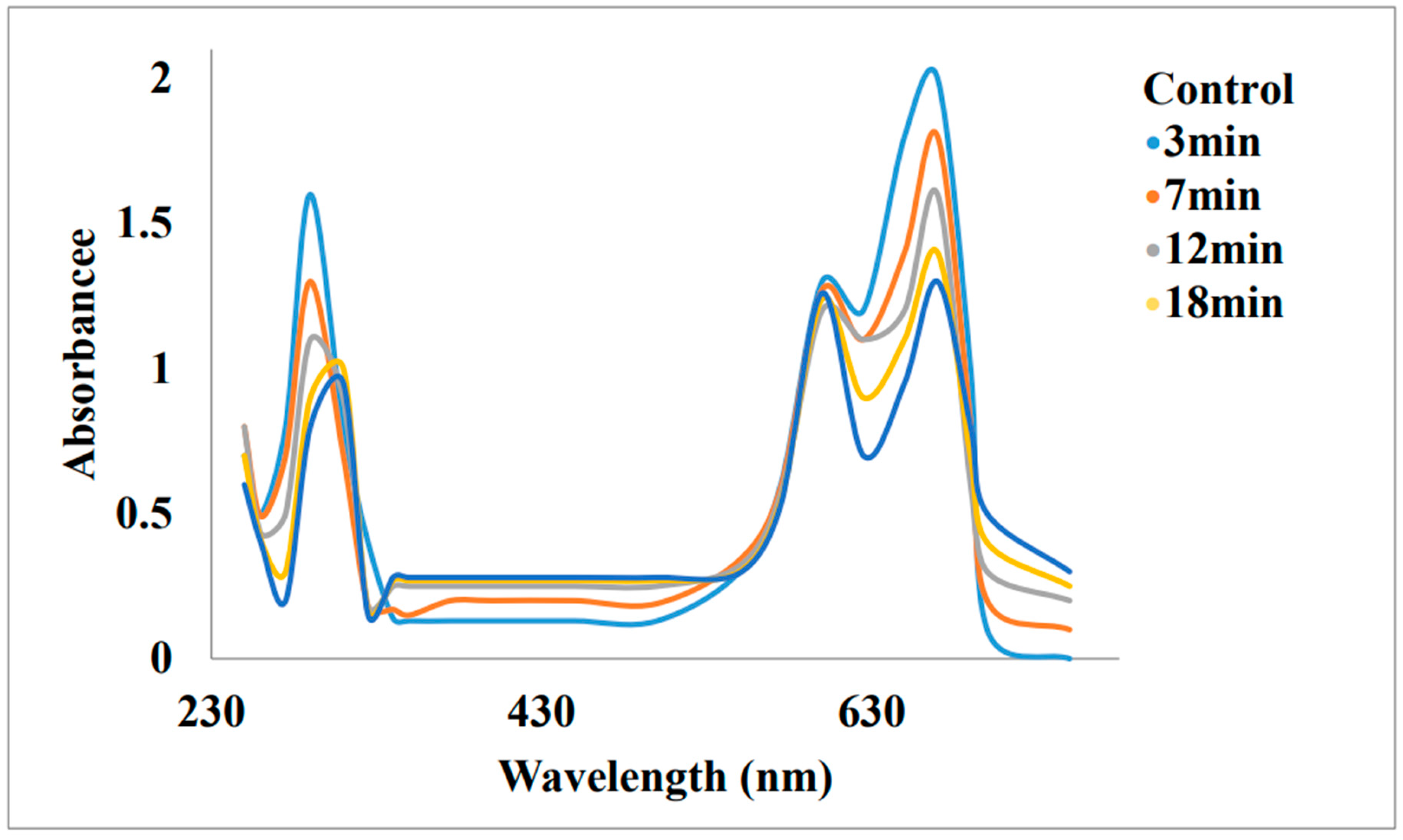
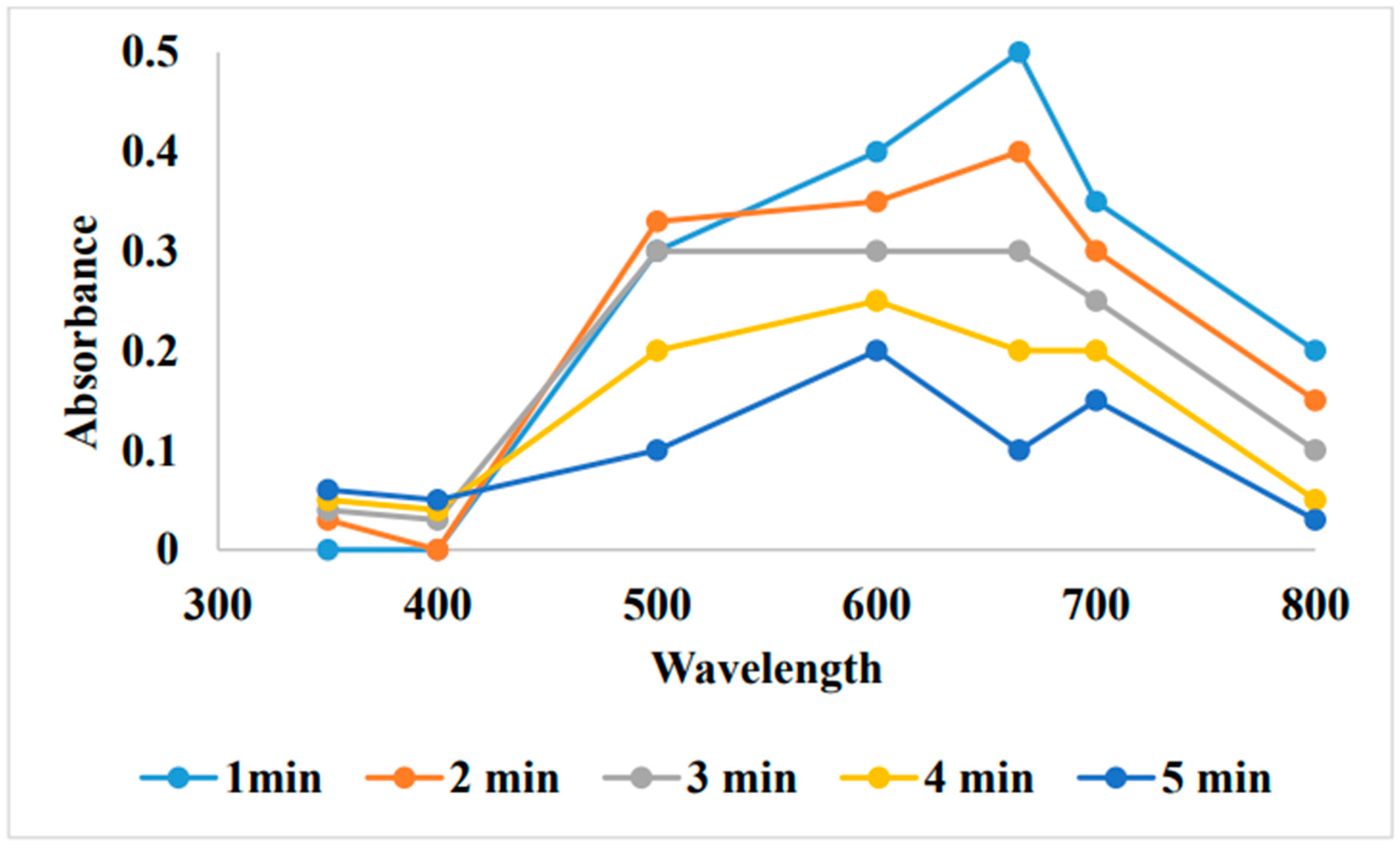
By absorption spectrophotometer the adsorption of MB on Au-NP particles was studied within the range of 400-800nm [8]. The characteristic absorption peaks of the aqueous MB containing samples obtained at various times after adsorption showed that the MB concentration profile decreased with an increase in the adsorption time. Concentration of dye was examined by photometric method. According to CCD (central composite design) method the efficiency of MB removal was examine at different experimental conditions [8].
Figure 13. Absorption spectrum of methylene blue on Au-NPs [8].
Preprints 99484 g013

Reduction of Methyl orange and Congo red from Dalbergia coromandeliana Au-NP:
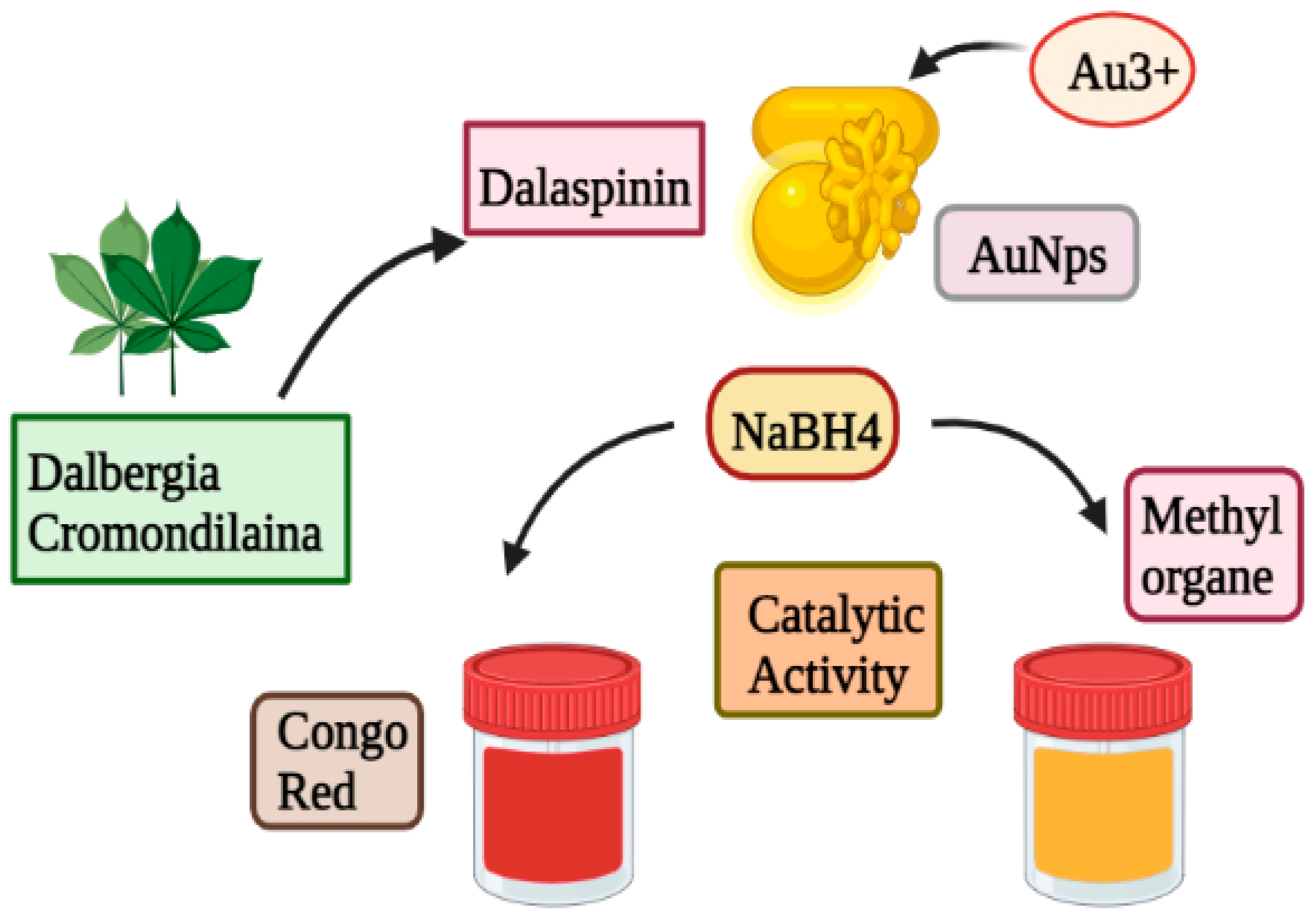
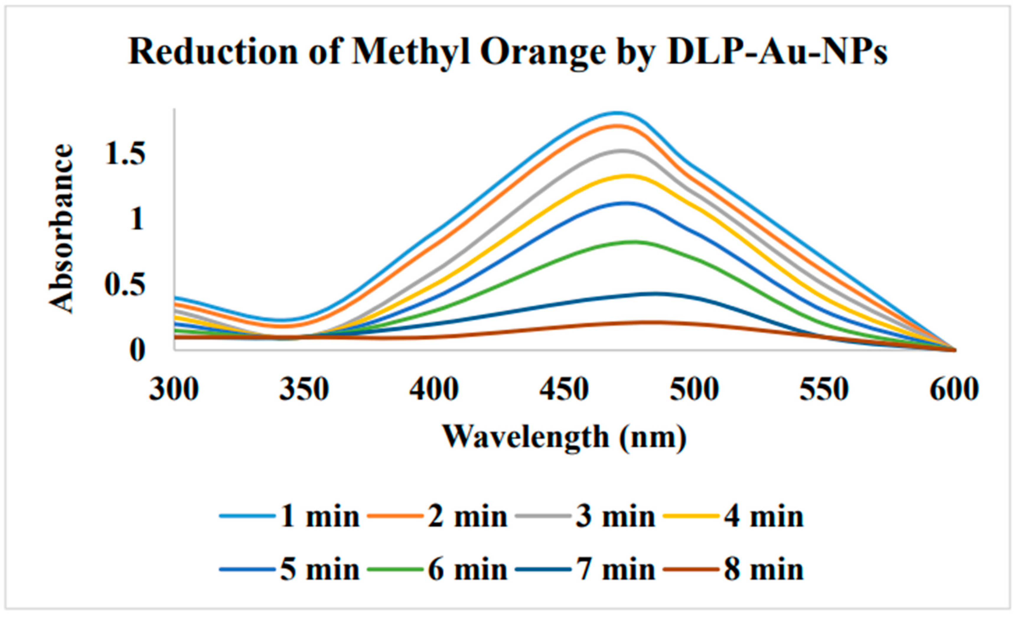
From the root of Dalbergia coromandeliana Au-NP was synthesized by using 5, 7-dihydroxy-6-metoxy-3, 4methylenedioxyisoflavone (Dalspinin). The average particle size of Au-NPs' was 10.5 nm. It was discovered that the prepared Au-NPs remained stable for more than 5 months without aggregating. The Au-NPs showed good activity for the reduction of Congo red and methyl orang dye. Initially a light yellow color of the gold salt solution changed to a wine red color when Dalspinin was added. That showed the synthesis of DLP-Au-NPs. The synthesized DLP-Au-NPs work well as a catalyst to degrade CR in the presence of NaBH4.
Figure 14. Reduction of Methyl orange and Congo Red from Dalbergia coromandeliana using gold nanoparticles.
Preprints 99484 g014
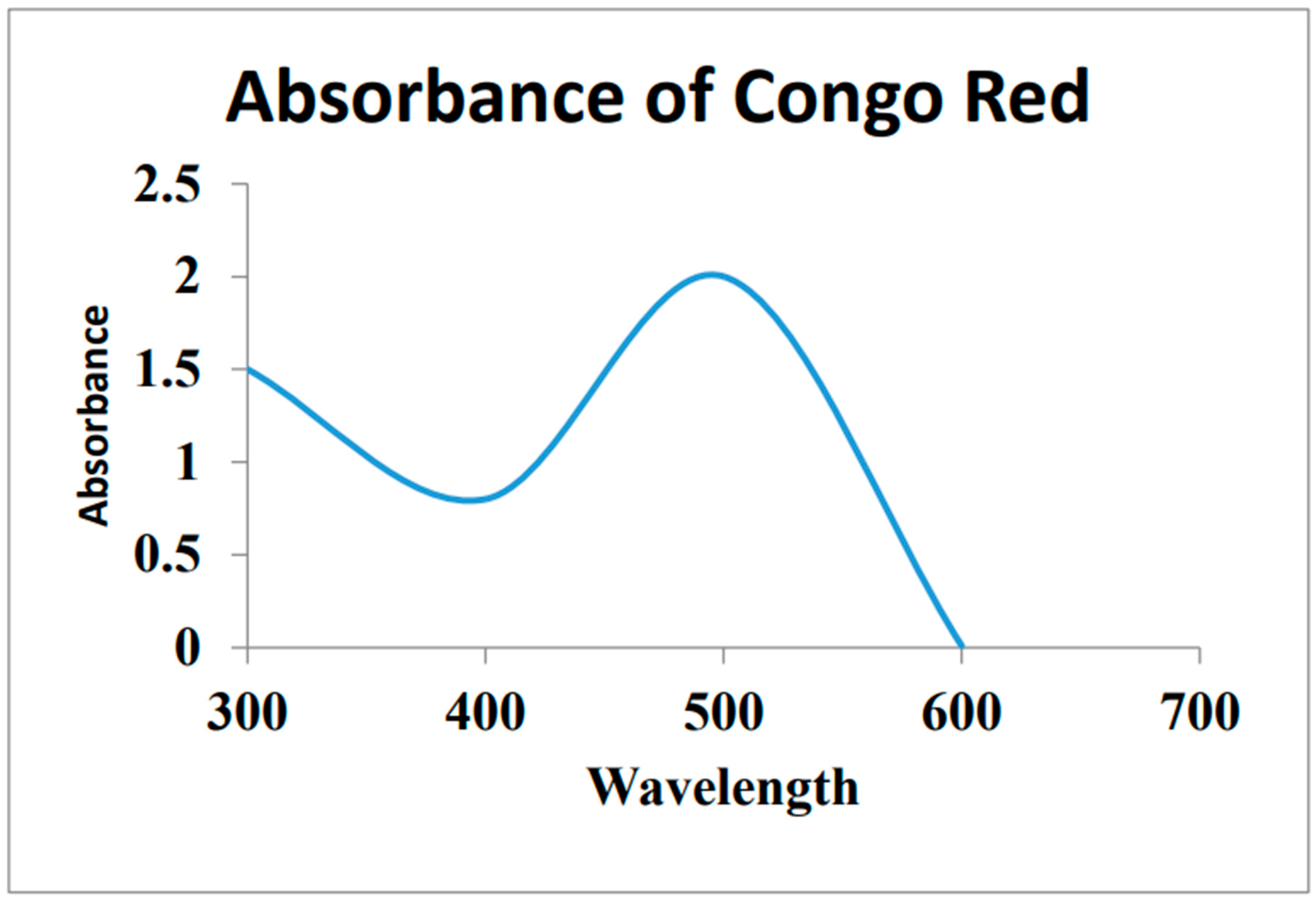
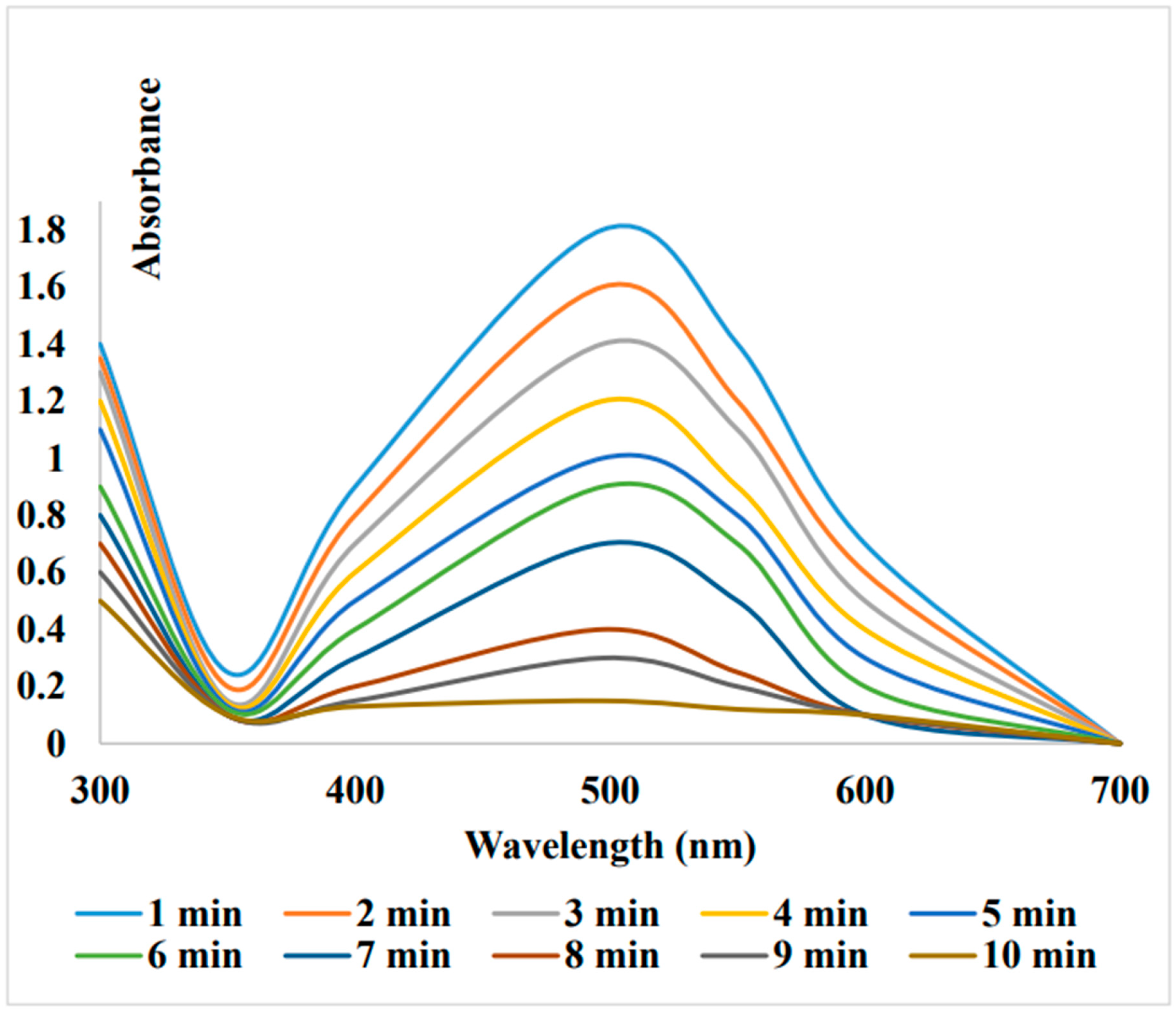
In an aqueous media, CR displayed an absorption band at 497 nm for the π -π*-transition and 350 nm for the n-π*-transition. When the Nano-catalyst was not present the (DLP-Au-NPs) showed wavelength maxima at 497nm for CR that remained un-changed after 2hours. The reaction mixture was changed after the addition of catalytic amount of DLP-Au-NPs. Within 10 minutes, the color changed from vivid red to colorless. This confirmed the considerable catalytic impact with DLP-Au-NPs [87].
Figure 15. UV-visible spectra of Congo red [87].
Preprints 99484 g015
Figure 16. UV-Visible spectra of Congo Red reduction by dalbergia coromandeliana Au-NP in the presence of NaBH4 [87].
Preprints 99484 g016
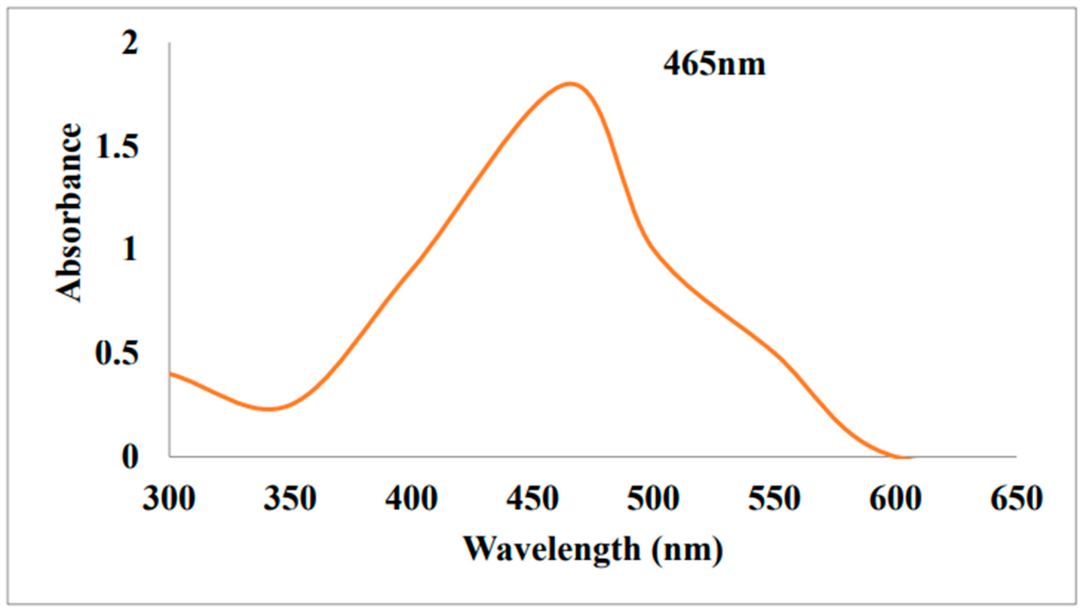
Additionally, the catalytic activity of Au-NPs for the degradation of MO was investigated the results revealed a maximum absorbance at 465 nm (Figure).When DLP-Au-NPs and NaBH4 were present, the dye colour changed from orange to colorless. Methyl orange was not reduced affectively only by NaBH4 in the absence of Au-NPS. But in the presence of DLP-Au-NPs MO was efficiently reduced within 8 minutes [87].
Figure 17. UV-Visible spectra of Methyl Orange [87].
Preprints 99484 g017
Figure 18. UV-Visible spectra of methyl orange reduction by DLP-Au-NPs [87].
Preprints 99484 g018

Biogenic Au-NPs on glass beads:

SnCl2 has been used as reducing agent, when gold nanoparticles are used as catalyst for treatment of MB. Sodium borohydride can also be used with gold NPs during removal of methylene blue. Addition to this, by using biogenic Au-NPs on glass beads in order to treat or remove MB, there is no need of any reducing agent. By following photoluminescence, formation of MB dye dimer has significant role in removal of NPs. Removal of MB has been done by utilizing maghemite fabricated CNTs that can be added with salt of 3,4,5-tricarboxybenzenediazonium. Release of chemicals in the environment does harmful effect that enforce us to the eco-friendly procedure such as by using sunlight, catalyst that has been prepared by green synthesis and adsorption.

Removal of Malachite green using Activated Carbon Gold Nanopartices (Au-NP-AC):
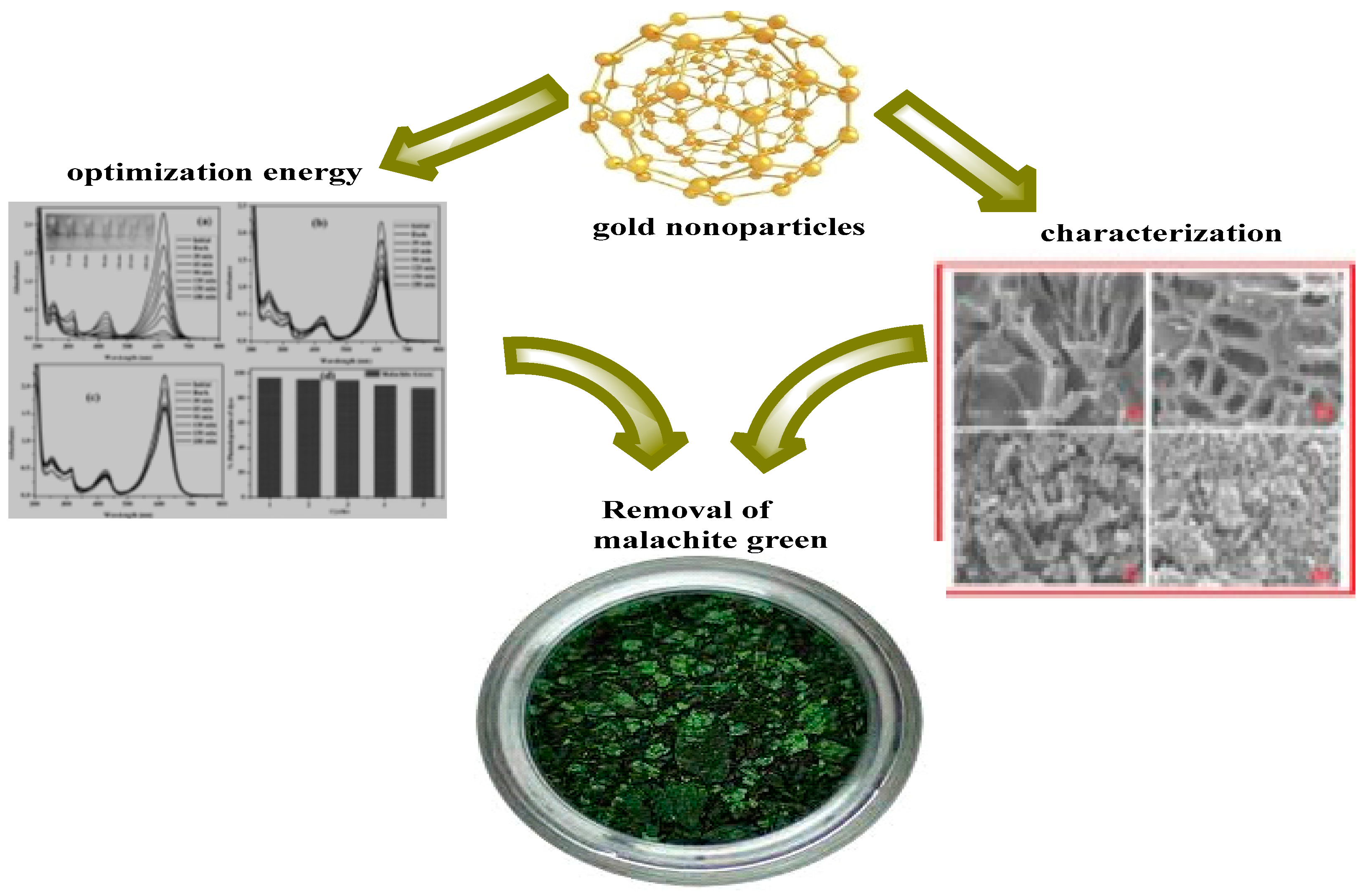
Malachite green is a basic, synthetic water-soluble N-methyl-di-amine-nitro-phenyl methane dye that is used in many industries such as silk, wool cotton, paper and leather industry for coloring purpose. It is also widely used in aquaculture across the globe eradicate fish parasites and mold growth. Activated carbon (AC) seems to be universal adsorbent for dye removal because it contain various reactive sites and having porous structure and large surface area. For the removal of MG an easy, affordable, quick, sensitive and aided adsorption technique with UV detection has been devised. With the help of central composite design and response surface methodology the effect of significant variables including time, pH, initial MG concentration, temperature and amount of adsorbent were examined and optimized. Gold nanoparticles loaded with activated carbon was prepared and characterized by different techniques. The adsorption kinetics and isotherms of MG removal on this adsorbent were then examined with ultrasound assistance serving a straight forward sensitive method. Ultrasound was used to examine dye solution clearance. In this experiment MG chemical was mixed with Au-NP-AC. The experiment was carried out by taking known amount of adsorbent dye and specified amount of dye solution in a flask at pH 7. The flask was then kept at required temperature for some time. The sample was centrifuged and examined after adsorption test. At maximum wavelength 619nm and working concentration the concentration of dye was examined by photometric method. On the activated carbon surface the activated sites were deprotonated by increase in pH and the OH group or COOH absorb MG by electrostatic interaction or hydrogen bonding. It was confirmed that with increase in time maximum absorption was observed. [78].
Figure 19. Degradation of Malachite green using Activated Carbon Gold Nanopartices (Au-NP-AC) [78].
Preprints 99484 g019

Synthesis of Gold Nanoparticles:

1. Synthesis of gold nanoparticles by using marine alga (Sargassum wightii):

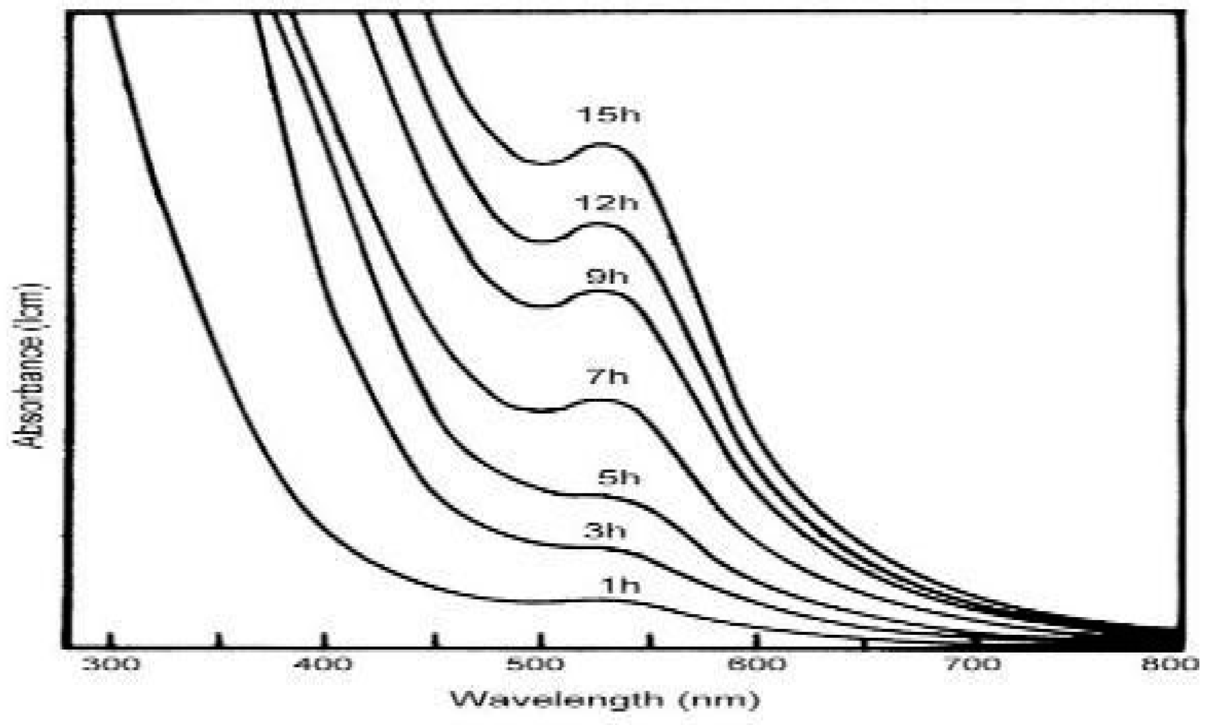
A significant advancement in the field of nanotechnology is the technique of creating environmentally acceptable metallic nanoparticles. Eco-friendly nanoparticles must be grown in order to avoid the synthesis of hazardous compounds. Sargassum wightii was used to produce gold nanoparticles through extracellular biosynthesis, and rapid formation of the particles was accomplished. The reduction of aqueous AuCl4 by the extract of the marine alga Sargassum led to the formation of high density, exceptionally stable gold nanoparticles with an average size of around 11 nm. These particles range in size from 8 to 12 nm. Seaweed is transported into a lab, cleaned with distilled water, dried, and then turned into powder. 95% bio-reduction of chloroauric acid (HAuCl4) required 12 hours to synthesise gold nanoparticles. [80].

Characterization Techniques:

The pattern of light absorption lies between 300nm and 800 nm. The band was seen in the UV-visible spectra at 527 nm. [80].
Figure 20. UV-Visible spectra of absorption of Au-NPs synthesized by marine alga [80].
Preprints 99484 g020

TEM micrograph

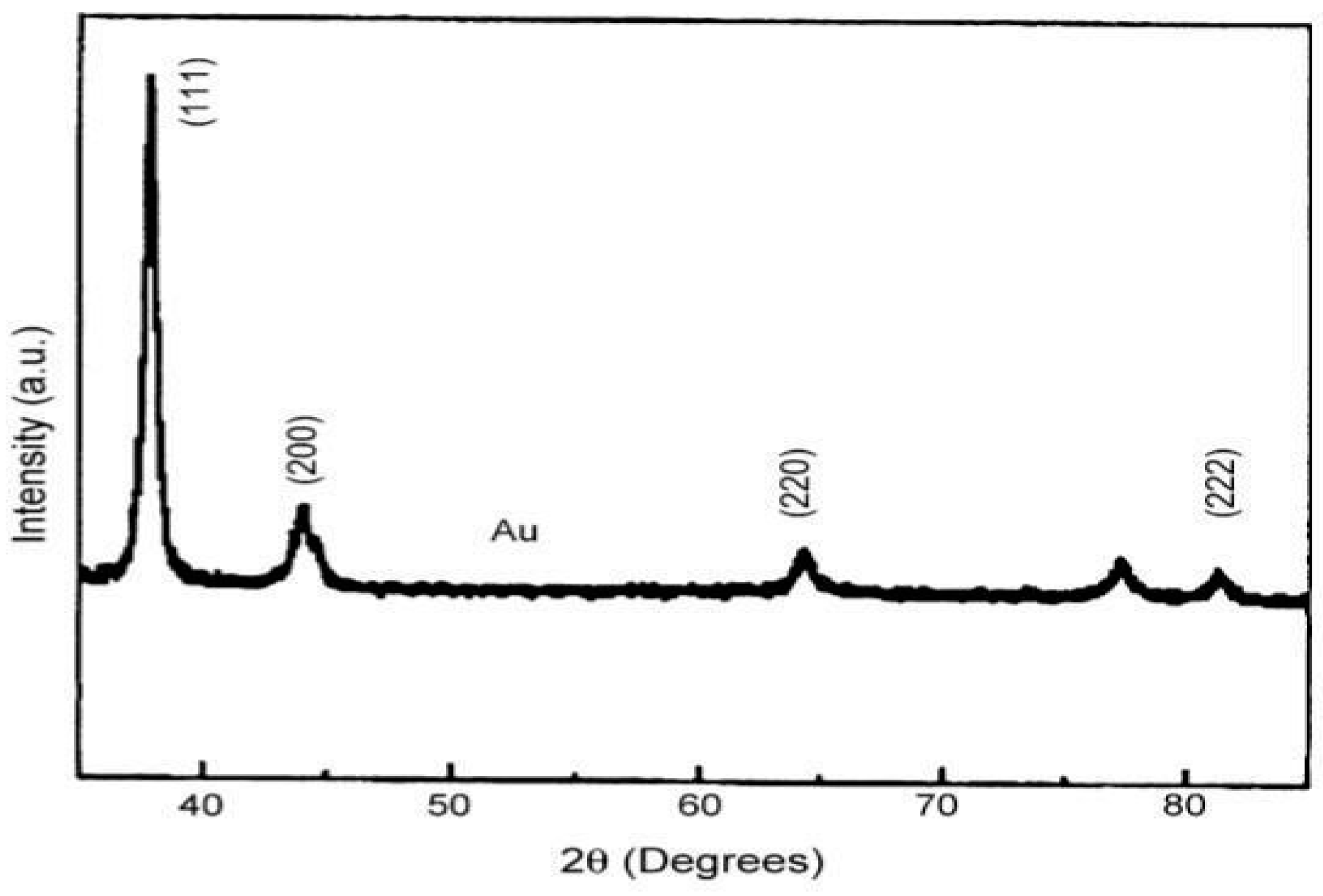
A large number of gold nanoparticles measured in the TEM micrograph ranged in size from 8 to 12 nm, and the shape of gold nanoparticles was thin planner structures rather than spherical structures. X-ray diffraction was used to verify the particle's crystalline nature and the resulting XRD pattern. [80].
Figure 21. X-ray diffraction of Au-NPs synthesized by marine alga [80].
Preprints 99484 g021

2. Synthesis of gold nanoparticles by using Scutellarin barbata:
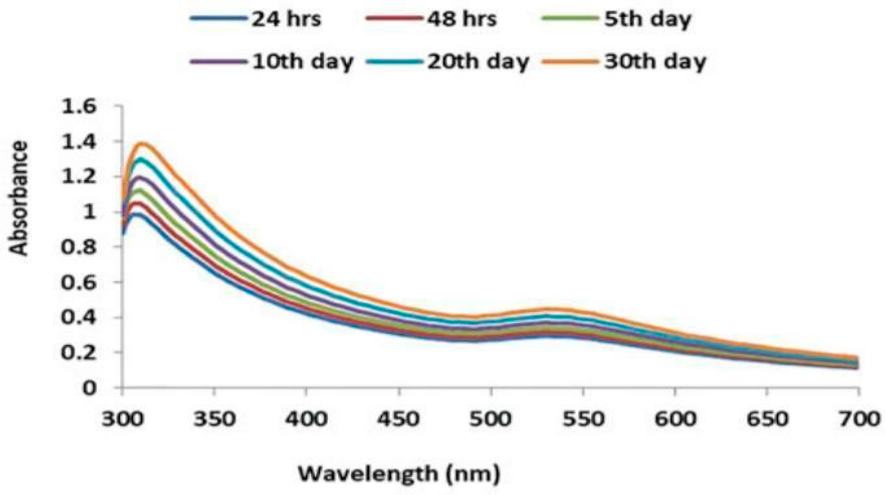
Leaves of Scutellaria barbata were collected, dried and finely powdered, mixed with 100ml deionized water. Shaked it for 10 min, then boil at 90°C for 40 min to obtain bioactive constituents. After boiling, the suspension was allowed to cool at room temperature and filtered it. After this process, the resulted crude aqueous extract was used for the synthesis of the gold nanoparticle. About 10 ml of aqueous extract of the plant was added to 50 ml of 0.1 M of an aqueous solution of chloroauric acid (HAuCl4). Then, this reaction mixture was constantly stirred by using magnetic stirrer for the reduction of gold ions. After 15–20 min, the reaction mixture turned from brown to reddish yellow and the suspension was constantly stirred for 3 h. The suspension was allowed to cool. By centrifugation at 10,000 rpm for 15 min, gold nanoparticles were obtained. Then, these gold nanoparticles were used for further characterization and analytical purposes. From UV-Visible spectroscopy range of gold nano particles were shown at the 540 nm range [81].
Figure 22. UV-Visible spectra of Au-NPs synthesized by Scutellarin barbata [81].
Preprints 99484 g022
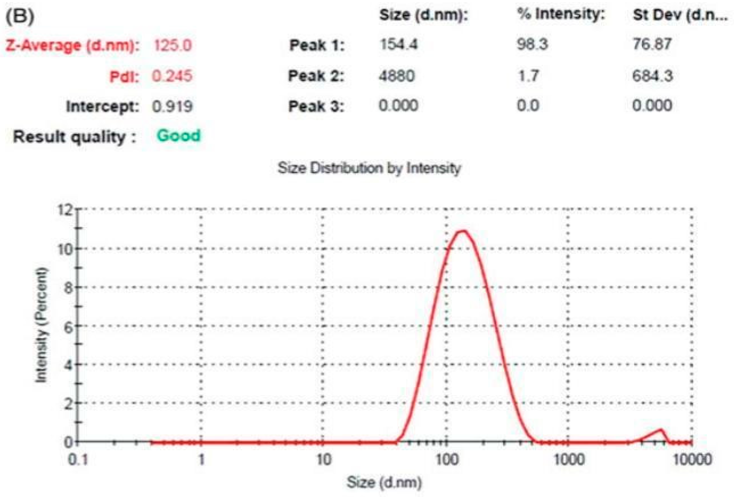
Dynamic light scattering (DLS) is a technique that showed synthesised gold nanoparticles may have an average size of 154 nm. [81].
Figure 23. Average Size of Au-NPs by DLS [81].
Preprints 99484 g023
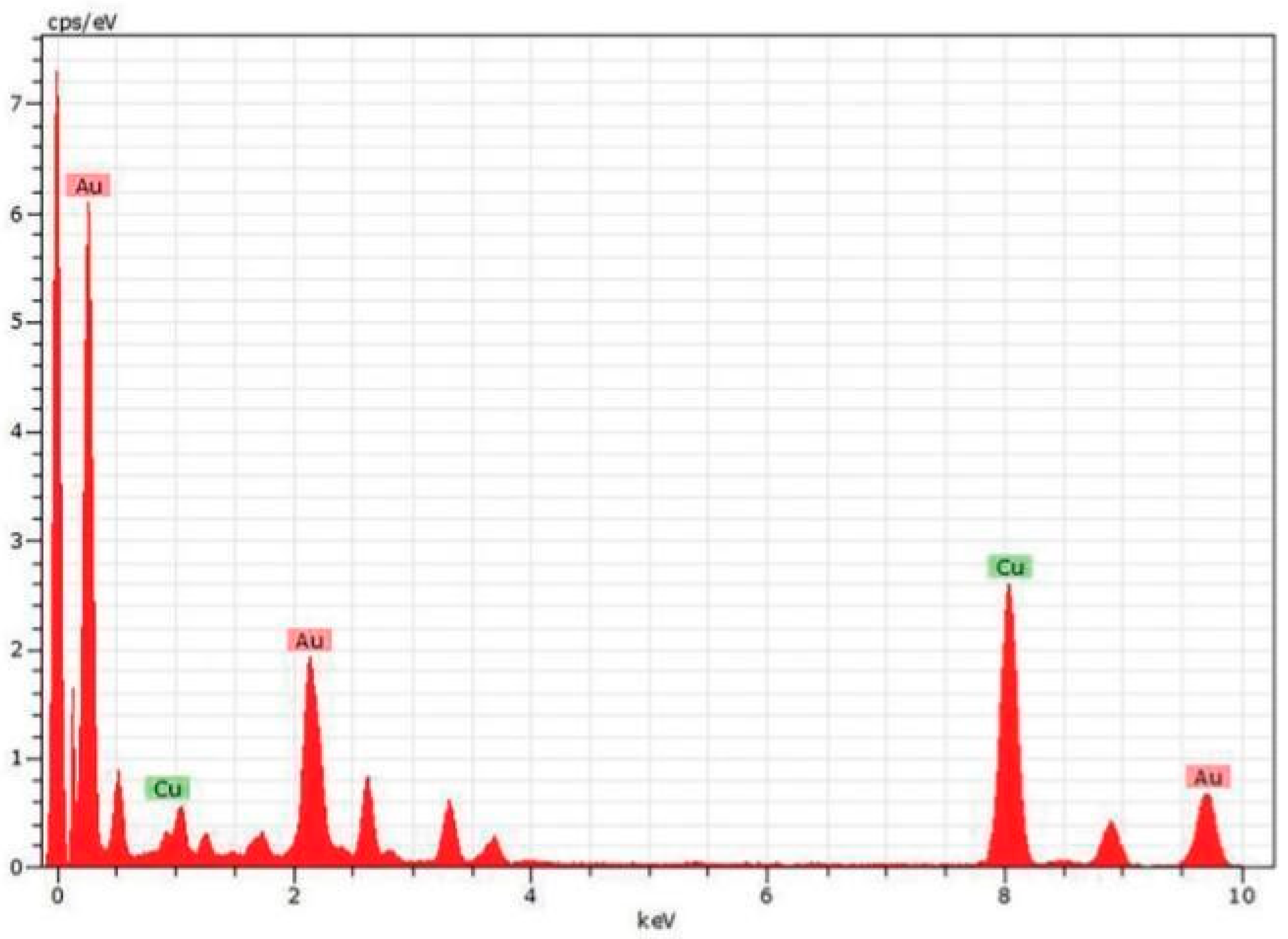
The EDX elemental analysis clearly showed the existence of the majority of gold (Au), followed by copper (Cu) This outcome demonstrates that there are gold nanoparticles present in the reaction media. [81].
Figure 24. Energy dispersive X-ray (EDX) and analysis of Scutellaria barbata-derived AuNPs [81].
Preprints 99484 g024
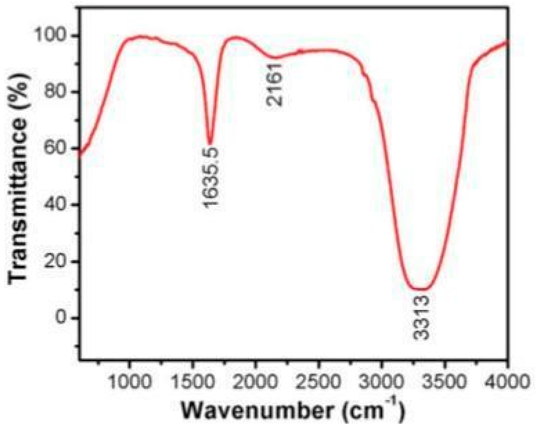
The profile of the biomolecules particularly present on the surface of the created gold nanoparticles was determined using Fourier transform infrared analysis (FT-IR). The FT-IR analysis's spectrum showed that the exterior of the synthesised gold nanoparticles contained carboxylic acid group, hydroxyl group, alkanes group, and carbonyl group.
Figure 25. Presence of different functional group on the surface of synthesized Au-NPs from Scutellaria barbata [81].
Preprints 99484 g025

3. Synthesis of gold nanoparticles by Alpinia nigra:

Leaves of A. nigra were transformed into powered form. Then mix them in distilled water 10g and 350ml respectively and then heated at 50°C for 30 minutes. After 30 min left for cooling. Upon cooling filter the extract, 1ml filtered aqueous extract of A. nigra leaves was purified with HPLC, for elution 60% methanol in 1% CH3COOH in water was used. (Flow rate at 0.8ml/min). Catechin was taken as the standard sample. Solution of 1mM HAuCl4.3H2O was taken. Heat for 5minutes with continuous stirring at 95°C. 12ml of ALW was added with continuous stirring. The color change from light yellow to purple red indicated the formation of nanoparticles. [82].

Removal of Rhodamine B and Methyl Orange dyes by using ANL-AnNPs:
200mg/L -1-solution of both methyl orange and Rhodamine B were treated with 5mg of ANL- AuNPs (Alpinia nigra). Stirred the solution for 30 minutes in the dark for absorption or desorption and then carried these mixtures in the sunlight to continue reaction. 5ml of each mixture collected at the interval of 15 minutes, 45 minute, 60 minute, 90 minutes, and 120 minutes and Nano particles were removed after centrifugation. The supernatant was observed for UV-Visible analysis for Rhodamine B (553 nm) and for Methyl orange (464 nm). Percentage of degradation was collected by the formula
%   degradation = A o A A o × 100
Where A° is the absorbance of a dye solution that has not been added with ANL-AuNPs and was exposed to sunlight, and A was the absorbance of the mixture [82].
Rhodamine B and methyl orange degradation was tracked with the use of a UV-visible spectrometer. Methyl orange's absorption maxima was observed at around 464 nm, while Rhodamine B absorption maxima was observed at about 553 nm. Rhodamine B and methyl orange have rate constants of 1.494 10-2 min-1 and 1.443 10-2 min-1, respectively. The regression coefficient values for methyl orange and rhodamine B are 0.985 and 0.965, respectively, indicating that the pseudo-first order kinetic model matches the data from the experiment. The presence of sunlight speed up the degradation process and led to the formation of low-energy electrons in gold atoms.
Figure 26. Percentage degradation of Rhodamine B [82].
Preprints 99484 g026

4. Synthesis of graphene/Au-NPs:

Mixture of graphene oxide (GeO) dispersion (0.5ml.10mg/L) and chloroauric (HAuCl4) (2ml.4pm) was prepared. Solution of NaOH (5ml 0.1M) was addes to the mixture Of GeO and HAuCl4. Then distilled water was added up to 100ml. Cover it with paper or tin foil to protect from light for 8 hrs. In clean PTFE (polytetrafluoroethylene) lining add ultrasonic solution. For reaction completion place it into reaction kettle for 10-12 hrs. Left it for cooling at room temperature. Graphene/Au-NPs were formed [83].

5. Adsorption of MB dye using Polyaniline coated gold aryl particles:
Polyaniline coated gold aryl particles were prepared by the reduction of aryldiazonium tetrachlorocuprate (III) electrochemically in addition to salt of polyaniline emeraldine. After the formation of NPs, the characterization was done through XPS, TEM, TGA. Gold nanoparticles were synthesized using electrochemistry. In order to stabilize the size of nanoparticles, different stabilizing agents were used. For example; surfactant of tetradodecylammonium bromide ion was used to make clusters of stabilized gold NPs. Polypyrene entrapped NPs have size less than 8nm. Addition of NaOH into electrolyte solution formed pure and uniform 5nm AuNPs. Gold and Patinum were used as anode and cathode respectively, in electrolysis to form gold nanoparticles. Tetraalkylammonium salts were employed as a stabilizer and organic medium served as the electrolyte. Because of different oxidation states polyaniline has been frequently studied for coating purpose in recent years. Emeraldine is suitable for catalytic purpose, super capacitors, storing data and in forensic research [77].
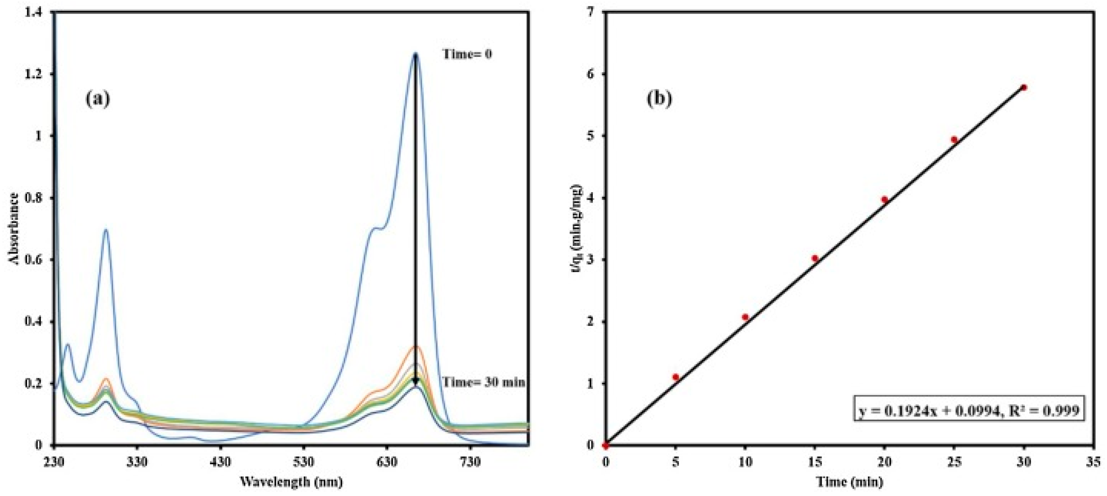
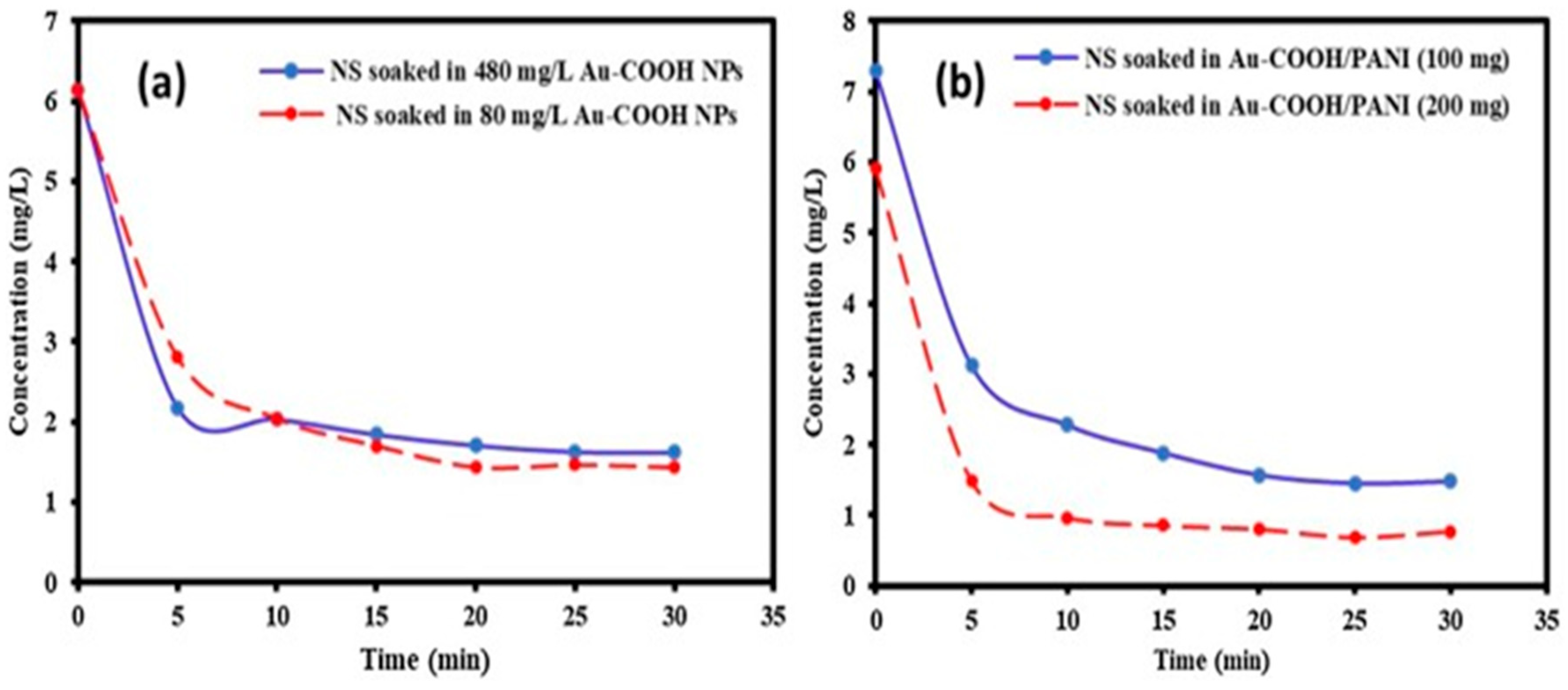
Adsorption of MB dye was done by polyaniline coated gold aryl NPs. During the adsorption of methylene blue, the major issue was that, it has the ability to stick on glass vessel that affects its amount. PANI coated nanoparticles nitrogen gives the formation of ion pairs with nitrogen present in methylene blue that reduces its ability to stick to glass surface. Adsorption of MB done on gold nanoparticles after soaking it for 24 hours. The time of soaking shows no significant effect on adsorption. UV-Visible measurements are used to analyze MB adsorption after each 5 minutes for 30 min [77].
Figure 27. UV-Vis monitoring of (a) 6 mg L-1 MB adsorption in 80 mg L-1 and 480 mg L-1 Au-COOH NPs for 24 hours in the presence of NS (nano sand) (b) Au-COOH/PANI (100 and 200 mg PANI) [77].
Preprints 99484 g027
After adsorption percentage of dye removal on Nano sand Au-COOH NPs and Au-COOH NPs /PANI nanocomposites are given below:
Table 7. Pseudo first and second order was applied to analyze the adsorption process.
Sample Removal (%)
Nano sand(5mg) 65.5
Au-COOH NPs

Au-COOH NPs(80mgl-1)
Au-COOH NPs (480mgL-1)


76.6
73.6
Au-COOH NPs/ PANI nanocomposites
Au-PANI (42mg PANI)
Au-PANI (20mg PANI)


80.0
85.0

Comparison of Reaction kinetics for both Au-COOH NPs and Au-COOH NPs/PANI
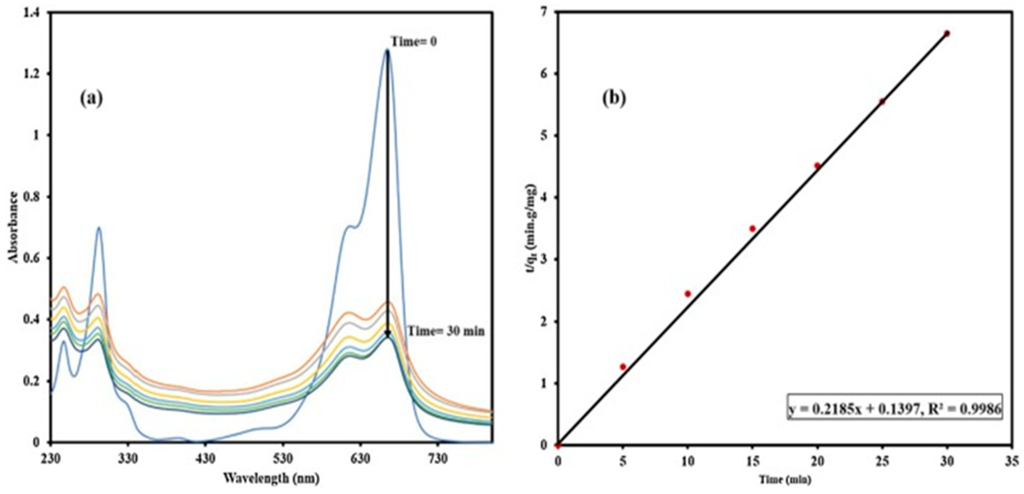
Pseudo second order rate constants has more correlation coefficient than first order. After analyzing result, it is concluded that more adsorption sites were available in nano composites than in NPs. After MB adsorption, FTIR of nano sand has been run, and it confirmed the MB adsorption by NPs. The adsorption peak of dye moved to 827, 883, and 1593 cm-1 from 828, 885, and 1596cm-1, these shifts symbolizes the π-π interaction in dye and ability of adsorption by NPs [77].
Figure 28. UV–vis spectrum of (a) 6 mg L−1 MB +5 mg NS soaked in Au−COOH NPs (480 mg L−1 ) for 24 h, (b) Pseudo-second order [77].
Preprints 99484 g028
Figure 29. UV–vis spectrum of (a) 6 mg L−1 MB +5 mg NS soaked in 5 mg of Au−COOH NPs/PANI for 24 h, (b) Pseudo-second order. Conditions: synthesis in PANI (200 mg) at 0.2 V [40].
Preprints 99484 g029

6. Synthesis of Asp-AuNPs:

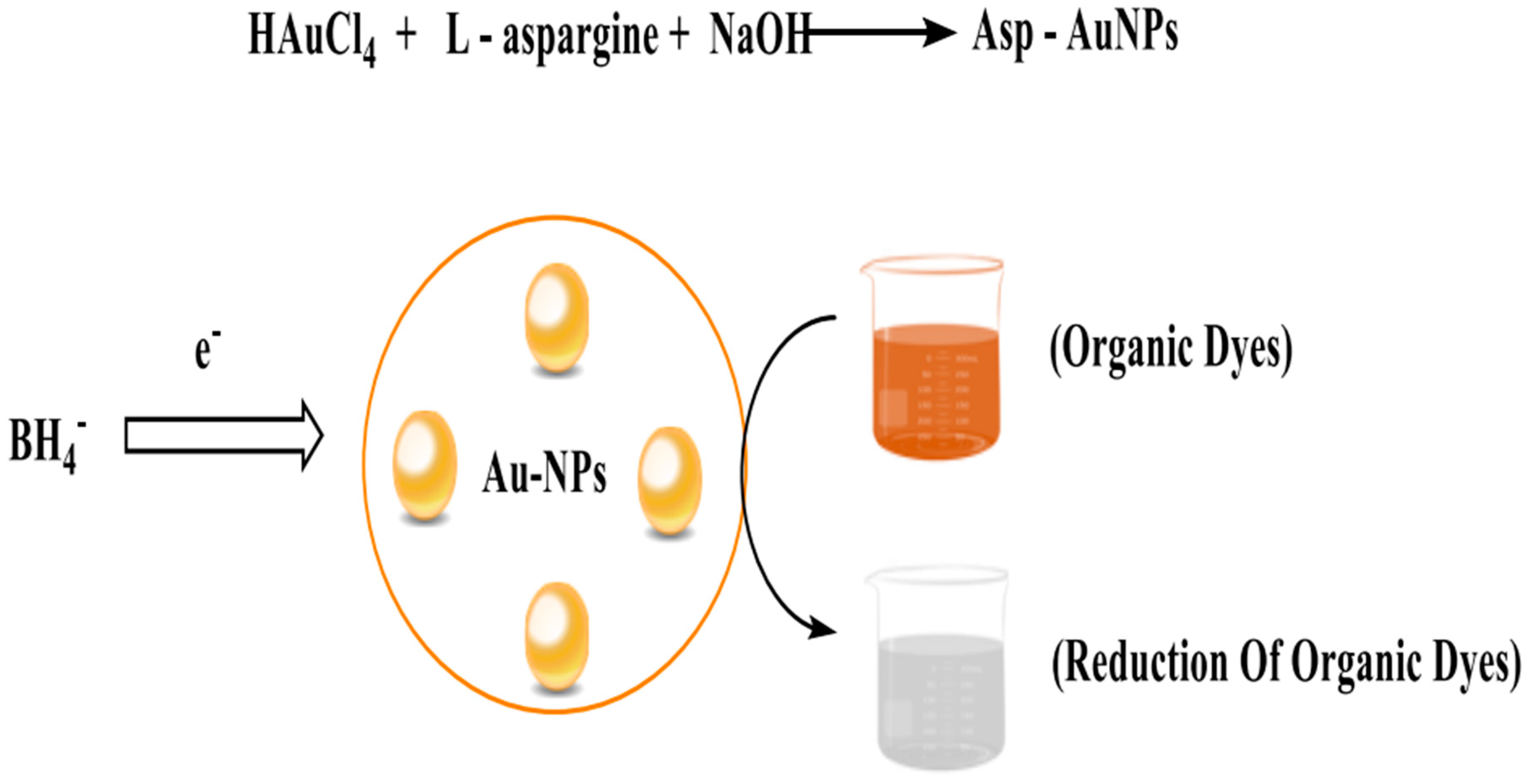
Au-NPs were prepared by one pot synthesis. Reduction of HAuCl4 with L-asparagine was done by addition of NaOH. L-asparagine solution was prepared in water (99ml) that has 2.5mM and 5mM NaOH, HAuCl4 (1mM of 100mM) was added in above mixture, mixture was stirred continuously at 70 degrees centigrade for almost 1 to 8 hours. After 15 to 20 min, red color showed the formation of NPs had begun. After synthesis UV-Visible absorption was used for characterization purpose of Asp-AuNPs. These were then further used for degradation and stored at ambient temperature [9].
Figure 30. Degradation of Organic Dyes using Asp-AuNPs synthesize from L-Aspargine.
Preprints 99484 g030

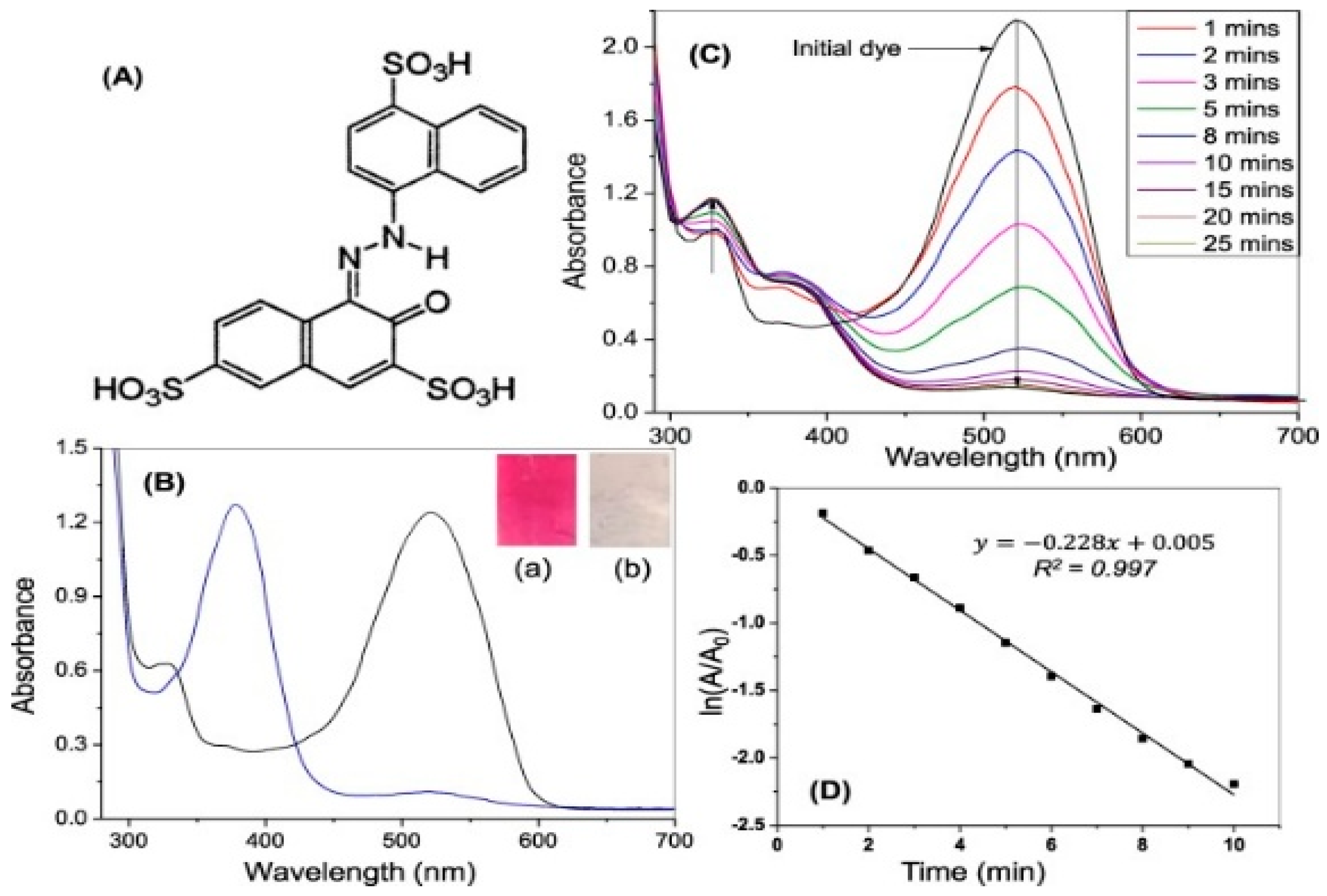
6.(. a) Degradation of Acid Red using Asp-AuNPs
Acid red that is amaranth has azo group as its chromophore and degradation of it was done by using reducing agent and catalyst. By using only reducing agent azo group become reduced but the breakage of its bond need some catalyst, so the degradation was not possible without gold nanoparticles. Degradation can be done in absence or presence of catalyst. We use NaBH4 for reduction, and asparagine gold nanoparticles as catalyst. Because of using Asp-Au-NPs, degradation process is more efficient and rapid. Dyes degradation could not happen without using reducing agent (NaBH4) because NaBH4 reduce chromophore present in dye. Degradation has two purposes, one is reducing chromophore, and other is conversion of large molecules to small eco-friendly organic molecules. Asp-AuNPs used for degradation of organic dyes [9].
Figure 31. Structure of Acid Red.
Preprints 99484 g031
Absorption maxima peak appeared at 520 nm, by reducing it with NaBH4 the peak shifted at 377nm. Reaction rate was faster at initial 10 min but after that due to the formation of products they influenced pH and reaction rate and degradation reaction became slow after that. [9].
Figure 32. (A) Structure of amaranth dye; (B) UV–visible absorption spectra of (a) amaranth, (b) amaranth in presence of NaBH4; (C) absorption spectra of amaranth at different times in presence of Asp-AuNPs and NaBH4; (D) pseudo first-order kinetics of degradation of amaranth. [9].
Preprints 99484 g032

7. Annona squamosa Au-NPs and its degradation activity:
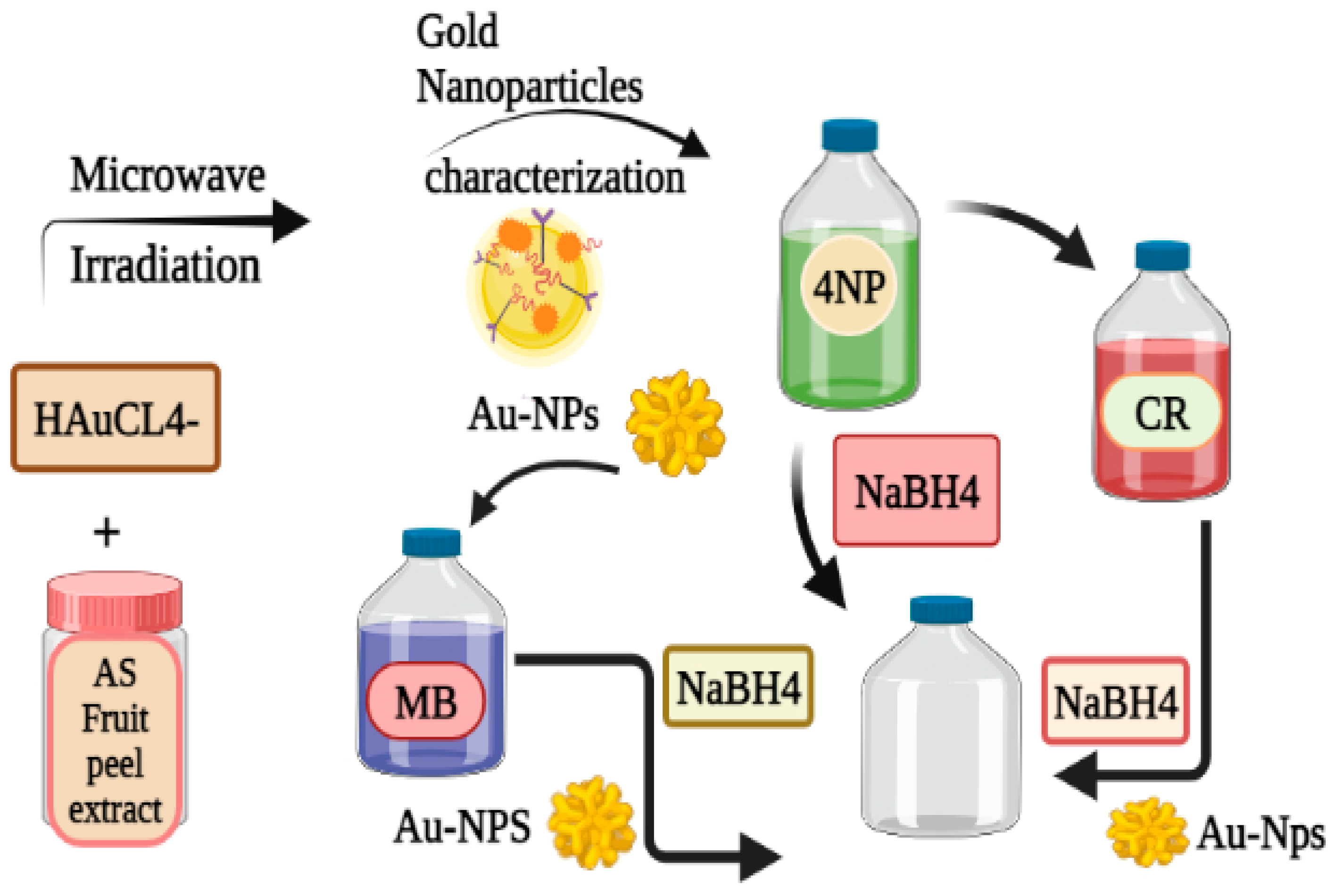
Au-NPs were synthesized from fruit peel extract from Annona squamosa (AS) using concentrated aqueous solution of chloro auric acid as a solvent. Aqueous AS fruit peel extract was dissolved to produce Au-NPs. Different analytical techniques including UV-Vis, FT-IR, XRD, DLS, and TEM [86] were used to examine these artificially coloured green Au-NPs. The Au NPs' potential as catalysts for the reduction of CR, MB, and 4-NP in water was examined using UV-Vis. [84,91].
Figure 33. Synthesis of Au-NPs from Annona squamosa and their catalytic activity for Congo Red, 4-Nitrophenol and Methylene Blue.
Preprints 99484 g033
For the preparation of TiO2/TiO2-Au nanocomposite, integrated copolymer was made from amylopectin and poly acrylic acid. A low temperature method was used for the preparation of Nano-composite. For the preparation of Nano-composite at 500C TiO2/TiO2-Au nanoparticles was formed in aqueous gelling by using ethanoic titanium isopropoxid and HAuCl4 as precursor [79].

8. Amylopectin-TiO2- Au Nanocomposite and its degradation activity:

Titania (TiO2) is widely used having great photo-catalytic activity. An integrated copolymer of amylopectin and polyacrylic acid is called TiO2/TiO2-Au. A less temperature method was used for the preparation of Nano-composite. For the preparation of Nano-composite at 50 ⁰C TiO2/TiO2-Au nanoparticles were formed in aqueous gelling by using ethanoic titanium isopropoxid and HAuCl4 as precursor. These non-composite efficiently degrade the methyl violet (MV) and anionic acidic orange (AO). Sunlight enhanced the degradation of MV [85].

Synthesis of Nano-composite:

To create a uniform gelling matrix, g-AP-pAA was disseminated in basic media (pH 9) at 50 °C with continuous stirring. In the homogeneous gelling matrix of g-AP-PAA a mixture ethanic titanium isoperoxide was added drop by drop under and stirred for 6 hours. The clear gelling matrix then turned milky white. After that ethanolic HAuCl4 solution was added and stirred for 12 hours at 50°C in a dark environment. Conversion of Au3⁺ to metallic Au⁰ was confirmed by the color change from milky white to distinct violet. The mixture was then precipitated in acetone and heated to 50 °C in a vacuum oven for 24 hours [81].

10. Gold nanoparticles synthesize on activated carbon:

Gold nanoparticles were synthesized by mixing of 200 microL aliquot of 0.05mol/L HAuCl4.3H2O aqueous solution and 500ml of aqueous soluble starch. Stirred it in presence of 0.05 mol/L NaOH for 1h. Starch act as capping agents because it stabilizes gold nanoparticles. Appearance of pink color showed the formation of gold nanoparticles, then mixture was maintained at 70°C for 6h, color of solution changed to wine red means Au-nanoparticles were completely synthesized. Au-nanoparticles (500ml) were mixed with 10g activated carbon and stirred up for 12h [90].

11. IPEI coated GNPs:

Synthesis of lPEI coated GNPs:

HAuCl4 stock solution was made for the synthesis of GNPs by mixing 20 ml of HAuCl4 with 980 ml of dH2O. For each molecular weight, an aqueous stock solution containing 10 mg ml 1 PEI was prepared.

Method-I

Added 10 mg lPEI into 1 ml deionized water. And then acetic acid was added dropwise until all lPEI was dissolved

Method-II

10mg of IPEI was dissolved in 1 ml of deionized water, which was then added to a warmed oil bah at at 950C. The effects of pH on the IPEI coated GNPs after was also studied [89].
Table 6. MW, pH, and mass ratio effects on the characteristics of GNPs.

Sample Name

PEI Mw(kDa)
lPEI: Au
(weight ratio)

Sizeα[nm]

PDIb
Zeta potential
[mV]
Particle formation Method I
Control-I 25 1:21 31.53 0.33 +44
lPEI2.5-Au 2.5 1:21 24.12 0.22 +51
Method II
Control-II 25 1:21 25.55 0.23 +52
pH 3.5 25 1:21 52.42 0.43 +53
pH 5 25 1:21 24.32 0.45 +47
lPEI2.5-Au 2.5 1:21 51.42 0.25 +50
Post-synthetic particle properties Method I
pH 3 25 1:21 51.55 0.44 +34
pH 5 25 1:21 46.10 0.47 +31
pH 7 25 1:21 49.27 0.47 +16
Method II
pH 3 25 1:21 50.50 0.46 +24
pH 5 25 1:21 41.62 0.41 +38
pH 7 25 1:21 50.97 0.52 +14

12. Biosynthesis gold nanoparticles using Bacillus marisflavi:
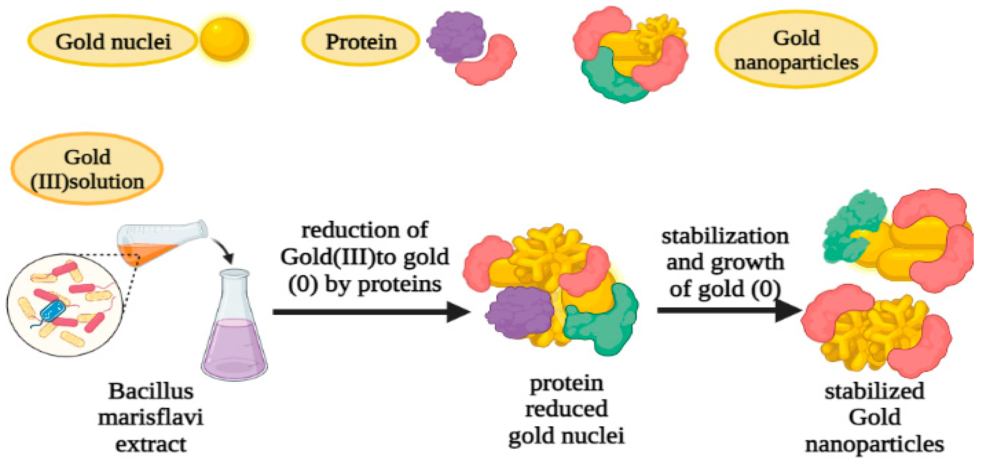
The sterile nutritional broth was inoculated with the pure culture of B. marisflavi and the mixture was then incubated for 24 hours at room temperature at 120 rpm. The biomass was gathered, thoroughly cleaned, suspended in sterile distilled water (DW), and stirred (at 120 rpm) for 24 hours at room temperature. NPs were synthesized using the supernatant (cell-free extract) that was collected after centrifugation. Cell-free extract (CFE) was combined with an equivalent volume of a 1 mM HAuCl4 solution, which was then stirred (at 120 rpm) in the dark and at room temperature. The reaction mixture's UV-visible spectra confirmed the synthesis of AuNPs. [88].
Figure 34. Formation and stabilization of gold nanoparticles.
Preprints 99484 g034

3). Catalytic activities of Asp-AuNPs in degradation of organic dyes:

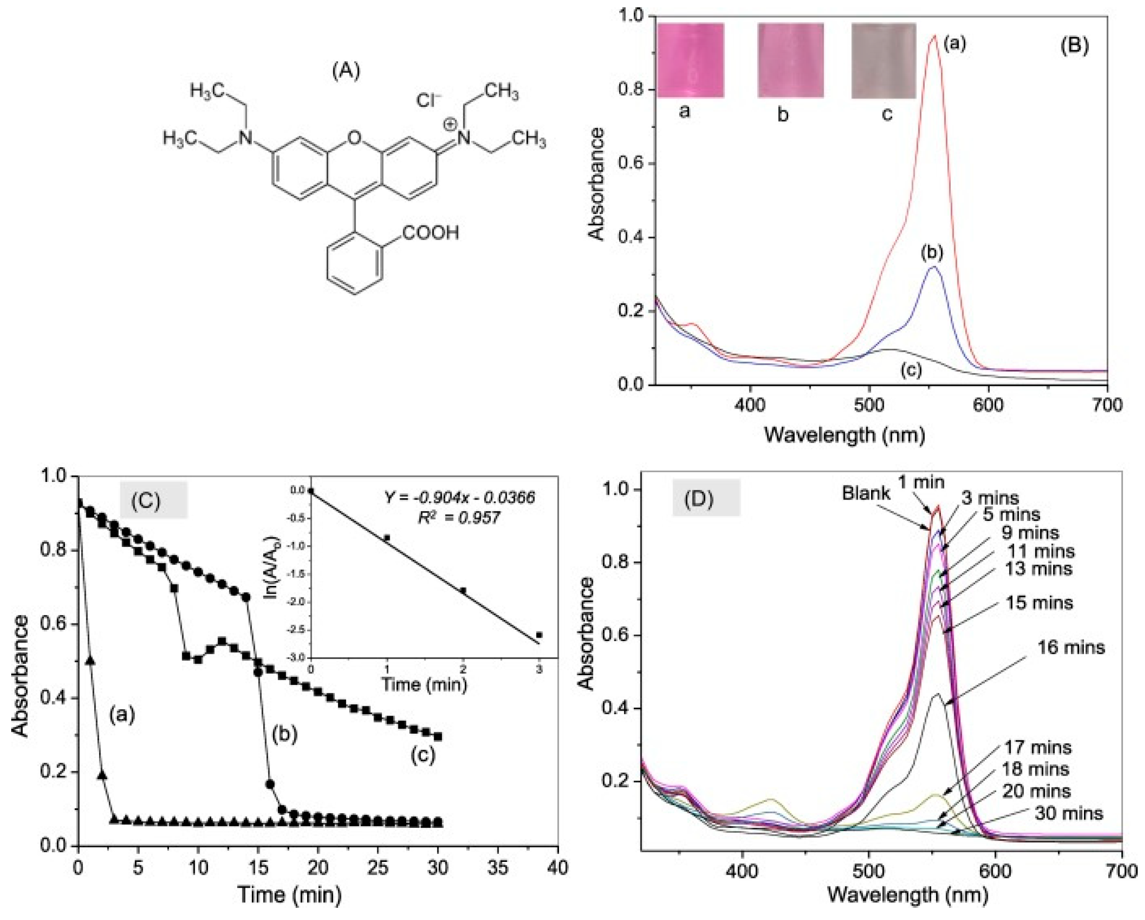
A) Rhodamine B (RB) Degradation
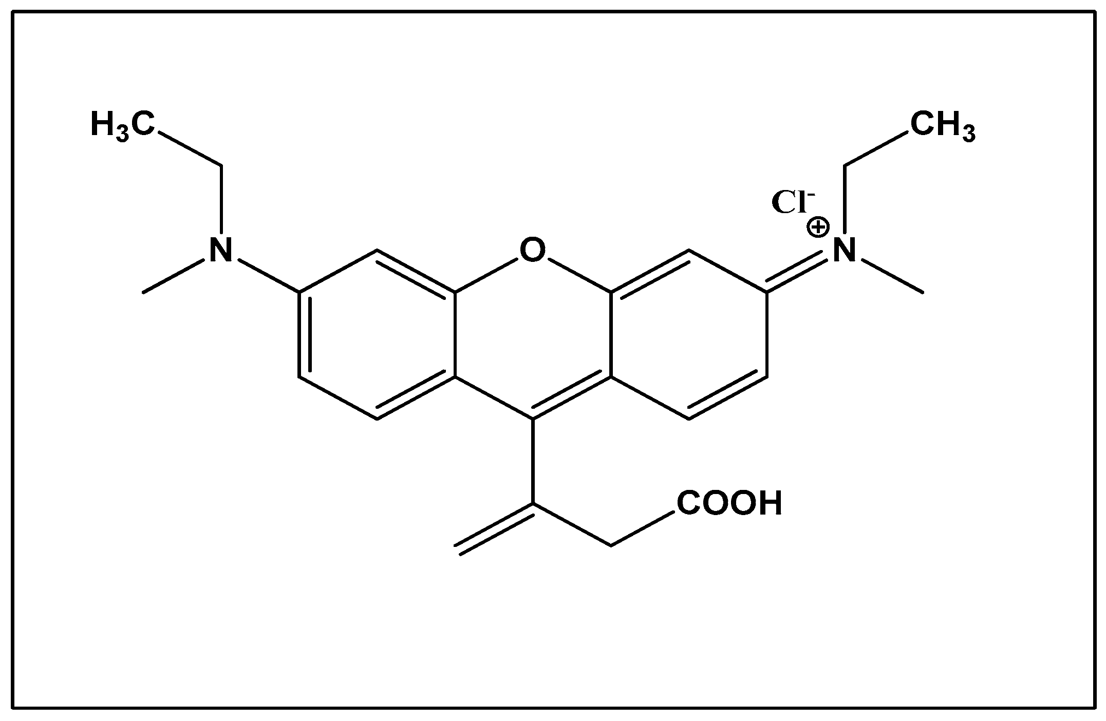
Figure 35. Structure of Rhodamine B.
Preprints 99484 g035
The most extensively used dye in textile mills is RB, it contains dibenzo-1, 4-pyran chromophore. Asp-AuNPs activity is analyzed for removal of RB by addition of strong reducing agent NaBH4. The absorption peak of RB is at 553nm for degradation by AuNPs. To get the maximum result by using minimum of the catalyst and AuNPs, the amount of reducing agent and gold nanoparticles were optimized by varying their concentration. Firstly, the experiment was performed without using Asp-AuNPs only in the presence of reducing agent by varying its amount but complete degradation could not have achieved. By adding the Asp-AuNPs, the process become fast and shows immediately color removal and disappearance of absorption peak, this is due to the electron transfer mechanism of gold nanoparticles from BH4 ion to RB that results in rapid degradation of dye. Due to the breakage of chromophore (dibenzo-1, 4-pyran) the decolorization occurs. The efficiency and time for dye degradation increases by accelerating amount of gold nanoparticles because it provides more surface area for degradation but up to certain level. Dye degradation is maximum by using 20 µL within 3 minutes, but at 50 µL the rate of degradation become decreases. Complete degradation and breakage of chromophore can be observed by complete discoloration of dyes. Beside the amount of gold nanoparticles, the pH also affects the degradation, maximum degradation occurs at pH 6. By using the higher initial concentration of dye, degradation reaction increases so it also effects its efficiency [9].
(A) Structure of RB dye; (B) UV–visible absorption spectra of (a) RB, (b) RB in presence of NaBH4, (c) RB in presence of Asp-AuNPs and NaBH4; (C) plots of RB dye characteristic
Figure 36. Absorbance maxima value (553 nm) vs time in presence of (a) 20 μL, (b) 15 μL and (c) 10 μL of Asp-AuNPs; NaBH4 concentration: 5 mM, inset shows the pseudo first-order kinetics of degradation for 20 μL of Asp-AuNPs, (D) absorbance spectra of RB at different times in the presence of Asp-AuNPs (15 μL) and NaBH4 [9].
Preprints 99484 g036
B) Degradation of Methyl Orange:
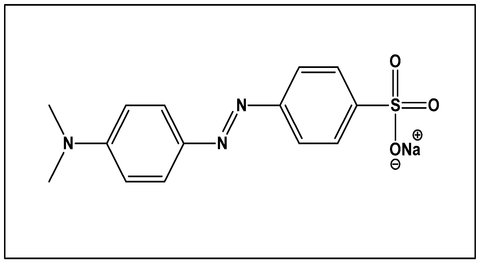
Figure 37. Structure of Methyl orange.
Preprints 99484 g037
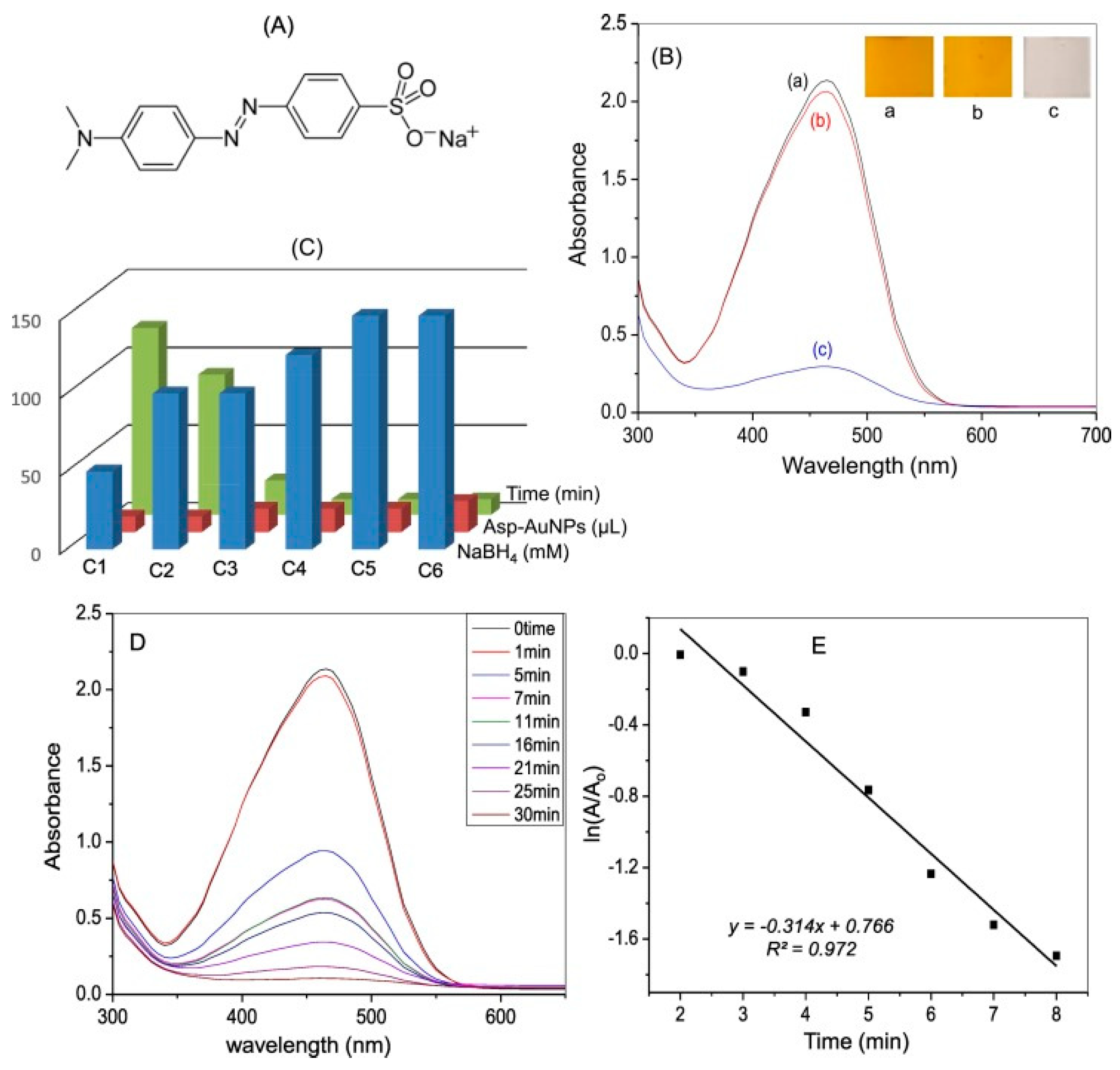
It is broadly used in different industries like paper, textile, and food and also in laboratories. It is non-biodegradable and has many health hazards it basically contains azo group that gives its characteristic color. Absorption peak of MO is at 465nm. The experiment was done firstly in absence of catalyst and there was no change observed in its peak even after 1 day, but in presence of gold nanoparticles the degradation done in 10minutes. After degradation methyl orange gives the biodegradable products that are sulfanilic acid and N, N-dimethyl-p-phenylenediamine. The reaction is pseudo first order reaction [9].
Figure 38. A) Structure of MO dye; (B) UV–visible absorption spectra of (a) MO, (b) MO in presence of NaBH4, (c) MO dye in presence of Asp-AuNPs and NaBH4; (C) optimization of NaBH4 and Asp-AuNPs concentration for degradation of MO; (D) absorbance spectra of MO at different times in the presence of optimized concentration of Asp-AuNPs and NaBH4; (E) pseudo first order kinetics of degradation [9].
Preprints 99484 g038

4. Degradation of 4-NP by Annona Squamosa Au-NPs:
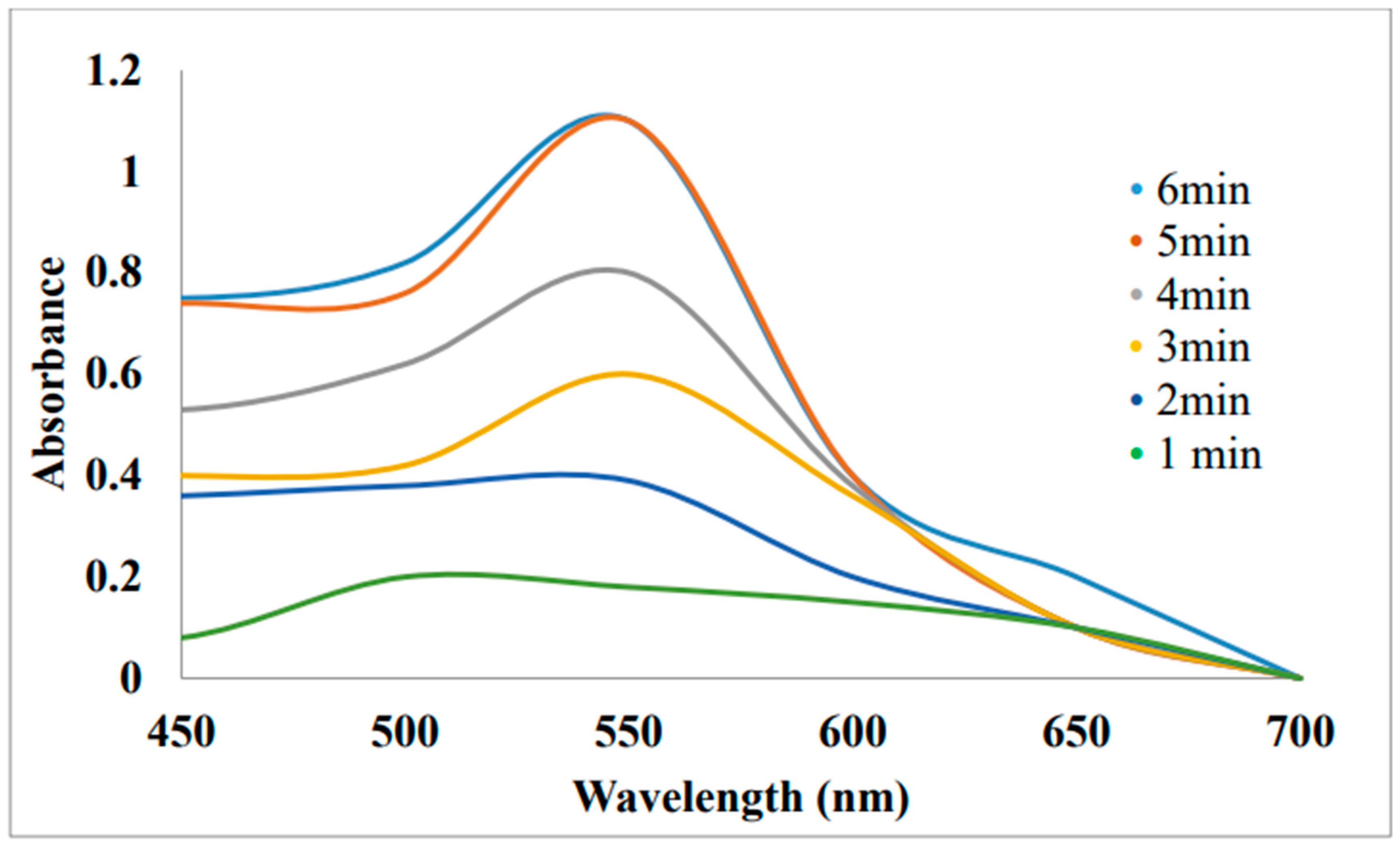
Au-NPs they were synthesized from Annona Squamosa. The reduction of 4-Nitrophenol to 4-Aminophenol (4-AP) by NaBH4 was selected as the model reaction. During this process the Au-NPs solution was mixed with 1ml of 2mM NaBH4 and 1.5ml of 0.2M 4-NP in a UV cuvette. Then, with a time interval of 1 minute, the UV-Vis absorption spectra was recorded at 650 nm in order to monitor the reaction [85].
Figure 39. UV-Visible spectra of degradation of 4-Aminophenol by Annona squamosa Au-NPs along with NaBH4 [85].
Preprints 99484 g039

Degradation of MB and CR:

Au-NPs were utilized as a catalyst in the reduction of MB and CR in the presence of NaBH4. The reaction mixture was diluted to 10ml with double distilled water and agitated for five minutes in accordance with standard technique. It was made up to 2 ml of 1mM MB solution and 1 ml of 10mM NaBH4 along with Annona squamosa Au-NPs. UV-Visible spectra was taken after 1 min. [85].
Figure 40. UV-visible spectra of Methylene Blue reduction by Annona squamosa Au-NPs.
Preprints 99484 g040
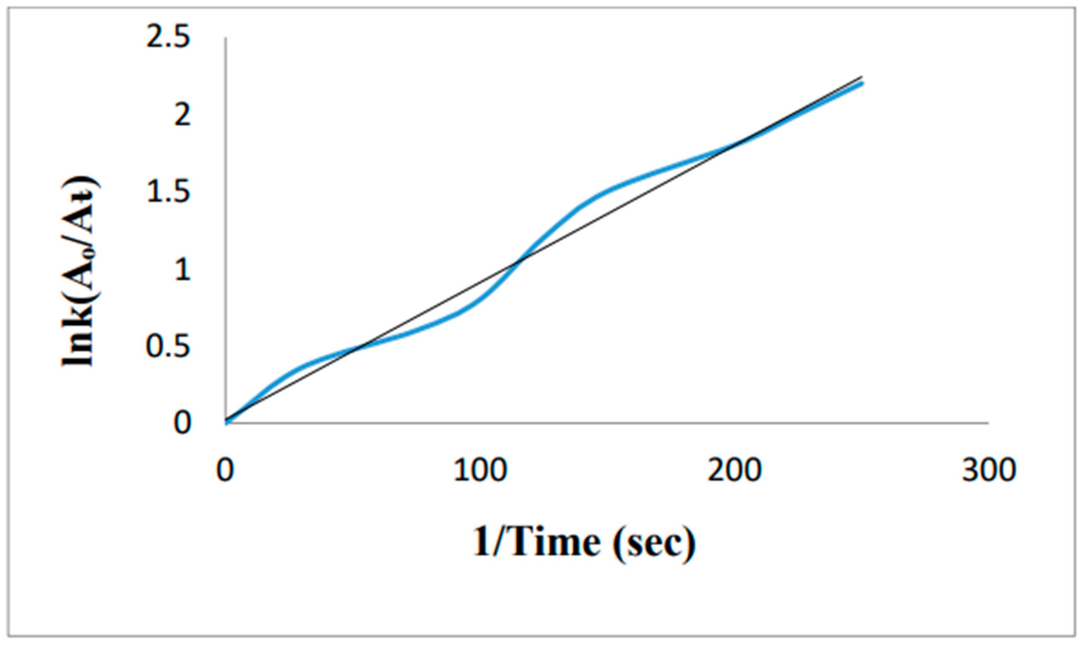
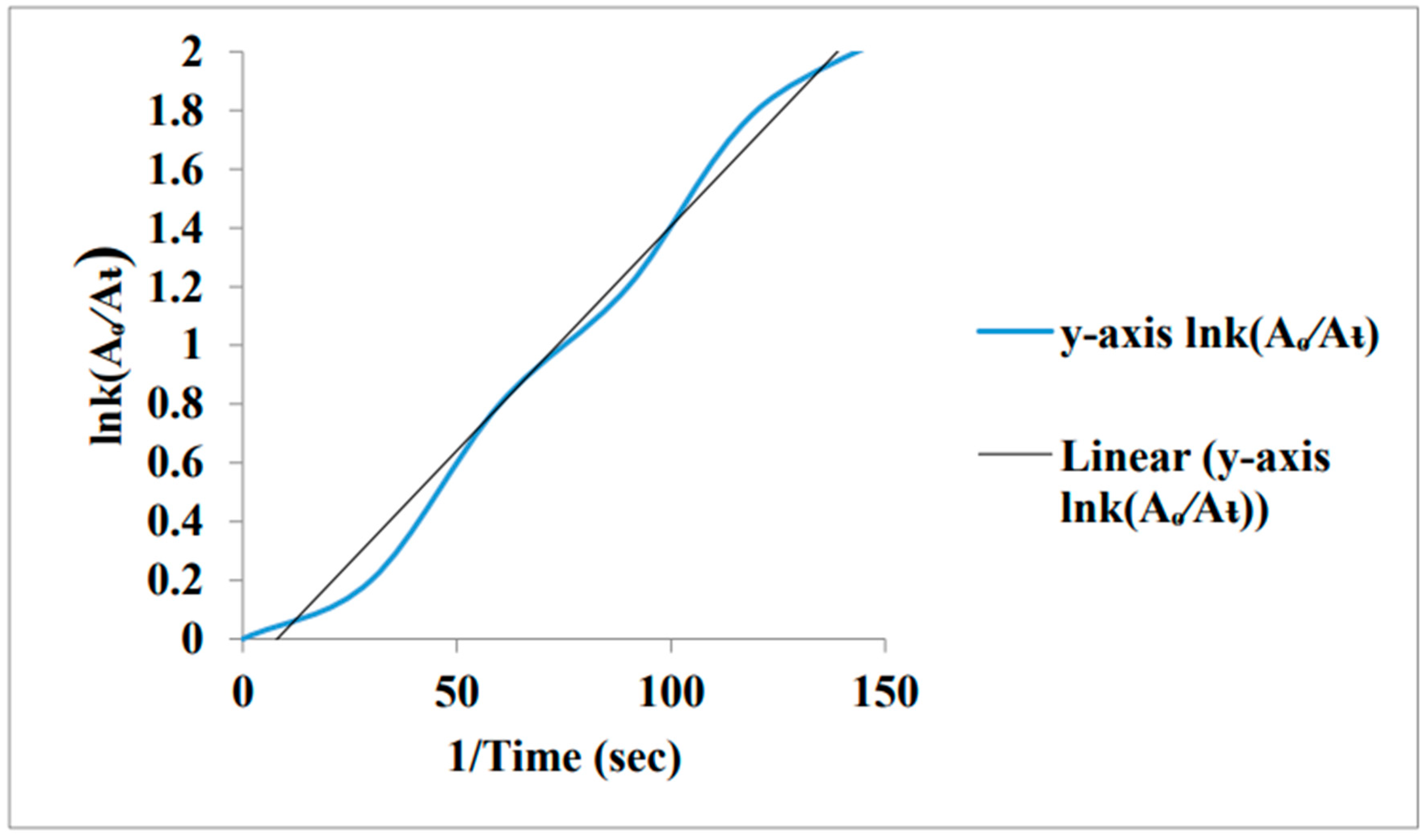
The absorption peaks of MB in aqueous media was recorded at 614 nm and 665 nm. The absorption peak of MB slightly decreases in the presence of NaBH4 after 60 mint. When AU-NPs were added in the reaction mixture MB dye was completely reduced after few minutes. For the reduction of MB dye the temperature used was 15-30 0C. A plot was drawn between Ink verses 1/T. It was shown that with the increase in temperature reaction rate increases [85].
Figure 41. Kinetics of Degradation Reaction of Methylene Blue.
Preprints 99484 g041
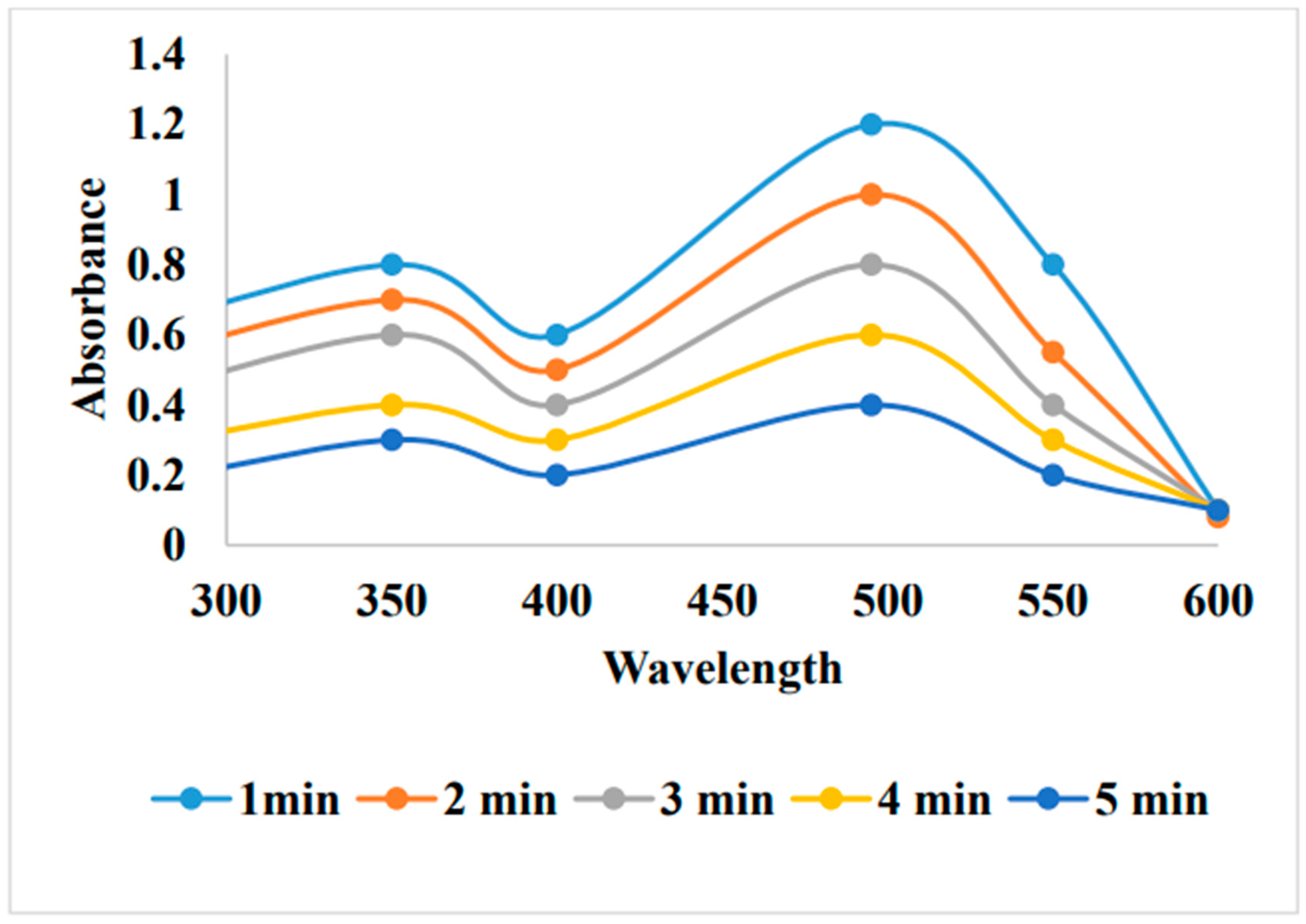
Two absorption peaks may be seen in the aqueous CR's UV-Vis spectrum at 495 nm and 342 nm. When NaBH4 Is used alone the CR was not reduced effectively after 120minutes but when AU-NPs was added in it the CR was completely reduced after 150 sec [85].
Figure 42. UV-visible spectra of Congo Red reduction by Annona squamosa Au-NPs [85].
Preprints 99484 g042
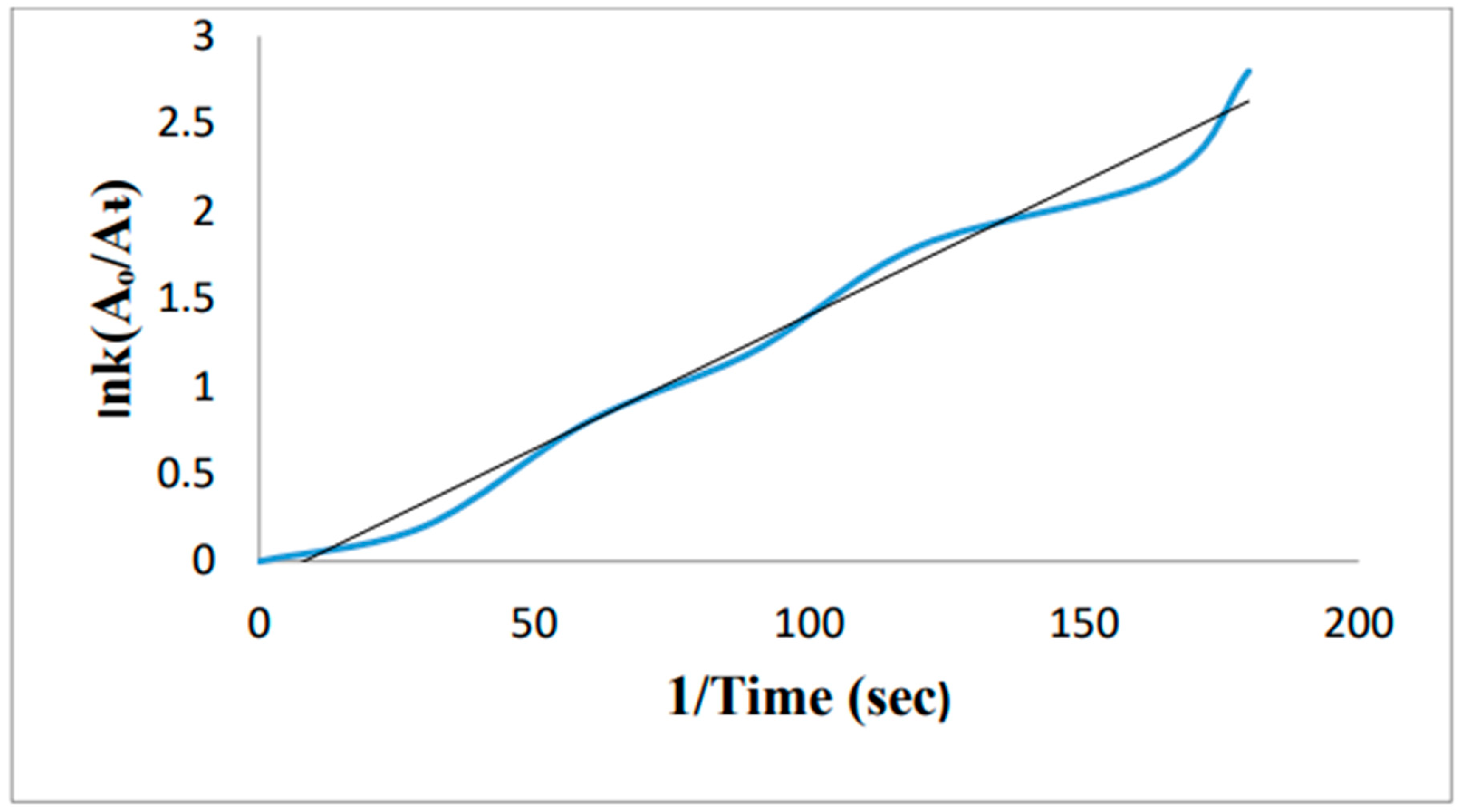
While maintaining the other parameters constant, the impact of temperature (15 to 30 C) on the lowering of CR was examined. A plot was drawn by Ink verses 1/T. by increasing the temperature the rate of reaction increase [85].
Figure 43. Kinetics of Degradation Reaction of Congo Red.
Preprints 99484 g043

5. Removal of methylene blue by using Au-NPs formed from activated carbon:
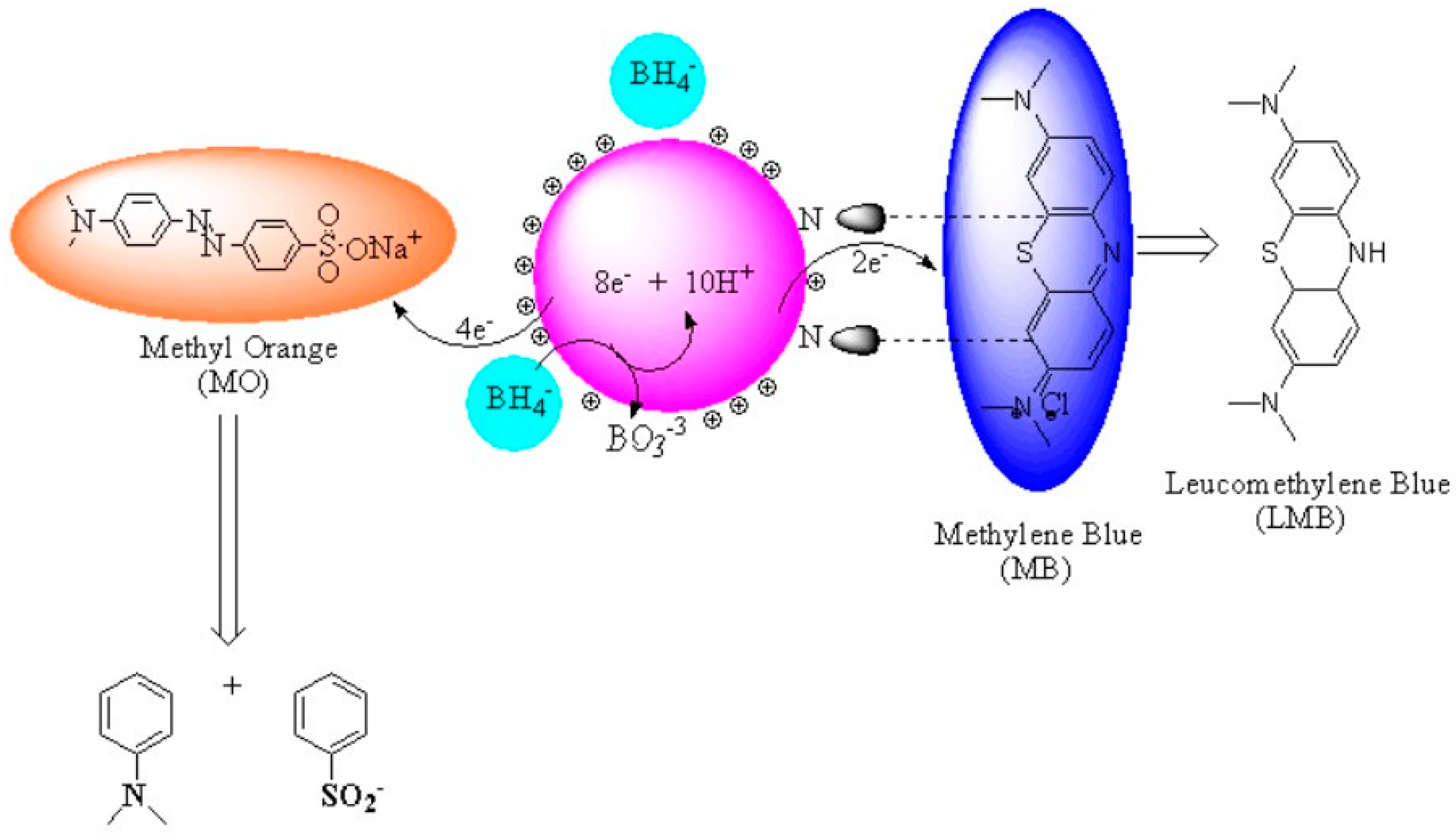
Removal of Methylene blue using Au-NP-AC was done by addition of dye solution (18mg/L) and 0.01g of adsorbent (Au-NP-AC) in flask at pH 7. Sonication time (1.6min) at optimum temperature (303.15k) showed excellent results. At end, sample was centrifuged and analyzed. Percentage degradation was calculated. [90].The % dye degradation was calculated by the following equation:
%   d y e   d e g r a d a t i o n = A A A × 100 %
Figure 44. Schematic representation of MO and MB degradation by cationic GNPs:.
Preprints 99484 g044

Conclusion:

The use of nanoparticles has pivotal role in different fields such as nano medicine, catalysis, green synthesis and in other biological applications that will advance the quality of life. Researchers have recently paid a lot of attention to the environment friendly synthesis of nanoparticles utilizing a variety of plants and their applications in many different industries. [91]. In the current review we focused on the synthesis of Au-NPs from a variety of Plants and microorganisms and their usage in removing the toxic dyes from waste water. [92] Green techniques for the production of Au-NPs using plant derived materials appear to be promising in this regard because they require non-toxic chemical compounds to reduce gold salt. [93] The current characterization methods employed to study the synthesis of Au-NPs had been suitably illustrated. In order to lower the common price, it is still necessary to broaden the protocols for the industrial synthesis of these nanoparticle and to understand the specific process by which nanoparticles derives from plant sources. Additionally, extensive testing for the removal of hazardous pollutants from water supplies desires to be investigated.

References

  1. Kaykhaii, M., Sasani, M., & Marghzari, S. (2018). Removal of dyes from the environment by adsorption process. Chem. Mater. Eng, 6(2), 31-35. [CrossRef]
  2. Sharma, S., & Kaur, A. (2018). Various methods for removal of dyes from industrial effluents-a review. Indian J. Sci. Technol, 11(1). [CrossRef]
  3. Natarajan, S., Bajaj, H. C., & Tayade, R. J. (2018). Recent advances based on the synergetic effect of adsorption for removal of dyes from waste water using photocatalytic process. Journal of Environmental Sciences, 65, 201-222. [CrossRef]
  4. Ruan, W., Hu, J., Qi, J., Hou, Y., Zhou, C., & Wei, X. (2019). Removal of dyes from wastewater by nanomaterials: a review. Advanced Materials Letters, 10(1), 9-20. [CrossRef]
  5. Hunger, K. (Ed.). (2007). Industrial dyes: chemistry, properties, applications. John Wiley & Sons.
  6. Mojsov, K. D., Andronikov, D., Janevski, A., Kuzelov, A., & Gaber, S. (2016). The application of enzymes for the removal of dyes from textile effluents. Advanced technologies, 5(1), 81-86. [CrossRef]
  7. Naeem, H., Ajmal, M., Muntha, S., Ambreen, J., & Siddiq, M. (2018). Synthesis and characterization of graphene oxide sheets integrated with gold nanoparticles and their applications to adsorptive removal and catalytic reduction of water contaminants. RSC advances, 8(7), 3599-3610. [CrossRef]
  8. Singh, R. K., Behera, S. S., Singh, K. R., Mishra, S., Panigrahi, B., Sahoo, T. R., ... & Mandal, D. (2020). Biosynthesized gold nanoparticles as photocatalysts for selective degradation of cationic dye and their antimicrobial activity. Journal of Photochemistry and Photobiology A: Chemistry, 400, 112704. [CrossRef]
  9. Sztandera, K., Gorzkiewicz, M., & Klajnert-Maculewicz, B. (2018). Gold nanoparticles in cancer treatment. Molecular pharmaceutics, 16(1), 1-23. [CrossRef]
  10. Garg, N., Bera, S., Rastogi, L., Ballal, A., & Balaramakrishna, M. V. (2020). Synthesis and characterization of L-asparagine stabilised gold nanoparticles: Catalyst for degradation of organic dyes. Spectrochimica Acta Part A: Molecular and Biomolecular Spectroscopy, 232, 118126. [CrossRef]
  11. Song, W. C., Kim, B., Park, S. Y., Park, G., & Oh, J. W. (2022). Biosynthesis of silver and gold nanoparticles using Sargassum horneri extract as catalyst for industrial dye degradation. Arabian Journal of Chemistry, 15(9), 104056. [CrossRef]
  12. Castillo-Henríquez, L., Alfaro-Aguilar, K., Ugalde-Álvarez, J., Vega-Fernández, L., Montes de Oca-Vásquez, G., & Vega-Baudrit, J. R. (2020). Green synthesis of gold and silver nanoparticles from plant extracts and their possible applications as antimicrobial agents in the agricultural area. Nanomaterials, 10(9), 1763. [CrossRef]
  13. Alle, M., Lee, S. H., & Kim, J. C. (2020). Ultrafast synthesis of gold nanoparticles on cellulose nanocrystals via microwave irradiation and their dyes-degradation catalytic activity. Journal of Materials Science & Technology, 41, 168-177. [CrossRef]
  14. Najahi-Missaoui, W., Arnold, R. D., & Cummings, B. S. (2020). Safe nanoparticles: Are we there yet?. International Journal of Molecular Sciences, 22(1), 385. [CrossRef]
  15. Martínez, G., Merinero, M., Pérez-Aranda, M., Pérez-Soriano, E. M., Ortiz, T., Villamor, E., ... & Alcudia, A. (2020). Environmental impact of nanoparticles’ application as an emerging technology: A review. Materials, 14(1), 166. [CrossRef]
  16. Yetisgin, A. A., Cetinel, S., Zuvin, M., Kosar, A., & Kutlu, O. (2020). Therapeutic nanoparticles and their targeted delivery applications. Molecules, 25(9), 2193. [CrossRef]
  17. Hosny, M., Fawzy, M., El-Borady, O. M., & Mahmoud, A. E. D. (2021). Comparative study between Phragmites australis root and rhizome extracts for mediating gold nanoparticles synthesis and their medical and environmental applications. Advanced Powder Technology, 32(7), 2268-2279. [CrossRef]
  18. Jamkhande, P. G., Ghule, N. W., Bamer, A. H., & Kalaskar, M. G. (2019). Metal nanoparticles synthesis: An overview on methods of preparation, advantages and disadvantages, and applications. Journal of drug delivery science and technology, 53, 101174. [CrossRef]
  19. Biao, L., Tan, S., Meng, Q., Gao, J., Zhang, X., Liu, Z., & Fu, Y. (2018). Green synthesis, characterization and application of proanthocyanidins-functionalized gold nanoparticles. Nanomaterials, 8(1), 53. [CrossRef]
  20. Dabhane, H., Chatur, S., Jadhav, G., Tambade, P., & Medhane, V. (2021). Phytogenic synthesis of gold nanoparticles and applications for removal of methylene blue dye: A review. Environmental Chemistry and Ecotoxicology, 3, 160-171. [CrossRef]
  21. Lu, H., Tang, S. Y., Yun, G., Li, H., Zhang, Y., Qiao, R., & Li, W. (2020). Modular and integrated systems for nanoparticle and microparticle synthesis—a review. Biosensors, 10(11), 165. [CrossRef]
  22. Yaqoob, S. B., Adnan, R., Rameez Khan, R. M., & Rashid, M. (2020). Gold, silver, and palladium nanoparticles: a chemical tool for biomedical applications. Frontiers in chemistry, 8, 376. [CrossRef]
  23. Nadaf, N. Y., & Kanase, S. S. (2019). Biosynthesis of gold nanoparticles by Bacillus marisflavi and its potential in catalytic dye degradation. Arabian Journal of Chemistry, 12(8), 4806-4814. [CrossRef]
  24. Parushuram, N., Ranjana, R., Harisha, K. S., Shilpa, M., Narayana, B., Neelakandan, R., & Sangappa, Y. (2022). Silk fibroin and silk fibroin-gold nanoparticles nanocomposite films: sustainable adsorbents for methylene blue dye. Journal of Dispersion Science and Technology, 43(8), 1161-1176. [CrossRef]
  25. Rather, M. Y., Shincy, M., & Sundarapandian, S. M. (2022). Photocatalytic degradation of Rhodamine-B by phytosynthesized gold nanoparticles. International Journal of Environmental Science and Technology, 1-12. [CrossRef]
  26. Shanmugaraj, K., Campos, C. H., Mangalaraja, R. V., Nandhini, K., Aepuru, R., Torres, C. C., ... & Vo, D. V. N. (2022). Gold nanoparticle–decorated earth-abundant clay nanotubes as catalyst for the degradation of phenothiazine dyes and reduction of 4-(4-nitrophenyl) morpholine. Environmental Science and Pollution Research, 1-12. [CrossRef]
  27. Diğdem, T. R. A. K., & ARSLAN, Y. Biosynthesis of Gold Nanoparticles (AuNPs) with Dimrit Raisin Extract and Their Degradation Activity for Water Contaminants. Düzce Üniversitesi Bilim ve Teknoloji Dergisi, 10(1), 117-127. [CrossRef]
  28. Trak, D. & Arslan, Y. (2022). Biosynthesis of Gold Nanoparticles (AuNPs) with Dimrit Raisin Extract and Their Degradation Activity for Water Contaminants . Düzce Üniversitesi Bilim ve Teknoloji Dergisi, 10 (1), 117-127.
  29. Alikhani, N., Hekmati, M., Karmakar, B., & Veisi, H. (2022). Green synthesis of gold nanoparticles (Au NPs) using Rosa canina fruit extractand evaluation of its catalytic activity in the degradation of organic dye pollutants of water. Inorganic Chemistry Communications, 139, 109351. [CrossRef]
  30. Singh, V. K., M. Yadav, S., Mishra, H., Kumar, R., Tiwari, R. S., Pandey, A., & Srivastava, A. (2019). WS2 quantum dot graphene nanocomposite film for UV photodetection. ACS Applied Nano Materials, 2(6), 3934-3942. [CrossRef]
  31. Kulkarni, R., Harip, S., Kumar, A. R., Deobagkar, D., & Zinjarde, S. (2018). Peptide stabilized gold and silver nanoparticles derived from the mangrove isolate Pseudoalteromonas lipolytica mediate dye decolorization. Colloids and Surfaces A: Physicochemical and Engineering Aspects, 555, 180-190. [CrossRef]
  32. Rabeea, M. A., Owaid, M. N., Aziz, A. A., Jameel, M. S., & Dheyab, M. A. (2020). Mycosynthesis of gold nanoparticles using the extract of Flammulina velutipes, Physalacriaceae, and their efficacy for decolorization of methylene blue. Journal of Environmental Chemical Engineering, 8(3), 103841. [CrossRef]
  33. Dheyab, M. A., Owaid, M. N., Rabeea, M. A., Aziz, A. A., & Jameel, M. S. (2020). Mycosynthesis of gold nanoparticles by the Portabello mushroom extract, Agaricaceae, and their efficacy for decolorization of Azo dye. Environmental nanotechnology, monitoring & management, 14, 100312. [CrossRef]
  34. Abdelghany, A. M., Oraby, A. H., & Asnag, G. M. (2019). Structural, thermal and electrical studies of polyethylene oxide/starch blend containing green synthesized gold nanoparticles. Journal of Molecular Structure, 1180, 15-25. [CrossRef]
  35. Huang, X., Jain, P. K., El-Sayed, I. H., & El-Sayed, M. A. (2006). Determination of the minimum temperature required for selective photothermal destruction of cancer cells with the use of immunotargeted gold nanoparticles. Photochemistry and photobiology, 82(2), 412-417. [CrossRef]
  36. Huang, X., & El-Sayed, M. A. (2010). Gold nanoparticles: Optical properties and implementations in cancer diagnosis and photothermal therapy. Journal of advanced research, 1(1), 13-28. [CrossRef]
  37. Deshmukh, A. R., & Kim, B. S. (2021). Bio-functionalized few-layer graphene for in situ growth of gold nanoparticles, improvement of polymer properties, and dye removal. Journal of Cleaner Production, 310, 127515. [CrossRef]
  38. Verma, N., Jagota, V., Alguno, A. C., Rakhra, M., Kumar, P., & Dugbakie, B. N. (2022). Characterization of fabricated gold-doped ZnO nanospheres and their use as a photocatalyst in the degradation of DR-31 dye. Journal of Nanomaterials, 2022, 1-8. [CrossRef]
  39. Ghaedi, M., Ramazani, S., & Roosta, M. (2011). Gold nanoparticle loaded activated carbon as novel adsorbent for the removal of Congo red. Indian Journal of Science and Technology, 4(10), 1208-1217. [CrossRef]
  40. Cui, W., Lu, W., Zhang, Y., Lin, G., Wei, T., & Jiang, L. (2010). Gold nanoparticle ink suitable for electric-conductive pattern fabrication using in ink-jet printing technology. Colloids and Surfaces A: Physicochemical and Engineering Aspects, 358(1-3), 35-41. [CrossRef]
  41. Bhowmik, T., Kundu, M. K., & Barman, S. (2015). Ultra small gold nanoparticles–graphitic carbon nitride composite: an efficient catalyst for ultrafast reduction of 4-nitrophenol and removal of organic dyes from water. RSC Advances, 5(48), 38760-38773. [CrossRef]
  42. Thomas, K. G., & Kamat, P. V. (2003). Chromophore-functionalized gold nanoparticles. Accounts of chemical research, 36(12), 888-898. [CrossRef]
  43. Qin, Z., & Bischof, J. C. (2012). Thermophysical and biological responses of gold nanoparticle laser heating. Chemical Society Reviews, 41(3), 1191-1217. [CrossRef]
  44. Wu, G., Liu, X., Zhou, P., Wang, L., Hegazy, M., Huang, X., & Huang, Y. (2019). A facile approach for the reduction of 4-nitrophenol and degradation of congo red using gold nanoparticles or laccase decorated hybrid inorganic nanoparticles/polymer-biomacromolecules vesicles. Materials Science and Engineering: C, 94, 524-533. [CrossRef]
  45. Katas, H., Moden, N. Z., Lim, C. S., Celesistinus, T., Chan, J. Y., Ganasan, P., & Suleman Ismail Abdalla, S. (2018). Biosynthesis and potential applications of silver and gold nanoparticles and their chitosan-based nanocomposites in nanomedicine. Journal of Nanotechnology, 2018, 1-13. [CrossRef]
  46. Roy, A., Murthy, H. A., Ahmed, H. M., Islam, M. N., & Prasad, R. (2022). Phytogenic synthesis of metal/metal oxide nanoparticles for degradation of dyes. Journal of Renewable Materials, 10(7), 1911. [CrossRef]
  47. Aljabali, A. A., Akkam, Y., Al Zoubi, M. S., Al-Batayneh, K. M., Al-Trad, B., Abo Alrob, O., ... & Evans, D. J. (2018). Synthesis of gold nanoparticles using leaf extract of Ziziphus zizyphus and their antimicrobial activity. Nanomaterials, 8(3), 174. [CrossRef]
  48. Nadaf, N. Y., & Kanase, S. S. (2019). Biosynthesis of gold nanoparticles by Bacillus marisflavi and its potential in catalytic dye degradation. Arabian Journal of Chemistry, 12(8), 4806-4814. [CrossRef]
  49. Choudhary, M. K., Kataria, J., & Sharma, S. (2017). A biomimetic synthesis of stable gold nanoparticles derived from aqueous extract of Foeniculum vulgare seeds and evaluation of their catalytic activity. Applied Nanoscience, 7, 439-447. [CrossRef]
  50. López-Miranda, J. L., Molina, G. A., Esparza, R., González-Reyna, M. A., Silva, R., & Estévez, M. (2021). Green Synthesis of Homogeneous Gold Nanoparticles Using Sargassum spp. Extracts and Their Enhanced Catalytic Activity for Organic Dyes. Toxics, 9(11), 280. [CrossRef]
  51. Baruah, D., Goswami, M., Yadav, R. N. S., Yadav, A., & Das, A. M. (2018). Biogenic synthesis of gold nanoparticles and their application in photocatalytic degradation of toxic dyes. Journal of Photochemistry and Photobiology B: Biology, 186, 51-58. [CrossRef]
  52. Islam, M. T., Dominguez, N., Ahsan, M. A., Dominguez-Cisneros, H., Zuniga, P., Alvarez, P. J., & Noveron, J. C. (2017). Sodium rhodizonate induced formation of gold nanoparticles supported on cellulose fibers for catalytic reduction of 4-nitrophenol and organic dyes. Journal of environmental chemical engineering, 5(5), 4185-4193. [CrossRef]
  53. Guo, M., Li, W., Yang, F., & Liu, H. (2015). Controllable biosynthesis of gold nanoparticles from a Eucommia ulmoides bark aqueous extract. Spectrochimica acta part A: molecular and biomolecular spectroscopy, 142, 73-79. [CrossRef]
  54. Note, C., Kosmella, S., & Koetz, J. (2006). Poly (ethyleneimine) as reducing and stabilizing agent for the formation of gold nanoparticles in w/o microemulsions. Colloids and Surfaces A: Physicochemical and Engineering Aspects, 290(1-3), 150-156. [CrossRef]
  55. Sarfraz, N., & Khan, I. (2021). Plasmonic gold nanoparticles (AuNPs): properties, synthesis and their advanced energy, environmental and biomedical applications. Chemistry–An Asian Journal, 16(7), 720-742. [CrossRef]
  56. Narayana, C., Upadhyay, R. K., Chaturvedi, R., & Sagar, R. (2017). A versatile carbohydrate based gelator for oil water separation, nanoparticle synthesis and dye removal. New Journal of Chemistry, 41(6), 2261-2267. [CrossRef]
  57. Carolin, C. F., Kumar, P. S., & Joshiba, G. J. (2021). Sustainable approach to decolourize methyl orange dye from aqueous solution using novel bacterial strain and its metabolites characterization. Clean Technologies and Environmental Policy, 23, 173-181. [CrossRef]
  58. Hanafi, M. F., & Sapawe, N. (2020). A review on the water problem associate with organic pollutants derived from phenol, methyl orange, and remazol brilliant blue dyes. Materials Today: Proceedings, 31, A141-A150. [CrossRef]
  59. Singh, A., Ahmed, A., Sharma, A., Sharma, C., Paul, S., Khosla, A., ... & Arya, S. (2021). Promising photocatalytic degradation of methyl orange dye via sol-gel synthesized Ag–CdS@ Pr-TiO2 core/shell nanoparticles. Physica B: Condensed Matter, 616, 413121. [CrossRef]
  60. Astuti, W., Sulistyaningsih, T., & Maksiola, M. (2017). Equilibrium and Kinetics of Adsorption of Methyl Violet from Aqueous Solutions Using Modified Ceiba pentandra Sawdust. Asian journal of chemistry, 29(1). [CrossRef]
  61. Srivastava, S., Sinha, R., & Roy, D. (2004). Toxicological effects of malachite green. Aquatic toxicology, 66(3), 319-329. [CrossRef]
  62. Raval, N. P., Shah, P. U., & Shah, N. K. (2017). Malachite green “a cationic dye” and its removal from aqueous solution by adsorption. Applied Water Science, 7, 3407-3445. [CrossRef]
  63. Al-Gheethi, A. A., Azhar, Q. M., Kumar, P. S., Yusuf, A. A., Al-Buriahi, A. K., Mohamed, R. M. S. R., & Al-Shaibani, M. M. (2022). Sustainable approaches for removing Rhodamine B dye using agricultural waste adsorbents: A review. Chemosphere, 287, 132080. [CrossRef]
  64. Bujdák, J., Martínez Martínez, V., López Arbeloa, F., & Iyi, N. (2007). Spectral properties of rhodamine 3B adsorbed on the surface of montmorillonites with variable layer charge. Langmuir, 23(4), 1851-1859. [CrossRef]
  65. Tian, Z., Tian, B., & Zhang, J. (2013). Synthesis and characterization of new rhodamine dyes with large Stokes shift. Dyes and Pigments, 99(3), 1132-1136. [CrossRef]
  66. Sulistina, D. R., & Martini, S. (2020). The effect of Rhodamine B on the cerebellum and brainstem tissue of Rattus norvegicus. Journal of Public Health Research, 9(2), jphr-2020. [CrossRef]
  67. Shen, K., & Gondal, M. A. (2017). Removal of hazardous Rhodamine dye from water by adsorption onto exhausted coffee ground. Journal of Saudi Chemical Society, 21, S120-S127. [CrossRef]
  68. Chakraborty, U., Bhanjana, G., Kaur, N., Sharma, R., Kaur, G., Kaushik, A., & Chaudhary, G. R. (2021). Microwave-assisted assembly of Ag2O-ZnO composite nanocones for electrochemical detection of 4-Nitrophenol and assessment of their photocatalytic activity towards degradation of 4-Nitrophenol and Methylene blue dye. Journal of hazardous materials, 416, 125771. [CrossRef]
  69. El-Aal, M. A., Ali, H. M., & Ibrahim, S. M. (2022). Cu-Doped 1D Hydroxyapatite as a Highly Active Catalyst for the Removal of 4-Nitrophenol and Dyes from Water. ACS omega, 7(30), 26777-26787. [CrossRef]
  70. Barreca, S., Colmenares, J. J. V., Pace, A., Orecchio, S., & Pulgarin, C. (2014). Neutral solar photo-Fenton degradation of 4-nitrophenol on iron-enriched hybrid montmorillonite-alginate beads (Fe-MABs). Journal of Photochemistry and Photobiology A: Chemistry, 282, 33-40. [CrossRef]
  71. Bhowmik, T., Kundu, M. K., & Barman, S. (2015). Ultra small gold nanoparticles–graphitic carbon nitride composite: an efficient catalyst for ultrafast reduction of 4-nitrophenol and removal of organic dyes from water. RSC Advances, 5(48), 38760-38773. [CrossRef]
  72. Ahsan, M. A., Deemer, E., Fernandez-Delgado, O., Wang, H., Curry, M. L., El-Gendy, A. A., & Noveron, J. C. (2019). Fe nanoparticles encapsulated in MOF-derived carbon for the reduction of 4-nitrophenol and methyl orange in water. Catalysis Communications, 130, 105753. [CrossRef]
  73. Liu, J., Wang, N., Zhang, H., & Baeyens, J. (2019). Adsorption of Congo red dye on FexCo3-xO4 nanoparticles. Journal of environmental management, 238, 473-483. [CrossRef]
  74. Oguzie, E. E. (2004). Influence of halide ions on the inhibitive effect of congo red dye on the corrosion of mild steel in sulphuric acid solution. Materials Chemistry and Physics, 87(1), 212-217. [CrossRef]
  75. Mittal, A., Mittal, J., Malviya, A., & Gupta, V. K. (2009). Adsorptive removal of hazardous anionic dye “Congo red” from wastewater using waste materials and recovery by desorption. Journal of colloid and interface science, 340(1), 16-26. [CrossRef]
  76. Khan, I., Saeed, K., Zekker, I., Zhang, B., Hendi, A. H., Ahmad, A., ... & Khan, I. (2022). Review on methylene blue: its properties, uses, toxicity and photodegradation. Water, 14(2), 242. [CrossRef]
  77. Kallel, F., Chaari, F., Bouaziz, F., Bettaieb, F., Ghorbel, R., & Chaabouni, S. E. (2016). Sorption and desorption characteristics for the removal of a toxic dye, methylene blue from aqueous solution by a low cost agricultural by-product. Journal of Molecular Liquids, 219, 279-288. [CrossRef]
  78. Zahoor, M., Nazir, N., Iftikhar, M., Naz, S., Zekker, I., Burlakovs, J., ... & Ali Khan, F. (2021). A review on silver nanoparticles: Classification, various methods of synthesis, and their potential roles in biomedical applications and water treatment. Water, 13(16), 2216. [CrossRef]
  79. Nadaf, N. Y., & Kanase, S. S. (2019). Biosynthesis of gold nanoparticles by Bacillus marisflavi and its potential in catalytic dye degradation. Arabian Journal of Chemistry, 12(8), 4806-4814. [CrossRef]
  80. AlMashrea, B. A., Abla, F., Chehimi, M. M., Workie, B., Han, C., & Mohamed, A. A. (2020). Polyaniline coated gold-aryl nanoparticles: Electrochemical synthesis and efficiency in methylene blue dye removal. Synthetic Metals, 269, 116528. [CrossRef]
  81. Roosta, M., Ghaedi, M., Shokri, N., Daneshfar, A., Sahraei, R., & Asghari, A. (2014). Optimization of the combined ultrasonic assisted/adsorption method for the removal of malachite green by gold nanoparticles loaded on activated carbon: experimental design. Spectrochimica Acta Part A: Molecular and Biomolecular Spectroscopy, 118, 55-65. [CrossRef]
  82. Sarkar, A. K., Saha, A., Tarafder, A., Panda, A. B., & Pal, S. (2016). Efficient removal of toxic dyes via simultaneous adsorption and solar light driven photodegradation using recyclable functionalized amylopectin–TiO2–Au nanocomposite. ACS Sustainable Chemistry & Engineering, 4(3), 1679-1688. [CrossRef]
  83. Singaravelu, G., Arockiamary, J. S., Kumar, V. G., & Govindaraju, K. (2007). A novel extracellular synthesis of monodisperse gold nanoparticles using marine alga, Sargassum wightii Greville. Colloids and surfaces B: Biointerfaces, 57(1), 97-101. [CrossRef]
  84. Wang, L., Xu, J., Yan, Y., Liu, H., Karunakaran, T., & Li, F. (2019). Green synthesis of gold nanoparticles from Scutellaria barbata and its anticancer activity in pancreatic cancer cell (PANC-1). Artificial cells, nanomedicine, and biotechnology, 47(1), 1617-1627. [CrossRef]
  85. Baruah, D., Goswami, M., Yadav, R. N. S., Yadav, A., & Das, A. M. (2018). Biogenic synthesis of gold nanoparticles and their application in photocatalytic degradation of toxic dyes. Journal of Photochemistry and Photobiology B: Biology, 186, 51-58. [CrossRef]
  86. Li, Q., Yu, D., Fan, C., Huang, Q., Tang, Y., Guo, R., ... & Lin, Y. (2021). Gold nanoparticles adsorbed on graphene as nanozymes for the efficient elimination of dye pollutants. ACS Applied Nano Materials, 5(1), 94-100. [CrossRef]
  87. Parushuram, N., Ranjana, R., Harisha, K. S., Shilpa, M., Narayana, B., Neelakandan, R., & Sangappa, Y. (2022). Silk fibroin and silk fibroin-gold nanoparticles nanocomposite films: sustainable adsorbents for methylene blue dye. Journal of Dispersion Science and Technology, 43(8), 1161-1176. [CrossRef]
  88. Das, J., & Velusamy, P. (2014). Catalytic reduction of methylene blue using biogenic gold nanoparticles from Sesbania grandiflora L. Journal of the Taiwan Institute of Chemical Engineers, 45(5), 2280-2285. [CrossRef]
  89. Umamaheswari, C., Lakshmanan, A., & Nagarajan, N. S. (2018). Green synthesis, characterization and catalytic degradation studies of gold nanoparticles against congo red and methyl orange. Journal of Photochemistry and Photobiology B: Biology, 178, 33-39. [CrossRef]
  90. Cavuslar, O., Nakay, E., Kazakoglu, U., Abkenar, S. K., Ow-Yang, C. W., & Acar, H. Y. (2020). Synthesis of stable gold nanoparticles using linear polyethyleneimines and catalysis of both anionic and cationic azo dye degradation. Materials Advances, 1(7), 2407-2417. [CrossRef]
  91. Roosta, M., Ghaedi, M., Daneshfar, A., Sahraei, R., & Asghari, A. (2014). Optimization of the ultrasonic assisted removal of methylene blue by gold nanoparticles loaded on activated carbon using experimental design methodology. Ultrasonics sonochemistry, 21(1), 242-252. [CrossRef]
  92. Gangapuram, B. R., Bandi, R., Alle, M., Dadigala, R., Kotu, G. M., & Guttena, V. (2018). Microwave assisted rapid green synthesis of gold nanoparticles using Annona squamosa L peel extract for the efficient catalytic reduction of organic pollutants. Journal of Molecular Structure, 1167, 305-315. [CrossRef]
  93. Khan, M., Ahmad, F., Koivisto, J. T., & Kellomäki, M. (2020). Green synthesis of controlled size gold and silver nanoparticles using antioxidant as capping and reducing agent. Colloid and Interface Science Communications, 39, 100322. [CrossRef]
Disclaimer/Publisher’s Note: The statements, opinions and data contained in all publications are solely those of the individual author(s) and contributor(s) and not of MDPI and/or the editor(s). MDPI and/or the editor(s) disclaim responsibility for any injury to people or property resulting from any ideas, methods, instructions or products referred to in the content.
Copyright: This open access article is published under a Creative Commons CC BY 4.0 license, which permit the free download, distribution, and reuse, provided that the author and preprint are cited in any reuse.
Prerpints.org logo

Preprints.org is a free preprint server supported by MDPI in Basel, Switzerland.

Subscribe

© 2024 MDPI (Basel, Switzerland) unless otherwise stated