Submitted:
21 February 2024
Posted:
23 February 2024
You are already at the latest version
Abstract
Keywords:
1. Introduction
2. Materials and Methods
2.1. Study Subjects
2.2. Instrumentation for gait analysis and data acquisition


2.3. Gait analysis
2.4. Statistical analysis
3. Results
3.1. Peak knee moments occurring in coronal motion plane
3.2. Moment results of ankle joint according to FPA condition
4. Discussion
5. Conclusions
References
- Yang, G.; Wang, J.; Liu, Y.; Lu, H.; He, L.; Ma, C.; Zhao, Z. Burden of knee osteoarthritis in 204 countries and territories, 1990–2019: Results from the global burden of disease study 2019. Arthritis Care Res. 2023, 75, 2489–2500. [Google Scholar] [CrossRef]
- Deshpande, B.R.; Katz, J.N.; Solomon, D.H.; Yelin, E.H.; Hunter, D.J.; Messier, S.P.; Suter, L.G.; Losina, E. Number of persons with symptomatic knee osteoarthritis in the us: impact of race and ethnicity, age, sex, and obesity: symptomatic knee OA in the US. Arthritis Care Res. 2016, 68, 1743–1750. [Google Scholar] [CrossRef]
- Kim, Y.; Richards, J.; Lidtke, R.H.; Trede, R. Characteristics of clinical measurements between biomechanical responders and non-responders to a shoe designed for knee osteoarthritis. Gait Posture 2018, 59, 23–27. [Google Scholar] [CrossRef]
- D’Souza, N.; Charlton, J.; Grayson, S.; Kobayashi, L.; Hutchison, M.; Hunt, M.; Simic, M. Are biomechanics during gait associated with the structural disease onset and progression of lower limb osteoarthritis? A systematic review and meta-analysis. Osteoarthritis Cartilage 2022, 30, 381–394. [Google Scholar] [CrossRef]
- Hutchison, L.; D'Souza, N.; Grayson, J.; Hiller, C.; Kobayashi, S.; Simic, M. Toe-in and toe-out gait retraining interventions to reduce proxy measures of medial knee joint load in people with medial knee osteoarthritis: Protocol for a randomised placebo-controlled trial. Contemp. Clin. Trials 2023, 134, 107355. [Google Scholar] [CrossRef]
- Guo, M.; Axe, M.J.; Manal, K. The influence of foot progression angle on the knee adduction moment during walking and stair climbing in pain free individuals with knee osteoarthritis. Gait Posture 2007, 26, 436–441. [Google Scholar] [CrossRef]
- Seagers, K.; Uhlrich, S.D.; Kolesar, J.A.; Berkson, M.; Kaneda, J.M.; Beaupre, G.S.; Delp, S.L. Changes in foot progression angle during gait reduce the knee adduction moment and do not increase hip moments in individuals with knee osteoarthritis. J. Biomech. 2022, 141, 111204. [Google Scholar] [CrossRef]
- Chehab, E.F.; Favre, J.; Erhart-Hledik, J.C.; Andriacchi, T.P. Baseline knee adduction and flexion moments during walking are both associated with 5-year cartilage changes in patients with medial knee osteoarthritis. Osteoarthritis Cartilage 2014, 22, 1833–1839. [Google Scholar] [CrossRef]
- Robbins, S.M.; Birmingham, T.B.; Callaghan, J.P.; Jones, G.R.; Chesworth, B.M.; Maly, M.R. Association of pain with frequency and magnitude of knee loading in knee osteoarthritis. Arthritis Care Res. 2011, 63, 991–997. [Google Scholar] [CrossRef]
- Hurwitz, D.E.; Ryals, A.B.; Case, J.P.; Block, J.A.; Andriacchi, T.P. The knee adduction moment during gait in subjects with knee osteoarthritis is more closely correlated with static alignment than radiographic disease severity, toe out angle and pain. J. Orthop. Res. 2002, 20, 101–107., 63, 405-426. [Google Scholar] [CrossRef]
- Hinman, R.S.; Wrigley, T.V.; Metcalf, B.R.; Hunter, D.J.; Campbell, P.; Paterson, K.; Staples, M.P.; Bennell, K.L. Unloading shoes for osteoarthritis of the knee: protocol for the SHARK randomised controlled trial. BMC Musculoskelet. Disord. 2014, 15, 48. [Google Scholar] [CrossRef]
- Simic, M.; Hinman, R.S.; Wrigley, T.V.; Bennell, K.L.; Hunt, M.A. Gait modification strategies for altering medial knee joint load: a systematic review. Arthritis Care Res. (Hoboken) 2011. [Google Scholar] [CrossRef]
- Kepple, T.M.; Segel, K.L.; Stanhope, S.J. Relative contributions of the lower extremity joint moments to forward progression and support during gait. Gait Posture 1997, 6, 1–8. [Google Scholar] [CrossRef]
- Ro, D.H.; Lee, J.; Lee, J.; Park, J.Y.; Han, H.S.; Lee, M.C. Effects of knee osteoarthritis on hip and ankle gait mechanics. Adv. Orthop. 2019, 2019, 9757369. [Google Scholar] [CrossRef]
- Wang, J.W.; Kuo, K.N.; Andriacchi, T.P.; Galante, J.O. The influence of walking mechanics and time on the results of proximal tibial osteotomy. J. Bone Joint Surg. Am. 1990, 72, 905–909. [Google Scholar] [CrossRef]
- Shull, P.B.; Shultz, R.; Silder, A.; Dragoo, J.L.; Besier, T.F.; Cutkosky, M.R.; Delp, S.L. Toe-in gait reduces the first peak knee adduction moment in patients with medial compartment knee osteoarthritis. J. Biomech. 2013, 46, 122–128. [Google Scholar] [CrossRef]
- Mündermann, A.; Dyrby, C.O.; Andriacchi, T.P. Secondary gait changes in patients with medial compartment knee osteoarthritis: Increased load at the ankle, knee, and hip during walking. Arthritis Rheum. 2005, 52, 2835–2844. [Google Scholar] [CrossRef]
- Legrand, T.; Younesian, H.; Equey, N.; Campeau-Lecours, A.; Turcot, K. Trunk lean and toe out gait strategies impact on lower limb joints. J. Biomech. 2021, 129, 110740. [Google Scholar] [CrossRef]
- Wesseling, M.; de Groote, F.; Meyer, C.; Corten, K.; Simon, J.P.; Desloovere, K.; Jonkers, I. Gait alterations to effectively reduce hip contact forces. J. Orthop. Res. 2015, 33, 1094–1102. [Google Scholar] [CrossRef]
- Charlton, J.M.; Hatfield, G.L.; Guenette, J.A.; Hunt, M.A. Ankle joint and rearfoot biomechanics during toe-in and toe-out walking in people with medial compartment knee osteoarthritis. PM R. 2019, 11, 503–511. [Google Scholar] [CrossRef]
- Kim, Y. Effects of foot-toe orthoses on moment and range of motion of knee joint in individuals with hallux valgus. Life (Basel) 2023, 13, 1162. [Google Scholar] [CrossRef]
- Collins, T.D.; Ghoussayni, S.N.; Ewins, D.J.; Kent, J.A. A six degrees-of-freedom marker set for gait analysis: repeatability and comparison with a modified Helen Hayes set. Gait Posture 2009, 30, 173–180. [Google Scholar] [CrossRef]
- Lynn, S.K.; Kajaks, T.; Costigan, P.A. The effect of internal and external foot rotation on the adduction moment and lateral-medial shear force at the knee during gait. J. Sci. Med. Sport 2008, 11, 444–451. [Google Scholar] [CrossRef]
- Dainese, P.; Wyngaert, K.V.; De Mits, S.; Wittoek, R.; Van Ginckel, A.; Calders, P. Association between knee inflammation and knee pain in patients with knee osteoarthritis: a systematic review. Osteoarthritis Cartilage 2022, 30, 516–534. [Google Scholar] [CrossRef]
- Hall, M.; Bennell, K.L.; Wrigley, T.V.; Metcalf, B.R.; Campbell, P.K.; Kasza, J.; Paterson, K.L.; Hunter, D.J.; Hinman, R.S. The knee adduction moment and knee osteoarthritis symptoms: relationships according to radiographic disease severity. Osteoarthritis Cartilage 2017, 25, 34–41. [Google Scholar] [CrossRef]
- Thorp, L.E.; Sumner, D.R.; Block, J.A.; Moisio, K.C.; Shott, S.; Wimmer, M.A. Knee joint loading differs in individuals with mild compared with moderate medial knee osteoarthritis. Arthritis Rheum. 2006, 54, 3842–3849. [Google Scholar] [CrossRef]
- Lindsey, B.; Eddo, O.; Caswell, S.V.; Prebble, M.; Cortes, N. Reductions in peak knee abduction moment in three previously studied gait modification strategies. Knee 2020, 27, 102–110. [Google Scholar] [CrossRef]
- Hunt, M.A.; Simic, M.; Hinman, R.S.; Bennell, K.L.; Wrigley, T.V. Feasibility of a gait retraining strategy for reducing knee joint loading: increased trunk lean guided by realtime biofeedback. J. Biomech. 2011, 44, 943–947. [Google Scholar] [CrossRef]
- Shull, P.B.; Silder, A.; Shultz, R.; Dragoo, J.L.; Besier, T.F.; Delp, S.L.; Cutkosky, M.R. Six-week gait retraining program reduces knee adduction moment, reduces pain, and improves function for individuals with medial compartment knee osteoarthritis. J. Orthop. Res. 2013, 31, 1020–1025. [Google Scholar] [CrossRef]
- Eddo, O.; Lindsey, B.; Caswell, S.V.; Cortes, N. Current evidence of gait modification with real-time biofeedback to alter kinetic, temporospatial, and function-related outcomes: a review. International Journal of Kinesiology and Sports Science 2017, 5, 35–55. [Google Scholar] [CrossRef]
- Wheeler, J.W.; Shull, P.B.; Besier, T.F. Real-time knee adduction moment feedback for gait retraining through visual and tactile displays. J. Biomech. Eng. 2011, 133, 041007. [Google Scholar] [CrossRef]
- Lynn, S.K.; Costigan, P.A. Effect of foot rotation on knee kinetics and hamstring activation in older adults with and without signs of knee osteoarthritis. Clin. Biomech. (Bristol, Avon) 2008, 23, 779–786. [Google Scholar] [CrossRef]
- Hurwitz, D.E.; Ryals, A.B.; Case, J.P.; Block, J.A.; Andriacchi, T.P. The knee adduction moment during gait in subjects with knee osteoarthritis is more closely correlated with static alignment than radiographic disease severity, toe out angle and pain. J. Orthop. Res. 2002, 20, 101–107. [Google Scholar] [CrossRef]
- Lin, C.J.; Lai, K.A.; Chou, Y.L.; Ho, C.S. The effect of changing the foot progression angle on the knee adduction moment in normal teenagers. Gait Posture 2001, 14, 85–91. [Google Scholar] [CrossRef]
- Baliunas, A.J.; Hurwitz, D.E.; Ryals, A.B.; Karrar, A.; Case, J.P.; Block, J.A.; Andriacchi, T.P. Increased knee joint loads during walking are present in subjects with knee osteoarthritis. Osteoarthritis Cartilage. 2002, 10, 573–579. [Google Scholar] [CrossRef]
- Costigan, P.A.; Deluzio, K.J.; Wyss, U.P. Knee and hip kinetics during normal stair climbing. Gait Posture 2002, 16, 31–37. [Google Scholar] [CrossRef]
- Gok, H.; Ergin, S.; Yavuzer, G. Kinetic and kinematic characteristics of gait in patients with medial knee arthrosis. Acta. Orthop. Scand. 2002, 73, 647–52. [Google Scholar] [CrossRef]
- Kaufman, K.R.; Hughes, C.; Morrey, B.F.; Morrey, M.; An, K.N. Gait characteristics of patients with knee osteoarthritis. J. Biomech. 2001, 34, 907–915. [Google Scholar] [CrossRef]
- Baghaei Roodsari, R.; Esteki, A.; Aminian, G.; Ebrahimi, I.; Mousavi, M.E.; Majdoleslami, B.; Bahramian, F. The effect of orthotic devices on knee adduction moment, pain and function in medial compartment knee osteoarthritis: A literature review. Disabil. Rehabil. Assist. Technol. 2017, 12, 441–449. [Google Scholar] [CrossRef]
- Simic, M.; Wrigley, T.V.; Hinman, R.S.; Hunt, M.A.; Bennell, K.L. Altering foot progression angle in people with medial knee osteoarthritis: the effects of varying toe-in and toe-out angles are mediated by pain and malalignment. Osteoarthritis Cartilage 2013, 21, 1272–1280. [Google Scholar] [CrossRef]

| Characteristics (N=35) | Mean (SD) |
|---|---|
| Age (yr) | 61.2 (2.1) |
| Height (cm) | 159.3 (9.5) |
| Weight (kg) | 64.8 (10.2) |
| Gender | Male: 9, Female: 26 |
| Foot rotation angle (deg) | NFP: 17.84 (7.12), IFP: -10.09 (5.55), EFP: 42.76 (6.80) |
| Velocity (m/s) | NFP: 1.19 (0.13), IFP: 1.16 (0.13), EFP: 1.20 (0.14) |
| Moment values | Level | F | p-value |
|---|---|---|---|
| Knee adduction moment ait 1st peak 0-25% stance (Nm/kg) |
Foot positions | 9.753 | 0.003 |
| Knee sides | 1.375 | 0.218 | |
| Positions*sides | 2.540 | 0.101 | |
| Knee adducton moment at mid stance 25-70% stance (Nm/kg) | Foot positions | 0.984 | 0.326 |
| Knee sides | 0.077 | 0.734 | |
| Positions*sides | 0.183 | 0.657 | |
| Knee adduction moment at 2nd peak 75-100% stance (Nm/kg) | Foot positions | 27.624 | 0.000 |
| Knee sides | 1.779 | 0.166 | |
| Positions*sides | 1.343 | 0.255 |
| Moment values (Nm∙kg-1) | NFP | IFP | EFP | p-value |
|---|---|---|---|---|
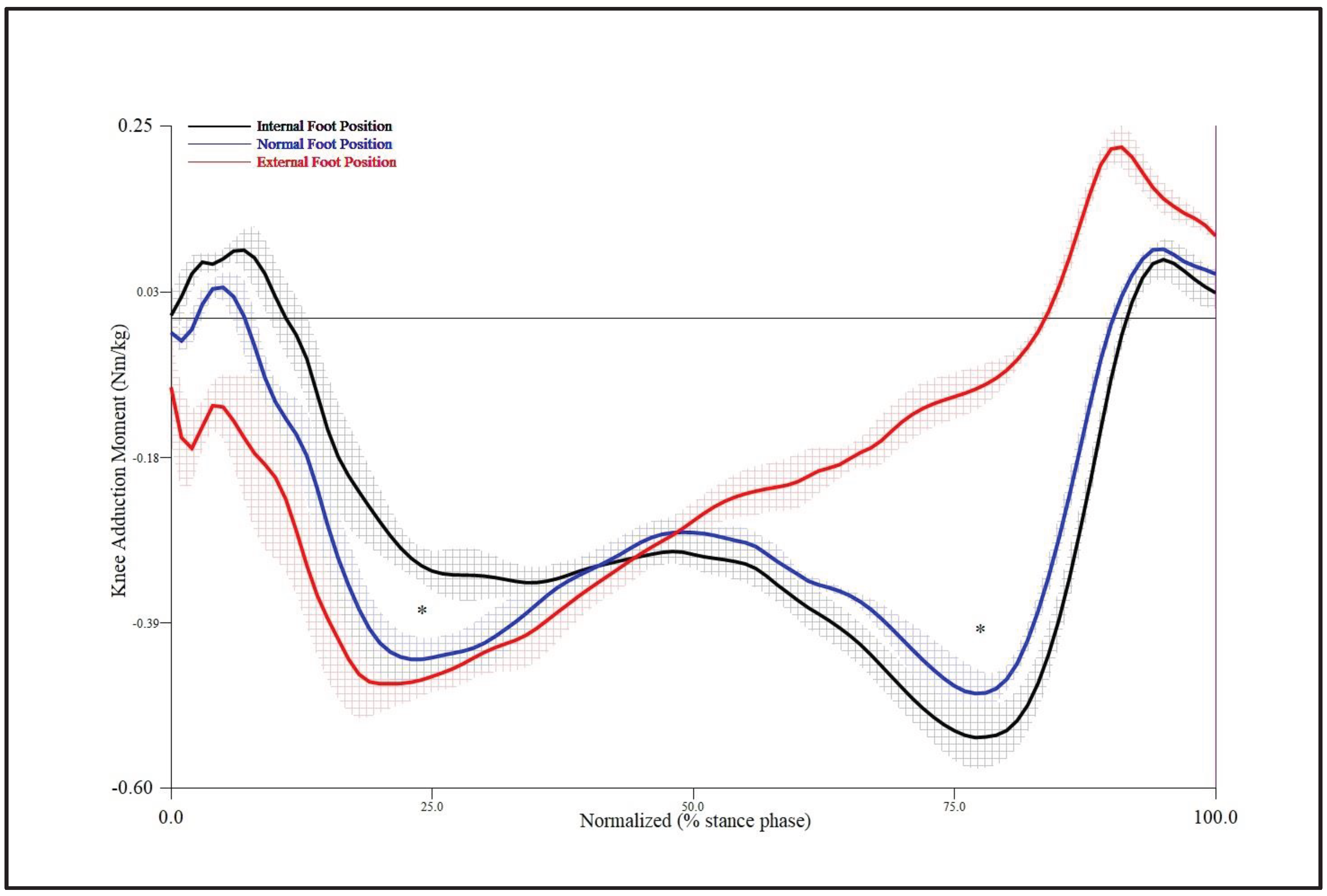
| 1st peak knee adduction moment at around 25% stance | -0.42* (0.11) | -0.34 (0.08) | -0.47 (0.10) | 0.003 |
| Knee adducton moment at mid 50% stance | -0.27 (0.09) | -0.30 (0.08) | -0.27 (0.09) | 0.326 |
| 2nd peak knee adduction moment at around 75% stance | -0.44† (0.14) | -0.50 (0.16) | -0.11 (0.11) | 0.000 |
| Ankle moment (Nm∙kg-1) | Level | F | p-value |
|---|---|---|---|
| Dorsiflexion moment peak 0-50% stance |
Foot position | 0.761 | 0.486 |
| Ankle sides | 1.122 | 0.299 | |
| Positions*sides | 2.744 | 0.078 | |
| Plantar flexion moment peak 50-100% stance |
Foot position | 0.529 | 0.571 |
| Ankle sides | 1.161 | 0.254 | |
| Positions*sides | 0.188 | 0.815 | |
| Inversion moment peak 50-75% stance |
Foot position | 6.107 | 0.019 |
| Ankle sides | 1.230 | 0.281 | |
| Positions*sides | 0.019 | 0.843 | |
| Eversion moment peak 0-50% stance |
Foot position | 2.347 | 0.102 |
| Ankle sides | 0.294 | 0.758 | |
| Positions*sides | 1.003 | 0.359 | |
| Internal rotation moment peak 0-50% stance |
Foot position | 0.888 | 0.390 |
| Ankle sides | 0.554 | 0.568 | |
| Positions*sides | 0.290 | 0.692 | |
| External rotation moment peak 50-100% stance |
Foot position | 1.453 | 0.197 |
| Ankle sides | 0.482 | 0.475 | |
| Positions*sides | 2.111 | 0.120 |
| Ankle moment (Nm∙kg-1) | NFP | IFP | EFP |
|---|---|---|---|
| Dorsiflexion peak 0-50% stance | 0.18 (0.08) | 0.18 (0.09) | 0.17 (0.09) |
| Plantarflexion peak 50-100% stance | -1.34 (0.26) | -1.34 (0.27) | -1.37 (0.26) |
| Inersion peak 50-75% stance | -0.09* (0.04) | -0.11 (0.06) | -0.04 (0.05) |
| Eversion peak 0-50% stance | 0.32 (0.26) | 0.32 (0.25) | 0.30 (0.26) |
| Internal rotation peak 0-50% stance | 0.05 (0.08) | 0.05 (0.07) | 0.03 (0.05) |
| External rotation peak 50-100% stance | -0.24 (0.16) | -0.27 (0.19) | 0.23 (0.16) |
Disclaimer/Publisher’s Note: The statements, opinions and data contained in all publications are solely those of the individual author(s) and contributor(s) and not of MDPI and/or the editor(s). MDPI and/or the editor(s) disclaim responsibility for any injury to people or property resulting from any ideas, methods, instructions or products referred to in the content. |
© 2024 by the authors. Licensee MDPI, Basel, Switzerland. This article is an open access article distributed under the terms and conditions of the Creative Commons Attribution (CC BY) license (http://creativecommons.org/licenses/by/4.0/).


