1. Introduction
With the evolution of the fast-paced modern workplace, occupational-related stress has notably increased [
1], particularly in careers involving persistent pressure and high-stake decision-making. Stress can be separated into three different categories: acute stress, episodic stress, and chronic stress [
2]. Acute stress results from short-term challenges, episodic stress is a more frequent form of acute stress that is still highly variable, and chronic stress is a constant severe stress resulting from constant demands [
3]. The chronic stress experienced by many professionals in high-pressure environments can lead to an increased rate of coronary heart disease [
4], an increase in depression and anxiety [
5], and decreased work productivity [
6]. The escalation of and dangers of workplace stress highlights the need for innovative approaches to monitor and manage the severity of stress.
Previously, the reliance on a survey to identify stress levels among professionals has been a common approach [
7]. However, this method is time inefficient and lacks the continuity of biometric monitoring techniques. In response to these limitations, prior studies have explored the use of physiological measures including electroencephalography (EEG) [
2], electrocardiography (ECG)-derived heart rate variability (HRV) [
8], galvanic skin response (GSR) [
9], and electromyography (EMG) [
10] to monitor experimentally-induced stress. Among these measures, EMG shows promise as an integrative measure to perpetually monitor stress with ease.
EMG is a diagnostic tool that utilizes the intrinsic electrical activity within muscles to monitor skeletal muscle activity and health [
11]. EMG hinges on the detection of local electrical charges caused by the neuromuscular junction. These charges trigger a wave of excitation that travels along the muscle fibers, leading to muscle contractions [
12]. Specifically, EMG records the action potentials generated when muscle fibers’ voltage rapidly exceeds a threshold, resulting in the development of a nerve signal. These myoelectric signals, produced by variations in muscle fiber membranes, allow for the analysis of motor units’ recruitment and firing characteristics within the measured muscle [
13].
Trapezius muscle EMG has been demonstrated to exhibit a strong association with stress [
14]. Previous literature depicts increased trapezius muscle activation during cognitive tasks [
15] and relates that blood flow to the trapezius muscles has the potential as an indicator of stress [
16]. Prior studies have also analyzed right and left trapezius muscles and erector spinae muscles for stress recognition [
17]. Asymmetries in left and right muscle activity have previously been recorded with stress [
14]. Determining the optimum sensor positioning and improving stress identification with enhanced features can bring us closer to more convenient, real-time stress detection. In this study, right trapezius muscle EMG features are compared with left trapezius muscle EMG features to assess the usefulness of obtaining bilateral EMG data and determine whether obtaining an asymmetry metric can improve cognitive state detection over a unilateral analysis.
Although current literature mostly considers EMG features above 20 Hz [
18,
19,
20], analyzing low-frequency (LF) EMG may hold potential in enhanced stress detection. Differing EMG frequencies corresponding to various oscillatory drives have been explored [
21] and LF EMG features can provide insight into various parts of the brain. Low-frequency oscillations in trapezius muscle EMG have been suggested as a tool to study neural mechanisms including slow-wave cortical oscillations represented in the descending cortico-spinal pathways and monoaminergic pathways originating in the brain stem reticular formation [
22]. Consequently, the 0.5-4 Hz range has revealed neurophysiological regularities in patients with Parkinson’s disease [
23] suggesting the possibility for improved diagnostic capabilities of cognitive functioning. Additionally, the 6-12 Hz frequency range has been associated with differing parts of the brain, including the cerebellar [
24], subcortical [
25], sensorimotor cortex [
26], and primary motor cortex [
27]. Given these interconnections between LF EMG and cognitive activity, our study seeks to investigate the relationship between LF EMG and cognitive states, particularly stress and meditation.
2. Materials and Methods
2.1. Experimental Protocol and Data Acquistion
After Institutional Review Board (IRB) approval, 15 healthy participants, 7 males, and 8 females, between the ages of 18 and 30 with no history of neurological diseases were recruited. All participants had no prior experience with meditation and were trained on how to meditate before starting the experiment.
The protocol involved a 5-minute resting data collection, followed by a questionnaire, then the 10-minute guided meditation, followed by a 5-minute mental stress segment,
Figure 1. Participants answered a stress survey between each segment. The questionnaire (QS) had participants’ input age, amount of sleep, waking time, gender, dominant hand, nationality, current profession, stress level at work, and caffeine and food intake. The stress surveys had the participants rank their current stress level on a visual scale from 1 to 10. At all gaps and intervals, participants were instructed to relax and avoid any physical activity or conversation.
The 5-minute stressor involved a Stroop color-word test (SCWT) for the first 2.5 minutes and a mental arithmetic (MA) task for the last 2.5 minutes. For the mental arithmetic, news was played in the background while subjects were asked to count backward from 2043 in integers of 17 [
28].
The meditation training involved a 10-minute guided meditation practice where participants were instructed to focus on their breathing and follow a visual breathing bubble that expanded with inhalation and deflated with exhalation. The visual was accompanied by a calming tone that matched the inhalation and exhalation patterns. This guided practice was repeated during data collection. There was a 20-minute gap time following the meditation training and the start of the experiment.
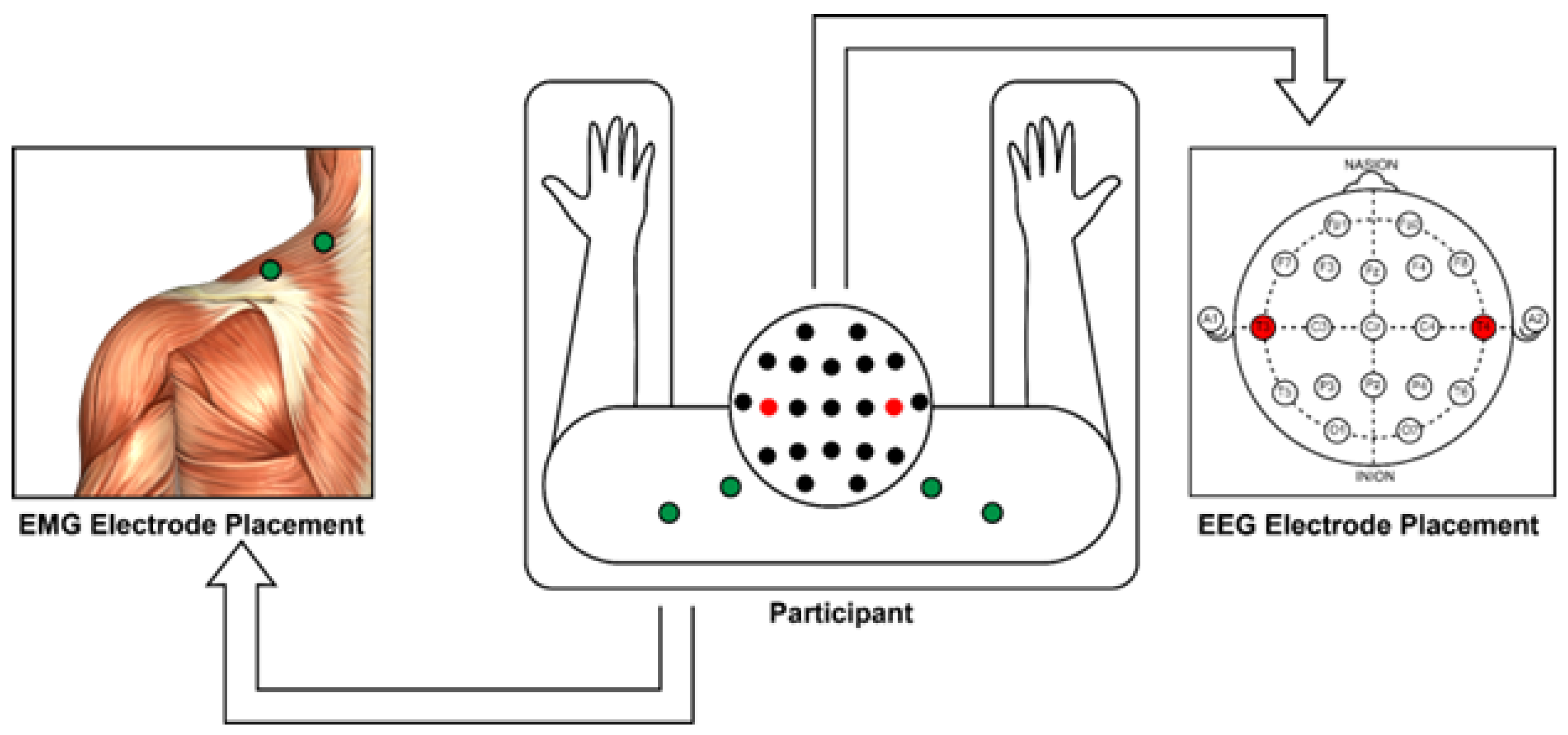
Electroencephalography (EEG) data was collected as a control to ensure that the expected cognitive results were achieved and was used as a comparison for EMG findings. A DSI-24 system was used for data collection and mid trapezius muscle EMG was collected at 300 samples/second. The electrode setup is shown in
Figure 2. The experiment was conducted in a naturally lit and noiseless environment to minimize signal interference and other artifacts. To reduce muscle artifacts in the trapezius muscle sensors, participants were instructed to sit in a natural position while keeping their elbows on the armrests and to maintain a neutral head position throughout the study.
2.2. Signal Processing
Stressful tasks have been linked to the development of pain and fatigue in the trapezius muscles [
29]. To investigate the relation between cognitive states and trapezius muscle EMG, a high pass filter at 20 Hz was applied to obtain EMG features above 20 Hz, referred to as high-frequency (HF) EMG, and a bandpass filter from 0.5 Hz - 20 Hz was applied to obtain LF EMG features. Min-max normalization was performed for each segment. EEG signals were preprocessed using EEGlab [
30]. Surface topology maps were created from the EEG electrodes in the DSI-24 system to assess the effect of stress and meditation. Arousal index [
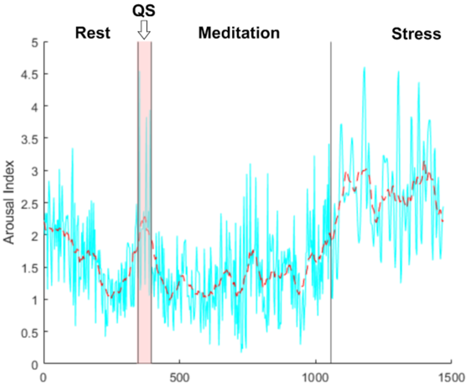
31] was calculated from averaged temporal electrodes T3 and T4, as shown in
Figure 2.
2.3. Feature Evaluation
Five EMG features were analyzed, namely median frequency (MDF), mean power spectral density (PSD), variance (VAR), Simple square integral (SSI), and asymmetry. Mean PSD can be highly informative [
10,
32] and has shown better performance when specifically assessing muscle fatigue [
33] and motor unit recruitment [
34]. Mean frequency (MNF) and MDF are understood as the ideal frequency domain features, frequently used to assess muscle fatigue [
35]. Although MNF and MDF are similar, MDF is less affected by signal noise and more sensitive to muscle fatigue [
36]. Muscle fatigue increases EMG signal amplitude, therefore, time-domain features based on energy information can also track this behavior. MNF and MDF, typically around 8 Hz, have previously been shown to decrease significantly during stress than during rest [
37]. Stress also tends to increase muscle tension and increase muscle contraction variations. SSI expresses the energy of the EMG signal; an increase in this energy indicates heightened muscle activity. Variance in EMG signals helps capture the variability in muscle activity.
The data was split into the 3 segments - rest, stress, and meditation - to determine whether a significant difference could be observed between these segments in either frequency range for any of the 4 features. Furthermore, an analysis of whether this observation was improved on one side more than the other was also performed. Asymmetry in either frequency range was assessed to determine whether a certain segment could be better differentiated using bilateral sensors to determine this metric. A paired t-test was performed for statistical significance between segments.
3. Results
Band power data from EEG was used as a control for the protocol to observe cognitive changes throughout the experiment.
Figure 3 depicts transient changes in the arousal index throughout the experiment for one subject. A decrease in this metric can be observed with meditation, followed by a significant increase during the stressor. Similar results were observed for other subjects.
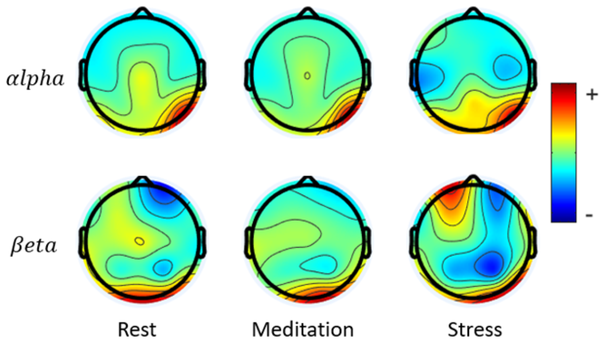
The surface topology maps shown in
Figure 4 further demonstrate the effects of stress and meditation and the overall reproducibility of the protocol in providing the desired results. With meditation, a slowing of brain activity is observed with decreased hemispheric asymmetry. Prior research has found that effective meditation led to slowed down synchronous alpha activity with an overall shift towards the parasympathetic system [
38]. The stress segment in this study increased both the beta and alpha band powers as expected, with an increase in hemispheric asymmetry. This parallels prior literature that has observed increased frontal asymmetry favoring the left during stress [
39].
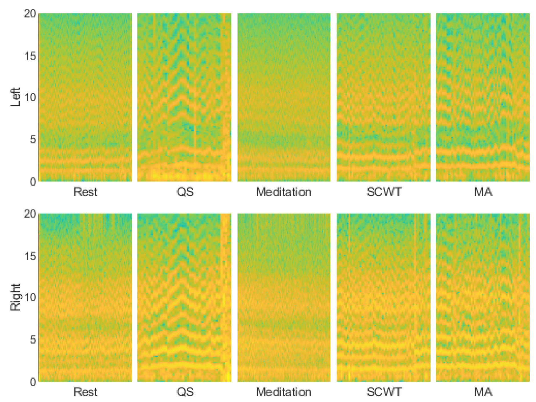
Table 1 indicates paired T-test p-values for the aforementioned parameters. A time-frequency distribution analysis using continuous wavelet transform throughout the study is shown for one subject in
Figure 5. Other subjects demonstrated similar results. Interestingly, the LF activity during the MA portion demonstrates fluctuations similar to the LF activity during the QS. The cognitive task of responding to questions in the QS and the task of mental arithmetic elicited similar variations, implicating that there may be a significant relationship between cognitive functioning and LF EMG. Less variation is observed during the SCWT compared to the MA segment likely due to task difficulty. Resting and meditation appeared similar in this frequency range whereas a stark difference can be observed during the stressor.
From
Table 1, both the left and right trapezius muscles appear capable of differentiating between segments on their own. An equal number of features were significant for either side. With the right trapezius muscle, there was an even split between LF and HF features in differentiating each segment. For the left trapezius, the LF features appeared to provide a greater differentiation than HF features between rest and stress and between stress and meditation, whereas HF features provided greater differentiation between meditation and rest. When asymmetry was considered, LF features allowed for differentiation between stress and meditation and between meditation and rest but not between rest and stress. HF features demonstrated an opposite trend, implicating that when meditation was involved, LF features were best at identifying a difference using trapezius muscle asymmetry. Therefore, analyzing asymmetry in the LF range can enable an improved differentiation between rest and meditation. Asymmetry in the HF range was only beneficial in differentiating between rest and stress.
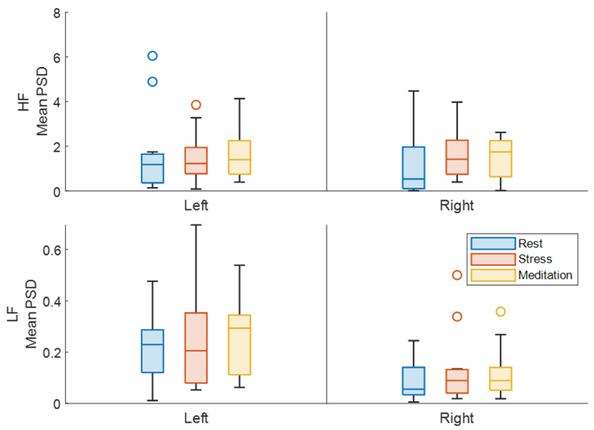
Overall, mean PSD was not useful in identifying varying cognitive states. As seen in
Figure 6, the mean value of this parameter did not vary significantly between each segment, regardless of which frequency range was considered. There was no preference for laterality in this feature either.
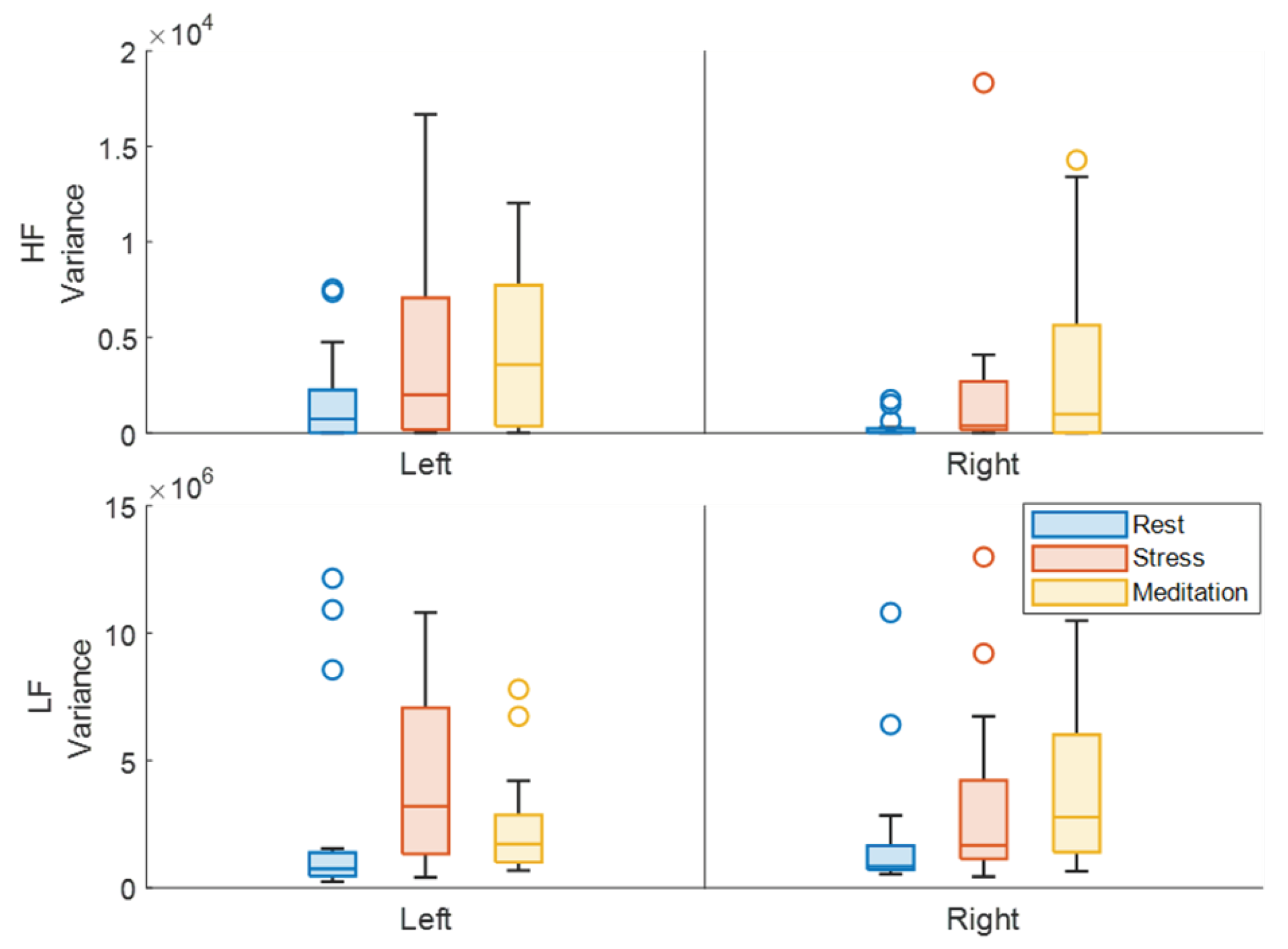
An analysis of EMG variance in unilateral trapezius muscles, shown in
Figure 7, indicates that LF features from the left trapezius were particularly useful in discerning rest from stress and stress from meditation but not in demarcating rest from meditation. Variance in the HF range made up for this deficit, providing significant differences between rest and meditation on the left. The right trapezius muscle allowed for differentiation only between rest and meditation in either frequency range. With this in mind, bilateral trapezius muscle analysis would be required to enable accurate identification of the various states.
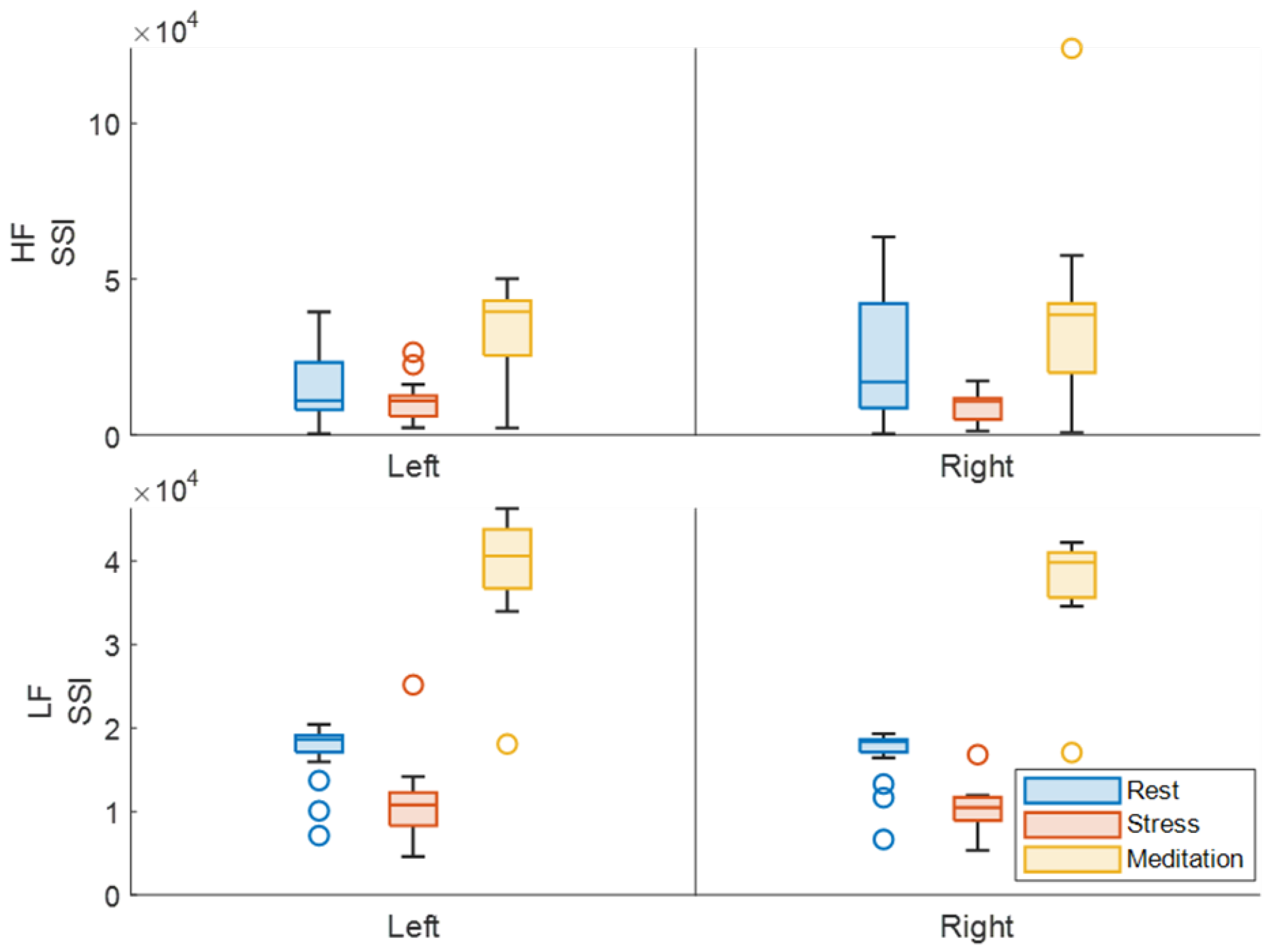
SSI is an amplitude parameter used to gauge the level of muscle activity. In the LF range, a significantly higher SSI is observed during meditation, (
Figure 8), which may seem counterintuitive but is secondary to the min-max normalization method applied to each segment. Applying min-max normalization to the whole dataset would have allowed for a different analysis between segments, but this method was chosen to focus more on the feature and its capability to identify the different states rather than highlighting expected trends throughout the experiment. The elevated value indicates that the LF amplitude during the meditation did not vary significantly. In that regard, stress consistently demonstrates the lowest SSI value in either frequency range bilaterally. The LF range did appear to highlight the difference between rest and stress better than the HF range as the p-values were much closer to 0. HF analysis of the SSI from the left trapezius muscle on the other hand did not allow for differentiation between the rest and stress segments. Overall, obtaining the SSI from the LF range allowed for the best characterization of the induced neurological states.
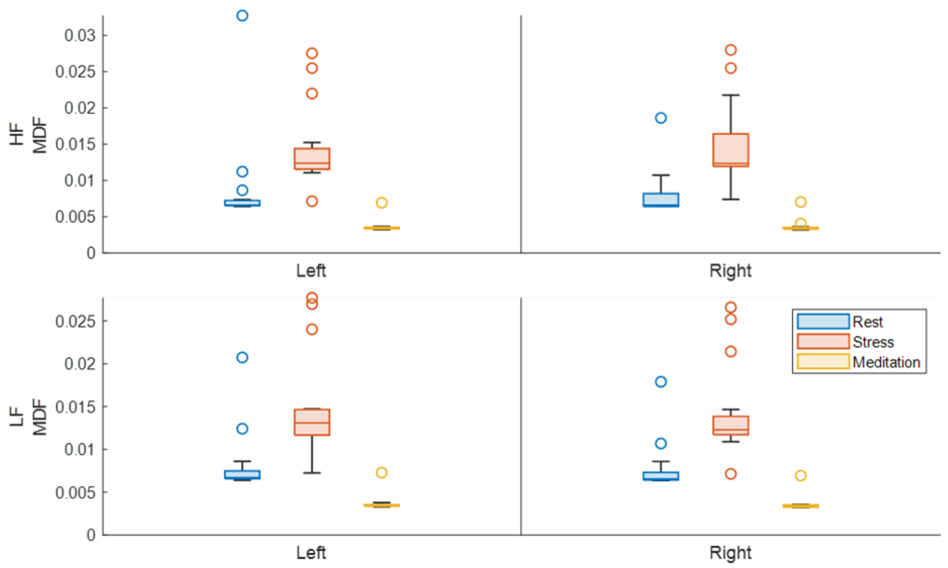
MDF was another feature that allowed for the identification of stress and meditation. From
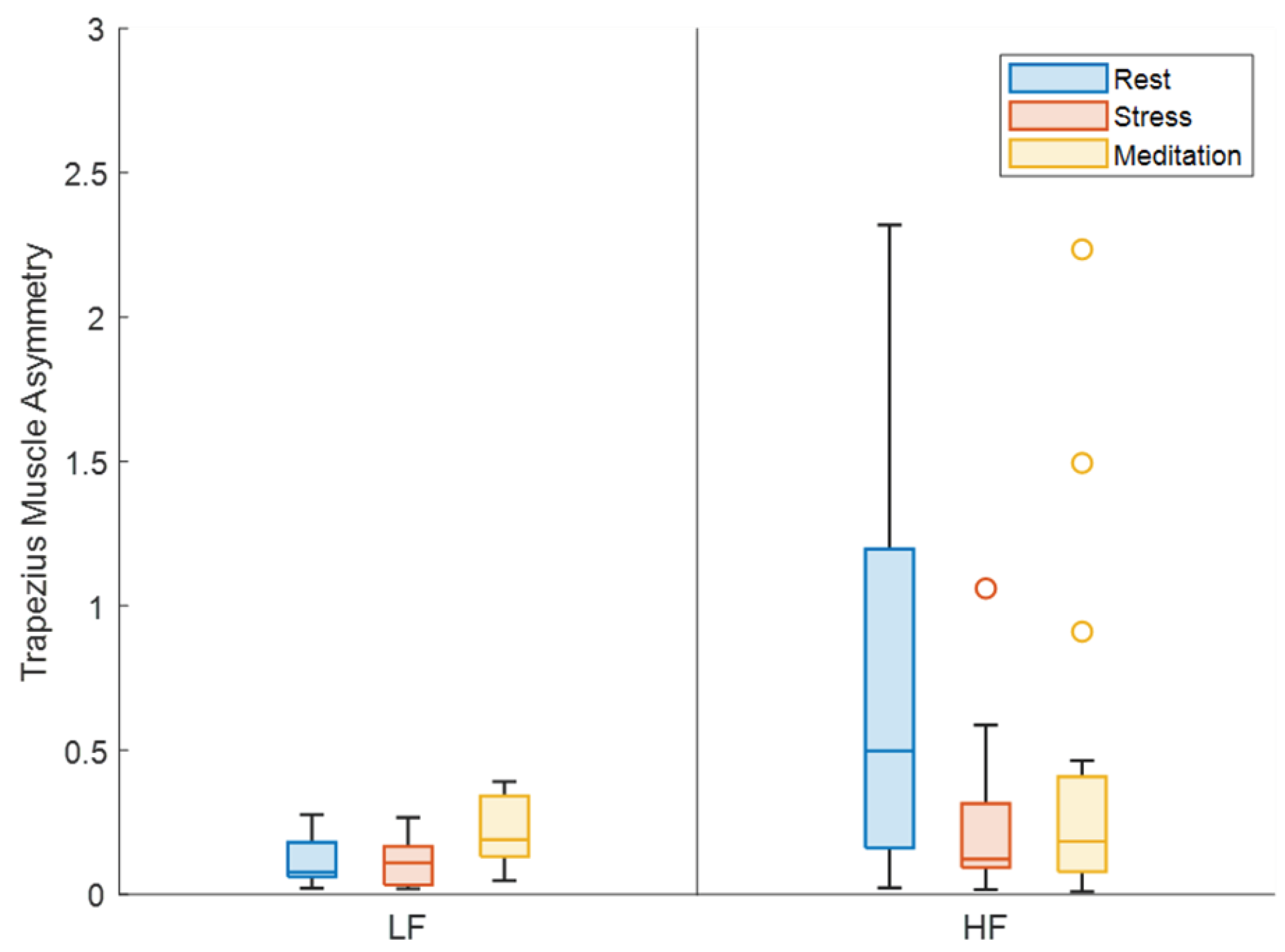
Figure 9, MDF appears to be elevated during the stressor and reduced during the meditation compared to the baseline resting. Both frequency ranges portrayed this relationship without a preference for laterality. LF asymmetry appeared beneficial in differentiating stress and meditation, and meditation from rest, but not stress from rest,
Figure 10. Asymmetry in the HF range provided the opposite result in that only the rest and stress segments were characterized with significant contrast. If asymmetry were to be applied to identify various states, results from both frequency ranges would be required for best results.
4. Discussion
From the results, LF EMG shows potential in identifying cognitive functioning and seemingly follows a pattern depending on the task performed. Greater variations in the PSD can be observed during stress whereas slowing of activity is portrayed during meditation. This parallels findings from the EEG topology maps where decreased activity was observed with meditation and greater activity was observed with stress. Meditation has been shown to decrease overall EEG power [
40] whereas stress results in significant increases in some spectral EEG indices [
41].
Similar to how EEG asymmetry enabled the characterization of rest, meditation and stress, trapezius muscle asymmetry provided accurate identification of these states using both LF and HF EMG analysis. However, given that both trapezius muscles were useful in identifying these states on their own, a bilateral analysis is not needed but can be used to obtain additional information. LF frequency features greatly increased the identification of stress and meditation unilaterally. Analyzing HF features improved the characterization of rest and meditation. Interpreting both frequency ranges in concert can lead to improved state detection when either the left or right trapezius muscle is assessed on its own.
Mean PSD showed the lowest ability in differentiating between segments. When considering EMG variance, the left trapezius muscle appeared best for differentiating between cognitive states. Although both left and right trapezius muscles appeared equally useful in identifying rest, stress, and meditation from the SSI parameter, characterization of these segments was improved with LF analysis. Similarly, the MDF feature did not appear to indicate a preference for laterality, and either muscle was able to effectively differentiate between the different states. All participants in this study were right-hand dominant, which may have influenced these results, however, previous work analyzing 9 different EMG features and various electrode positioning determined that the EMG signal of the right trapezius muscle recognized stress better than other muscles [
10]. Based on previous findings and results from this study, the right trapezius muscle can be used on its own for accurate stress detection. For better observations of meditational states, a bilateral EMG signal can be beneficial.
As a more direct measure of brain activity, EEG can provide better indications regarding stress and arousal [
42]. However, wearing scalp electrodes is not feasible for daily real-time monitoring. Small movements can disrupt the electrode contacts and introduce artifacts into the signal [
43], preventing accurate detection. In contrast, trapezius muscle sensors can easily be placed and can provide stress-related information despite physical movements. Studies have shown that when physical stressors are present in addition to mental stressors, there is an increase in trapezius muscle EMG [
14], demonstrating the effectiveness of trapezius muscle EMG in detecting stress regardless of daily activities.
5. Conclusions
Trapezius muscle EMG can allow for convenient, non-invasive monitoring of cognitive states. Low-frequency EMG features provided enhanced detection of stress and sensor configurations can be minimized to the right trapezius muscle for accurate stress identification. Additionally, the introduction of a novel method for determining asymmetry in EMG features demonstrates that applying sensors on bilateral trapezius muscles improved the detection of stress and meditation.
Author Contributions
Conceptualization, Mohammad Ahmed, Mehmet Kaya, and Peshala T. Gamage; methodology, Mohammad Ahmed; validation, Amirtaha Taebi, Mehmet Kaya, and Peshala T. Gamage; formal analysis, Mohammad Ahmed; investigation, Mohammad Ahmed and Michael Grillo; writing—original draft preparation, Mohammad Ahmed and Michael Grillo; writing—review and editing, Mohammad Ahmed, Michael Grillo, Amirtaha Taebi, Mehmet Kaya, Peshala T. Gamage; visualization, Amirtaha Taebi, Mehmet Kaya, and Peshala T. Gamage; supervision, Mehmet Kaya and Peshala T. Gamage.
Funding
This research received no external funding.
Institutional Review Board Statement
This study was approved by the Institutional Review Board (IRB) at Florida Institute of Technology, IRB number 23-019. The research protocol was reviewed and approved by the IRB Chairperson Dr. Jignya Patel. Per federal regulations, 45 CFR 46.110, this study has been determined to involve no more than minimal risk for human subjects. Federal regulations define minimal risk to mean that the probability and magnitude of harm are no more than would be expected in the daily life of a normal, healthy person. A written consent form was obtained from all participants. All data, including signed consent form documents, must be retained in a locked file cabinet for a minimum of three years (six if HIPAA applies) past the completion of this research. Any links to the identification of participants should be maintained on a password-protected computer if electronic information is used. Access to data is limited to authorized individuals listed as key study personnel.
Informed Consent Statement
Informed consent was obtained from all subjects involved in the study
Data Availability Statement
The data presented in this study are available on request from the corresponding author (accurately indicate status).
Conflicts of Interest
The authors declare no conflicts of interest.
References
- Boyd, D. Workplace Stress.
- Katmah, R.; Al-Shargie, F.; Tariq, U.; Babiloni, F.; Al-Mughairbi, F.; Al-Nashash, H. A Review on Mental Stress Assessment Methods Using EEG Signals. Sensors 2021, 21, 5043. [Google Scholar] [CrossRef] [PubMed]
- Sincero, S. Three Different Kinds of Stress., 2012.
- Levine, G.N. Psychological Stress and Heart Disease: Fact or Folklore? The American Journal of Medicine 2022, 135, 688–696. [Google Scholar] [CrossRef]
- Melchior, M.; Caspi, A.; Milne, B.J.; Danese, A.; Poulton, R.; Moffitt, T.E. Work stress precipitates depression and anxiety in young, working women and men. Psychological Medicine 2007, 37, 1119–1129. [Google Scholar] [CrossRef] [PubMed]
- Bui, T.; Zackula, R.; Dugan, K.; Ablah, E. Workplace Stress and Productivity: A Cross-Sectional Study. Kansas Journal of Medicine 2021, 14. [Google Scholar] [CrossRef] [PubMed]
- Vagg, P.R.; Spielberger, C.D. The Job Stress Survey: Assessing perceived severity and frequency of occurrence of generic sources of stress in the workplace. Journal of Occupational Health Psychology 1999, 4, 288–292. [Google Scholar] [CrossRef] [PubMed]
- Kim, H.G.; Cheon, E.J.; Bai, D.S.; Lee, Y.H.; Koo, B.H. Stress and Heart Rate Variability: A Meta-Analysis and Review of the Literature. Psychiatry Investigation 2018, 15, 235–245. [Google Scholar] [CrossRef] [PubMed]
- Fernandes, A.; Helawar, R.; Lokesh, R.; Tari, T.; Shahapurkar, A.V. Determination of stress using Blood Pressure and Galvanic Skin Response. 2014 International Conference on Communication and Network Technologies; IEEE: Sivakasi, India, 2014; pp. 165–168. [Google Scholar] [CrossRef]
- Pourmohammadi, S.; Maleki, A. Stress detection using ECG and EMG signals: A comprehensive study. Computer Methods and Programs in Biomedicine 2020, 193, 105482. [Google Scholar] [CrossRef]
- Wang, J.; Sun, Y.; Sun, S. Recognition of Muscle Fatigue Status Based on Improved Wavelet Threshold and CNN-SVM. IEEE Access 2020, 8, 207914–207922. [Google Scholar] [CrossRef]
- Mukund, K.; Subramaniam, S. Skeletal muscle: A review of molecular structure and function, in health and disease. WIREs Systems Biology and Medicine 2020, 12, e1462. [Google Scholar] [CrossRef]
- Kuo, I.Y.; Ehrlich, B.E. Signaling in Muscle Contraction. Cold Spring Harbor Perspectives in Biology 2015, 7, a006023. [Google Scholar] [CrossRef]
- Luijcks, R.; Hermens, H.J.; Bodar, L.; Vossen, C.J.; Os, J.v.; Lousberg, R. Experimentally Induced Stress Validated by EMG Activity. PLoS ONE 2014, 9, e95215. [Google Scholar] [CrossRef] [PubMed]
- Schleifer, L.M.; Spalding, T.W.; Kerick, S.E.; Cram, J.R.; Ley, R.; Hatfield, B.D. Mental stress and trapezius muscle activation under psychomotor challenge: A focus on EMG gaps during computer work. Psychophysiology 2008, 45, 356–365. [Google Scholar] [CrossRef] [PubMed]
- Larsson, S.E.; Larsson, R.; Zhang, Q.; Cai, H.; Ake Oberg, P. Effects of psychophysiological stress on trapezius muscles blood flow and electromyography during static load. European Journal of Applied Physiology and Occupational Physiology 1995, 71, 493–498. [Google Scholar] [CrossRef] [PubMed]
- Sharma, N.; Gedeon, T. Objective measures, sensors and computational techniques for stress recognition and classification: A survey. Computer Methods and Programs in Biomedicine 2012, 108, 1287–1301. [Google Scholar] [CrossRef]
- Sarilho De Mendonça, F.; De Tarso Camillo De Carvalho, P.; Biasotto-Gonzalez, D.A.; Calamita, S.A.P.; De Paula Gomes, C.A.F.; Amorim, C.F.; Fumagalli, M.A.; Politti, F. Muscle fiber conduction velocity and EMG amplitude of the upper trapezius muscle in healthy subjects after low-level laser irradiation: a randomized, double-blind, placebo-controlled, crossover study. Lasers in Medical Science 2018, 33, 737–744. [Google Scholar] [CrossRef] [PubMed]
- Oberg, T.; Sandsjo, L.; Kadefors, R. Electromyogram mean power frequency in non-fatigued trapezius muscle. European Journal of Applied Physiology and Occupational Physiology 1990, 61, 362–369. [Google Scholar] [CrossRef] [PubMed]
- Hummel, A.; Läubli, T.; Pozzo, M.; Schenk, P.; Spillmann, S.; Klipstein, A. Relationship between perceived exertion and mean power frequency of the EMG signal from the upper trapezius muscle during isometric shoulder elevation. European Journal of Applied Physiology 2005, 95, 321–326. [Google Scholar] [CrossRef]
- Grosse, P.; Cassidy, M.; Brown, P. EEG–EMG, MEG–EMG and EMG–EMG frequency analysis: physiological principles and clinical applications. Clinical Neurophysiology 2002, 113, 1523–1531. [Google Scholar] [CrossRef]
- Westgaard, R.H.; Bonato, P.; Holte, K.A. Low-Frequency Oscillations (<0.3 Hz) in the Electromyographic (EMG) Activity of the Human Trapezius Muscle During Sleep. Journal of Neurophysiology 2002, 88, 1177–1184. [Google Scholar] [CrossRef]
- Kotelnikov Institute of Radioengineering and Electronics of RAS.; Sushkova, O.S.; Morozov, A.A.; Kotelnikov Institute of Radioengineering and Electronics of RAS.; Gabova, A.V.; Institute of Higher Nervous Activity and Neurophysiology of RAS.; Karabanov, A.V.; Research Center of Neurology of RF Ministry of Education and Science.; Chigaleychik, L.A.; Research Center of Neurology of RF Ministry of Education and Science. NVESTIGATION OF THE 0.5-4 HZ LOW-FREQUENCY RANGE IN THE WAVE TRAIN ELECTRICAL ACTIVITY OF MUSCLES IN PATIENTS WITH PARKINSON’S DISEASE AND ESSENTIAL TREMOR. Radioelectronics. Nanosystems. Information Technologies 2019, 11, 225–236. [Google Scholar] [CrossRef]
- McAuley, J.H. Physiological and pathological tremors and rhythmic central motor control. Brain 2000, 123, 1545–1567. [Google Scholar] [CrossRef] [PubMed]
- Raethjen, J.; Lindemann, M.; Morsnowski, A.; Dümpelmann, M.; Wenzelburger, R.; Stolze, H.; Fietzek, U.; Pfister, G.; Elger, C.E.; Timmer, J.; Deuschl, G. Is the rhythm of physiological tremor involved in cortico-cortical interactions? Movement Disorders 2004, 19, 458–465. [Google Scholar] [CrossRef] [PubMed]
- Salenius, S.; Portin, K.; Kajola, M.; Salmelin, R.; Hari, R. Cortical Control of Human Motoneuron Firing During Isometric Contraction. Journal of Neurophysiology 1997, 77, 3401–3405. [Google Scholar] [CrossRef] [PubMed]
- Marsden, J.F.; Brown, P.; Salenius, S. Involvement of the sensorimotor cortex in physiological force and action tremor:. Neuroreport 2001, 12, 1937–1941. [Google Scholar] [CrossRef]
- Ahmed, M.H.; Kaya, M.; Taebi, A.; Thibbotuwawa Gamage, P. Feasibility of Trapezius Muscle Electromyography and Electrocardiography to Monitor Stress Levels in High Demand Positions. Volume 5: Biomedical and Biotechnology; American Society of Mechanical Engineers: New Orleans, Louisiana, USA, 2023. [Google Scholar] [CrossRef]
- Sjörs, A.; Larsson, B.; Dahlman, J.; Falkmer, T.; Gerdle, B. Physiological responses to low-force work and psychosocial stress in women with chronic trapezius myalgia. BMC Musculoskeletal Disorders 2009, 10, 63. [Google Scholar] [CrossRef]
- Hanke, M.; Halchenko, Y.O. Neuroscience Runs on GNU/Linux. 5. [CrossRef]
- Ahmed, M.H.; Kaya, M.; Taebi, A.; Thibbotuwawa Gamage, P. The Role of Meditation in Stress Recovery and Performance: An EEG Study. Volume 5: Biomedical and Biotechnology; American Society of Mechanical Engineers: New Orleans, Louisiana, USA, 2023. [Google Scholar] [CrossRef]
- Roman-Liu, D.; Konarska, M. Characteristics of power spectrum density function of EMG during muscle contraction below 30%MVC. Journal of Electromyography and Kinesiology 2009, 19, 864–874. [Google Scholar] [CrossRef]
- R., M.; Sepulveda, F.; Colley, M. sEMG Techniques to Detect and Predict Localised Muscle Fatigue. In EMG Methods for Evaluating Muscle and Nerve Function; Schwartz, M., Ed.; InTech, 2012. [Google Scholar] [CrossRef]
- Oskoei, M.; Huosheng, Hu. Support Vector Machine-Based Classification Scheme for Myoelectric Control Applied to Upper Limb. IEEE Transactions on Biomedical Engineering 2008, 55, 1956–1965. [Google Scholar] [CrossRef]
- Phinyomark, A.; Thongpanja, S.; Hu, H.; Phukpattaranont, P.; Limsakul, C. The Usefulness of Mean and Median Frequencies in Electromyography Analysis. In Computational Intelligence in Electromyography Analysis - A Perspective on Current Applications and Future Challenges; Naik, G.R., Ed.; InTech, 2012. [Google Scholar] [CrossRef]
- Stulen, F.B.; De Luca, C.J. Frequency Parameters of the Myoelectric Signal as a Measure of Muscle Conduction Velocity. IEEE Transactions on Biomedical Engineering 1981, BME-28, 515–523. [Google Scholar] [CrossRef]
- Wijsman, J.; Grundlehner, B.; Penders, J.; Hermens, H. Trapezius muscle EMG as predictor of mental stress. ACM Transactions on Embedded Computing Systems 2013, 12, 1–20. [Google Scholar] [CrossRef]
- Dobrakowski, P.; Blaszkiewicz, M.; Skalski, S. Changes in the Electrical Activity of the Brain in the Alpha and Theta Bands during Prayer and Meditation. International Journal of Environmental Research and Public Health 2020, 17, 9567. [Google Scholar] [CrossRef]
- Berretz, G.; Packheiser, J.; Wolf, O.T.; Ocklenburg, S. Acute stress increases left hemispheric activity measured via changes in frontal alpha asymmetries. iScience 2022, 25, 103841. [Google Scholar] [CrossRef] [PubMed]
- Hinterberger, T.; Schmidt, S.; Kamei, T.; Walach, H. Decreased electrophysiological activity represents the conscious state of emptiness in meditation. Frontiers in Psychology 2014, 5. [Google Scholar] [CrossRef] [PubMed]
- Vanhollebeke, G.; De Smet, S.; De Raedt, R.; Baeken, C.; Van Mierlo, P.; Vanderhasselt, M.A. The neural correlates of psychosocial stress: A systematic review and meta-analysis of spectral analysis EEG studies. Neurobiology of Stress 2022, 18, 100452. [Google Scholar] [CrossRef] [PubMed]
- Chand, T.; Alizadeh, S.; Jamalabadi, H.; Herrmann, L.; Krylova, M.; Surova, G.; van der Meer, J.; Wagner, G.; Engert, V.; Walter, M. EEG revealed improved vigilance regulation after stress exposure under Nx4 – A randomized, placebo-controlled, double-blind, cross-over trial. IBRO Neuroscience Reports 2021, 11, 175–182. [Google Scholar] [CrossRef]
- Kappel, S.L.; Looney, D.; Mandic, D.P.; Kidmose, P. Physiological artifacts in scalp EEG and ear-EEG. BioMedical Engineering OnLine 2017, 16, 103. [Google Scholar] [CrossRef]
|
Disclaimer/Publisher’s Note: The statements, opinions and data contained in all publications are solely those of the individual author(s) and contributor(s) and not of MDPI and/or the editor(s). MDPI and/or the editor(s) disclaim responsibility for any injury to people or property resulting from any ideas, methods, instructions or products referred to in the content. |
© 2024 by the authors. Licensee MDPI, Basel, Switzerland. This article is an open access article distributed under the terms and conditions of the Creative Commons Attribution (CC BY) license (http://creativecommons.org/licenses/by/4.0/).

