Submitted:
22 March 2024
Posted:
25 March 2024
You are already at the latest version
Abstract
Mycobacteria can be one of the main contaminants of biological products, and their presence can have serious consequences on patients’ health. Thus, specific testing is necessitated by regulations such as the European Pharmacopoeia. The current pharmacopeial reference i.e., microbial culture method cannot ensure an exhaustive detection of mycobacteria due to their growth characteristics. Additionally, the method is time-consuming and requires a continuous supply of culture media, which could be further challenging. Thus, to overcome these challenges, pharmaceutical industries need to consider alternative non-microbiological techniques to detect these fastidious, slow-growing contaminating agents. This review provides an overview of alternative methods, which could be applied within a quality control environment of biological products, and underlines advantages and limitations of these methods. Nucleic acid amplification techniques or direct measurement of mycobacteria stands out as the most suitable alternatives for mycobacterial testing in biological products.
Keywords:
Mycobacteria
; Mycobacteria detection
; contaminants
; biological products
; biopharmaceutical QC control
; nucleic acid amplification techniques
; alternative methods
1. Introduction
Mycobacterial contamination of biological products refers to the presence of mycobacterial species such as Mycobacterium (M.) tuberculosis, M. avium and others in the products derived from biological sources, such as vaccines, cell cultures, blood products, and other biologicals. Although most mycobacterial species are nonpathogenic, their survivability and ubiquitous presence across environments make them an ever-present potential source of contamination in eukaryotic cell-based bioproducts [1]. The mycobacteria are also known for their resistance to disinfection methods, which makes their elimination during the production of biologicals challenging. Contamination of biologicals poses a significant risk to patients’ health considering the potential infections and serious adverse effects [2,3,4]. Such contaminants can be introduced at any stage of the production, with sources varying from raw materials and environments to operators. Therefore, detection and isolation of mycobacteria is a regulatory requirement to ensure the efficacy and safety of the biologicals [5].
Various methods, including culture-based techniques, nucleic acid amplification techniques (NATs, e.g., polymerase chain reaction [PCR]), and immunological assays, can be employed to detect and/or identify the presence of mycobacteria in biologicals. The reference ‘compendial assay’ method is based on the detection of mycobacteria by the microbial culture method. Considering the slow growth of some of the species e.g., Mycobacterium tuberculosis complex (MTBC), culture incubation can be notoriously long and can last for over 56 days. Accordingly, the duration of mycobacterial testing is the longest of all bacteriological quality control tests for biologicals. It is twice the duration of mycoplasma culture tests and nearly four times the duration of sterility tests. Additionally, the method requires a continuous supply of culture media, which again could be challenging.
Thus, for products with a limited shelf-life, this prolonged duration of process can be critical to ensure their timely and efficient release. These challenges could be addressed using the alternative culture-free detection methods, which could drastically reduce the required time and the requirement of raw materials, such as culture media [6,7].
According to the guidelines on the implementation of alternative methods, an alternative candidate method must demonstrate the non-inferiority and, ideally should be superior to the reference method [6,7,8], where superiority can mean, an overall improvement of product’s safety by early detection, increased sensitivity and/or increased specificity.
Available alternative methods are based on different detection mechanisms such as nucleic acid amplification, flow cytometry, or electrochemical detection. While a considerable number of studies are focused on mycobacterial testing to assist diagnostics in a clinical setting, the information on mycobacterial testing for quality control of biologicals is limited. The method deployed in a quality control setting needs not only to be sensitive but also exhaustive enough to enable detection of the lowest number of cells of any mycobacterial species.
The purpose of this review article is to outline the alternative mycobacterial detection methods that could potentially replace the microbial culture method for quality control of biologicals in an industrial setting.
2. Compendial Assay
2.1. Compendial Assay: The Gold Standard for Mycobacterial Detection
The microbial culture method or compendial assay is used for detecting mycobacteria in diagnostics as well as quality control of biologicals [5,9,10]. Although the limit of detection (LOD) of this method has never been published, it is reported that this method is highly sensitive and can thus detect even the lowest number of mycobacteria. The method can be paired with various identification techniques which can be used for further investigations following a positive result. These identification techniques can be based on either microscopic morphology with Ziehl–Neelsen staining, deoxyribonucleic acid [DNA] sequencing, or matrix-assisted laser desorption/ionization time of flight mass spectrometry (MALDI-TOF MS) [10].
Different regulatory texts describe the detection of mycobacteria by culture with varying degrees of details for the quality control of biologicals. European Pharmacopoeia 2.6.2 requires the product to be assessed on three different media, which includes two solid and one liquid media, in triplicates, for a period of 56 days. If any of these nine samples is positive i.e., establishment of mycobacterial growth during the incubation period, the result is considered positive. Whereas, if none of these media show any mycobacterial growth after the incubation period, the result is considered negative, and the product passes the quality control test [5].
Mycobacteria specific media exist in both solid and liquid forms and are complementary to each other. Most of these media are developed for mycobacterial detection in sputum, tissues, or body fluid samples and contain bacterial and fungal growth inhibitors, which allows the growth of specific target mycobacterial species [10]. Different media are commercially available depending on their applications (Table 1). Overall, liquid media enable an early detection of mycobacterial growth and have a better recovery rate than solid media [10,11]. Egg-based and agar-based media are the two commonly used solid media. Due to the long incubation period for mycobacterial recovery, solid media are prepared in slant tubes. However, counting and numeration of mycobacterial colonies are extremely difficult due to the limited slant area and non-homogenous spread of colonies [10,11]. Apart from the requirement of two solid and one liquid media, there are no specific recommendations in European Pharmacopoeia [5].
2.2. Advantages and Limitations of Compendial Assay
The microbial culture method is a simple assay and does not require any expensive equipment or skilled personnel. Besides the low hands-on time, this assay offers a high level of sensitivity, which is of particular importance to detect potential mycobacterial contaminations in biologicals.
However, in a quality control setting, the method remains a time-consuming affair. Besides, the method is not suitable for all mycobacterial species, as some of these species are not easily cultured on media (e.g., M. lepraemurinum), or have specific nutritional requirements (e.g., M. haemophilum), whereas some do not grow on the media at all, (e.g., M. leprae) [11]. Despite their selectivity, these media are not specific to mycobacteria and can support the growth of other actinomycetes such as Nocardia. This could lead to false positive results for the identification of mycobacteria using the Ziehl–Neelsen coloration method due to the similar cell wall compositions i.e., mycolic acid in Nocardia and others [9].
3. NAT: Alternative Mycobacterial Detection Methods
Over the past 20 years, molecular biology methods for the detection, isolation, and identification of mycobacteria have developed significantly. These methods not only avoid fastidious culturing and incubations, but can also render results in few hours.
3.1. NATs
The NATs can be developed to improve the diagnostics of mycobacterial infections (MTBC or nontuberculous mycobacteria) and environmental control [12]. In addition to mycobacterial diagnostics, numerous applications have been developed using nucleic acid detection as the marker of bacterial, viral, or fungal contamination or infection. These include detection of non-cultivable microorganisms [13], rapid diagnostics [14], environmental contaminant search [15,16,17], and food safety [18]. NATs can also be used for testing the presence of adventitious agents in biological products [19]. As outlined in European Pharmacopoeia 5.1.6, NATs can be used as an alternative method for the detection of microorganisms [7]. Expending on European Pharmacopoeia 2.6.21, NATs could represent as a suitable alternative to the culture method for detecting mycobacterial contaminants in the quality control of biological products, provided that such an assay is validated [20,21,22,23,24]. The sensitivity and specificity of NATs depend on both the efficiency of the nucleic acid extraction method and the quality of extracts. The purity level of extracts is equally important to avoid the presence of PCR inhibitors.
Different extraction methods are documented. However, the choice of the extraction method, and of the extraction protocol, will depend on several factors, such as the nature, size, and number of samples to be tested. The optimization of these techniques is crucial for the success of microbial detection and identification [25,26,27,28,29,30].
3.1.1. Nucleic Acid Extraction Methods
Compared with other commercially available kits or automatized extraction methods, organic solvent extraction is the most sensitive and efficient nucleic acids extraction method available [26,29,30,31,32], and the phenol-chloroform method is considered as the gold standard for mycobacterial nucleic acids extraction [33]. As the method involves the use of volatile and hazardous chemicals, there is a need for specialized equipment and skilled personnel. Moreover, the method is not suitable for quality check testing of all biologicals due to its limitation in assay standardization and throughput [34,35].
Spin column extraction is a solid phase extraction method. It is widely used in molecular biology and is effective in recovering even the minute amounts of DNA with a high level of purity [35,36].
Magnetic beads extraction is based on the binding of specific DNA with magnetic nanoparticles. The DNA-magnetic bead complexes can then be isolated from the lysate using an external magnet [35]. This method can be used for mycobacterial DNA extraction [37]. Both spin column and magnetic beads extraction methods can be more standardized and/or automatized than the phenol-chloroform method, and also required less skilled worker.
Mycobacterial nucleic acids can also be extracted without any downstream purification step. This approach is usually adopted while handling fresh cultures. However, it cannot be used for mycobacterial detection in biologicals due to the low number of cells expected and the complexity of matrix.
Based on the current literature, extraction with magnetic beads and extraction with spin column appear such the two most appropriate techniques for quality control testing of biologicals.
3.1.2. Membrane Lysis Methods
For the selection of cell lysis technique, the sample to be assessed and the nucleic acids extraction method to be used are the key parameters [30]. Different methods can be combined to increase the efficiency of cell lysis. Mechanical lysis is the simplest and one of the most efficient techniques to break down the mycobacterial cell walls [17,38]. The most commonly used mechanical lysis methods include bead beating [30,39], boiling [40,41], or sonication [39].
A chemical lysis of mycobacterial cell wall can be performed with cetyltrimethylammonium bromide, which induces cell lysis by precipitation of cell wall components such as free lipoglycans and polysaccharides. However, the technique needs to be coupled with organic solvent extraction (phenol-chloroform), and thus needs specialized equipment and skilled personnel [28,38]. Detergents such as sodium dodecyl sulphate and Triton X-100 can be used for dissolving the bacterial cell wall [29]. Several kits with lysis buffers containing these detergents are commercially available.
Another lysis method is enzymatic lysis, which is often combined with other membrane lysis methods. Proteinase K induces lysis of proteins. This enzymatic digestion allows the purification of DNA extracts. The method is frequently used in nucleic acids extraction protocols [17,26,29,35] and is often combined with lysis buffers. Lysozymes have a specific lytic activity on the peptidoglycan (PDG) layer of the bacterial cell wall. Once the PDG layer is exposed by a prior lysis method, it becomes accessible to lysozymes for further lysis [29,30].
The selection of method for mycobacterial cell wall lysis depends on the sample type and purpose of the test [26,30]. In clinical diagnostics, the mycobacterial load in patient samples (sputum, blood, body fluids, exudates from abscess, tissues from biopsies) is generally higher and can be detected easily. Whereas for biologicals, the test should be able to detect even the lowest number of mycobacterial cells in product samples (cell cultures, bulk harvest). Both cases need a performant lysis method [23].
3.1.3. Choice of Nucleic Acids Extraction Method for Quality Control of Biologicals
As previously discussed, the preferred methods should have an efficient lysis step for disruption of mycobacterial cell wall and a DNA purification step to limit the presence of compounds that could interfere with the extraction of nucleic acids.
The choice of lysis method must take the nature of matrices to be analyzed (e.g., master seed lot and cell cultures for vaccine production) into consideration. Chemical, enzymatical, and mechanical lysis methods can be combined to increase the rate of extraction [17].
For DNA purification, silica columns kits and magnetic bead extraction appear as the most suitable techniques. Both methods do not involve any use of hazardous chemicals and can also be automatized thus providing quick and reproducible results. The selected extraction method should not impede the sensitivity of the detection method.
3.2. PCR Techniques
There are several techniques based on the principle of PCR for the quality control of biologicals. Most of these techniques were developed for the diagnosis of M. tuberculosis and helped reducing the time-to-result, thus allowing faster initiation of appropriate treatments [12,42,43,44].
3.2.1. Gene Target
The choice of gene target is particularly important as it defines the specificity of amplification and accordingly the reliability and inclusivity of the results. Many PCR methods rely on the amplification of the 16S ribosomal RNA (16S rRNA) gene as it presents both regions conserved across the mycobacterium genus and species-specific variable domains. [42,45]. Thus, depending on the 16S rRNA gene region targeted by the primer, this approach can be used either to detect the presence of mycobacteria or to identify specific mycobacterial strains or species [42,46,47,48]. Additionally, rpoB gene [49] and the 16S-23S rRNA gene spacer region are also used as targets for amplification of mycobacterial DNA [50].
3.2.2. PCR Amplification
Once the target gene is selected and the corresponding primer is available, the nucleic acids extract can be amplified using PCR [51]. Touchdown PCR is a variation of the classic PCR method with an improved specificity. It is based on the principle “the higher hybridization temperature entails a more specific pairing between primer and template.” Thus, in the first steps of touchdown PCR, only regions of highest specificity are amplified, thus limiting the amplification of non-specific products. In the following cycles, as the hybridization temperature is incrementally reduced, the target products will be further amplified and outcompetes any remaining non-specific products [52].
Nested-PCR is designed to increase the specificity and sensitivity of detection. It does so by amplifying a first target template, one that could be either rare or, on the contrary, that yields different amplification product if the selected pair of primers has different binding sites. This approach can be especially useful in the context of high background extraction samples. Then, using a second pair of ‘nested’ primers (i.e., primers targeting a region within initial template), the second run amplifies the target region, resulting in a higher specificity. Using the 16S rRNA gene, nested-PCR approaches have been developed to assist in the diagnosis of both tuberculous and nontuberculous mycobacterial infections [53].
3.2.3. Endpoint PCR Detection
There are several approaches for the detection of mycobacterial nucleic acids following amplification. The first and oldest method involves migration of the amplicons on an agarose gel. This technique is reliable for checking the purity of DNA extracts and is often coupled with other detection methods for diagnostics. Additionally, to further improve the detection of specific amplicons, hybridization probes can be used. These probes consist of short oligonucleotides (usually 13–25 nucleotides long) labelled with a fluorescent dye. After PCR amplification, the amplified DNA can be hybridized on a microarray spotted with an array of specific oligonucleotide probes [42]. However, considering the handling time of post-amplification analyses and the risk of contamination, endpoint PCR techniques are not suitable in a quality control setting. Also, these techniques are focused on diagnostics, and their lack of exhaustivity and sensitivity is prohibitive for mycobacterial testing in biologicals.
3.2.4. Real-Time PCR
Real-time PCR has been a breakthrough in molecular biology technology. The technique is based on the detection of amplified products generated during PCR cycles. As this technique avoids post-PCR manipulations, it limits the risks of contamination of amplified products, while also reducing the time-to-result. Most real-time PCR instrumentation can analyze 96- or 384-well plates, which further improves the analysis throughput. Other advantages include an increased sensitivity and specificity for the target DNA. However, the detection of amplified products using DNA intercalator dye such as SYBR®Green is not specific [54]. The detection of mycobacteria can be achieved by the use of specific probes.
Due to its ability to quantify the amount of DNA in analyzed samples against a range of dilutions of a calibrated reference, real-time PCR is also known as quantitative PCR (qPCR). This technology has broadened possibilities in the field of mycobacterial detection. In addition to diagnostics, mycobacterial detection by real-time PCR can also be used for environmental analysis, e.g., water quality testing [15,16,55]. Real-time PCR is also used for testing other bacterial or viral contaminations [19]. Compared to other PCR-based approaches, this technique offers several advantages such as increased sensitivity, specificity, efficiency, and exhaustivity (i.e., the capacity to detect broadest possible number of mycobacterial species) while testing biologicals. However, efficiency and sensitivity remain dependent on the selected nucleic acids extraction method. Whereas the specificity and exhaustivity depend on the design of primer and probe.
3.2.5. Digital PCR
Digital PCR (dPCR) is an endpoint PCR technique that allows an absolute quantification of a sample’s DNA content. It is based on the partitioning of a sample into numerous independent PCR reactions such that each partition contains either a few or no target sequences [56]. This technique requires a post-amplification analysis, which is more time-consuming than real-time PCR. Though this technique is less sensitive (i.e., the limit of detection is higher) than real-time PCR due to partitioning for detecting low contamination rates, it has a better limit of quantification [57].
3.3. Next-Generation Sequencing
The advent of next-generation sequencing (NGS) techniques brings a new opportunity for the detection of mycobacterial nucleic acids [58,59]. The limitations of previous- generation sequencing techniques [60] did not allow the use of sequencing as a routine method for mycobacterial detection. The first-generation sequencing is usually reserved for genetic studies. Now, NGS techniques can be an interesting alternative as these are faster, easier to perform, and more financially accessible than previous-generation technologies [61].
Like other mycobacterial nucleic acids detection techniques, NGS techniques have diagnostic applications. The technology is useful to identify the species or strains involved in a disease and can provide information about possible drug resistances [59].
The NGS technology can detect all species of mycobacteria within a sample. However, such indiscriminate sequencing can generate a vast amount of sequencing data, including a potentially large number of irrelevant sequences which would further complicate data management and analysis. Furthermore, these analyses often require high-capacity instrumentation which comes at a higher cost. The development of non-whole sequencing techniques targeting specific mycobacterial DNA region could lead to cost effective solutions. However, NGS-based techniques still need to be assessed for their sensitivity, specificity, and exhaustivity.
3.4. Choice of NATs
NATs have rapidly evolved from the assessment of PCR products on agarose gel to NGS in the span of a few years, as have their applications. These methods have in common that they allow bypassing the lengthy process of mycobacterial culture and significantly reduce the time-to-result. However, it is worth noting that nucleic acid detection does not provide any information about the viability of contaminants.
A comparison of NATs is presented in Table 2. Among all the NATs, real-time PCR with mycobacteria-specific probe detection appears to be the most suitable technique for mycobacterial testing. The method is not only rapid (low handling time and time to result) but also offers a high sensitivity and specificity. As the method does not require any post PCR analyses, it also limits the risks of contamination of amplified products. However, to our knowledge, no applications of real-time PCR have been described in the literature for the quality control of biologicals.
4. Other Methods for Mycobacterial Testing
4.1. Protein Detection by HPLC or MALDI-TOF MS
Protein detection methods are widely used for the identification of mycobacterial species for diagnostic purposes. The development of a “protein identity cards” allows identifying individual mycobacteria species. One of the first techniques developed based on this approach is high-performance liquid chromatography (HPLC) [62]. HPLC targets mycolic acids from the cell wall for the identification of mycobacterial species [62,63]. More recently, MALDI-TOF MS protocols have also been used for mycobacterial identification [64]. The MS analysis is based on the principle that individual mycobacterial species has a unique spectrum [65]. It is important to note that the quality and performance of a detection test based on this method is highly dependent on the protein extraction protocols [9]. Moreover, the main aim of techniques based on mycobacterial proteins detection is the identification of mycobacterial species for diagnostic purposes. In the context of the quality control of biologicals, detection of mycobacteria is the main purpose, and the identification of species is rarely required, except in cases of investigation with a proven contamination. As there is not yet a spectrum for each mycobacterial species, an exhaustive detection cannot be demonstrated with HPLC or MALDI-TOF MS analysis.
4.2. Viable Mycobacteria Detection
Cytometry techniques can be used for the detection and numeration of both viable and non-viable bacteria cells, depending on the selected labels [66]. All cytometry techniques involve labelling cells with fluorescent dyes, scanning each cell with a laser, and collecting the emission levels of each fluorescent label from each cell. This can be done on either a circulating liquid phase (flow cytometry) or a solid phase (scanning cytometry).
Flow cytometry allows detection and numeration of mycobacteria in real-time. Due to the variations in labeling strategies, the difference between live and dead mycobacteria can also be performed without any culture preparation [67]. Whereas some labels bind to the cell DNA and highlight the damaged membranes [68], others can specifically recognize intact membranes using a labelled antibody or labels of esterase activity [67]. Flow cytometry numeration is precise and accurate [69]. However, it is generally used for the preparation of reference strains and diagnostic applications, especially when the cells concentration is expected to be high.
In the solid-phase cytometry, samples presumed to contain mycobacteria are filtrated on a solid membrane. The filtration is followed by labelling of bacteria. Fluorescence signals can be collected using laser-based scanning cytometry of the solid phase surface [70] or a charge-coupled device (i.e., a CCD camera) [71]. Mycobacteria can be gated depending on their size and fluorescence and differentiated from background noise.
Overall, cytometry techniques are interesting candidates for quality check of biologicals as they allow a direct measurement of samples. However, they lack some key features to qualify as an alternative method for mycobacterial testing. The main drawback of cytometry techniques is their lack of sensitivity and, for this reason, they are mostly used on samples containing a high number of mycobacteria, such as cultures [69] or biological samples for diagnostics [67]. Moreover, if the mycobacterial testing is performed on samples from a complex matrix, the matrix composition can generate noise which will hinder the interpretation of results.
4.3. Electrochemical Detection
The electrochemical detection technique is based on the measurement of acetyltransferase activity of the Antigen 85 (Ag85) complex [72]. This Ag85 protein complex is involved in the construction of mycobacterial cell wall. The enzymatic activity of this species-specific protein complex is determined by voltametric measurement. As both substrate and products can be detected as separate peaks, their presence confirms the Ag85 enzymatic activity, and in turn, the presence of viable mycobacteria.
The main advantage of this technique is that it allows the direct detection of viable mycobacteria. However, samples cannot undergo a real-time analysis as it requires a 24-hour incubation period. It is important to note that the duration of incubation has been set to include a range of mycobacterial species and it might not be adequate to detect the enzymatic activity of all the species. In any case, the technique is not expected to be exhaustive as some species require their specific culture conditions (i.e., not adapted for intracellular mycobacteria species, such as M. leprae). Finally, the method has an LOD around 103-104 cells/mL and thus lacks the required level of sensitivity for a quality check of biologicals.
4.4. Immunodetection
Antigens specific to a mycobacterium can be targeted by an adapted antibody. Rapid and simple devices have been developed for the point-of-care diagnosis of tuberculosis [73]. The main drawbacks of required sensitivity and exhaustivity in a quality control setting.
5. Implementation of an Alternative Method for Biopharmaceutical QC Control
5.1. Validation of Alternative Method for Release Testing
The validation of an alternative microbiological method is a mandatory request from health authorities and has been described in different regulatory guidelines such as the European Pharmacopeia (Chapter 5.1.6) [7] the United States Pharmacopoeia (Chapter <1223>) [8], and PDA technical report 33 [74]
According to these regulatory requirements, an alternative method must give a proof of its efficiency, it must be at least comparable to the compendial assay and, ideally, show superiority over it. Validation criteria depend on the type of microbiological tests. In any cases, the alternative method must bring a benefit against the compendial assay to justify the replacement.
5.2. Comparison of Techniques Compliant to Biopharmaceutical QC Control
The current culture assay for mycobacterial testing of biological products is simple and sensitive. However, as mentioned before, the method is time-consuming and not exhaustive as (i.e., the limit of detection is higher)
it cannot detect non-cultivable mycobacteria. An ideal method for mycobacterial testing should be simple, rapid, exhaustive, sensitive, and specific. Therefore, a reduction in time-to-result and the achievement of exhaustivity could represent advantages of a candidate alternative method over the current compendial assay.
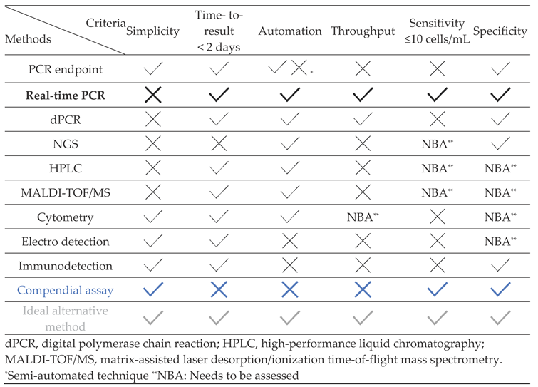
The best combination can be identified by comparing available alternatives presented in Table 3.
5.2.1. Simplicity
The criterion of simplicity is defined by the requirements of instrumentations/operator expertise, and the ease of use. A test should not require any specific qualifications for operators, or expensive materials and also be easy to perform. The compendial assay is a simple test. Similarly, electro- [72] or immunodetection methods [73] rely on simple ready-to-use equipment. Whereas, PCR, NGS, MALDI-TOF MS approaches and cytometry methods require specific expensive equipment and skilled operators.
5.2.2. Time-to-Result
The time-to-result is another challenge with the compendial assay due to the long incubation period i.e., 56 days. Most of the proposed alternative methods have much shorter time-to-results, with results available within few hours to 2 days except for the NGS method, which may need longer due to data analysis.
5.2.3. Automation
Automation can be an advantage to improve the efficiency of tests (cost reduction, early results, reproducibility). It also offers an advantage by reducing the handling time by an operator and thereby reducing the required full-time equivalents. Extraction of nucleic acids and amplification can be automated, however endpoint analysis cannot be automatized, thus making it semi-automated. Automatized instruments are available for HPLC, MALDI-TOF MS, and cytometry approaches. However, direct measurement techniques such as electro-detection and immunodetection cannot be automated.
5.2.4. Throughput
A method’s throughput determines the number of samples that can be analyzed at the same time. For real-time PCR and dPCR with an automatized extraction, the number of samples that can be analyzed at the same time is defined by the plate capacity (from 96, up to 384 samples). Additionally, depending on the technology selected, multiple plates can be analyzed at the same time to increase the throughput. As expected, analyzing amplicons on agarose gels has a much lower output.
Data management and analysis can also be a limiting factor for the processing of large number of samples. For instance, protein analysis by HPLC or NGS analysis generate an important amount of data which, in turn, can be fastidious to analyze. Although the throughput of analysis can be improved with new and more performant software, it is by no mean sufficient to reach the pace of real-time PCR and dPCR [75]. Cytometry techniques allow analysis in 24- or 96-well plates, but their use case must be assessed for the detection of mycobacteria [76]. The direct analysis with electro-detection or immuno-detection cannot be automatized, but their short turnaround time allows repeating the procedure to perform multiple analyses. However, even these methods cannot reach the throughput levels of real-time PCR and dPCR.
5.2.5. Sensitivity
With regards to mycobacterial detection, the sensitivity of any technique corresponds to the lowest detectable concentration of viable bacteria. It is defined by the compendial assay and an alternative technique needs to be at least as sensitive as the reference technique. However, it is to note that the compendial assay’s sensitivity involves a degree of variability as the growth time mycobacteria differs from one species to the other, ranging from 2 days to 8-12 weeks. The real-time PCR is the most sensitive NAT approach, but it also depends on the efficiency of extraction. The NGS sensitivity must still be reported and there are difficulties to detect low contamination levels in complex samples. The mycobacterial protein detection by HPLC or MALDI-TOF MS depends on the quality of protein purification. In the current literature, there are no data or reports on the sensitivity of cytometry, electro-detection, or immunodetection for mycobacteria detection. Based on the principle of these techniques, it is likely that they are not sensitive enough.
5.2.6. Specificity
The specificity is the ability of the test to detect mycobacteria among a range of different microorganisms [8]. Ideally, a positive result should be coming from a mycobacterial contamination without any nonspecific interferences. In theory, NATs are highly specific due to their primer design and their specificity can be increased with a probe detection. Other techniques can also be specific depending on the test design and the samples to be assessed.
6. Conclusions
Based on this review, NATs combined with an efficient extraction method appears to be the most suitable alternative to the compendial assay for mycobacterial testing in biological products. Real-time PCR seems to be the closest to the ideal alternative method. To our knowledge, no other NATs method is available for such an exhaustive, specific, and sensitive detection of mycobacteria in biologicals. Additionally, real-time PCR provides results in few hours leading to significant reduction of time to result as compared to the compendial assay. The development of this type of technique could be the best alternative method.
In any case, the prior validation of an alternative method is the way leading to the potential replacement of the compendial assay. It is important to note that even if a NAT method has the potential to overcome some of the limitations of the compendial assay, it most likely will come with its own set of drawbacks. Therefore, the choice of an alternative method should be carefully made and, ideally, integrated into an overall contamination control strategy.
Author Contributions
MM: Supervision, writing – review & editing. CF: Literature search, writing – review & editing.
Acknowledgments
The authors thank Thierry Bonnevay, Sanofi for his invaluable review support, and Atharva Moghe, and Manojkumar Patel, both from Sanofi, for their medical writing assistance. Coordination of the development of this manuscript, facilitation of author discussion and critical review was provided by Jean-Sébastien Bolduc, Sanofi. The authors were responsible for all content and editorial decisions, and received no honoraria related to the development of this publication.
Conflicts of Interest
MM is a Sanofi employee and may hold shares and/or stock options in the company. CF is a contractor with Sanofi and has no conflict of interest.
References
- Boyer: S.D.; Marino, A.; Chun, A.; Nims, R. Nontuberculosis Mycobacterium contamination of a mammalian cell bioreactor process. BioPharm International 2011, 24, 30-34.
- Borghans, J.G.; Stanford, J.L. Mycobacterium chelonei in abscesses after injection of diphtheria-pertussis-tetanus-polio vaccine. Am Rev Respir Dis 1973, 107, 1-8. [CrossRef]
- Kavitha, K.; Ragunathan, L.; Elantheriyan, P.; Gopalakrishnan, K.; Gopala, K.A.; Balamurugan, I.D.; Navya, R.R.; Marcella, S.S.; Venkatachalam, G.K. The burden of mycobacteria species among children from postvaccination abscess in Southern India. Int J Mycobacteriol 2021, 10, 358-363. [CrossRef]
- Galil, K.; Miller, L.A.; Yakrus, M.A.; Wallace Jr, R.J.; Mosley, D.G.; England, B.; Huitt, G.; McNeil, M.M.; Perkins, B.A. Abscesses due to mycobacterium abscessus linked to injection of unapproved alternative medication. Emerging infectious diseases 1999, 5, 681.
- 2.6.2. Mycobacteria - European Pharmacopoeia 11.2 n.d. Accessed August 23, 2023. Available at: https://pheur.edqm.eu/app/11-2/content/default/20602E.htm.
- United States Pharmacopoeial Convention. USP 41-NF 36, chapter 〈1225〉 Validation of compendial procedures. United States Pharmacopoeia: North Bethesda, 2018.
- 5.1.6. Alternative Methods for control of microbiological quality. European Pharmacopoeia 11.2 n.d. Accessed August 23, 2023. Available at: https://pheur.edqm.eu/app/11-2/content/default/50106E.htm.
- United States Pharmacopoeial Convention. USP 41-NF 36, chapter <1223> Validation of alternative microbiological methods. United States Pharmacopoeia: North Bethesda, 2018.
- Forbes, B.A.; Hall, G.S.; Miller, M.B.; Novak, S.M.; Rowlinson, M.C.; Salfinger, M.; Somoskövi, A.; Warshauer, D.M.; Wilson, M.L. Practical guidance for clinical microbiology laboratories: Mycobacteria. Clin Microbiol Rev 2018, 31. [CrossRef]
- CLSI. M48. Laboratory detection and identification of Mycobacteria, 2nd ed.; 2018.
- arish, T.; Stoker, N.G. Mycobacteria protocols; Springer: 1998; Volume 101.
- Mokrousov, I. Current topics of molecular mycobacteriology. Infection, Genetics and Evolution 2019, 73, 132-138. [CrossRef]
- Scheler, O.; Glynn, B.; Kurg, A. Nucleic acid detection technologies and marker molecules in bacterial diagnostics. Expert review of molecular diagnostics 2014, 14, 489-500. [CrossRef]
- Greub, G.; Sahli, R.; Brouillet, R.; Jaton, K. Ten years of R&D and full automation in molecular diagnosis. Future microbiology 2016, 11, 403-425. [CrossRef]
- Kaevska, M.; Slana, I. Comparison of filtering methods, filter processing and DNA extraction kits for detection of mycobacteria in water. Annals of Agricultural Environmental Medicine 2015, 22. [CrossRef]
- Radomski, N.; Lucas, F.S.; Moilleron, R.; Cambau, E.; Haenn, S.; Moulin, L. Development of a real-time qPCR method for detection and enumeration of Mycobacterium spp. in surface water. Applied Environmental Microbiology 2010, 76, 7348-7351.
- Käser, M.; Ruf, M.-T.; Hauser, J.; Marsollier, L.; Pluschke, G. Optimized method for preparation of DNA from pathogenic and environmental mycobacteria. Applied environmental microbiology 2009, 75, 414-418. [CrossRef]
- Ceuppens, S.; Li, D.; Uyttendaele, M.; Renault, P.; Ross, P.; Ranst, M.V.; Cocolin, L.; Donaghy, J. Molecular methods in food safety microbiology: interpretation and implications of nucleic acid detection. Comprehensive Reviews in Food Science Food Safety 2014, 13, 551-577. [CrossRef]
- Morris, C.; Lee, Y.S.; Yoon, S. Adventitious agent detection methods in bio-pharmaceutical applications with a focus on viruses, bacteria, and mycoplasma. Current Opinion in Biotechnology 2021, 71, 105-114. [CrossRef]
- 2.6.21 Nucleic acid amplification techniques. European Pharmacopoeia 11.2 n.d. Accessed September 18, 2023. Available at: https://pheur.edqm.eu/app/11-2/content/11-2/20621E.htm?highlight=on&;terms=2.6.21.
- 2.6.16. Tests for extraneous agents in human viral vaccines. European pharmacopoeia 2020. Accessed September 18, 2023. Available at: http://uspbpep.com/ep60/2.6.16.%20tests%20for%20extraneous%20agents%20in%20viral%20vaccines%20for%20human%20use%2020616e.pdf.
- 5.2.3 Cell substrates for the production of vaccines for human use. European Pharmacopoeia 2018. Accessed September 18, 2023. Available at: http://www.uspbpep.com/ep50/5.2.3.%20Cell%20substrates%20for%20the%20production%20of%20vaccines%20for%20human%20use.pdf.
- World Health Organization. WHO expert committee on biological standardization. World Health Organ Tech Rep Ser. 2013;(979):1-366.
- FDA. Guidance for Industry Characterization and Qualification of Cell Substrates and Other Biological Materials Used in the Production of Viral Vaccines for Infectious Disease Indications (Rockville, MD, 2010). .
- Hermann, C.; Karamchand, L.; Blackburn, J.M.; Soares, N.C. Cell envelope proteomics of mycobacteria. Journal of Proteome Research 2020, 20, 94-109. [CrossRef]
- Hosek, J.; Svastova, P.; Moravkova, M.; Pavlik, I.; Bartos, M. Methods of mycobacterial DNA isolation from different biological material: a review. Veterinarni medicina 2006, 51, 180-192. [CrossRef]
- Jagatia, H.; Cantillon, D. DNA isolation from Mycobacteria. Mycobacteria Protocols 2021, 59-75.
- anya Parish, A.C.B. Mycobacteria Protocols 2009.
- Amita, J.; Vandana, T.; Guleria, R.; Verma, R. Qualitative evaluation of mycobacterial DNA extraction protocols for polymerase chain reaction. Mol Biol Today 2002, 3, 43-49.
- 3Radomski, N.; Kreitmann, L.; McIntosh, F.; Behr, M.A. The critical role of DNA extraction for detection of mycobacteria in tissues. PLoS One 2013, 8, e78749. [CrossRef]
- Belisle, J.T.; Sonnenberg, M.G. Isolation of genomic DNA from mycobacteria. In Mycobacteria protocols; Springer: 1998; pp. 31-44.
- Husakova, M.; Kralik, P.; Babak, V.; Slana, I. Efficiency of DNA isolation methods based on silica columns and magnetic separation tested for the detection of Mycobacterium avium subsp. paratuberculosis in milk and faeces. Materials 2020, 13, 5112.
- Salgado, M.; Verdugo, C.; Heuer, C.; Castillo, P.; Zamorano, P. A novel low-cost method for Mycobacterium avium subsp. paratuberculosis DNA extraction from an automated broth culture system for real-time PCR analysis. Journal of veterinary science 2014, 15, 233-239.
- Köchl, S.; Niederstätter, H.; Parson, W. DNA extraction and quantitation of forensic samples using the phenol-chloroform method and real-time PCR. Forensic DNA typing protocols 2005, 13-29.
- Dairawan, M.; Shetty, P.J. The evolution of DNA extraction methods. Am. J. Biomed. Sci. Res 2020, 8, 39-45. [CrossRef]
- Katevatis, C.; Fan, A.; Klapperich, C.M. Low concentration DNA extraction and recovery using a silica solid phase. PloS one 2017, 12, e0176848.
- Caldarelli-Stefano, R.; Vago, L.; Bonetto, S.; Nebuloni, M.; Costanzi, G. Use of magnetic beads for tissue DNA extraction and IS6110 Mycobacterium tuberculosis PCR. Molecular Pathology 1999, 52, 158. [CrossRef]
- Mohammadi, S.; Esfahani, B.N.; Moghim, S.; Mirhendi, H.; Zaniani, F.R.; Safaei, H.G.; Fazeli, H.; Salehi, M. Optimal DNA isolation method for detection of nontuberculous mycobacteria by polymerase chain reaction. Advanced biomedical research 2017, 6. [CrossRef]
- Hansen, S.; Roller, M.; Alslim, L.M.; Böhlken-Fascher, S.; Fechner, K.; Czerny, C.-P.; Abd El Wahed, A. Development of rapid extraction method of Mycobacterium avium subspecies paratuberculosis DNA from bovine stool samples. Diagnostics 2019, 9, 36. [CrossRef]
- Richardson, E.; Samson, D.; Banaei, N. Rapid identification of Mycobacterium tuberculosis and nontuberculous mycobacteria by multiplex, real-time PCR. Journal of clinical microbiology 2009, 47, 1497-1502.
- Tortoli, E.; Nanetti, A.; Piersimoni, C.; Cichero, P.; Farina, C.; Mucignat, G.; Scarparo, C.; Bartolini, L.; Valentini, R.; Nista, D. Performance assessment of new multiplex probe assay for identification of mycobacteria. Journal of Clinical Microbiology 2001, 39, 1079-1084. [CrossRef]
- Kox, L.; Van Leeuwen, J.; Knijper, S.; Jansen, H.; Kolk, A. PCR assay based on DNA coding for 16S rRNA for detection and identification of mycobacteria in clinical samples. Journal of Clinical Microbiology 1995, 33, 3225-3233. [CrossRef]
- Simon, A.; Onya, O.; Mazza-Stalder, J.; Nicod, L.; Gilbert, G.; Katia, J. Added diagnostic value of 16S rRNA gene pan-mycobacterial PCR for nontuberculous mycobacterial infections: a 10-year retrospective study. European Journal of Clinical Microbiology Infectious Diseases 2019, 38, 1873-1881.
- Chen, R.; Gao, X.-B.; Liu, Z.-H.; Shen, X.-B.; Guo, A.-Z.; Duan, Y.-Y.; Liu, Z.-L.; Wu, X.-W.; Zhu, D.-Z. Combination of multiplex PCR with denaturing high-performance liquid chromatography for rapid detection of Mycobacterium genus and simultaneous identification of the Mycobacterium tuberculosis complex. Diagnostic microbiology infectious disease 2013, 77, 53-57. [CrossRef]
- Böddinghaus, B.; Rogall, T.; Flohr, T.; Blöcker, H.; Böttger, E. Detection and identification of mycobacteria by amplification of rRNA. Journal of clinical microbiology 1990, 28, 1751-1759. [CrossRef]
- Tringe, S.G.; Hugenholtz, P. A renaissance for the pioneering 16S rRNA gene. Current opinion in microbiology 2008, 11, 442-446. [CrossRef]
- Chae, H.; Han, S.J.; Kim, S.-Y.; Ki, C.-S.; Huh, H.J.; Yong, D.; Koh, W.-J.; Shin, S.J. Development of a one-step multiplex PCR assay for differential detection of major Mycobacterium species. Journal of clinical microbiology 2017, 55, 2736-2751. [CrossRef]
- Rocchetti, T.T.; Silbert, S.; Gostnell, A.; Kubasek, C.; Widen, R. Validation of a multiplex real-time PCR assay for detection of Mycobacterium spp., Mycobacterium tuberculosis complex, and Mycobacterium avium complex directly from clinical samples by use of the BD max open system. Journal of Clinical Microbiology 2016, 54, 1644-1647. [CrossRef]
- Adékambi, T.; Drancourt, M.; Raoult, D.J.T.i.m. The rpoB gene as a tool for clinical microbiologists. Trends in microbiology 2009, 17, 37-45. [CrossRef]
- Xiong, L.; Kong, F.; Yang, Y.; Cheng, J.; Gilbert, G.L. Use of PCR and reverse line blot hybridization macroarray based on 16S-23S rRNA gene internal transcribed spacer sequences for rapid identification of 34 mycobacterium species. Journal of Clinical Microbiology 2006, 44, 3544-3550. [CrossRef]
- Valones, M.A.A.; Guimarães, R.L.; Brandão, L.A.C.; Souza, P.R.E.d.; Carvalho, A.d.A.T.; Crovela, S. Principles and applications of polymerase chain reaction in medical diagnostic fields: a review. Brazilian Journal of Microbiology 2009, 40, 1-11. [CrossRef]
- Korbie, D.J.; Mattick, J.S. Touchdown PCR for increased specificity and sensitivity in PCR amplification. Nature protocols 2008, 3, 1452-1456. [CrossRef]
- Miyazaki, Y.; Koga, H.; Kohno, S.; Kaku, M. Nested polymerase chain reaction for detection of Mycobacterium tuberculosis in clinical samples. Journal of Clinical Microbiology 1993, 31, 2228-2232. [CrossRef]
- Cross, L.J.; Anscombe, C.; McHugh, T.D.; Abubakar, I.; Shorten, R.J.; Thorne, N.; Arnold, C. A rapid and sensitive diagnostic screening assay for detection of mycobacteria including Mycobacterium tuberculosis directly from sputum without extraction. International journal of bacteriology 2015, 2015. [CrossRef]
- Ichijo, T.; Izumi, Y.; Nakamoto, S.; Yamaguchi, N.; Nasu, M. Distribution and respiratory activity of mycobacteria in household water system of healthy volunteers in Japan. PLoS One 2014, 9, e110554. [CrossRef]
- Quan, P.-L.; Sauzade, M.; Brouzes, E. dPCR: A technology review. Sensors 2018, 18, 1271. [CrossRef]
- Song, N.; Tan, Y.; Zhang, L.; Luo, W.; Guan, Q.; Yan, M.-z.; Zuo, R.; Liu, W.; Luo, F.-l.; Zhang, X.-L.J.E.m. Detection of circulating Mycobacterium tuberculosis-specific DNA by droplet digital PCR for vaccine evaluation in challenged monkeys and TB diagnosis. Emerging microbes infections 2018, 7, 1-9. [CrossRef]
- Mohamed, S.; Köser, C.U.; Salfinger, M.; Sougakoff, W.; Heysell, S.K. Targeted next-generation sequencing: a Swiss army knife for mycobacterial diagnostics? European Respiratory Journal 2021, 57.
- Li, Y.; Jiao, M.; Liu, Y.; Ren, Z.; Li, A. Application of Metagenomic Next-Generation Sequencing in Mycobacterium tuberculosis Infection. Frontiers in Medicine 2022, 9, 802719. [CrossRef]
- Heather, J.M.; Chain, B. The sequence of sequencers: The history of sequencing DNA. Genomics 2016, 107, 1-8. [CrossRef]
- Smith, C.; Halse, T.A.; Shea, J.; Modestil, H.; Fowler, R.C.; Musser, K.A.; Escuyer, V.; Lapierre, P. Assessing nanopore sequencing for clinical diagnostics: a comparison of next-generation sequencing (NGS) methods for Mycobacterium tuberculosis. Journal of Clinical Microbiology 2020, 59. [CrossRef]
- Butler, W.R.; Guthertz, L.S. Mycolic acid analysis by high-performance liquid chromatography for identification of Mycobacterium species. Clinical microbiology reviews 2001, 14, 704-726. [CrossRef]
- Butler, W.; Jost Jr, K.; Kilburn, J. Identification of mycobacteria by high-performance liquid chromatography. Journal of Clinical Microbiology 1991, 29, 2468-2472.
- El Khechine, A.; Couderc, C.; Flaudrops, C.; Raoult, D.; Drancourt, M. Matrix-assisted laser desorption/ionization time-of-flight mass spectrometry identification of mycobacteria in routine clinical practice. PloS one 2011, 6, e24720. [CrossRef]
- 6La'Tonzia, L.A.; Salee, P.; Dionne, K.; Carroll, K.; Parrish, N. A novel protein extraction method for identification of mycobacteria using MALDI-ToF MS. Journal of microbiological methods 2015, 119, 1-3.
- Alvarez-Barrientos, A.; Arroyo, J.; Cantón, R.; Nombela, C.; Sánchez-Pérez, M. Applications of flow cytometry to clinical microbiology. Clinical microbiology reviews 2000, 13, 167-195. [CrossRef]
- Soejima, T.; Iida, K.-i.; Qin, T.; Taniai, H.; Yoshida, S.-i. Discrimination of live, anti-tuberculosis agent-injured, and dead Mycobacterium tuberculosis using flow cytometry. FEMS microbiology letters 2009, 294, 74-81.
- Qin, D.; He, X.; Wang, K.; Tan, W. Using fluorescent nanoparticles and SYBR Green I based two-color flow cytometry to determine Mycobacterium tuberculosis avoiding false positives. Biosensors Bioelectronics 2008, 24, 626-631. [CrossRef]
- Barr, D.A.; Omollo, C.; Mason, M.; Koch, A.; Wilkinson, R.J.; Lalloo, D.G.; Meintjes, G.; Mizrahi, V.; Warner, D.F.; Davies, G. Flow cytometry method for absolute counting and single-cell phenotyping of mycobacteria. Scientific Reports 2021, 11, 18661. [CrossRef]
- Pina-Vaz, C.; Costa-Oliveira, S.; Rodrigues, A.G.; Salvador, A. Novel method using a laser scanning cytometer for detection of mycobacteria in clinical samples. Journal of clinical microbiology 2004, 42, 906-908. [CrossRef]
- Microorganisms: Automated & instantaneous enumeration with Red OneTM. Accessed August 24, 2023. Available at. https://www.a3p.org/enumeration-viable-microorganisms/.
- Rochelet, M.; Barbier, É.; Hartmann, A. Method for electrochemical detection of mycobacteria. 2022.
- Nour-Neamatollahi, A.; Siadat, S.D.; Yari, S.; Tasbiti, A.H.; Ebrahimzadeh, N.; Vaziri, F.; Fateh, A.; Ghazanfari, M.; Abdolrahimi, F.; Pourazar, S. A new diagnostic tool for rapid and accurate detection of Mycobacterium tuberculosis. Saudi journal of biological sciences 2018, 25, 418-425. [CrossRef]
- 7PDA Technical Report 33 (Revised). Evaluation, validation and implementation of alternative and rapid microbiological methods; Parenteral Drug Association: Bethesda, MD, 2013; 30–33.
- Jansen, B.C.; Hafkenscheid, L.; Bondt, A.; Gardner, R.A.; Hendel, J.L.; Wuhrer, M.; Spencer, D.I.R.J.P.o. HappyTools: a software for high-throughput HPLC data processing and quantitation. PloS one 2018, 13, e0200280. [CrossRef]
- Al-Attar, A.; Kumar, K.R.; Untersee, D.; O'Driscoll, M.; Ventura, M.F.S.; Lin, L. Automation in flow cytometry: Guidelines and review of systems. Cytometry Part B: Clinical Cytometry 2023. [CrossRef]

Table 1.
Different commercial media.
| Liquid media | |
|---|---|
| Proskauer and Beck medium | Not available commercially Needs to be prepared in-house based on empirical formulation Preparation process is fastidious and can induce variability between batches |
| Sauton medium | |
| Kircher medium | Used for the diagnosis of tuberculosis |
| Dubos medium | Allows the development of most cultivable mycobacteria |
| Middlebrook medium | Middlebrook 7H9 is one of the most useful media for the recovery of all mycobacterial species |
| Solid media | |
| Lowenstein–Jensen medium | Most commonly used and oldest egg-based medium Selective; inhibits bacterial and fungal growth. Strongly supports the growth of MTBC |
| Middlebrook 7H10 and 7H11 media | Standard agar-based media Mycobacterial growth time on these media is shorter than that on egg-based media but longer than that on liquid media Expensive and have a short storage life |
MTBC, Mycobacterium tuberculosis complex.
Table 2.
Comparison of NAT.
| Techniques | PCR Endpoint | Real-Time PCR | Digital PCR | NGS | ||||
|---|---|---|---|---|---|---|---|---|
| Detection | ICA | Probes | ICA | Probes | ICA | Probes | ||
| Sensitivity | NO | YES | YES | * | ||||
| Specificity | NO | YES | NO | YES | YES | YES | YES | |
| Handling-time | 1-2h | 3h | 30 min | 30 min | 1h | 4h | ||
| Time-to-result | 3h | 4h | 4h | 3h | 4-5h | >2 days | ||
| Amplified production contamination risk | YES | NO | NO | YES | ||||
| Automation/ high throughput analysis capacity | NO | YES | YES | YES | ||||
| ICA: intercalating agent of DNA like ethidium bromide, SYBR® Green etc.; NAT, nucleic acid amplification technique; NGS, next-generation sequencing; PCR, polymerase chain reaction. *Must be assessed. | ||||||||
Table 3.
Alternative methods comparison.
 |
Disclaimer/Publisher’s Note: The statements, opinions and data contained in all publications are solely those of the individual author(s) and contributor(s) and not of MDPI and/or the editor(s). MDPI and/or the editor(s) disclaim responsibility for any injury to people or property resulting from any ideas, methods, instructions or products referred to in the content. |
© 2024 by the authors. Licensee MDPI, Basel, Switzerland. This article is an open access article distributed under the terms and conditions of the Creative Commons Attribution (CC BY) license (http://creativecommons.org/licenses/by/4.0/).
Copyright: This open access article is published under a Creative Commons CC BY 4.0 license, which permit the free download, distribution, and reuse, provided that the author and preprint are cited in any reuse.


