Preprint
Review

Biophysical Mechanisms of Vaginal Smooth Muscle Contraction: Role of the Membrane Potential and Ion Channels

Altmetrics

Downloads

150

Views

104

Comments

0

A peer-reviewed article of this preprint also exists.

This version is not peer-reviewed

Submitted:

27 March 2024

Posted:

27 March 2024

You are already at the latest version

Alerts
Abstract
The vagina is an essential component of the female reproductive system and is responsible for providing female sexual satisfaction. Vaginal smooth muscle contraction plays a crucial role in various physiological processes, including sexual arousal, childbirth, and urinary continence. In pathophysiological conditions such as pelvic floor disorders, aberrations in vaginal smooth muscle function can lead to urinary incontinence and pelvic organ prolapse. A set of cellular and sub-cellular physiological mechanisms regulates the contractile properties of the vaginal smooth muscle cells. Calcium influx is a crucial determinant of smooth muscle contraction, facilitated through voltage-dependent calcium channels and calcium release from intracellular stores. Comprehensive reviews on smooth muscle biophysics are relatively scarce within the scientific literature, likely due to the complexity and specialized nature of the topic. The objective of this review is to provide a comprehensive description of alterations in the cellular physiology of vaginal smooth muscle contraction. The benefit associated with this particular approach is that conducting a comprehensive examination of the cellular mechanisms underlying contractile activation will enable the creation of more targeted therapeutic agents to control vaginal contraction disorders.
Keywords: 
Subject: Biology and Life Sciences  -   Life Sciences

1. Introduction

The vagina is a female reproductive organ that undergoes physiological changes throughout a woman's existence. It exhibits diverse physiological responses to hormonal fluctuations, such as those observed during puberty, menstruation, pregnancy, and menopause. The vagina is also an essential component of the female reproductive system and is responsible for providing female sexual satisfaction [1]. Smooth muscle is found throughout the body and serves a variety of functions [2]. Smooth muscle is an involuntary, non-striated muscle divided into two subgroups: single-unit and multi-unit smooth muscle. Within single-unit muscle, the whole bundle or sheet of smooth muscle cells contracts as a syncytium [3]. Vascular smooth muscle is found in the walls of hollow organs, including the stomach, intestines, bladder, and uterus, as well as walls of blood vessels and lymph vessels.
Vaginal smooth muscle (VSM) contraction plays a crucial role in various physiological processes, including sexual arousal, childbirth, and urinary continence. Smooth muscle cells in the vaginal wall also play a pivotal role in maintaining tissue integrity and responding to mechanical and biochemical stimuli [4]. A set of cellular and sub-cellular physiological mechanisms regulates the contractile properties of the VSM cells. Calcium (Ca2+) influx is a key determinant of smooth muscle contraction, facilitated through voltage-dependent calcium channels (VDCC) and Ca2+ release from intracellular stores [2]. The value of intracellular Ca2+ concentration is modulated by the coordinated action of ion channels, neurotransmitters, and hormonal signals, leading to cyclic changes in vaginal tone and elasticity [4,5,6]. During sexual arousal, pelvic nerve stimulation triggers the relaxation of vaginal smooth muscles, facilitating intercourse [7].
Figure 1 shows the schematic illustration of an isolated vaginal smooth muscle cell in both relaxed and contracted states.
In pathophysiological conditions such as pelvic floor disorders, aberrations in vaginal smooth muscle function can lead to urinary incontinence and pelvic organ prolapse. Dysregulation of VSM tone and contractility may result from factors such as aging, hormonal changes, childbirth trauma, or neurological disorders [8,9]. Insights into the biophysical mechanisms governing smooth muscle contraction not only enhance our fundamental understanding of muscle physiology but also hold promise for developing novel therapeutic strategies targeting smooth muscle disorders and dysfunctions [10]. Therefore, it is crucial to understand the underlying cellular and sub-cellular biophysical processes in VSM physiology for developing targeted therapeutic interventions to manage pelvic floor disorders and addressing pathological conditions related to reproductive health effectively.
Smooth muscle contraction relies on intricate interactions between cytoskeletal elements, ion channels, hormones, and biochemical and calcium signaling pathways [8,11,12,13]. The Ca2+ serves as a critical mediator, initiating cross-bridge cycling between actin and myosin, ultimately leading to force development [8,14,15]. Ion channels play a pivotal role in regulating membrane potential, intracellular Ca2+ concentration, and, ultimately, smooth muscle contractility [16,17,18]. Furthermore, the roles of sodium (Na+), chloride (Cl-), and potassium (K+) channels in controlling the membrane potential are crucial for deciphering the underlying mechanisms of smooth muscle function and its dysregulation [19,20,21,22]. Therefore, understanding the interplay between membrane potential and ion channels in VSM contraction is paramount for elucidating reproductive physiology and addressing associated disorders. Additionally, the involvement of smooth muscle tissue's morphological and structural aspects is also imperative for comprehending its contractile properties and functionality [11]. The alterations in the structure and function of VSM, such as those induced by prolapse mesh, underscore the importance of understanding mechanical stimulation in elucidating disease mechanisms [23].
Comprehensive reviews on smooth muscle biophysics are relatively scarce within the scientific literature, likely due to the complexity and specialized nature of the topic. While research articles and studies delve into specific aspects of smooth muscle physiology and biophysics [2], a consolidated overview that encompasses all facets of smooth muscle biophysics is less common. This scarcity may stem from the interdisciplinary nature of biophysics, which requires expertise in both biology and physics [24]. However, the importance of understanding smooth muscle biophysics in various physiological and pathological conditions underscores the necessity for more comprehensive reviews to bridge the gap between basic science and clinical applications. To bridge this divide, the current review aims to enhance our understanding of the biophysical facets of VSM contraction. It includes experimental shreds of evidence for biophysical processes in VSM and other smooth muscles and establishes a model for VSM cell contraction. This understanding offers valuable insights into typical physiological mechanisms, guiding the development of therapeutic interventions for addressing reproductive disorders and promoting optimal women's health

2. Materials and Methods

We conducted a thorough investigation using the MEDLINE database via PubMed [25], focusing on English articles published at any time. We aimed to explore the associations among all types of smooth muscle ion channels, biophysics, gap junctions, neurotransmitters, neuromodulators, calcium dynamics, intracellular electrical activities (depolarization, hyperpolarization, action potential, and slow waves), and experimental and computational studies. Then, we meticulously screened all these relevant studies. Exclusion criteria comprised non-English articles, conference/symposium abstracts, and studies duplicating information from other sources. Priority was given to the latest and most comprehensive manuscripts in cases of overlap. Our selection criteria encompassed original research articles, including randomized and non-randomized clinical trials, experimental studies, prospective observational studies, retrospective cohort studies, and case-control studies, along with review articles exploring the potential impact of ion channels on smooth muscle excitability and contraction development. Each included article underwent thorough scrutiny, and supplementary references were consulted to ensure comprehensive coverage. Ultimately, we designed a model workflow diagram to elaborate on all the essential steps necessary for vaginal smooth muscle contraction.

3. Membrane Potential in Smooth Muscle Contraction

The electrical terms used in excitable cells are membrane potential, resting membrane potential (RMP), depolarization, hyperpolarization, action potential (AP), slow wave (SW), and bursting firing patterns [26]. Conduction velocity is also another property to measure the propagation of membrane potential in all types of excitable cells [27]. The membrane potential, a fundamental aspect of cellular physiology, plays a crucial role in various cellular processes beyond merely facilitating ion transport [28]. Recent research highlights its significance in governing diverse cellular functions, including cell cycle regulation, cell volume control, proliferation, and muscle activity [29]. While traditionally associated with the generation of AP in excitable cells, such as neurons and muscle cells, emerging evidence suggests that the RMP exerts profound effects on cell behavior across different cell types [29]. Biophysical signaling mediated by membrane potential serves as a key regulator of long-term cellular behavior, contributing significantly to both excitable and non-excitable cell functions [30]. Furthermore, the membrane potential acts as a pivotal determinant of cell-to-cell signaling and coordination, ultimately influencing broader physiological processes within organisms. It serves as a medium through which cells communicate with each other and the central nervous system, facilitating the transmission of messages critical for maintaining homeostasis and orchestrating various bodily functions [31].
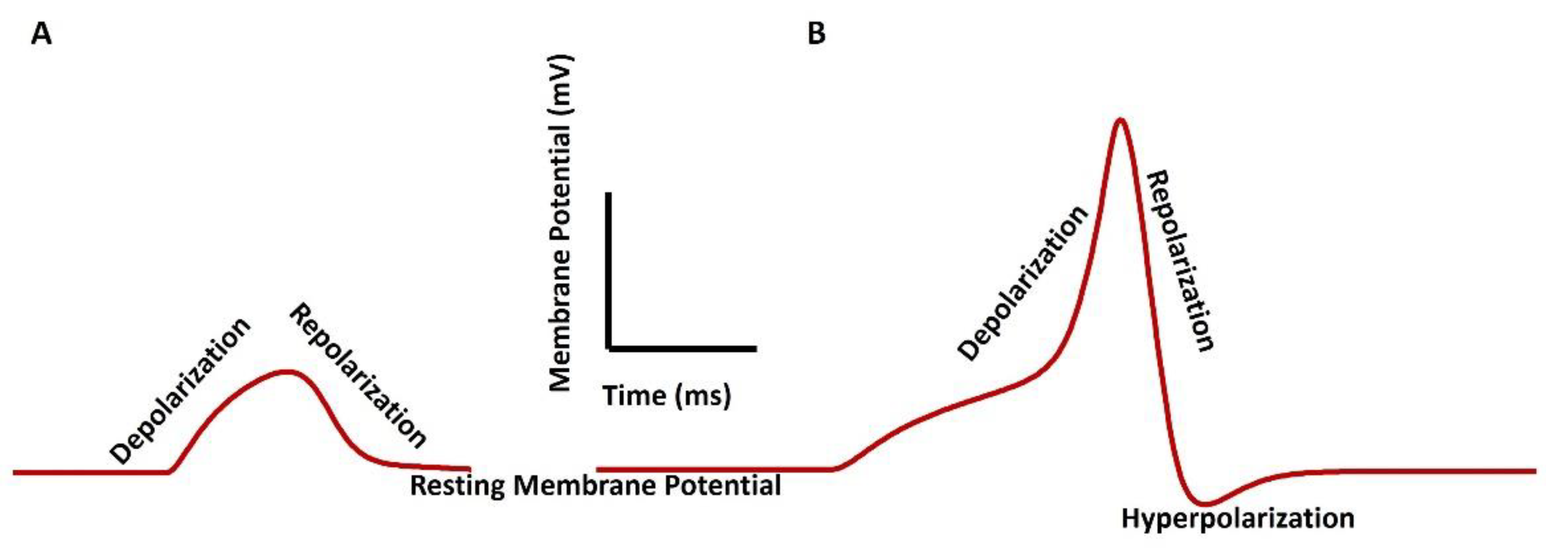
Smooth muscle cells exhibit distinct patterns of electrical activity, including AP and SW, which play crucial roles in triggering muscle contraction. APs in smooth muscle cells are characterized by slower kinetics than skeletal muscle, with durations nearly fifty times longer [2]. APs are generated in the urinary bladder [18,32], vas deferens [33,34], urethra [35,36], and ureter [37,38] smooth muscles. Slow waves, rhythmic electrophysiological events in the gastrointestinal (GI) tract, are generated and propagated by interstitial cells of Cajal (ICC), which are electrically coupled to smooth muscle cells [39]. These slow waves organize primary electrical activity in GI smooth muscles and are essential for regulating peristalsis and digestive processes [40]. Figure 2 illustrates SW (a) and pacemaking AP in smooth muscle cells. The RMP is the membrane potential when the cell is not excitable. The depolarization is the positive shift of the membrane potential from the RMP. When the membrane potential returns to the RMP from the peak value of SW/AP, it is known as repolarization. Any negative membrane potential shift from the RMP is known as hyperpolarization.
Table 1 illustrates the value of RMP, and types of electrical properties generated in major important smooth muscle cells.
Electrical activity in VSM plays a pivotal role in various physiological functions, including sexual arousal and childbirth. Research has shown that electrical stimulation can induce contractions in VSM strips, suggesting the presence of nerve-mediated pathways regulating muscle activity [50]. These contractions are believed to be mediated by cholinergic nerves, indicating the involvement of neurotransmitter release in modulating smooth muscle contractility [50]. Furthermore, the electrical properties of VSM contribute to its viscoelastic response, with smooth muscle contraction leading to changes in vaginal creep behavior, highlighting the dynamic nature of electrical activities in regulating tissue mechanics [4].
The membrane potential, primarily controlled by ion channel activity, is a critical determinant of cellular excitability and contractility in VSM cells. Research indicates that changes in membrane potential, induced by various stimuli such as hormonal fluctuations or mechanical stretch, play a significant role in modulating the responsiveness of vaginal smooth muscle [19]. For instance, depolarization of the smooth muscle cell membrane can increase cellular responsiveness, leading to enhanced contractile activity, while hyperpolarization may reduce smooth muscle relaxation [51]. Unfortunately, like the urinary bladder, ureter, vas deference, and gut smooth muscle, intracellular electrophysiology for VSM cells is not well investigated. Shafik, One lab [52,53] have recorded both extracellular SW and AP bursting from the VSM using the electroretinogram method. They have also suggested that the RMP of the VSM be like that of the uterine smooth muscle, i.e., at – 50 mV. The mean velocity for the human VSM cell is mentioned as 4.3 cm/s.
Recent studies have explored the therapeutic potential of electrical stimulation for pelvic floor rehabilitation, indicating its predictive role in enhancing muscle contraction force and detrusor function [54]. Moreover, advancements in non-invasive techniques for measuring electrical signals in uterine smooth muscle have contributed to a deeper understanding of the role of electrical activity in reproductive physiology [55]. Pathophysiological conditions can lead to modifications in the signaling cascades involved in initiating and sustaining contraction and relaxation [56].

4. Ion Channel Biophysics in Smooth Muscle

Ion channels are integral membrane proteins that regulate the flow of ions across cell membranes, thereby playing fundamental roles in various physiological processes, including neuronal signaling, muscle contraction, and hormone secretion. These channels exhibit diverse structural and functional characteristics, with significant types including voltage-gated, ligand-gated, and mechanically-gated ion channels [57]. Voltage-gated ion channels, such as voltage-gated Na+ channels, voltage-gated K+ channels, voltage-gated Cl- channels, and voltage-gated Ca2+ channels respond to changes in membrane potential, enabling rapid AP generation and propagation in excitable cells like neurons and muscle cells [58]. Specific neurotransmitters or ligands, such as nicotinic acetylcholine receptors, activate ligand-gated ion channels, leading to ion flux and subsequent cellular responses. Mechanically gated ion channels, like those found in sensory neurons, open in response to physical stimuli such as pressure or stretching, transducing mechanical signals into electrical signals [59]. Various mechanisms, including protein phosphorylation, protein-protein interactions, and changes in intracellular ion concentrations, tightly regulate the activity of ion channels. Protein phosphorylation, for instance, can modulate ion channel function by altering their conformation or membrane localization [59].
Additionally, intracellular ion concentrations, such as Ca2+, play critical roles in regulating ion channel activity. Changes in Ca2+ levels can directly influence ion channel gating or indirectly affect channel function by activating Ca2+-sensitive signaling pathways [60]. Overall, the intricate regulation of ion channels ensures precise control over cellular excitability and function, highlighting their importance in maintaining physiological homeostasis [61].
Various types of ion channels play crucial roles in regulating membrane potential and intracellular Ca2+ concentration in smooth muscle cells. Notably, the differential expression patterns of ion channels and their regulation by signaling pathways can lead to distinct contractile or relaxant responses, influencing muscle tone and contraction [62]. These channels, embedded within the sarcolemma, control the flow of ions across the cell membrane, influencing membrane potential and intracellular Ca2+ concentrations, which are critical determinants of muscle contraction [16]. Sodium ion channels are integral to the physiological function of smooth muscle, including that of the vagina. These channels regulate membrane potential, intracellular calcium concentration, and contractility [16]. Voltage-gated K+ (Kv) channels contribute to repolarization during the AP, aiding in maintaining RMP and controlling excitability [63]. Voltage-dependent Ca2+-activated K+ (KCa) channels also influence membrane potential and intracellular Ca2+ concentration, affecting smooth muscle excitability and contractility [63]. Moreover, transient receptor potential (TRP) channels, including TRPC, TRPM, and TRPV subtypes, regulate smooth muscle excitability by modulating Ca2+ influx and membrane potential [64]. The significance of ion channels in contraction dynamics extends beyond their role in initiating and propagating electrical signals. These channels contribute to fine-tuning contractile responses by integrating various signaling pathways and responding to mechanical forces. For example, TRP channels, activated by mechanical stimuli, participate in cellular Ca2+ signaling and homeostasis, potentially influencing smooth muscle contraction [65]. Chloride ion channels play a crucial role in the physiological function of smooth muscle cells, including those found in the vagina. These channels regulate ion movement across the cell membrane, influencing membrane potential and contractility [66]. Leak ion channels, a non-specific ion channel, play a crucial role in the electrical activity of smooth muscle, particularly in uterine smooth muscle during pregnancy [67]. The store-operated system is an intriguing mechanism that takes place within cells. Upon activation of intracellular Ca2+ reserves, a significant amount of Ca2+ will exit the cell. Research has revealed that emptying the reserves does initiate the entry of Ca2+ ions. The channels in this process are called calcium release-activated channels (CRAC) in various smooth muscles [68]. According to several reports, the lower urinary tract which includes VSM consists of a set of ion channels, such as voltage-gated Ca2+ channel (T and L type), voltage-gated K+ channel (Kir, KATP, Kv1, Kdr), voltage-gated Na+ channel, voltage-gated Cl- channel Ca2+-activated K+ (KCa) channels (large conductance and small conductance) CRAC channel, TRPM channel, and leak channels [69,70,71]. The Kir, KATP, KA, and Kdr are inward rectifying, ATP-gated, A-type, and delayed rectifier-type voltage-gated K+ channels. The large and small conductance-based Ca2+-activated K+ channels are also known as BKCa and SKCa.
Interstitial cells of Cajal (ICC) are the leading specialized cells responsible for pace-making activities in smooth muscle cells. These cells act as pacemaker cells and transmit electrical signals through gap junctions to nearby smooth muscle cells [39]. These ICCs produce regular depolarizations called slow waves, which trigger contractions in neighboring smooth muscle cells. Gap junctions are of utmost importance in facilitating the transmission of electrical activities between ICCs and smooth muscle cells, hence permitting synchronous contractions, and coordinated physiological responses [72].
Table 2 illustrates the role of various ion channels in smooth muscle AP/SW generation.

5. Calcium Dynamics in VSM Contraction

Calcium signaling pathways are pivotal in various cellular processes, serving as a universal signaling mechanism in eukaryotic cells [73]. These pathways regulate diverse functions such as muscle contraction, neurotransmitter release, gene expression, and cell proliferation and differentiation [74]. Intracellular Ca2+ levels are tightly controlled by a delicate balance between Ca2+ influx through various channels and Ca2+ efflux through pumps and exchangers. The dynamic changes in Ca2+ concentration are a molecular switch, triggering downstream signaling events that orchestrate cellular responses to internal and external stimuli [75]. Changes in membrane potential, induced by factors such as the firing of AP or activation of stretch-dependent ion channels, can trigger or regulate smooth muscle contraction [8]. This phenomenon, known as excitation-contraction coupling, involves linking alterations in membrane potential to intracellular Ca2+ concentration changes, ultimately leading to force generation [76].
Sarcoplasmic reticulum (SR) calcium dynamics are pivotal in regulating smooth muscle contraction. The SR, a specialized organelle in smooth muscle cells, serves as the primary intracellular calcium store and orchestrates calcium signaling events crucial for contractility. Dynamic changes in SR calcium levels, governed by calcium release and reuptake mechanisms, finely tune smooth muscle contraction and relaxation. Calcium release from the SR into the cytoplasm initiates muscle contraction by binding to contractile proteins, while subsequent calcium reuptake into the SR via calcium-ATPase pumps facilitates muscle relaxation. This intricate interplay between SR calcium release and reuptake ensures precise control over smooth muscle contractile activity, contributing to various physiological functions, including vascular tone regulation and organ motility [77]. The process of sarcoplasmic calcium-induced calcium release (CICR) is a critically important mechanism that regulates smooth muscle contraction, playing a vital role in numerous physiological processes. Following the depolarization of the cell membrane, voltage-gated calcium channels facilitate the entry of calcium ions into the smooth muscle cell, thereby activating ryanodine receptors (RYRs) located on the sarcoplasmic reticulum (SR) membrane. This triggers a series of events in which calcium released from the SR triggers the activation of nearby RYRs, resulting in a coordinated and intensified release of calcium called CICR. The elevated calcium concentration in the cytosol triggers contractile protein activation, leading to smooth muscle contraction. The CICR protein is crucial in controlling vascular tone, gastrointestinal motility, and other functions that rely on smooth muscle. This emphasizes its importance in physiological processes [76,78].
Furthermore, dysregulation of calcium signaling pathways has been implicated in numerous diseases, including cancer, neurodegenerative disorders, and cardiovascular diseases [79]. For instance, aberrant calcium signaling contributes to uncontrolled cell proliferation, apoptosis resistance, and metastasis in cancer [79]. Understanding the intricate mechanisms of calcium signaling and its dysregulation in disease states holds promise for developing targeted therapies to restore cellular homeostasis and treat a wide range of pathologies [80].

6. The Model of Generation of Tension Generation in VSM Cell

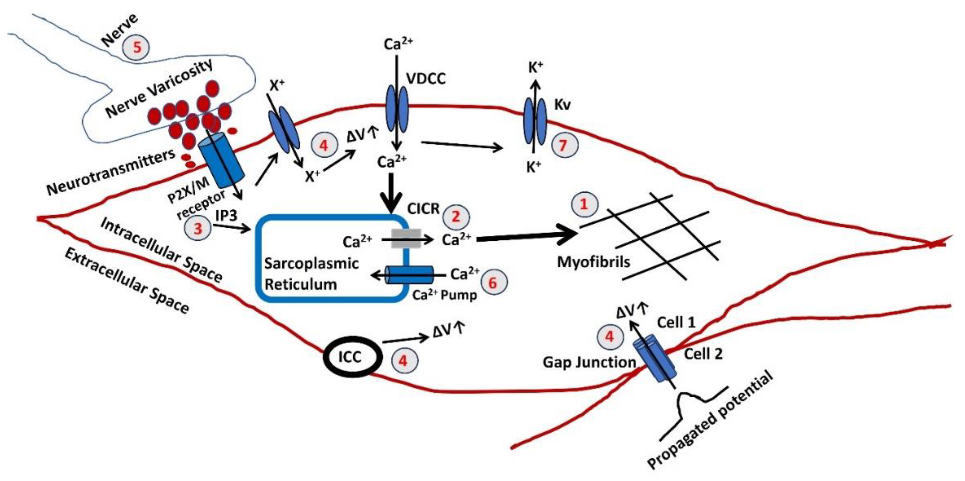
Figure 3 illustrates a schematic representation of the sequential processes entailed in the production of active tension within the vaginal smooth muscle. Both variables possess the potential for modulation and may play a role in the development of abnormal vaginal contractile function. Nevertheless, the pathophysiology of each step in the development of vaginal smooth muscle has not been thoroughly investigated. Instead, these findings are based on analyzing other smooth muscle models that have been experimentally validated. A red color number in the circle denotes the steps. The ΔV is known as a rise in membrane potential.
  • The final stage in generating tension involves an increase in the sarcoplasmic concentration of Ca2+. The myofibrils exhibit a comparable sensitivity to Ca2+, as shown in other muscles, necessitating a Ca2+ concentration of around one mmol/L for half-maximal activation. Calcium ions Ca2+ form complexes with a soluble protein called calmodulin. This complex triggers a series of processes that activate a part of the myosin molecule by phosphorylation. As a result, actin and myosin can interact, requiring ATP.
  • Sarcoplasmic Ca2+ is derived from the SR, an intracellular reservoir. Ca2+ ions are transported from the storage site to the sarcoplasm by Ca2+ channels, which intracellular agents’ control. The formation of tension is influenced by various factors that affect the buildup or release of calcium in the SR. Any disruption to the cellular metabolic mechanisms that produce ATP would undermine their effectiveness. The release of Ca2+ from the SR can often be accomplished through one of two methods. An increase in the Ca2+ concentration near the SR triggers further release of Ca2+. The CICR mechanism is usually initiated by a Ca2+ flux across the surface membrane, although this is not always true.
  • There is a possibility of an elevation in the concentration of a diffusible second messenger, which connects the surface membrane with the release of intracellular Ca2+. The primary mechanism in typical human vagina smooth muscle involves the binding of purinergic or acetylcholine (ACh) to the P2X or M3 muscarinic receptor, which triggers a series of membrane-bound processes resulting in the synthesis of inositol trisphosphate (IP3). Alterations can significantly influence the release of intracellular Ca2+ in the sensitivity or gain of this mechanism.
  • Three reasons can cause a rise in the membrane potential ΔV. The membrane potential can be propagated from cell 2 to cell one via the gap junction as the VSM behaves like a syncitium. Activating pacemaking cell ICC can also trigger a rise in membrane potential. The extracellular ATP might bind to the purinergic receptor (P2X) and open a non-specific cation channel to permit the influx of any positive ion (X+), which can cause a rise in membrane potential. The resultant depolarization can open L-type Ca2+ channels, initiate Ca2+ influx, and trigger AP.
  • The parasympathetic nerves innervate the smooth muscle, and varicosites are the sites where the neurotransmitters are released. The number and distribution of excitatory nerves or the quantity of transmitter released modulate the membrane potential of the VSM. The neurotransmitted might be purinergic or cholinergic cotransmitters.
  • The Ca2+ is filled in the SR lumen through a highly efficient ATP-dependent calcium pump, which transports calcium against a concentration gradient.
  • The decaying of the Ca2+ transient, which occurs after the generation of AP or SW, ceases the VSM contraction. The activation of the Ca2+ channel and generation of AP/SW open the various K+ channels to repolarize the membrane, bringing the membrane potential to the RMP. After completing the contraction, the VSM cell returns to the relaxed state.

7. Experimental and Computational Techniques for Studying VSM Contraction

In vitro studies utilizing experimental techniques offer valuable insights into understanding vaginal smooth muscle contraction. These techniques involve the isolation of vaginal tissue strips, which are then subjected to controlled conditions to evaluate contractility. For instance, Cellai et al. conducted in vitro contractility studies on vaginal strips to assess the influence of testosterone on nitric oxide-induced relaxation [81]. By measuring the contractile responses under various experimental conditions, researchers can elucidate the signaling pathways and regulatory mechanisms involved in vaginal smooth muscle contraction. Such studies contribute to the development of pharmacological interventions targeting vaginal disorders associated with smooth muscle dysfunction.
Furthermore, methodologies such as inflation-extension testing provide a comprehensive evaluation of vaginal tissue mechanics and contractility. Utilizing in vivo pressure measurements and biaxial testing, studies have explored the viscoelastic properties and contractile behavior of vaginal smooth muscle [4]. These experimental approaches enable the characterization of mechanical properties and responses to various stimuli, shedding light on the physiological and pathological mechanisms underlying vaginal function. Overall, in vitro studies employing experimental techniques play a crucial role in advancing our understanding of vaginal smooth muscle contraction and hold promise for future therapeutic strategies.
In vivo, approaches utilizing experimental techniques offer valuable insights into understanding vaginal smooth muscle contraction within its physiological context. Studies have employed methodologies such as in vivo pressure measurements and multiaxial loading to evaluate the contribution of smooth muscle to vaginal viscoelastic response [4]. By conducting experiments directly within living organisms, researchers can assess the dynamic interactions between smooth muscle fibers and surrounding tissues, providing a comprehensive understanding of vaginal function and mechanics. Moreover, investigations utilizing in vivo models allow for studying smooth muscle contraction under physiological conditions, considering factors such as pregnancy and aging. These studies contribute to elucidating the role of smooth muscle in maintaining vaginal integrity and function throughout various life stages [4]. By combining in vivo experimental approaches with clinical observations, researchers can develop more effective therapeutic strategies for addressing vaginal disorders associated with smooth muscle dysfunction.
Computational modeling offers a powerful approach to studying vaginal smooth muscle contraction, allowing researchers to simulate and analyze complex physiological processes with precision. There are a lot of computational models established for different types of smooth muscle electrophysiology [10,82,83,84,85,86,87,88,89,90,91,92,93,94]. By integrating experimental data and physiological parameters, these models can elucidate the underlying mechanisms governing smooth muscle function in the vaginal wall. For instance, finite element analysis (FEA) has been employed to characterize the mechanical behavior of pelvic floor muscles during vaginal contractions, providing insights into the strains induced in the vaginal tissue [95]. These computational approaches enable researchers to explore various factors influencing smooth muscle contraction, such as hormonal changes, pregnancy, and aging, providing a comprehensive understanding of vaginal biomechanics.
Furthermore, computational models allow for the prediction of the effects of different interventions or pathologies on vaginal smooth muscle function. By simulating scenarios in silico, researchers can assess the efficacy of potential treatments for conditions like pelvic organ prolapse or urinary incontinence, guiding the development of targeted therapeutic strategies [95]. These models serve as valuable tools for both basic research and clinical applications, offering a cost-effective and non-invasive means of studying vaginal smooth muscle contraction and its implications for women's health.

8. Clinical Implications and Future Directions

The pathophysiology of vaginal disorders holds significant clinical implications for women's health. The therapeutic targeting of membrane potential and ion channels holds significant clinical implications across various medical fields. For instance, ion channels play crucial roles in cellular excitability, neurotransmission, and muscle contraction, making them attractive targets for pharmacological interventions [96]. Modulating ion channel activity can lead to the development of novel therapies for neurological disorders, cardiovascular diseases, and cancer [97]. Additionally, targeting mitochondrial ion channels shows promise in cancer therapy, offering a potential strategy to eliminate cancer cells selectively [98]. These advancements underscore the clinical relevance of understanding ion channel function and developing targeted therapeutics to address various pathological conditions.
Looking ahead, future directions in therapeutic targeting of membrane potential and ion channels involve exploring innovative approaches to modulate ion channel activity with higher specificity and fewer side effects. Advancements in drug discovery technologies, such as high-throughput screening and computational modeling, enable the identification of novel ion channel modulators [99]. Moreover, personalized medicine approaches may facilitate the development of tailored ion channel-targeted therapies based on individual patient profiles, optimizing treatment efficacy and minimizing adverse effects.
Future research directions in the pathophysiology of vaginal smooth muscle contraction aim to deepen our understanding of the mechanisms underlying pelvic floor disorders and to identify novel therapeutic targets. Investigating the molecular signaling pathways involved in regulating vaginal smooth muscle tone and contractility could provide insights into the pathogenesis of conditions such as pelvic organ prolapse (POP) and stress urinary incontinence (SUI) [100]. Additionally, exploring the role of hormonal influences, neurotransmitters, and extracellular matrix components in modulating vaginal smooth muscle function may offer new avenues for intervention and treatment strategies [7]. Furthermore, future research efforts could focus on developing innovative diagnostic techniques and therapeutic modalities targeting vaginal smooth muscle dysfunction. Advancements in imaging technologies, such as high-resolution ultrasound and magnetic resonance elastography, may enable non-invasive assessment of vaginal biomechanics and contractile function [101]. Moreover, developing precision medicine approaches tailored to individual patient characteristics and disease phenotypes holds promise for optimizing treatment outcomes and minimizing adverse effects [7]. Collaborative efforts between clinicians, researchers, and industry stakeholders are essential for translating these insights into clinically relevant applications, ultimately improving the management and outcomes of vaginal disorders.

9. Conclusion

The vagina plays a pivotal role in sexual intercourse, serving as a conduit for sperm to travel to the uterus and fallopian tubes for fertilization. Additionally, it functions as the birth canal during childbirth, facilitating the passage of the baby from the uterus to the outside world. Vaginal smooth muscle significantly contributes to regulating vaginal tone, thereby maintaining vaginal structure and function. Its contractile activity is essential for various physiological processes within the vagina, including supporting childbirth and sexual function. Fundamental research on vaginal smooth muscle contraction is crucial for understanding its role in vaginal viscoelasticity and structural integrity, which informs potential treatments for conditions like pelvic floor disorders. The study of electrophysiology in excitable cells enhances our comprehension of cellular function, uncovering ion channel biophysics, calcium dynamics, and neural control mechanisms as potential drug targets. However, smooth muscle electrophysiology research lags behind cardiac and neuronal electrophysiology due to challenges in isolating viable cells and applying electrophysiological techniques. Moreover, there lacks a single comprehensive review of vaginal smooth muscle electrophysiology to elucidate underlying ion channel biophysics and calcium dynamics. To address this gap, we present a detailed review of vaginal smooth muscle contraction electrophysiology, drawing from experimental, clinical, and computational studies to develop a model explaining the essential steps in vaginal smooth muscle contraction. These steps provide valuable directions for future research, enabling the exploration of new pharmacological targets for vaginal smooth muscle disorders.

Author Contributions

C.M conceived the idea of draft, wrote manuscript. R.K also help in writing, editing and formatting the final draft. C.M also responded to reviewers’ comments.

Funding

This research received no external funding.

Institutional Review Board Statement

Not applicable.

Informed Consent Statement

Not applicable.

Data Availability Statement

Not applicable.

Acknowledgments

Chitaranjan Mahapatra is thankful to UCSF, San Francisco for providing space and other required facilities for completion of this manuscript. RK is thankful to UTHSC, Memphis for providing facilities and space for completion of the manuscript.

Conflicts of Interest

The authors declare no conflicts of interest.

References

  1. Gold, Joann M., and Isha Shrimanker. "Physiology, vaginal." (2019).
  2. Hafen, Brant B., Micah Shook, and Bracken Burns. "Anatomy, Smooth Muscle." (2018).
  3. Banerjee D, Das PK. Check for updates Muscular System Dipak Banerjee, Pradip Kumar Das, and Joydip Mukherjee. Textbook of Veterinary Physiology. 2023 Aug 31:235.
  4. Clark-Patterson GL, Buchanan LM, Ogola BO, Florian-Rodriguez M, Lindsey SH, De Vita R, Miller KS. Smooth muscle contribution to vaginal viscoelastic response. Journal of the mechanical behavior of biomedical materials. 2023 Apr 1;140:105702. [CrossRef]
  5. Wong PY, Fong Z, Hollywood MA, Thornbury KD, Sergeant GP. Regulation of nerve-evoked contractions of the murine vas deferens. Purinergic Signalling. 2024 Feb 20:1-1. [CrossRef]
  6. Mahapatra, Chitaranjan, and Inna Samuilik. "A Mathematical Model of Stochastic Synaptic Noise Dynamics Based Spontaneous Action Potential in Non-neural Cell." (2024).
  7. Clark GL, Pokutta-Paskaleva AP, Lawrence DJ, Lindsey SH, Desrosiers L, Knoepp LR, Bayer CL, Gleason Jr RL, Miller KS. Smooth muscle regional contribution to vaginal wall function. Interface Focus. 2019 Aug 6;9(4):20190025.
  8. Webb RC. Smooth muscle contraction and relaxation. Advances in physiology education. 2003 Dec;27(4):201-6.
  9. Huntington A, Abramowitch SD, Moalli PA, De Vita R. Strains induced in the vagina by smooth muscle contractions. Acta Biomaterialia. 2021 Jul 15;129:178-87. [CrossRef]
  10. Mahapatra C, Brain KL, Manchanda R. A biophysically constrained computational model of the action potential of mouse urinary bladder smooth muscle. PloS one. 2018 Jul 26;13(7):e0200712. [CrossRef]
  11. Maleiner B, Spadiut O, Fuchs C. The importance of biophysical and biochemical stimuli in dynamic skeletal muscle models. Frontiers in Physiology. 2018 Aug 22;9:342859. [CrossRef]
  12. Jorge S, Chang S, Barzilai JJ, Leppert P, Segars JH. Mechanical signaling in reproductive tissues: mechanisms and importance. Reproductive Sciences. 2014 Sep;21(9):1093-107. [CrossRef]
  13. Mahapatra C, Dave V, Manchanda R. A Mathematical Modeling of Voltage gated Calcium ion channel based Calcium Transient Response in UrinaryBladder Smooth Muscle Cell. International Journal of Pure and Applied Mathematics. 2017;117(9):71-5.
  14. Berridge MJ. Smooth muscle cell calcium activation mechanisms. The Journal of physiology. 2008 Nov 1;586(21):5047-61. [CrossRef]
  15. Dave V, Mahapatra C, Manchanda R. A mathematical model of the calcium transient in urinary bladder smooth muscle cells. In2015 37th Annual International Conference of the IEEE Engineering in Medicine and Biology Society (EMBC) 2015 Aug 25 (pp. 5359-5362). IEEE.
  16. KS T. Ion channels in smooth muscle: regulators of intracellular calcium and contractility. Can J Physiol Pharmacol. 2005;83:215-42.
  17. Pereira da Silva EA, Martín-Aragón Baudel M, Navedo MF, Nieves-Cintrón M. Ion channel molecular complexes in vascular smooth muscle. Frontiers in Physiology. 2022 Aug 26;13:999369.
  18. Mahapatra C, Brain K, Manchanda R. Biophysically Realistic Modles of Detrusor Ion Channels: role in shaping spike and excitavility. InUrinary Bladder Physiology: Computational Insights 2024 Jan 1. Publ Narosa Publishing House.
  19. Riemer RK, Heymann MA. Regulation of uterine smooth muscle function during gestation. Pediatric research. 1998 Nov;44(5):615-27. [CrossRef]
  20. Brading A. Spontaneous activity of lower urinary tract smooth muscles: correlation between ion channels and tissue function. The Journal of physiology. 2006 Jan;570(1):13-22. [CrossRef]
  21. Amberg GC, Koh SD, Imaizumi Y, Ohya S, Sanders KM. A-type potassium currents in smooth muscle. American journal of physiology-cell physiology. 2003 Mar 1;284(3):C583-95.
  22. Jackson WF. Potassium channels in regulation of vascular smooth muscle contraction and growth. Advances in Pharmacology. 2017 Jan 1;78:89-144.
  23. Jallah Z, Liang R, Feola A, Barone W, Palcsey S, Abramowitch SD, Yoshimura N, Moalli P. The impact of prolapse mesh on vaginal smooth muscle structure and function. BJOG: An International Journal of Obstetrics & Gynaecology. 2016 Jun;123(7):1076-85. [CrossRef]
  24. Hille B. A Life of Biophysics. Annual Review of Biophysics. 2022 May 9;51:1-7.
  25. Motschall E, Falck-Ytter Y. Searching the MEDLINE literature database through PubMed: a short guide. Oncology Research and Treatment. 2005 Aug 19;28(10):517-22. [CrossRef]
  26. Armstrong CM, Hille B. Voltage-gated ion channels and electrical excitability. Neuron. 1998 Mar 1;20(3):371-80. [CrossRef]
  27. Seidl AH. Regulation of conduction time along axons. Neuroscience. 2014 Sep 12;276:126-34. [CrossRef]
  28. Hille B. Evolutionary origin of electrical excitability. InCellular Mechanisms of Conditioning and Behavioral Plasticity 1988 (pp. 511-518). Boston, MA: Springer US.
  29. Abdul Kadir L, Stacey M, Barrett-Jolley R. Emerging roles of the membrane potential: action beyond the action potential. Frontiers in physiology. 2018 Nov 21;9:419544. [CrossRef]
  30. Sundelacruz S, Levin M, Kaplan DL. Role of membrane potential in the regulation of cell proliferation and differentiation. Stem cell reviews and reports. 2009 Sep;5:231-46. [CrossRef]
  31. Endresen LP, Hall K, Høye JS, Myrheim J. A theory for the membrane potential of living cells. European Biophysics Journal. 2000 May;29:90-103. [CrossRef]
  32. Hashitani H, Brading AF. Electrical properties of detrusor smooth muscles from the pig and human urinary bladder. British journal of pharmacology. 2003 Sep;140(1):146-58. [CrossRef]
  33. Mahapatra C, Manchanda R. Modeling Vas Deferens Smooth Muscle Electrophysiology: Role of Ion Channels in Generating Electrical Activity. InSoft Computing for Problem Solving: SocProS 2017, Volume 2 2019 (pp. 655-663). Springer Singapore.
  34. Holman ME, Tonta MA, Parkington HC, Coleman HA. Tetrodotoxin-sensitive action potentials in smooth muscle of mouse vas deferens. Journal of the autonomic nervous system. 1995 Apr 8;52(2-3):237-40. [CrossRef]
  35. Mahapatra C. Computational Study of Action Potential Generation in Urethral Smooth Muscle Cell. InComputational Advances in Bio and Medical Sciences: 10th International Conference, ICCABS 2020, Virtual Event, December 10-12, 2020, Revised Selected Papers 10 2021 (pp. 26-32). Springer International Publishing.
  36. Brading A. Spontaneous activity of lower urinary tract smooth muscles: correlation between ion channels and tissue function. The Journal of physiology. 2006 Jan;570(1):13-22. [CrossRef]
  37. Mahapatra C, Manchanda R. Computational studies on ureter smooth muscle: modeling ion channels and their role in generating electrical activity. InProceedings of the 2019 Summer Simulation Conference 2019 Jul 22 (pp. 1-6).
  38. Burdyga T, Wray S. Action potential refractory period in ureter smooth muscle is set by Ca sparks and BK channels. Nature. 2005 Jul 28;436(7050):559-62. [CrossRef]
  39. Sanders KM. Spontaneous electrical activity and rhythmicity in gastrointestinal smooth muscles. Smooth muscle spontaneous activity: Physiological and pathological modulation. 2019:3-46.
  40. Van Helden DF, Laver DR, Holdsworth J, Imtiaz MS. Generation and propagation of gastric slow waves. Clinical and Experimental Pharmacology and Physiology. 2010 Apr;37(4):516-24.
  41. WAKUI M, FUKUSHI Y. Evidence for suppression of potassium conductance by noradrenaline in smooth muscle of guinea-pig vas deferens. The Tohoku Journal of Experimental Medicine. 1986;150(4):365-71. [CrossRef]
  42. Kobayashi M, Irisawa H. Effect of sodium deficiency of the action potential of the smooth muscle of ureter. American Journal of Physiology-Legacy Content. 1964 Jan 1;206(1):205-10. [CrossRef]
  43. Shmigol AV, Eisner DA, Wray S. Properties of voltage-activated [Ca2+] i transients in single smooth muscle cells isolated from pregnant rat uterus. The Journal of physiology. 1998 Sep;511(3):803-11.
  44. Thorneloe KS, Nelson MT. Properties of a tonically active, sodium-permeable current in mouse urinary bladder smooth muscle. American Journal of Physiology-Cell Physiology. 2004 Jun;286(6):C1246-57. [CrossRef]
  45. Kuriyama H, Ohshima K, Sakamoto Y. The membrane properties of the smooth muscle of the guinea-pig portal vein in isotonic and hypertonic solutions. The Journal of Physiology. 1971 Aug;217(1):179.
  46. Clapp LH, Gurney AM. Outward currents in rabbit pulmonary artery cells dissociated with a new technique. Experimental Physiology: Translation and Integration. 1991 Sep 1;76(5):677-93. [CrossRef]
  47. Cauvin C, Lukeman S, Cameron J, Hwang O, Meisheri K, Yamamoto H, Van Breemen C. Theoretical bases for vascular selectivity of Ca2+ antagonists. Journal of Cardiovascular Pharmacology. 1984;6: S630-8. [CrossRef]
  48. Furness JB. An electrophysiological study of the innervation of the smooth muscle of the colon. The Journal of Physiology. 1969; 205:549-62.
  49. Kajimoto N, Kirpekar SM, Wakade AR. An investigation of spontaneous potentials recorded from the smooth-muscle cells of the guinea-pig seminal vesicle. The Journal of Physiology. 1972 Jul 1;224(1):105-19. [CrossRef]
  50. van Helden DF, Kamiya A, Kelsey S, Laver DR, Jobling P, Mitsui R, Hashitani H. Nerve-induced responses of mouse vaginal smooth muscle. Pflügers Archiv-European Journal of Physiology. 2017; 469:1373-85. [CrossRef]
  51. Parkington HC, Tonta MA, Davies NK, Brennecke SP, Coleman HA. Hyperpolarization and slowing of the rate of contraction in human uterus in pregnancy by prostaglandins E2 and F2α: involvement of the Na+ pump. The Journal of physiology. 1999; 514:229-43.
  52. Shafik A, El Sibai O, Shafik AA, Ahmed I, Mostafa RM. The electrovaginogram: study of the vaginal electric activity and its role in the sexual act and disorders. Archives of Gynecology and Obstetrics. 2004; 269:282-6. [CrossRef]
  53. Shafik A, Shafik IA, El Sibai O, Shafik AA. An electrophysiologic study of female ejaculation. Journal of sex & marital therapy. 2009; 35:337-46. [CrossRef]
  54. Sarmento AL, Sá BS, Vasconcelos AG, Arcanjo DD, Durazzo A, Lucarini M, Leite JR, Sousa HA, Kückelhaus SA. Perspectives on the Therapeutic Effects of Pelvic Floor Electrical Stimulation: A Systematic Review. International Journal of Environmental Research and Public Health. 2022; 19:14035. [CrossRef]
  55. Blanks AM, Eswaran H. Measurement of uterine electrophysiological activity. Current Opinion in Physiology. 2020; 13:38-42. [CrossRef]
  56. Kuo IY, Ehrlich BE. Signaling in muscle contraction. Cold Spring Harbor perspectives in biology. 2015;7(2): a006023.
  57. Camerino DC, Tricarico D, Desaphy JF. Ion channel pharmacology. Neurotherapeutics. 2007; 4:184-98. [CrossRef]
  58. KS T. Ion channels in smooth muscle: regulators of intracellular calcium and contractility. Can J Physiol Pharmacol. 2005; 83:215-42.
  59. Alberts B, Johnson A, Lewis J, Raff M, Roberts K, Walter P. Ion channels and the electrical properties of membranes. InMolecular Biology of the Cell. 4th edition 2002. Garland Science.
  60. LEWIS DL, LECHLEITER JD, KIM D, NANAVATI C, CLAPHAM DE. Intracellular regulation of ion channels in cell membranes. InMayo Clinic Proceedings 1990, 65, 1127-1143. [CrossRef]
  61. Balse E, Steele DF, Abriel H, Coulombe A, Fedida D, Hatem SN. Dynamic of ion channel expression at the plasma membrane of cardiomyocytes. Physiological reviews. 2012; 92:1317-58. [CrossRef]
  62. Daghbouche-Rubio N, Pérez-García MT, Cidad P. Vascular smooth muscle ion channels in essential hypertension. Frontiers in physiology. 2022; 13:1016175. [CrossRef]
  63. Firth AL, Remillard CV, Platoshyn O, Fantozzi I, Ko EA, Yuan JX. Functional ion channels in human pulmonary artery smooth muscle cells: Voltage-dependent cation channels. Pulmonary circulation. 2011; 1:48-71. [CrossRef]
  64. Syed AU, Le T, Navedo MF, Nieves-Cintrón M. Ion channels and their regulation in vascular smooth muscle. InBasic and Clinical Understanding of Microcirculation 2019. IntechOpen.
  65. Martinac B. The ion channels to cytoskeleton connection as potential mechanism of mechanosensitivity. Biochimica et Biophysica Acta (BBA)-Biomembranes. 2014; 1838:682-91. [CrossRef]
  66. Bulley S, Jaggar JH. Cl− channels in smooth muscle cells. Pflügers Archiv-European Journal of Physiology. 2014; 466:861-72.
  67. Reinl EL, Cabeza R, Gregory IA, Cahill AG, England SK. Sodium leak channel, non-selective contributes to the leak current in human myometrial smooth muscle cells from pregnant women. MHR: Basic science of reproductive medicine. 2015 Oct 1;21(10):816-24. [CrossRef]
  68. Brading AF, Brain KL. Ion channel modulators and urinary tract function. Urinary Tract. 2011:375-93.
  69. Mahapatra C, Manchanda R. Modulating Properties of Hyperpolarization-Activated Cation Current in Urinary Bladder Smooth Muscle Excitability: A Simulation Study. InRecent Findings in Intelligent Computing Techniques: Proceedings of the 5th ICACNI 2017, Volume 1 2019 (pp. 261-266). Springer Singapore.
  70. Mahapatra C, Brain KL, Manchanda R. Computational study of Hodgkin-Huxley type calcium-dependent potassium current in urinary bladder over activity. In2018 IEEE 8th international conference on computational advances in bio and medical sciences (ICCABS) 2018 Oct 18 (pp. 1-4). IEEE.
  71. Mahapatra C, Brain KL, Manchanda R. Computational study of ATP gated Potassium ion channel in urinary bladder over activity. In2016 International Conference on Inventive Computation Technologies (ICICT) 2016,2, 1-4.
  72. Lee HT, Hennig GW, Fleming NW, Keef KD, Spencer NJ, Ward SM, Sanders KM, Smith TK. The mechanism and spread of pacemaker activity through myenteric interstitial cells of Cajal in human small intestine. Gastroenterology. 2007; 132:1852-65. [CrossRef]
  73. Bootman MD, Collins TJ, Peppiatt CM, Prothero LS, MacKenzie L, De Smet P, Travers M, Tovey SC, Seo JT, Berridge MJ, Ciccolini F. Calcium signalling—an overview. InSeminars in cell & developmental biology 2001,12, 3-10. Academic Press.
  74. Bootman MD, Bultynck G. Fundamentals of cellular calcium signaling: a primer. Cold Spring Harbor perspectives in biology. 2020;12: a038802. [CrossRef]
  75. Ureshino RP, Erustes AG, Bassani TB, Wachilewski P, Guarache GC, Nascimento AC, Costa AJ, Smaili SS, da Silva Pereira GJ. The interplay between Ca2+ signaling pathways and neurodegeneration. International journal of molecular sciences. 2019; 20:6004. [CrossRef]
  76. Matthew A, Shmygol A, Wray S. Ca2+ entry, efflux and release in smooth muscle. Biological research. 2004;37(4):617-24. [CrossRef]
  77. Wray S, Burdyga T. Sarcoplasmic reticulum function in smooth muscle. Physiological reviews. 2010; 90:113-78. [CrossRef]
  78. Collier ML, Ji G, Wang YX, Kotlikoff MI. Calcium-induced calcium release in smooth muscle: loose coupling between the action potential and calcium release. The Journal of general physiology. 2000; 115:653-62.
  79. Park YJ, Yoo SA, Kim M, Kim WU. The role of calcium–calcineurin–NFAT signaling pathway in health and autoimmune diseases. Frontiers in immunology. 2020; 11:455685.
  80. Lin W, Wang Y, Chen Y, Wang Q, Gu Z, Zhu Y. Role of calcium signaling pathway-related gene regulatory networks in ischemic stroke based on multiple WGCNA and single-cell analysis. Oxidative medicine and cellular longevity. 2021;2021:1-35. [CrossRef]
  81. Cellai I, Filippi S, Comeglio P, Cipriani S, Maseroli E, Di Stasi V, Todisco T, Marchiani S, Tamburrino L, Villanelli F, Vezzani S. Testosterone positively regulates vagina NO-induced relaxation: an experimental study in rats. Journal of Endocrinological Investigation. 2022; 45:1161-72. [CrossRef]
  82. Mahapatra C, Manchanda R. Simulation of In Vitro-Like Electrical Activities in Urinary Bladder Smooth Muscle Cells. Journal of Biomimetics, Biomaterials and Biomedical Engineering. 2017; 33:45-51. [CrossRef]
  83. Mahapatra C, Brain KL, Manchanda R. Computational studies on urinary bladder smooth muscle: Modeling ion channels and their role in generating electrical activity. In2015 7th International IEEE/EMBS Conference on Neural Engineering (NER) 2015 Apr 22 (pp. 832-835). IEEE.
  84. Aliev RR, Richards W, Wikswo JP. A simple nonlinear model of electrical activity in the intestine. Journal of theoretical biology. 2000; 204:21-8. [CrossRef]
  85. Bursztyn L, Eytan O, Jaffa AJ, Elad D. Mathematical model of excitation-contraction in a uterine smooth muscle cell. American Journal of Physiology-Cell Physiology. 2007;292:C1816-29. [CrossRef]
  86. Rihana S, Terrien J, Germain G, Marque C. Mathematical modeling of electrical activity of uterine muscle cells. Medical & biological engineering & computing. 2009; 47:665-75. [CrossRef]
  87. Tong WC, Choi CY, Karche S, Holden AV, Zhang H, Taggart MJ. A computational model of the ionic currents, Ca2+ dynamics and action potentials underlying contraction of isolated uterine smooth muscle. PloS one. 2011;6:e18685. [CrossRef]
  88. Poh, Yong Cheng, Alberto Corrias, Nicholas Cheng, and Martin Lindsay Buist. "A quantitative model of human jejunal smooth muscle cell electrophysiology." (2012): e42385. [CrossRef]
  89. Corrias A, Buist ML. A quantitative model of gastric smooth muscle cellular activation. Annals of biomedical engineering. 2007; 35:1595-607. [CrossRef]
  90. Corrias A, Buist ML. Quantitative cellular description of gastric slow wave activity. American Journal of Physiology-Gastrointestinal and Liver Physiology. 2008;294: G989-95.
  91. Kapela A, Bezerianos A, Tsoukias NM. A mathematical model of Ca2+ dynamics in rat mesenteric smooth muscle cell: agonist and NO stimulation. Journal of theoretical biology. 2008; 253:238-60. [CrossRef]
  92. Miftakhov RN, Abdusheva GR, Wingate DL. Model predictions of myoelectrical activity of the small bowel. Biological cybernetics. 1996; 74:167-79.
  93. Cha CY, Earm KH, Youm JB, Baek EB, Kim SJ, Earm YE. Electrophysiological modelling of pulmonary artery smooth muscle cells in the rabbits—special consideration to the generation of hypoxic pulmonary vasoconstriction. Progress in biophysics and molecular biology. 2008; 96:399-420. [CrossRef]
  94. Jacobsen JC, Aalkjær C, Nilsson H, Matchkov VV, Freiberg J, Holstein-Rathlou NH. A model of smooth muscle cell synchronization in the arterial wall. American Journal of Physiology-Heart and Circulatory Physiology. 2007;293:H229-37. [CrossRef]
  95. Yang M, Chen C, Wang Z, Long J, Huang R, Qi W, Shi R. Finite element analysis of female pelvic organ prolapse mechanism: current landscape and future opportunities. Frontiers in Medicine. 2024;11. [CrossRef]
  96. Hutchings CJ, Colussi P, Clark TG. Ion channels as therapeutic antibody targets. InMAbs 2019, 11, 265-296. Taylor & Francis. [CrossRef]
  97. Lang F, Stournaras C. Ion channels in cancer: future perspectives and clinical potential. Philosophical Transactions of the Royal Society B: Biological Sciences. 2014 Mar 19;369(1638):20130108.
  98. Szabo, I., Zoratti, M. and Biasutto, L., Targeting mitochondrial ion channels for cancer therapy. Redox Biology, 2021 42, p.101846. [CrossRef]
  99. Haustrate A, Hantute-Ghesquier A, Prevarskaya N. Monoclonal antibodies targeting ion channels and their therapeutic potential. Frontiers in pharmacology. 2019; 10:435761. [CrossRef]
  100. Mei S, Ye M, Gil L, Zhang J, Zhang Y, Candiotti K, Takacs P. The role of smooth muscle cells in the pathophysiology of pelvic organ prolapse. Urogynecology. 2013 Sep 1;19(5):254-9. [CrossRef]
  101. Huntington A, Abramowitch SD, Moalli PA, De Vita R. Strains induced in the vagina by smooth muscle contractions. Acta Biomaterialia. 2021; 129:178-87. [CrossRef]
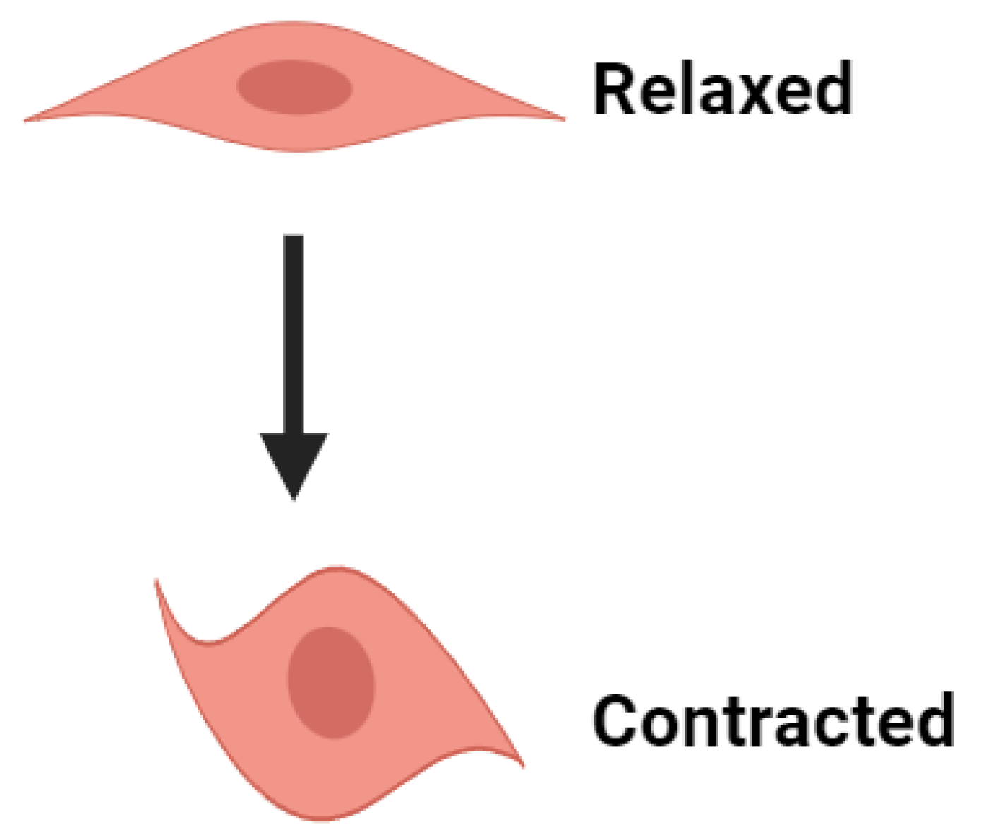
Figure 1. Schematic diagram of vaginal smooth muscle in relaxed and contracted states (Created with BioRender.com).
Preprints 102378 g001
Figure 2. Illustration of slow wave (A) and pace-making type action potential (B) in smooth muscle cells.
Preprints 102378 g002
Figure 3. A schematic representation of the factors involved in contractile activation and relaxation of vaginal smooth muscle. The numbers refer to the steps explained in the text.
Preprints 102378 g003
Table 1. Values of RMP in various smooth muscle cells.
Smooth muscle type RMP (mV) AP/SW Reference
Urinary bladder -45 to -55 AP [10]
Vas deferens -60 AP [41]
Ureter -45 AP [42]
Uterine -50 AP [43]
Urethra -40 AP [44]
Portal vein -50 SW [45]
Pulmonary artery -55 SW [46]
Aorta -50 SW [47]
Colon (GI tract) -60 SW [48]
Seminal vesicles -50 SW [49]
Table 2. Role of ion channels in AP/SW generations.
Ion channel type Role in AP/SW
Ca2+ channels Depolarization, RMP, AP firing
Na+ channels Depolarization, AP firing
K+ channels Repolarization, Hyperpolarization, RMP
Cl- channels Repolarization, RMP
TRP channels Depolarization, RMP, AP firing
Leak channels RMP
Disclaimer/Publisher’s Note: The statements, opinions and data contained in all publications are solely those of the individual author(s) and contributor(s) and not of MDPI and/or the editor(s). MDPI and/or the editor(s) disclaim responsibility for any injury to people or property resulting from any ideas, methods, instructions or products referred to in the content.
Copyright: This open access article is published under a Creative Commons CC BY 4.0 license, which permit the free download, distribution, and reuse, provided that the author and preprint are cited in any reuse.
Prerpints.org logo

Preprints.org is a free preprint server supported by MDPI in Basel, Switzerland.

Subscribe

© 2024 MDPI (Basel, Switzerland) unless otherwise stated