Preprint
Review

Carbon Dots for Future Prospects: Synthesis, Characterizations and Recent Applications: A Review (2019–2023)

Altmetrics

Downloads

179

Views

125

Comments

0

A peer-reviewed article of this preprint also exists.

Submitted:

02 April 2024

Posted:

03 April 2024

You are already at the latest version

Alerts
Abstract
Nanotechnology sparks discussions and concerns about the impacts of new nanomaterials on health and the environment. It holds importance in various domains due to its unique struc-tures. Carbon dots (C-dots) are versatile nanomaterials with applications in bioimaging, sensing, catalysis, polymers, solar cells, and more. They offer desirable characteristics such as stability, cost-effectiveness, biocompatibility, and high photoluminescent quantum yield. C-dots have the potential to replace expensive fluorophores in solar cells. They are catego-rized as carbon quantum dots, graphene quantum dots, and carbonized polymer dots. C-dots have outstanding optical and photoelectric properties with low toxicity. Bottom-up and top-down approaches are used for the synthesis of carbon dots (CDs), with each method im-pacting their physicochemical characteristics. The choice of synthesis method depends on de-sired properties and application requirements. Researchers combine these methods and ex-plore new approaches to enhance synthesis efficiency and tailor CD properties. CDs have di-verse chemical structures with modified oxygen, polymer-based, or amino groups on their surface. Various characterization methods such as HRTEM, XPS, and optical analysis (PL, UV) are used to determine CD structure. Carbon dots (CDs) are cutting-edge fluorescent nano-materials with remarkable qualities such as biocompatibility, low toxicity, environmental friendliness, high water solubility, and photo-stability. They are easily adjustable in terms of their optical properties, making them highly versatile in various fields. CDs find applications in bio-imaging, nanomedicine, drug delivery, solar cells, LEDs, photo-catalysis, elec-tro-catalysis, and other related areas.
Keywords: 
Subject: Chemistry and Materials Science  -   Materials Science and Technology

1. Introduction

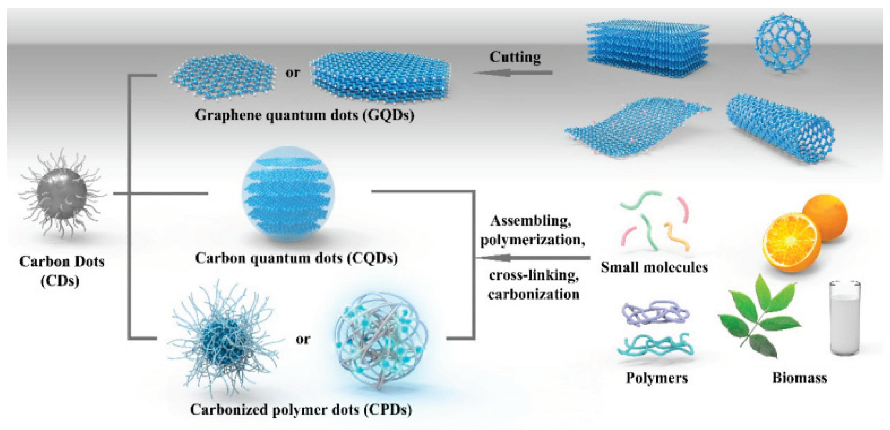
Nanotechnology is a rapidly developing field that has sparked extensive discussions [1,2] and raised concerns about the potential impacts of new nanomaterials on human health and the environment [3,4]. This technology holds significant importance across various technological domains due to its unique and well-defined structures [5]. Since the start of the 21st century, nanotechnology has garnered considerable interest for its ease of synthesis and its applications in diverse fields, including astronomy and environmental protection [6,7]. Carbon dots, also known as C-dots, belong to a novel fluorescent category within the carbon nanomaterial family. They have emerged as a versatile and potent platform with diverse applications [8] . C-dots have gotten a lot of attention and incredible interest in the field of nanotechnology [9], including bioimaging [10], sensing [11,12], catalysis [13], solar cells [14], long-term chemical stability, cost-effectiveness [15], excellent biocompatibility and light-emitting diodes [16], enhanced electron transferability [17], photobleaching, high photoluminescent quantum yield [18], and good aqueous solubility are all desirable characteristics [6,11,19,20]. Because of its outstanding optical properties, C-dots have been developed to replace expensive, heavy-metal-based fluorophores in some photovoltaic solar cells [21]. Various C-dots are also used as doping into the photoanode, counter electrode, hole transport layer, and electron transport layer of dye-sensitized solar cells (DSSC) [22,23,24,25]. Furthermore, many C-dots advantages have recently been reported about their own distinguishing characteristics over other C-dots materials (for example, the crystallinity of the core determines the presence (or absence) of quantum confinement in C-dots, where quantum confinement is identified in C-dots with a crystalline core but not in C-dots with an amorphous core) [8,26,27]. Along with different kinds of carbon-based nanomaterials, C-dots are new advanced nanomaterials and (zero-dimensional) photolumenescent carbons with typical sizes <10 nm. Recently, significant advances in C-dots applications were introduced graping energy conversion (e.g, super-capacitors,PV,LED), optical properties (e,g anticounterfeiting) and promising biomedical are the most underlined. Although, according to their virility formation, nanostructures, properties and mechanism; C-dots [28] are mostly categorised into carbon quantum dots (CQDs),graphene quantum dots (GQDs), and carbonized polymer dots (CPDs) [29,30]. C-dots as a special type of fluorescent material with outstanding optical and photoelectric properties and low toxicity [6,31,32]. The difficulties and potential of these nanomaterials are reviewed, with a focus on how carbon dots can be used to improve the efficiency of photovoltaics and white LED. Advancements in materials chemistry have significantly impacted human lifestyles, with carbon dots emerging as a promising carbon-based nanomaterial[33,34,35]. However, their structural diversity and incomplete field require further research. The review examines the chemistry, history of beginning, classification, design fundamentals, applications, and bright futures of carbon dot-based materials[35]. One of the hottest issues right now in the area of nanomaterials is carbon dots (CDs). Their preparation has typically relied on hydrothermal syntheses. Additionally, the materials that are produced frequently have poor repeatability and are challenging to purify [9,36]. Revolutionary progress in materials chemistry has led to the development of carbon-based nanomaterials like fullerene, carbon nanotubes, and graphene. Carbon dots (CDs), small carbon nanoparticles under 10 nm, are becoming a hot topic due to their superior properties[33]. This article provides a comprehensive review of the carbon dot synthesis process, characterization methods, Optical properties, and current applications. Additionally, it examines the influence of doping and surface engineering on the optical properties of carbon dots and their potential future uses.
Figure 1. Classification of CDs: including graphene quantum dots (GQDs), carbon quantum dots (CQDs), and carbonized polymer dots (CPDs), and their main preparation approaches [29]. Copyright 2020, American Chemical Society.
Preprints 102893 g001

2. Synthesis Method for Carbon Dots

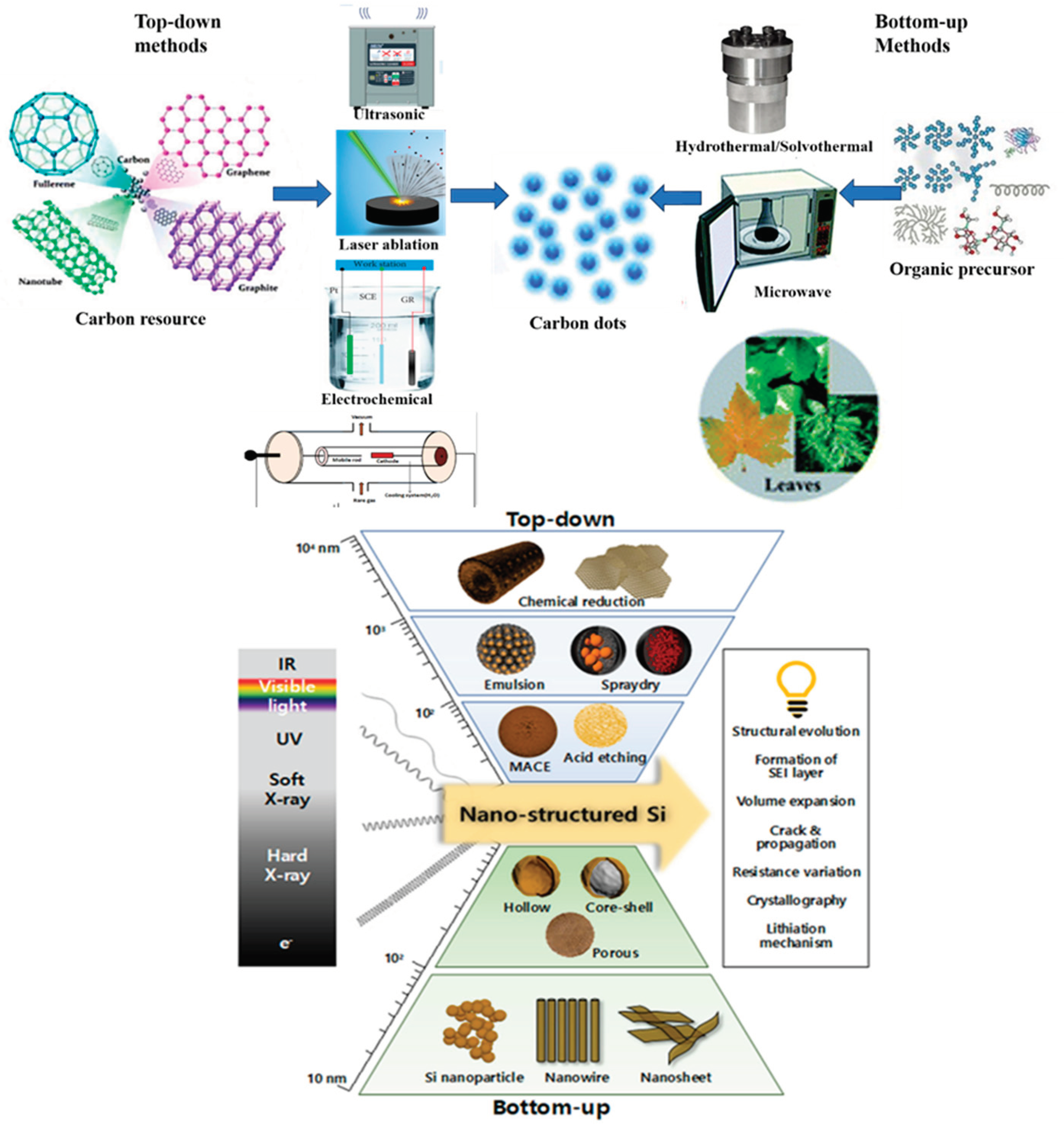
The bottom-up and top-down approaches(seen in Figure 2), which have been schematically shown, represent two broad categories that encompass the many methods for the synthesis of CDs[37]. Although CD production offers convenience, it comes with various significant challenges. These include the nanoparticles' tendency to aggregate during carbonization, the need to control size and homogeneity, and the requirement to modify surface attributes [38]. There are numerous methods for creating CDs from a range of complex-structured raw materials. CDs don't display a suitable graphitic domain, in contrast to GQDs[39]. Which is more fascinating is that each synthesis method significantly affects the physicochemical characteristics of CDs [14], enabling the use of CDs in a variety of applications, such as size dependent PL for biological imaging, and optoelectronic, etc. applications. Bottom-up synthesis involves the construction of carbon dots from smaller molecular precursors or carbon sources[40,41]. This approach typically involves the following steps:Carbonization, Surface passivation, Size control, Purification[42,43]. Bottom-up synthesis methods offer precise control over the size, surface chemistry, and optical properties of carbon dots[44]. Top-down synthesis involves the fragmentation or exfoliation of larger carbonaceous materials to obtain carbon dots[45]. Top-down synthesis methods offer a scalable and cost-effective approach for the production of carbon dots[46]. They utilize readily available carbon sources and can yield carbon dots in large quantities. However, they may have limited control over the surface chemistry and functionalization compared to bottom-up methods[47]. Both bottom-up and top-down synthesis approaches have their advantages and limitations[48,49]. The choice of synthesis method depends on the desired properties, scalability, and specific application requirements of the carbon dots[50]. Researchers employ a combination of these methods and continue to explore new approaches to enhance the synthesis efficiency and tailor the properties of carbon dots for various applications In top-down techniques, various carbon materials such as graphene oxide sheets, graphene, carbon nanotubes, and carbon fibers are processed chemically and electrochemically. This process involves breaking them down into smaller particles using methods like ultrasonic treatment, laser ablation, arc discharge, and electrochemical techniques [51,52].
Figure 2. illustrates the bottom-up and top-down approaches of carbon dots synthesized[53]. Copyright 2018, Wiley..
Preprints 102893 g002

2.1. Ultrasonic Approaches

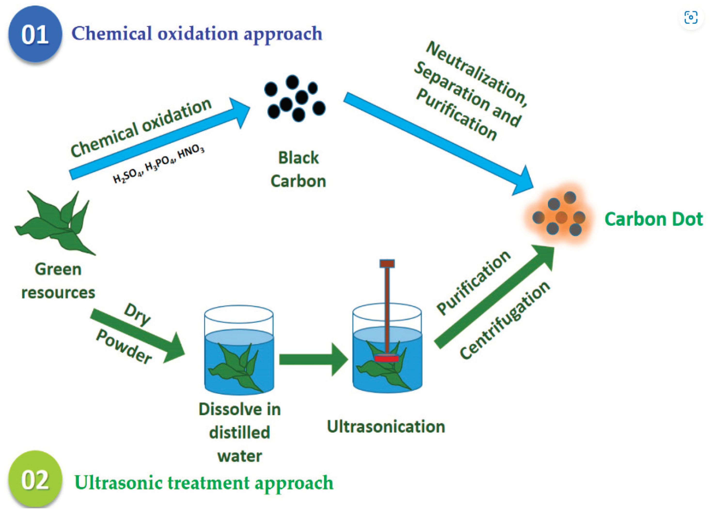
The ultrasonic approach is one kind of "top-down" technique. It breaks down large carbon molecules into smaller CD particles by applying high-energy ultrasonic sound pulses[54]. This approach has been suggested because of its exceptional advantages, which include being cheap, strong penetration, uniform in impact, and environmentally friendly [55]. Ultrasonic approaches are a set of techniques used to synthesize carbon dots (CDs) [39]. Carbon dots are nanoscale carbon-based materials with unique optical and electronic properties[56]. They have gained significant attention in various fields, including optoelectronics, bioimaging, sensing, and drug delivery, due to their excellent photoluminescence, biocompatibility, and low toxicity[57]. Ultrasonic approaches offer several advantages for the synthesis of carbon dots, such as simplicity, low cost, scalability, and control over the size, shape, and surface properties of the resulting CDs[58]. These approaches involve the use of ultrasound, which is a form of mechanical energy in the form of sound waves with frequencies above the audible range (>20 kHz)[59].
Figure 3. Top-down approaches in green synthesis of carbon dots [60]. Copyright 2023, mdpi.com.
Preprints 102893 g003
There are different ultrasonic approaches for synthesizing carbon dots, including the direct ultrasonic method and the indirect ultrasonic method [61]. Let's discuss each of these approaches in detail; The direct ultrasonic method involves the synthesis of carbon dots directly from carbon precursor materials using ultrasound[62]. The process typically starts with the preparation of a precursor solution containing carbon sources such as organic molecules, carbohydrates, or polymers[63]. The precursor solution is then subjected to ultrasonic treatment, usually by immersing an ultrasound probe or horn directly into the solution or by using an ultrasonic bath. The ultrasonic waves induce cavitation, which is the formation and implosion of tiny bubbles in the liquid[64]. The collapse of these bubbles generates localized high temperatures and pressures, leading to the fragmentation and carbonization of the precursors. The resulting carbon atoms reassemble into carbon dots due to the rapid cooling and quenching effects of the surrounding liquid.The direct ultrasonic method offers several advantages, including rapid synthesis, high yield, and tunable optical properties of the resulting carbon dots[65,66]. However, it may require the use of toxic or hazardous precursors and may suffer from low stability and reproducibility[67]. The indirect ultrasonic method provides better control over the synthesis process and allows for the incorporation of additional functional groups or dopants into the carbon dots. It also offers improved stability and reproducibility compared to the direct ultrasonic method. However, it may require more complex reaction conditions and longer reaction times[68,69].

2.2. Laser Ablation

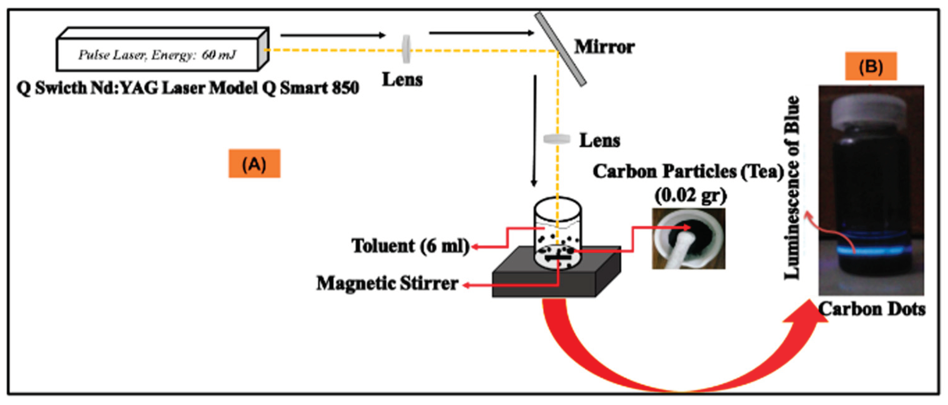
Laser ablation is a synthesis approach that comprises the use of a laser beam and a source of carbon to destroy it into a single material (seen in Figure 4). It is an effective, simple method that does not require excessive chemicals. The timescale of material interaction with the laser beam is the most critical factor for creating perfect structures[70]. CD nanomaterials are created using the laser ablation process at a wavelength of 1064 nm. For instance, Kaczmarek, A., et al. [71] synthesized Carbon Dots (CDs) by focusing the laser beam on carbon (Tea) material in colloid toluent for 3 hours, as illustrated in Figure 3 The prepared CDs were utilized as fluorescent materials for the applications of bioimaging. One of the main advantages of laser ablation is that functional groups can be controlled without requiring hazardous solvents and a straightforward, efficient technique that doesn't require a lot of chemicals[70]. Limitation: 4.5–18% low quantum yield[72].
Figure 4. schematetic illustration of (a) synthesis of CDs by laser ablation approch and (b) Luminance of CDs[73]. Copyright 2023.
Preprints 102893 g004

2.3. Electrochemical Oxidation

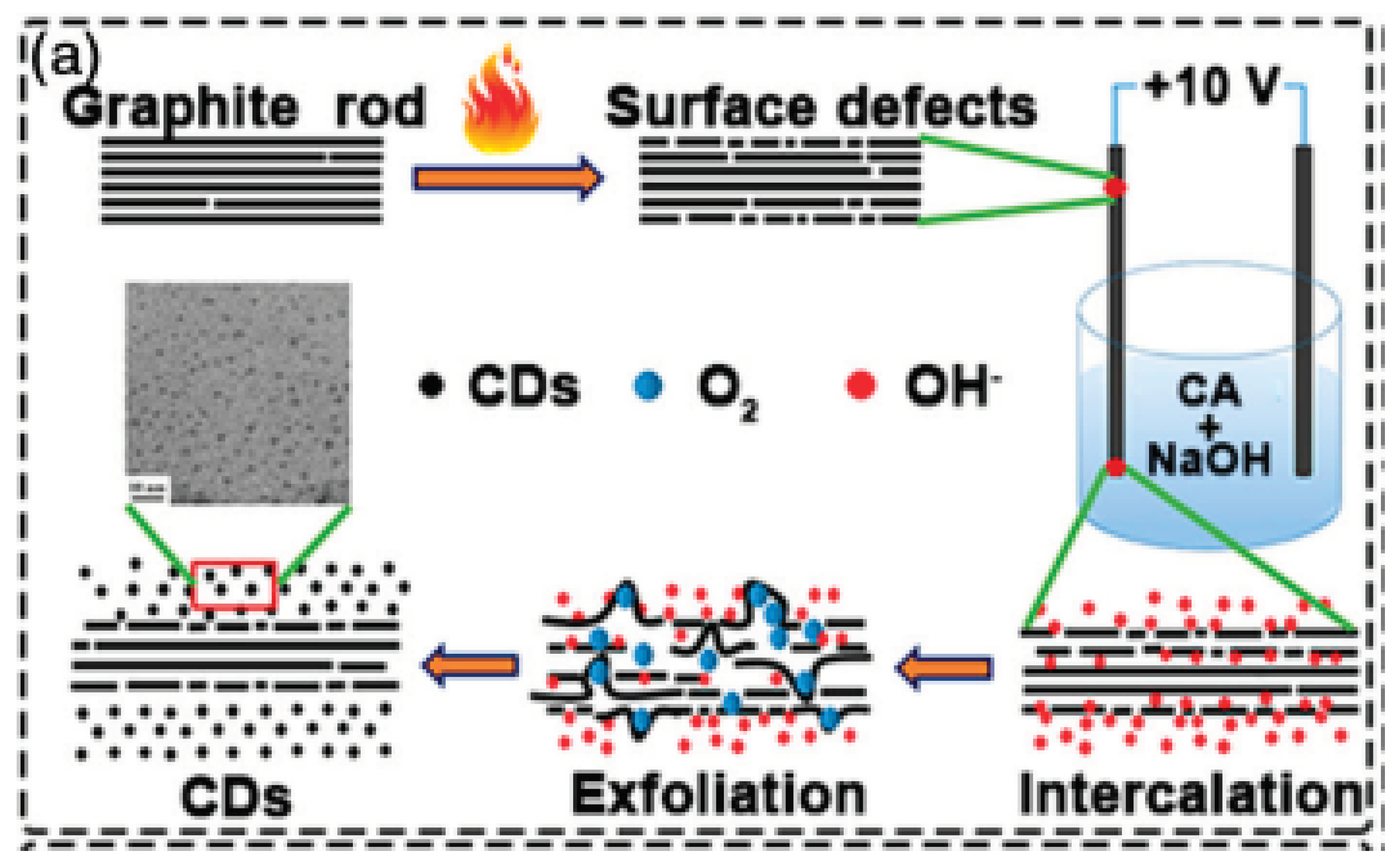
The electrochemical approach is a facile technique for manufacturing CQDs with controllable optical properties and sizes. Typically, it follows a top-down method, wherein larger carbon materials, like graphite electrodes, undergo severing into CQDs under an electrical potential to produce a high quality CQDs [74]. It can be a very effective method and enables heterogeneous redox reactions, in which it is simple to control the potential between two electrodes and is used to monitor the current that passes through an electrolytic cell and use that information as an indicator of certain CD properties. Furthermore, the electron is a redox reagent that is inexpensive, inherently non-polluting, and simple to administer. For these reasons, it is regarded as a "green" reactant in chemical reactions. CDs made using this method are typically plentiful in functional groups, they have more extensive uses in the sensor. Advantages of this approche including large production yield , low-cost easy size control and high purity[75]. Limitation, it required longer time.
Figure 5. Synthesis of CDs by top-down methods Electrochemical oxidation[76]. Copyright 2022,Wiley.
Preprints 102893 g005

2.1.4. Arc Discharge

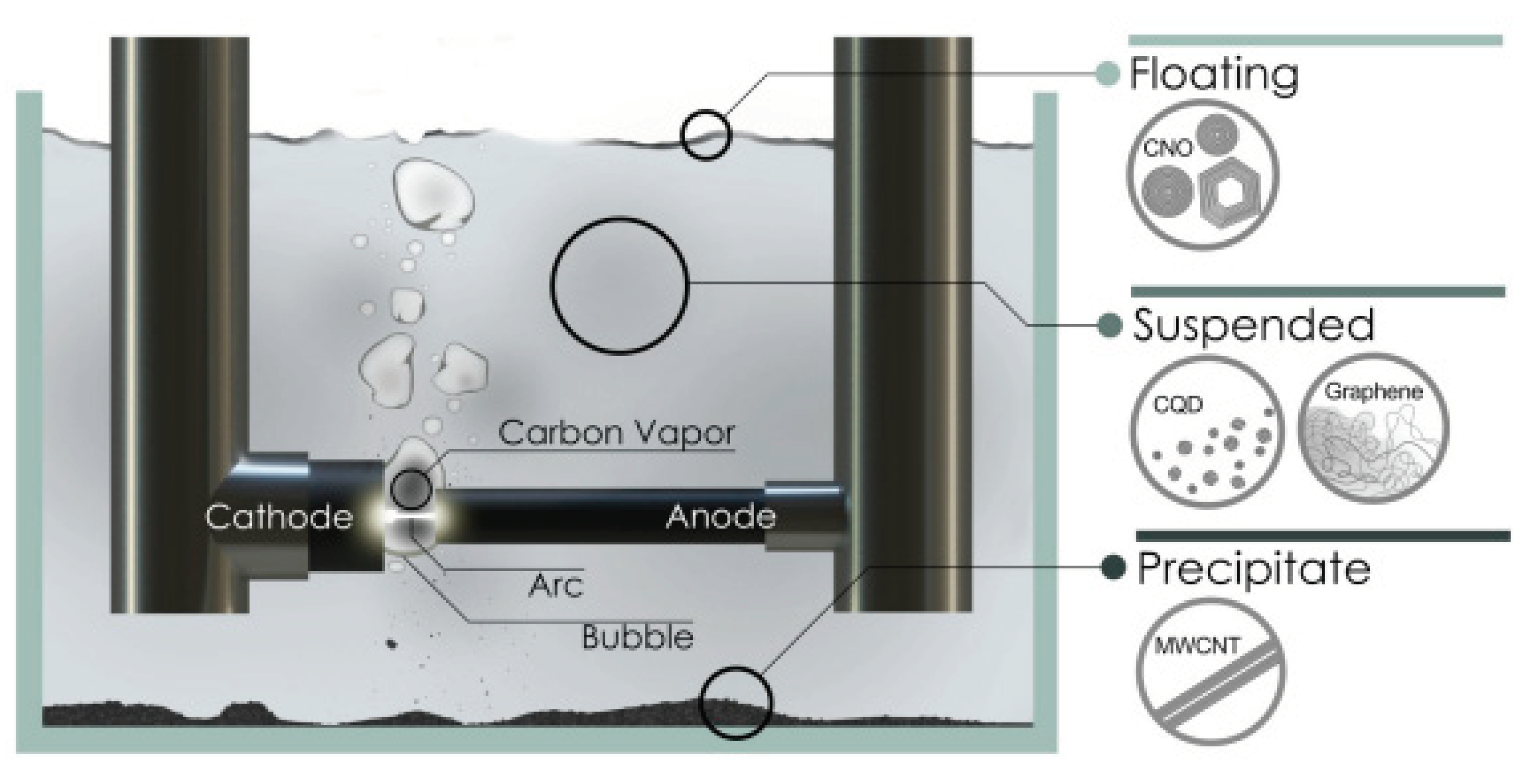
Arc discharge synthesis is a top-down method used to produce carbon dots (CDs). It involves the fragmentation of larger carbon structures through an electric arc discharge. Here is a detailed explanation of the arc discharge synthesis method[77,78,79]. Carbon nanoparticles were produced in very small quantities by arc discharge. Arc discharge dust frequently contains a variety of intricate, challenging-to-extract elements[80]. The numerous investigations done for the arc discharge technique's synthesis of C-dots. Limitation of this approach low quantum yield 2.3 to 8.7% [50]. A high voltage is applied between the two electrodes, creating an electric arc discharge. The high electric field generated during the discharge leads to the vaporization and fragmentation of the graphite electrode[81]. A high voltage is applied between the two electrodes, creating an electric arc discharge. The high electric field generated during the discharge leads to the vaporization and fragmentation of the graphite electrode[81]. Within these carbon nanoparticles, a fraction of the carbon atoms undergo further rearrangement and bond formation, leading to the formation of carbon dots[82]. These dots are typically composed of a core made of sp2-hybridized carbon atoms and a surface functionalized with various chemical groups[83]. The resulting mixture contains a variety of carbon nanostructures, including carbon dots of different sizes. To isolate and purify the carbon dots, various separation techniques such as centrifugation, filtration, or chromatography can be employed[84,85]. However, the size distribution of the carbon dots produced by arc discharge (Figure 6) is often uneven, making separation and purification challenging[86]. The carbon dots obtained from the arc discharge synthesis method are then characterized using various[61,87]. Arc discharge synthesis has been widely used in the early stages of carbon dot research[88]. However, due to the challenges associated with size distribution and purification, other methods such as laser ablation, oxidative cracking, and electrochemical approaches have gained popularity in the synthesis of carbon dots in recent years[86]. Arc discharge synthesis is a top-down method that breaks down larger carbon structures to produce carbon dots (CDs). However, it has limitations such as low yield and the presence of complex elements. The process involves using high-voltage electric arc discharge to vaporize and fragment a graphite electrode, resulting in the formation of carbon nanoparticles.
The top-down techniques, which include arc discharge, laser ablation, oxidative cracking, and electrochemical oxidation, are predicated on the fragmentation of larger carbon structures[77,89,90].When Be Manoj et al. (2020) were purifying single-walled carbon nanotubes generated from arc discharge soot, they discovered an unidentified luminous carbon substance. This is regarded as the CD's initial discovery[91]. This method's drawback is that the size of the CDs is uneven, making separation and purification challenging because a variety of variously sized carbon nanostructures coexist[92]. Moreover, this method has a very low CDs yield. Strong acid passivation of the CD surface is used in laser ablation and oxidative cracking processes. Electrochemical approaches have superseded them due to the hazardous chemicals needed[93]. However, the drawback of this method is the uneven size distribution of the CDs, which makes separation and purification challenging. Additionally, the yield of CDs using arc discharge is low. Laser ablation and oxidative cracking methods involve using strong acid passivation to treat the CD surface, but these techniques have been surpassed by electrochemical approaches due to the use of hazardous chemicals.
Figure 6. Arc Discharging synthesis scheme of Carbon quantum dots[88]. Copyright 2021, AIP publishing.
Preprints 102893 g006

2.4. Hydrothermal Method

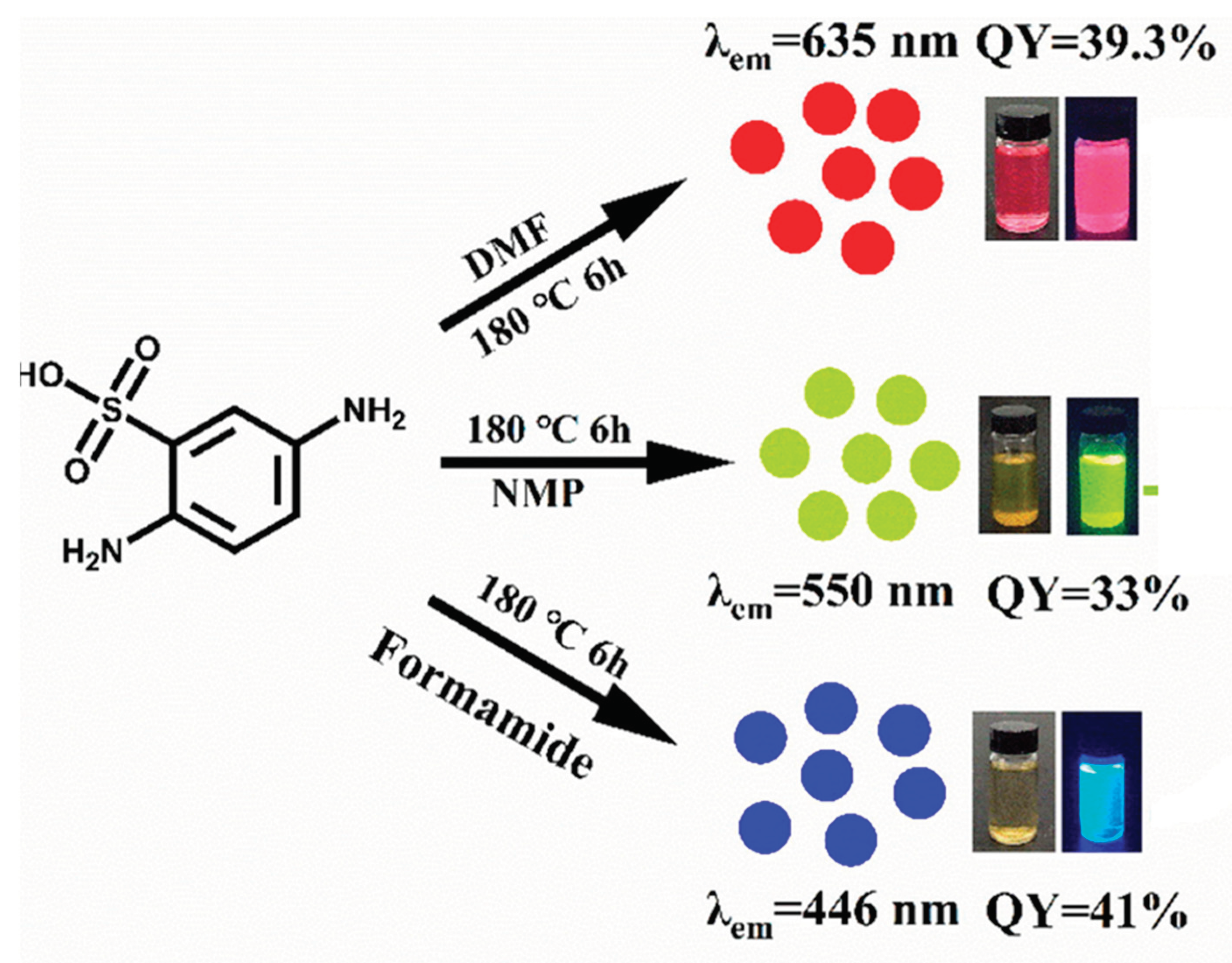
The hydrothermal synthesis approach is the most green method of CD preparation. It uses water as a solvent. In the typical synthesis, the reaction time and temperature range from 3–12 h and 120 °C–240 °C, respectively [94]. It is characterized by the reactions of carbon dots precursors under autoclave conditions at high temperatures and pressures; the temperature should be above the boiling point of the water[95]. Because it is straightforward, low-cost, rapid, requires moderate reaction conditions, can be used for large-scale synthesis and yields highly tunable carbon dots. The hydrothermal method is the most widely used to synthesize C-Dots from renewable resources [96]. The solvothermal synthesis procedure is similar to the hydrothermal method, except that organic solvents, like ethanol, are utilized in the solvothermal method [97]. However, the optical properties, quantum yield (QY), and purity of CDs greatly depend on the solvent utilized during synthesis[97]. For example, Chen, X., et al. [98] synthesized trichromatic CDs such as green (G), red (R), and blue (B) by solvothermal method from a single precursor 2,5-dimethylbenzenesulfonic acid by changing the solvents NMP, DMF, and formamide, respectively, and keeping other conditions the same. Not only the color but also the quantum yield varied as the solvent was changed, as illustrated in Figure 7. Limitations of these approaches include long time and high energy consumption.
Figure 7. Schematic synthesis and detection of the green, red, and blue CDs[99]. Copyright 2023, Elsevier.
Preprints 102893 g007
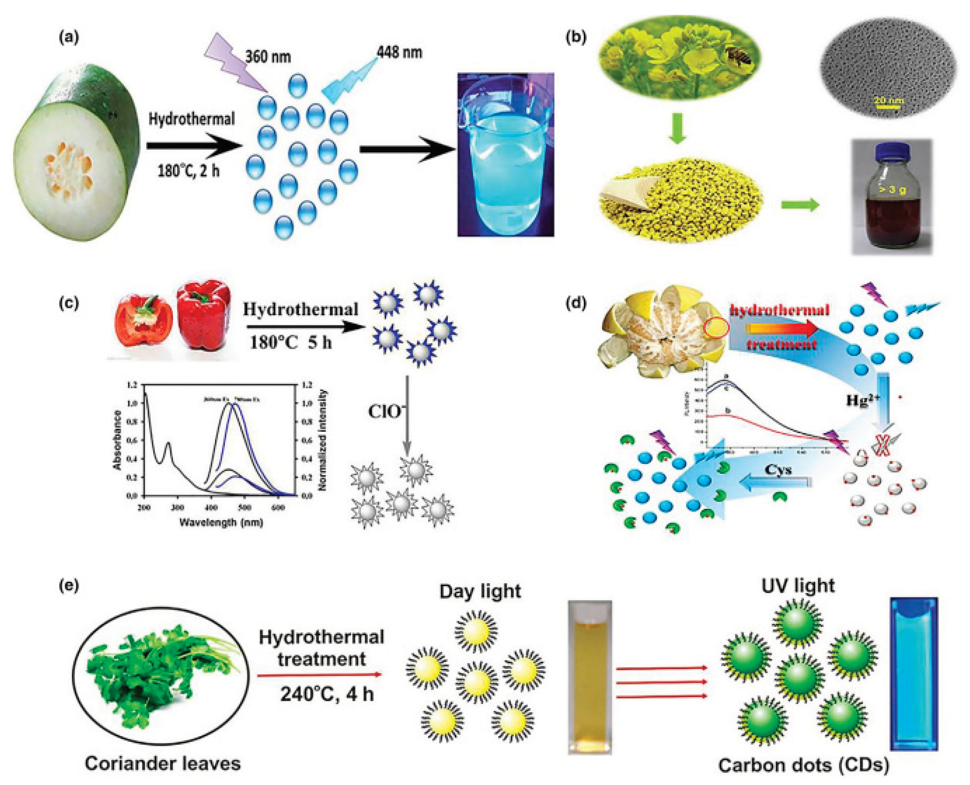
As revealed from Figure 8 below, beyond fruits and vegetables, a wide variety of biomass can be utilised as a carbon source to create BCDs, such as soy milk, black tea, ginkgo leaves, bamboo leaves[100,101] (Figure 8 (e)) and other plants, grass, and so on. When BCDs are made using these carbon sources, the size distribution is rather uniform and the dispersion is good. The majority of the particle sizes fall within the range of 1 to 5 nm, while some BCDs made using oats[102] as a carbon source have a size distribution that ranges from 20 to 40 nm. A few years back, Khalifa et al.[103]suggested using bee pollen as a carbon source to increase the synthesis of BCDs; from 10 g of bee pollen, more than 3 g of BCDs could be created (Figure 8b). High-quality BCDs with up-conversion fluorescence were prepared by hydrothermal treatment in an autoclave set at 180 °C using sweet pepper [104] as a carbon source (Figure 8c). BCDs have a diameter dispersion spanning from 2 to 7 nm with a QY of 19.3% [105]. Simultaneously, cabbage was hydrothermally treated at 140 °C for five hours to produce BCDs[106,107]. The BCDs' QY is 16.5%, and their limited diameter distribution spans from 2 to 6 nm[104,107]. The size distribution of BCDs made with sweet pepper and cabbage as carbon sources is comparatively narrower than that of the aforementioned carbon sources, and as a result, their quantum yield[108,109].
The produced BCDs had a quantum yield of 6.9% and a particle size that varied from 2 to 4 nm [110](Figure 8d). However, compared to BCDs made from the majority of other carbon sources, the quantum yield of BCDs made from orange juice was 26% greater[111,112]. Additionally, the prepared BCDs have a size of 2.5 nm. Citrus lemon juice was used as a carbon source in NaOH solution to make BCDs with a fluorescence QY of 12.1%, as reported by W. Meng et al. [106].
Figure 8. Preparation of biomass carbondots (BCDs) by hydrothermal methods from a) winter melon, b) bee pollens, c) sweet pepper, d) pomelo peel and e) coriander leaves[106]. Copyright 2019. Wiley.
Preprints 102893 g008

2.5. Microwave Synthesize Approach

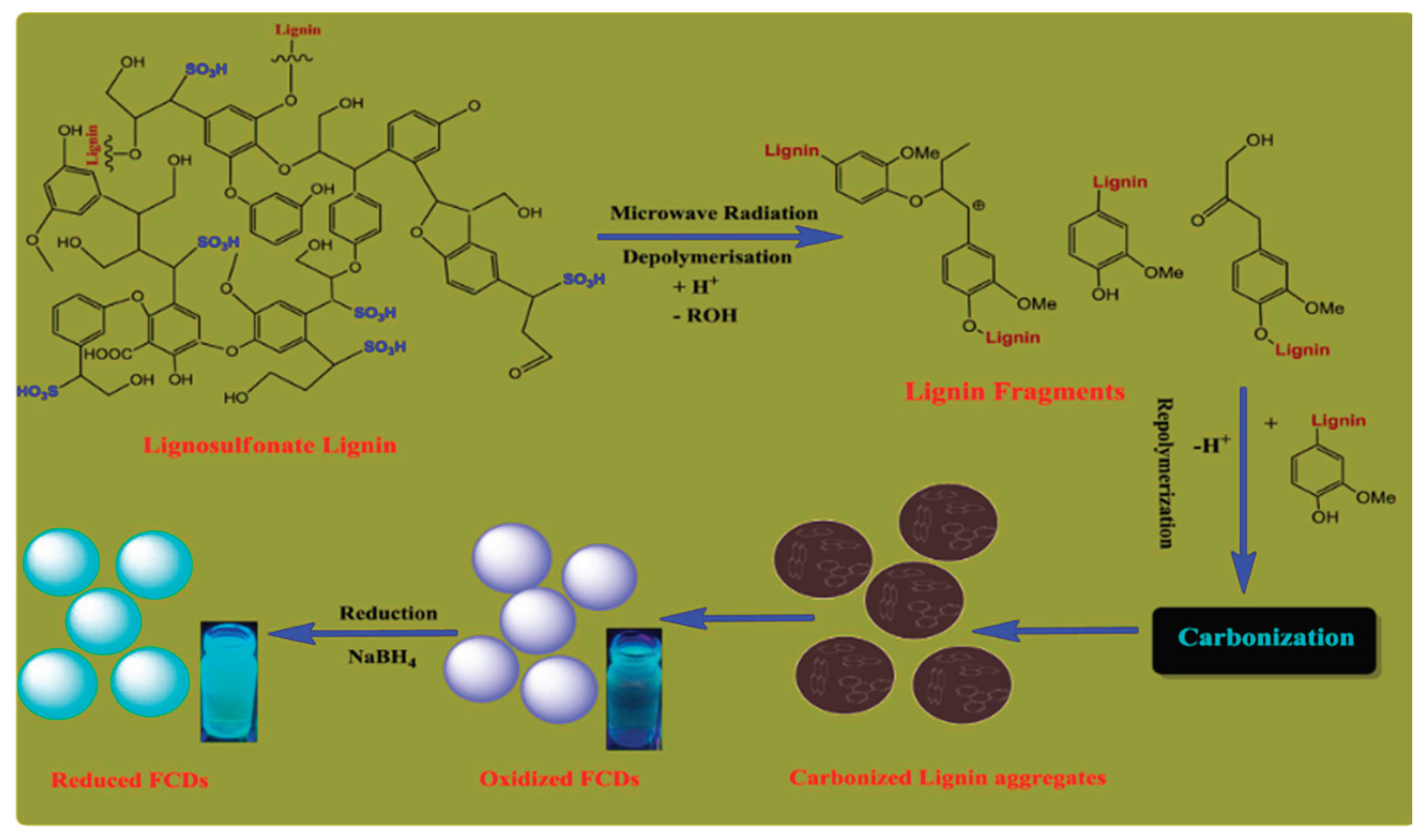
The microwave synthesis approach is a bottom-up process in which CQD precursors are mixed in an aqueous solution as seen below in Figure 9. And exposed to microwave radiation for regulated amounts of time. Compared to the hydrothermal process, it works at far lower temperatures [113]. For example, Vikneswaran et al. used banana peels to synthesize CDs by the microwave method. The synthesized CDs were used to selectively detect Fe3+ ions. This CD displayed excitation fluorescence in the range 280–460 nm, with a fluorescence band at 438–521 nm Rai, S., et al.,[114] have also reported microwave-assisted synthesis of fluorescent carbon dots ( FCDs) from sulfur containing lignosulfonate lignin as a carbon source, in which sulfur acts as doping agents. The synthesized CDs were further converted into reduced carbon dots (r-FCDs) by simply adding NaBH4 to the solution, as illustrated in Figure 5. CDs that have been synthesized can be used to load and release drugs. Due to the many kinds of functional groups that lignin CDs have on their surface. It was verified that manufactured CDs with a narrow particle size distribution exhibit excellent stability and fluorescence properties.
Figure 9. Preparation procedure of reduced carbon dots by microwave synthesize approach [114]. Copyright 2017, Elsevier.
Preprints 102893 g009

2.6. Pyrolysis Synthesis Approach

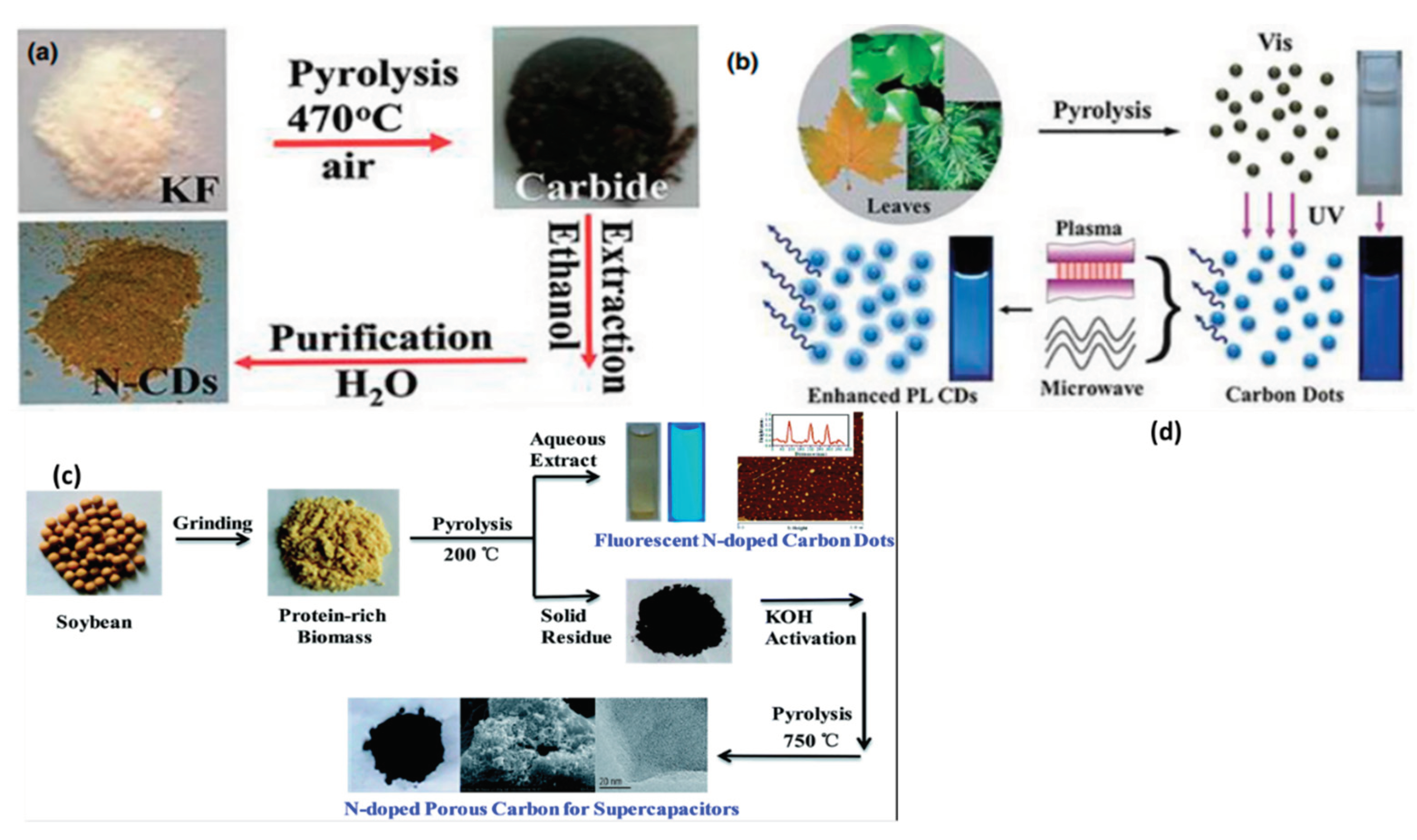
The most popular "bottom-up" technique, known as carbonization or pyrolysis synthesis, was initially put out by Xu et al.[115] during the discovery of CDs and has since been used by several researchers. Speight[116]defines carbonization as the process of thermally breaking down carbon-containing materials, especially natural goods, to produce carbonaceous residue. It is a low-cost, environmentally beneficial process that breaks down a chemical at high temperatures (Figure 10 (a) and (b)) It solid compounds with a greater carbon content from organic sources by continuously pyrolyzing them in an inert atmosphere[117].The advantage of this approach is Easy operation, low cost, solvent-free , scalable production and fast reaction time. The main problem with pyrolysis is aggregation, which occurs during carbonization. Here, we present a unique two-step carbonization process (Figure 10(c)) that allows for the simultaneous extraction of porous carbon (SC) and N-doped fluorescent carbon dots (SCDs) from naturally nitrogen-rich soybean biomass. Fluorescent carbon dots were produced by a first low-temperature carbonization process (20 oC), and porous carbon with a unique network of linked micropores and mesopores and a high specific surface area (1663.1 m2 ·g -1) was produced by a second high-temperature carbonization process (750 o C). The schematic diagram for the SCD and SC preparation is shown in Figure10(c). The carbonisation process consists of two treatment steps: a low-temperature pre-carbonization (200 °C) that yields fluorescent carbon dots and preserves the microstructure and heteroatoms (such as N, O, and S) of the raw material soybean; a high-temperature carbonisation (750 °C) that involves KOH activation that partially graphitizes the pre-carbonization residue and yields highly porous active carbon.
Figure 10. Different Pyrolysis synthesis of Carbon dots [106,118,119]. Copyright 2019•Wiley Copyright 2016,Wiley , Copyright 2016•pubs.rsc.org .
Preprints 102893 g010
As seen from Table 1, hydrothermally synthesized carbon dots exhibit excellent photoluminescence properties, making them suitable for biomedical imaging applications. They can be used as fluorescent probes for imaging various biological structures, including cells, tissues, and even specific biomarkers. Hydrothermal synthesis produces carbon dots with unique surface properties and functional groups. These properties make them ideal for sensing applications, such as chemical and biological sensing. Carbon dots can be used as fluorescent probes or integrated into sensing platforms for detecting analytes, including heavy metals, gases, and biomolecules. Carbon dots synthesized through other methods may also find similar applications, but the specific properties and characteristics of hydrothermal synthesis make them particularly suitable for these applications. Microwave synthesis allows for rapid heating and reaction, leading to shorter synthesis times compared to other methods. This can be beneficial for large-scale production or time-sensitive applications. Microwave synthesis offers easy control over reaction parameters such as temperature, power, and reaction time. This control allows researchers to optimize the synthesis conditions for specific environmental remediation requirements. By examining this Table 1, researchers can compare the advantages, disadvantages, and applications of different synthesis methods. This information helps in selecting the most suitable method based on specific research goals, cost considerations, equipment availability, and desired properties of the carbon do. It's important to note that these applications are not limited to hydrothermally or microwave synthesized carbon dots alone.

3. Characterizations Tools for Carbon Dots and Its Optical Properties
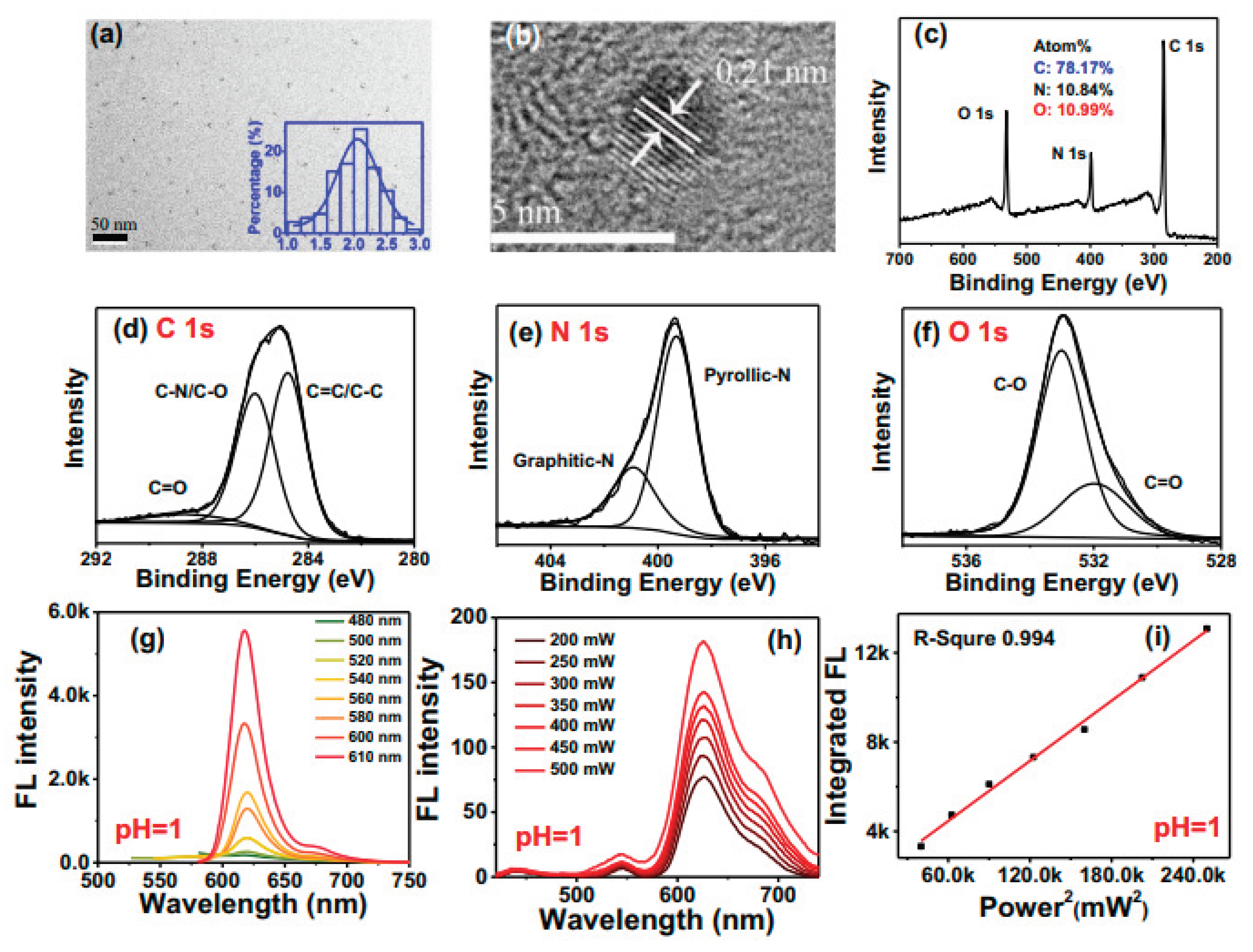
Characterization tools play a crucial role in the study and understanding of carbon dots (CDs)[68], which are nanoscale carbon-based materials with unique optical and electronic properties[135]. These tools enable researchers to investigate various aspects of CDs, such as their structure, size, surface chemistry, optical properties, and surface morphology[136]. Characterization tools are essential for gaining a comprehensive understanding of carbon dots[137]. They enable researchers to explore the structural, optical, surface, and electrical properties of CDs, which are crucial for tailoring their properties and optimizing their performance in various applications ranging from optoelectronics and biomedicine to energy storage and sensing. Because of various synthesis approaches, the chemical structure of each CD will also be different. Nevertheless, all have modified oxygen, polymer-based, or amino groups on their surface. Thus, numerous characterization methods have been developed to determine the structure of CDs[138].
TEM pictures at 50 nm resolution, as displayed in Figure 11a, reveal that the manufactured CDs are evenly distributed in aqueous solution. According to the inset image of Figure 1a, CD particle sizes range from 1 to 3 nm, with an average dimension of 2 nm. According to high-resolution photos shown in Figure 11b, CDs are linked to the (1, 0, 0) plane of graphite with a lattice distance of 0.21 nm. The CDs' recorded Raman spectra is displayed in Supplementary Figure 11(b). The structure of sp3 (D) and sp2 (G) is represented by the vibration peaks at 1374 and 1607 cm−1, respectively, and the ID/IG ratio of CDs is 0.80. The investigated CDs' graphitic structure is shown by the prominent strength of the G peak. The manufactured CDs' chemical composition is examined by the use of XPS examination. The full scale XPS spectra analysis results, as displayed in Figure 11c, show that the produced CDs.
CD have atomic ratios of 78.17, 10.84, and 10.99% for the elements carbon (C), nitrogen (N), and oxygen (O), respectively. XPS spectra with high resolution are displayed in Figure 11(d–f). The presence of sp2 C=C carbon (284.76 eV), sp3 C−N/C−O carbon (286.0 eV), and C=O groups (288.6 eV) can be seen in the high resolution C 1s spectra (see Figure 11d). The high-resolution N 1s spectra displayed in Figure 1e have two peaks that stand for graphitic nitrogen (400.9 eV) and pyrrolic nitrogen (399.3 eV). The O 1s band (refer to Figure 1f) can be disassembled into two peaks, denoting the C=O, C−O−C/C−O−H groups, at 531.9 and 533.0.
The solvent environment can have a significant impact on the PL property of CDs. Therefore, the FL emission is determined by dissolving the protonated CDs samples in deionized water in order to gain a better knowledge of the PL properties of produced CDs in biological water-soluble environments. The red FL emission of protonated CDs, as depicted in Figure 11g, appears at 620 nm with an FWHM of 24 nm, which is 34% narrower than the most recent discovery of high-color-purity CDs with an FWHM of 35 nm16, 17. An excitation-independent PL behavior is displayed by the produced CDs when excited by wavelengths ranging from 480 to 610 nm. Experiments additionally show that up conversion FL emission of CDs produces red emission lines at 630 and 680 nm with FWHM of 70 nm when excited by near-infrared (NIR) light of an 808 nm femtosecond (fs) laser (see Figure11h). The FL intensity of CDs increases in accordance with a linear relationship (R = 0.994) with the square of laser power increase from 200 to 570 mW, as illustrated in Figure 11i. This suggests that two-photon excitation-induced FL emission is responsible for the red up-conversion FL of CDs.
Figure 11. Structure, composition, and optical properties of protonated CDs. (a) TEM image of CDs (inset, particle size distribution of CDs); (b) high-resolution TEM image of CDs; (c−f) full scale XPS spectra, high-resolution C 1s, N 1s, and O 1s XPS spectra of CDs; (g, h) one-photon and two-photon fluorescence (FL) spectra of protonated CDs in deionized water; (i) relationship of two-photon FL intensity and femtosecond (fs) laser power[139]. Copyright 2021, nature communications.
Preprints 102893 g011
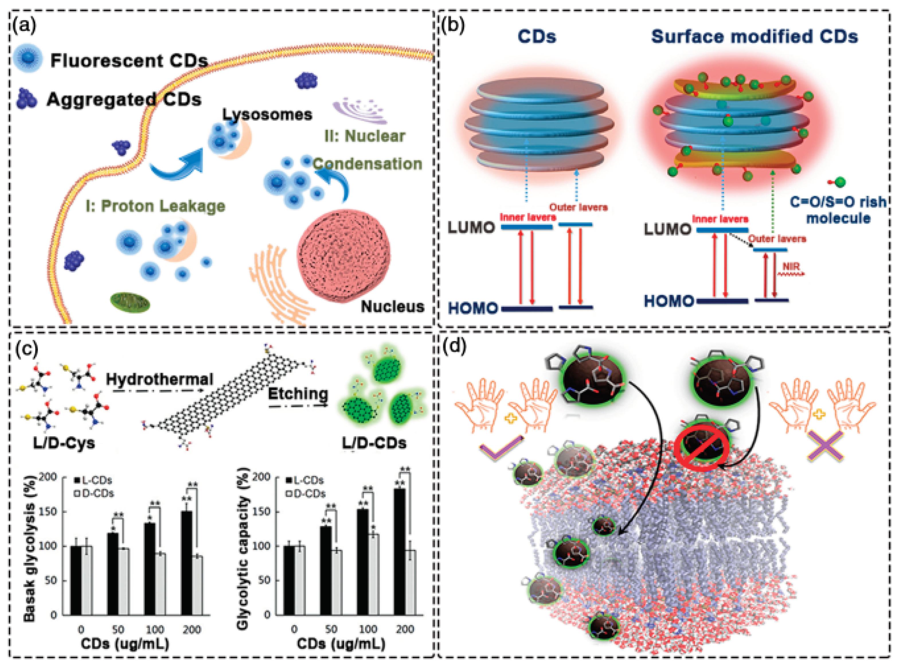
CQDs typically exhibit optical absorption in the UV with a tail that extends into the visible spectrum Regardless of how they are made, the majority of CDs have an absorption band that ranges from 260 to 323 nm[140,141]. However, the π–π* transition of the C=C bonds or the n-π* transition of the C=O bonds of CDs may be linked to some absorption shoulders in the absorption spectra. It has been demonstrated that surface passivation of CQDs containing different molecules alters CD absorption and causes an absorbance shift to a longer wavelength[142]. Research on the optical characteristics of CDs is currently quite popular. Features including red/NIR emission, up-conversion photoluminescence (UCPL), high QYs of fluorescence, and NIR-driven (seen in Figure 12 (a). The use of CDs in biology and biomedicine involves the exploitation of photothermal heating and chiral luminescence. A high QYs is highly advantageous for CD tracking in vivo.Because short wavelength light penetrates biological tissues poorly, most CDs absorb in the UV area and produce blue-green light, making detection challenging. To some extent, this limitation can be circumvented using blue-green emission CDs with high QYs. By taking advantage of the acidophilic characteristics of the CDs, lysosomes in living cells can be passively seen. Using branched polyethyleneimine (bPEI) and rose bengal (RB) as precursors, green emission CDs with QYs of 90.49% were produced.
As revealed Figure 12 (b), Red and NIR emitting CDs are currently receiving a lot of attention because of their low autofluorescence interference, deep tissue penetration, and low tissue absorption. Numerous applications related to the in vitro and in vivo observation of biological systems have been investigated. Red/NIR-CDs can be cultured with different cancer cells to evaluate their potential for labeling applications in in vitro cell imaging. Red/NIR-CDs primarily aggregate in the cytoplasm and cell membrane, according to the data.[101] Using o-phenylenediamine as the precursor, our lab has successfully created redemitting CDs with high QYs. These CDs have been employed as fluorescent probes for both in vitro and in vivo imaging. We then created a range of red-emissive CDs based on o-phenylenediamine, which made bioimaging possible. From Figure 12(c), the resulting L-CDs are appropriate for long-term in situ imaging of the Golgi because they show high light stability and biocompatibility. The dynamic alterations of the Golgi apparatus during the early stages of viral infection can be seen with the aid of the produced L-CDs. Furthermore, chiral cysteine has been extensively employed as a chiral ligand and stabilizer to modify the characteristics of nanomaterials. When L (or D)-cysteine generated chiral CDs were used to treat human bladder cancer T24 cells, L-CDs demonstrated up-regulated glycolysis while D-CDs had no such impact (Figure 12c). For instance, D-proline with inverted chirality preferentially interacts with liposome mimics or the cell membrane (Figure 12d).
Figure 12. Optical properties of CDs. (a) High quantum yield CDs for visualization of lysosomes b) NIR-emitting CDs for bio-imaging (c) Chiral CDs for the treatment of T24 cells (d) Chiral CDs demonstrating high selectivity for cell membranes[76]. Copyright 2022,Wiley.
Preprints 102893 g012
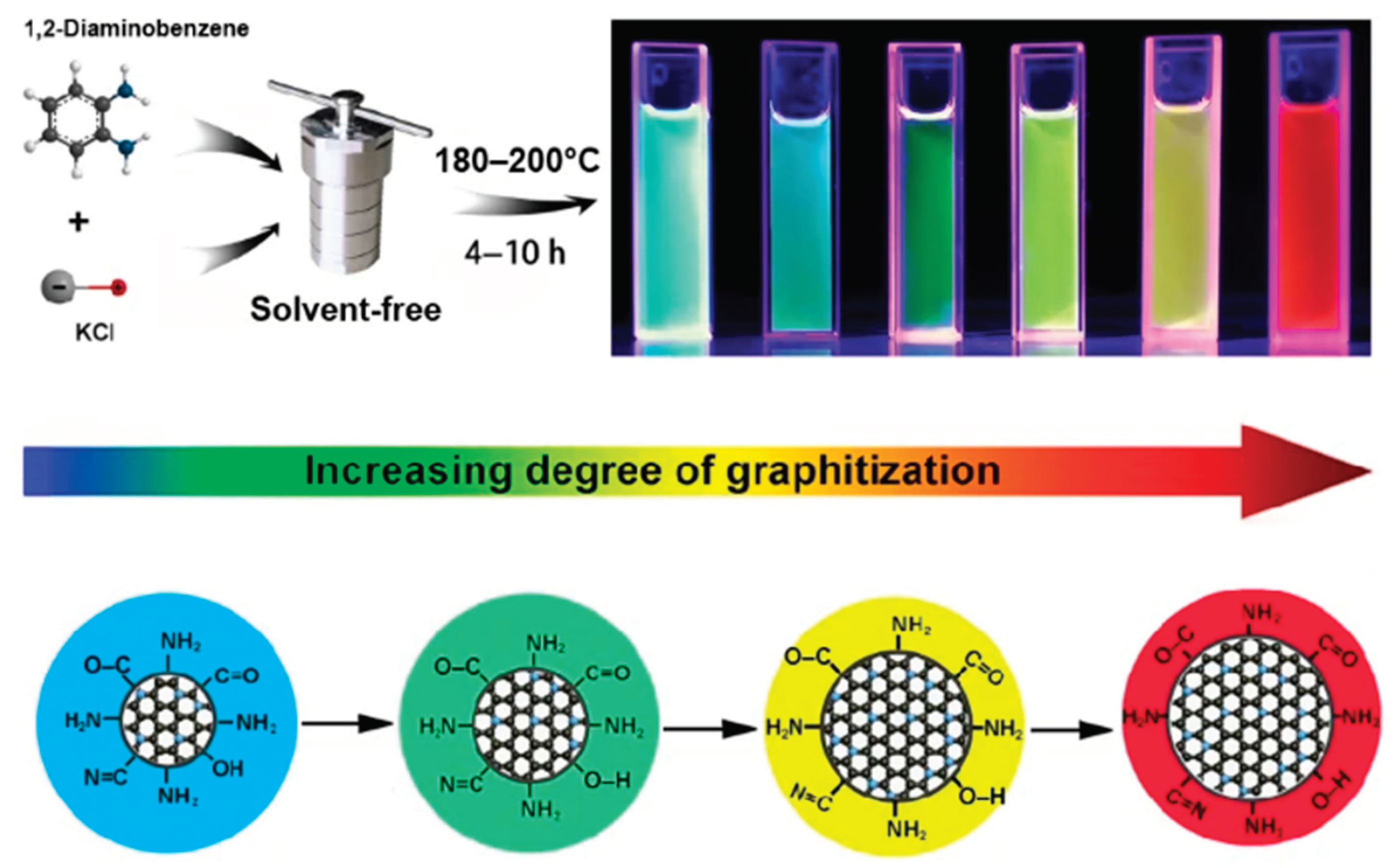
Excitation-dependent photoluminescence, or excitation-dependent fluorescence emission, is one of the most remarkable characteristics of CQDs (seen in Figure 13). The district's dependence on the emission wavelength and intensity is a characteristic of the PL forCQDs generally[143]. Zhang's group conducted a study to investigate the emission behavior in this regard. of CQDs at various concentrations exposed to 470 nm light[144]. They discovered that when the excitation wavelength and CD concentration were raised, the PL intensity of the yellow-emitting CQDs first climbed to a maximum λex and then declined. This work demonstrated that CDs exhibit similar excitation-dependent photoluminescence activity to other luminous carbon nanoparticles[144].Similarly, by heating o-phenylenediamine under the catalysis of KCl, a series of CDs with tunable PL emission from 442 to 621 nm, QY of 23%–56%, and production yield within 34%–72% are produced by Ding, H., et al[145]. Extensive analyses reveal that the variations among these CDs concerning the extent of graphitization, the amount of graphitic nitrogen in them, and the functional groups that contain oxygen are in charge of their unique optical characteristics as illustrated in Figure 13. These variations can be adjusted by managing the deamination and dehydrogenation procedures that take place during reactions. To create blue, green, yellow, and red emissive films and LEDs, the appropriate CDs are dispersed into polyvinyl alcohol (PVA). Red, green, and blue emissive CDs are also mixed to create all varieties of warm, standard, and cool white LEDs (WLEDs) with high color rendering index (CRI).
Figure 13. A CDs with tunable PL emission range from 442 - 621 nm[145]. Copyright2022, Springer.
Preprints 102893 g013

4. Application of Carbon Dots

As a cutting-edge fluorescent a nanomaterial, carbon dots display remarkable qualities like biocompatibility, low toxicity, environmental friendliness, high water solubility, and photo-stability. These nano-sized can also be made easily, quickly, and in accordance with green chemistry concepts. With their easily adjustable optical properties, CDs are used in a variety of fields, including bio-imaging, nanomedicine, drug delivery, solar cells, LEDs, photo-catalysis, electro-catalysis, and other closely related ones[23,32,89,146,147].

4.1. Solar Cell

The experimental results presented in Figure 14 (A) and (B)[148] demonstrate the significant impact of varying weight percentages of C-dots deposited on ZnO NPs and NiO NPs on the performance enhancement of Dye Sensitized Solar Cells (DSSCs). To investigate this, composite DSSCs consisting of ZnO@Cdots/dye nanostructures were tested under blue light illumination with an intensity of 156 w/m2. The comparisons were conducted by adjusting the Cdots concentrations and the ethylenediamine/citric acid mole ratios. Figure 14 (A) illustrates the I-V characteristics of different DSSC combinations employing ZnO@Cdots (1) as well as sensitizers, while varying the weight percentage of Cdots content from 0 to 50. The I-V curves showed varying strengths depending on the Cdots content, with weaker curves observed for weight percentages above a certain threshold, and more intense curves within the range of 0 to 10 weight percent. Notably, the ZnO(20)@Cdots DSSC sensitized with N719 demonstrated the highest performance among all combinations. Additionally, the ZnO(20)@Cdots DSSC exhibited a current 1.2-1.5 times higher than that of the ZnO(100)@Cdots DSSC. Furthermore, when the sensitizer was changed from RhB to N719, both ZnO(100)@Cdots and ZnO(20)@Cdots DSSCs experienced an increase in both voltage (1.3-2.0 times) and current (almost one order of magnitude). These observations emphasize the superior efficiency achieved by the optimized conditions.
The I-V curves for NiO@C-dots DSSCs are shown in Figure 14 (B) with a 12.5 weight percent C-dot content, but with different EDA/CA molar ratios for the N719 and Rh6G sensitizers (0.5:1, 1.0:1, 1.5:1, 2.0:1, and 2.5:1). Plotted in Figure 14 (B), the electrochemical parameters determined from these I-V curves are provided[149]. The EDA/CA molar ratio for both sensitizers affected four parameters: Since the rise in the EDA/CA molar ratio enhances the amine content relative to the carboxylic acid content, all metrics were usually maximum at an EDA/CA molar ratio of 1.5:1 and higher for N719 DSSC than for Rh6G DSSC, suggesting the effect of the amine (NH2) moiety in the C-dots. Overall, the experimental results demonstrated that optimizing the weight percentages of C-dots deposited on ZnO NPs and NiO NPs, as well as the EDA/CA molar ratio, can significantly enhance the performance of DSSCs by improving current, voltage, and overall efficiency.
Figure 14. (A) C-dots content dependency on I-V characteristics of ZnO@Cdots/dye DSSCs (B) I−V curves and calculated electrochemical parameters of NiO@C-dots DSSCs at different EDA/CA molar ratios at 12.5 wt % C-dot content. The numerals in I−V curves indicate EDA/CA mole ratio[148,149]. Copyright2019 Elsevier, Copyright 2020, American Chemical Society.
Preprints 102893 g014

4.2. Photo Catalysis

Through the environmentally benign and sustainable process of photocatalysis, light stimulates a catalyst to produce energetic electrons and holes that initiate other reactions. Energy and environmental applications have seen the biggest use of it. Numerous studies have been conducted to investigate the photocatalytic potential of CDs due to their wide light absorption, PL characteristics, and electron transfer capabilities. CDs can function as a single photocatalyst in the absence of a co-catalyst or combine with other materials to create composite catalysts (such as conventional semiconductor photocatalysts).
Due to the rapid population growth and increasing demand for everyday consumables, the two main obstacles to human sustainable development are the energy crisis and environmental pollution. Modern and sustainable technologies can aid in resolving the problems. The pure nature of photocatalysis and its endless supply of solar energy make it an attractive solution that won't add to the already heavy environmental load. Usually, photocatalysts are semiconductors that may be activated by light irradiation to produce highly oxidative hole charge carriers and reductive electron charge carriers for various redox processes. The most promising choices have been thought to be II–VI group quantum dots (QDs) and Cd-free I–III–VI QDs because of their exceptional optical capabilities, wide surface area, and unique quantum confinement effect. Certain conventional QDs have garnered increased interest in photocatalysis because of their composition-tunable band gap. These photocatalysts, however, exhibit poor stability and low production efficiency. This deficiency can be attributed to I-III-VI QD's limited charge separation and photo-corrosion capabilities.
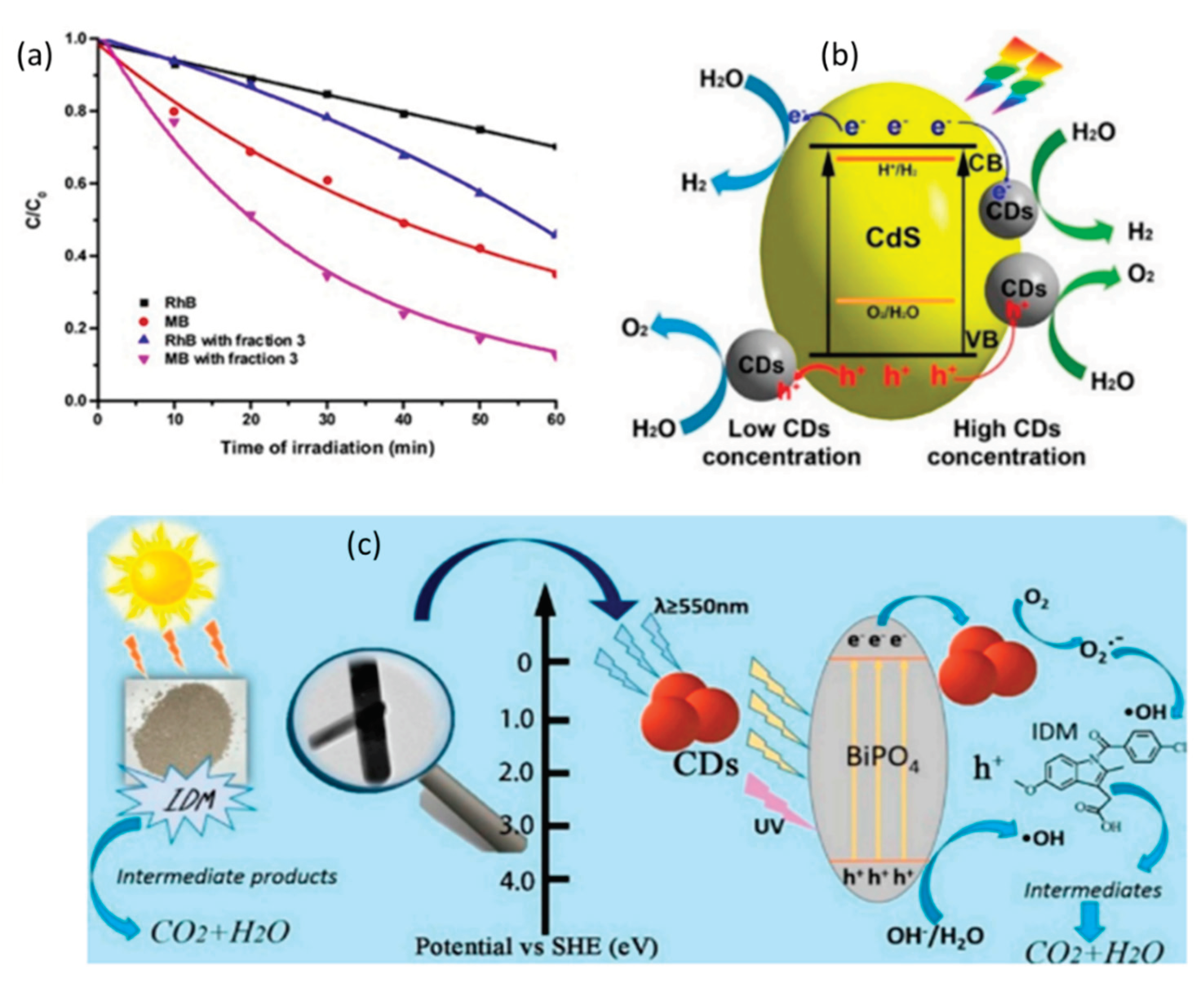
According to Figure 15 (a), fraction 3 (or around 2 nm CDs) was able to rapidly degrade MB (60%) and RhB (20%) through photocatalysis in less than 60 minutes. It's interesting to note that exposure to visible light can cause RhB molecules to change into RhB* radicals. When CdS and CDs were combined (seen in Figure 15 (b)), the resulting nanocomposites significantly improved the production of hydrogen and oxygen from water splitting and improved catalytic stability without the need for sacrificial agents. By regulating the CDs content in the CDs-CdS composite, the evolution of hydrogen and oxygen was able to be achieved at approximately 2.55 and 0.52 µmol h−1, respectively. The findings did not demonstrate a 2:1 stoichiometric ratio (H2:O2). In addition, the CDs-CdS nanocomposite outperformed the previously published CdS catalyst in terms of cyclic stability (8 cycles of catalytic tests). The suggested reaction mechanism of CDs-CdS is depicted in Figure 15 (b). Following the incorporation of carbon dots with CdS, the reaction demonstrates good stability due to enhanced light absorption and electron-hole pair separation. This approach opens up a new avenue for the development and investigation of stable CdS photocatalyst, notwithstanding its ongoing shortcomings (such as limited gas generation). According to a study by Liu et al., Rhodamine B (RhB) underwent photodegradation, and CDs effectively trap electrons and prevent photoexcited electrons and holes from recombining. The degrading efficiency of RhB by pure CdS after one hour of radiation exposure was approximately 50%, while the efficiency was increased to 90% by 1% CDs/CdS nanocomposites. It is anticipated that the hydrothermal approach of integrating carbon dots with Bi2MoO6 nanosheets will improve the nanosheet structure's photocatalytic activity (seen in Figure 15 (c). It was looked into how structure and photocatalytic activity related to them other. They discovered that the as-prepared CDs modified Bi2MoO6 had a larger BET surface area, which increased the amount of photocatalyst-pollutant interaction and the amount of active species absorbed. This is demonstrated by the photodegradation of ciprofloxacin (CIP), where 2 weight percent CDs modified Bi2MoO6 showed a 5-fold increase over pure Bi2MoO6. The wider visible light absorption zone, slower electron-hole recombination rate, and more active adsorption sites and photocatalytic reaction centers are only a few benefits of the change.
Figure 15. (a) ) Photocatalytic degradation of MB and RhB along with illuminating time[150,151] (b) the proposed reaction mechanism of carbon dots-cadmium sulfide (CDs-CdS) under[150] (c) the schematic photocatalytic mechanism of indometacin (IDM) degradation by the CDs-doped BiPO4 composite under irradiation of simulation visible light irradiation[150]. Copyright2019, mdpi.com.
Preprints 102893 g015

4.3. Biomass

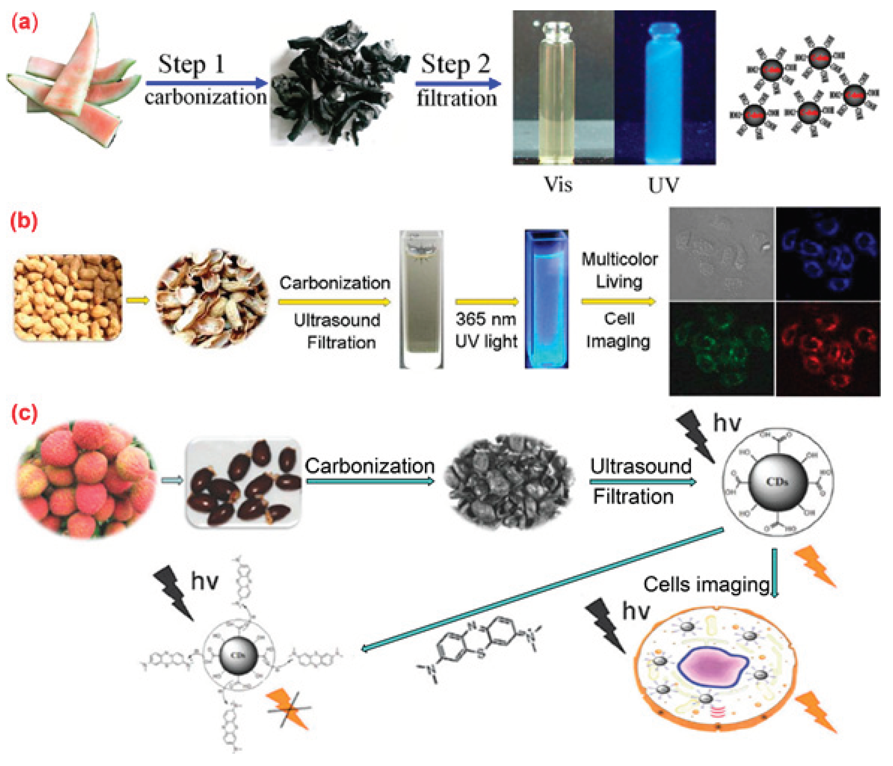
Biomass Carbon dots (BCDs) made from discarded organic materials or biomass are currently the subject of research into solar cell applications due to their size and excitation-dependent fluorescence emission. A fluorescence quenching mechanism was reported by Zhang et al. [117] to have a large positive impact on the conversion efficiency of BCD-sensitized aqueous solar cells. Grass-based synthetic BCDs were used as the test case to verify the theory attributed to improving the quenching of fluorescence [152,153]. The results of this study confirm a theory that may have broad applications in improving the efficiency of solar cells using different fluorescent quantum dot/nanodot sensitizers. Pyrolysis is a commonly used method for producing C-dots[154]. It involves converting organic material in a carbon source into carbon dots through heating, dehydration, degradation, and carbonization under high temperatures in a vacuum or inert atmosphere. The pyrolysis method typically utilizes high-concentration acid or alkali to break down the carbon precursors into nanoparticles[102]. Various biomass materials, such as watermelon peel, sago waste, coffee grounds, and plant leaves, can be used as carbon sources for producing C-dots through pyrolysis. The properties of the resulting C-dots can be adjusted by changing the pyrolysis conditions, including temperature, duration, and pH value of the reaction systems[155]. The resulting C-dots have outstanding water solubility, intense blue luminescence, and strong stability in solutions with high salinity and a broad pH range. HeLa cell imaging was accomplished with success using the carbon dots when prepared (Figure 16a). Pyrolysis of lychee seeds produced fluorescence C-dots with low intrinsic cytotoxicity and a quantum yield of 10.6%, which were then utilized for fluorescence imaging of living HepG2 cells (Figure 16b). A straightforward and affordable pyrolysis technique for the production of fluorescent C-dots from peanut shell waste was presented by Xue et al. [54].
Figure 16. Preparation of fluorescence C-dots from agriculture wastes by pyrolysis method. (a) Synthesis of water-soluble C-dots from watermelon peel; (b) the preparation and application of fluorescent C-dots from lychee seeds; (c) the preparation and application of C-dots from peanut shells [156]. Copyright 2020, mdpi.com.
Preprints 102893 g016

4.4. Biomedical

CDs have shown excellent properties, such as good photostability, excellent photoluminescence,
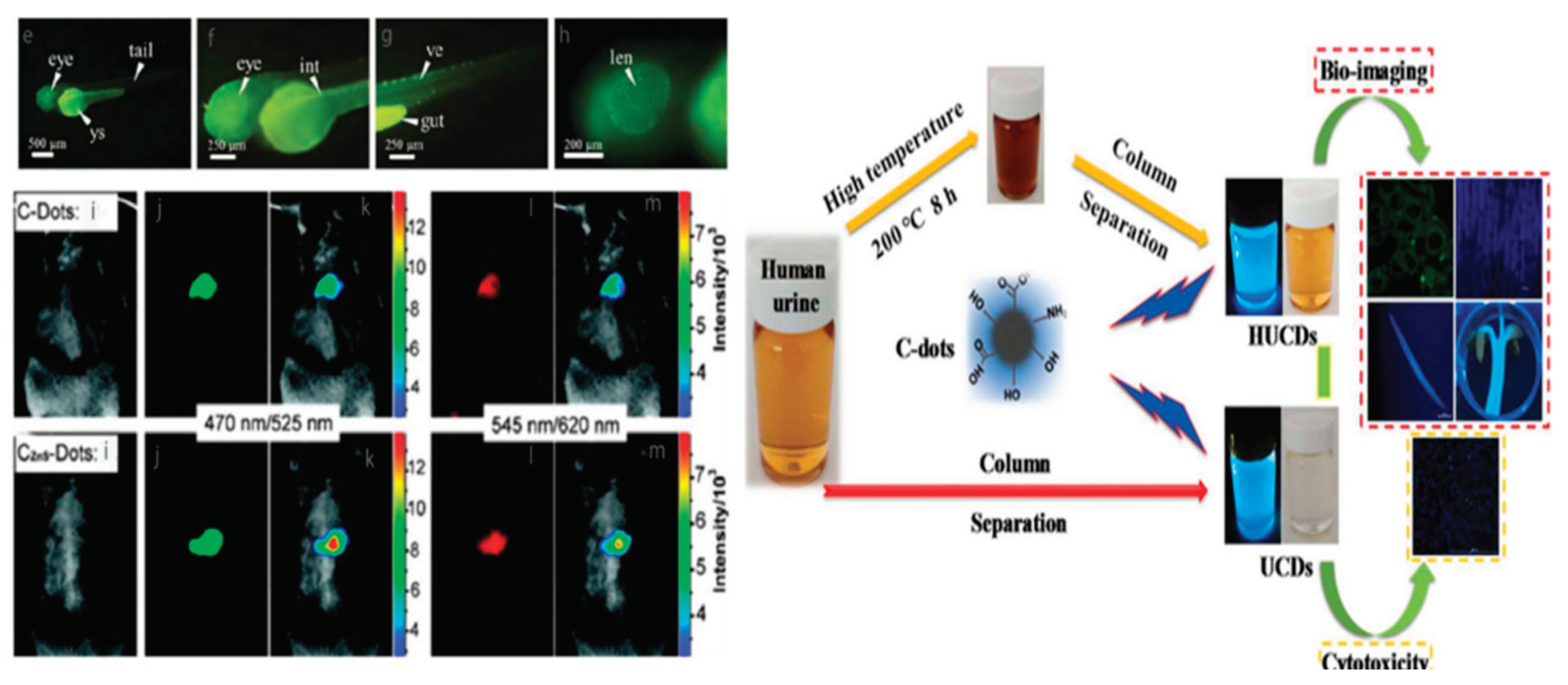
high chemical inertness and outstanding dispersibility in both organic and aqueous media. Coupled with these out-standing features, CDs have gained great attention in bioimaging research areas due to their facile synthesis, excellent biocompatibility and low toxicity. CDs for bioimaging applications use confocal microscopy or confocal laser scanning Microscopy (CLSM). The intensity of light emission or fluorescence absorbed by cells upon stimulation of red, blue and green wavelengths is measured by microscopy (CLSM) [71]. For example Zebrafish were used to study several important medical sciences, including illness progression, improved mechanism, and pattern formation because of their clearly defined developmental stages and optical imaging amenability. Zebrafish were chosen because of the fluorescent imaging model; CDs were discovered to accumulate preferentially in the yolk sac and eyes. CDs were kept intact for more than 60 hours, they are appropriate for studying the long-term stages of zebrafish development as illustrated in Figure 17(a). Zhang et al.also obtained urine-based CDs through the hydrothermal route and the sephadex filtration approach, as displayed in Figure 17(b). Various characterization revealed that both types of CDs comprised of oxygen and carbon directed the existence of many functional groups such as carboxylate, amino, hydroxy, and carbonyl. The cytotoxicity study revealed good applicability and biocompatibility in both in vivo and in vitro imaging.
Figure 17. (a) Fluorescence image of zebrafish with CDs (b) Graphical representation of how CDs are made from human urine and used in bioimaging [157]. Copyright 2022, beilstein-journals.org.
Preprints 102893 g017

4.5. Antibacterial Applications of CDs
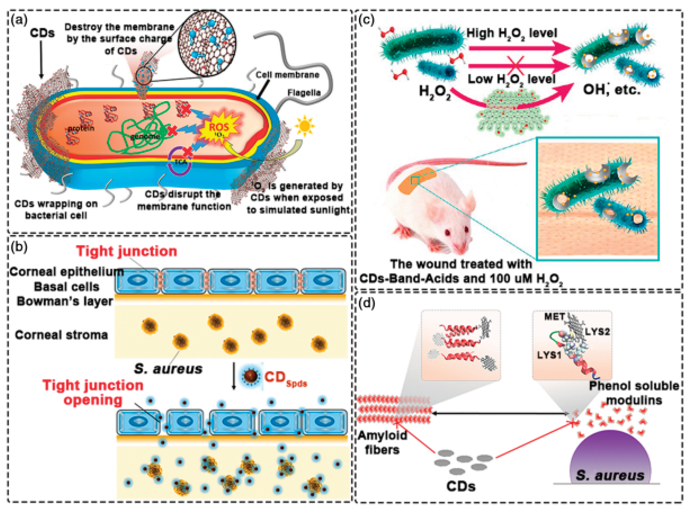
In recent years, CDs have also been explored for their antimicrobial activity, which refers to their ability to inhibit the growth of microorganisms such as bacteria, fungi, and viruses. The antimicrobial mechanism of CDs involves various physical and chemical interactions, which revealed as shown Figure 18(a). The surface of CDs can carry positive or negative charges depending on their surface functional groups. These charged surfaces can interact with the negatively charged cell membranes of microorganisms, leading to membrane disruption and cell death Figure 18(b). CDs can possess various chemical functional groups on their surface, such as carboxyl, amino, hydroxyl, or sulfhydryl groups. These functional groups can interact with microbial cells through chemical reactions, interfering with their vital processes and inhibiting their growth. CDs can interact with the cell membrane of microorganisms, causing disruption and destabilization. They can penetrate the lipid bilayer of the membrane, leading to leakage of intracellular contents and ultimately cell death. Reactive Oxygen Species (ROS) Generation: CDs can generate reactive oxygen species such as singlet oxygen, hydroxyl radicals, and superoxide ions when exposed to light or in the presence of oxygen. These ROS can induce oxidative stress in microorganisms, damaging their DNA, proteins, and lipids, and impairing their viability(seen in Figure 18 (c). Some CDs can exhibit metal ion chelation properties, which means they can bind to metal ions present in microorganisms. This can disrupt essential metal-dependent enzymatic processes in microorganisms, leading to their inactivation. It's important to note that the specific antimicrobial mechanism of CDs can vary depending on their size, surface chemistry, and the type of microorganism they are targeting. Additionally, the antimicrobial activity of CDs can be enhanced by functionalizing them with antimicrobial agents or coupling them with other materials to create hybrid nanocomposites Figure 18(d). Overall, the antimicrobial activity of carbon dots is a complex interplay of physical and chemical interactions, making them a promising candidate for various biomedical and environmental applications. However, further research is still needed to fully understand and optimize their antimicrobial properties.
Figure 18. (a) Antimicrobial activity mechanism of CDs through various physical and chemical interactions. (b) Antibacterial activity using the surface charge on CDs (c) Antibacterial activity using ROS produced by CDs (d) Antibacterial activity using the anti-biofilm properties of CDs[76] Copyright 2022, Wiley.
Preprints 102893 g018

4.6. Drug Delivery

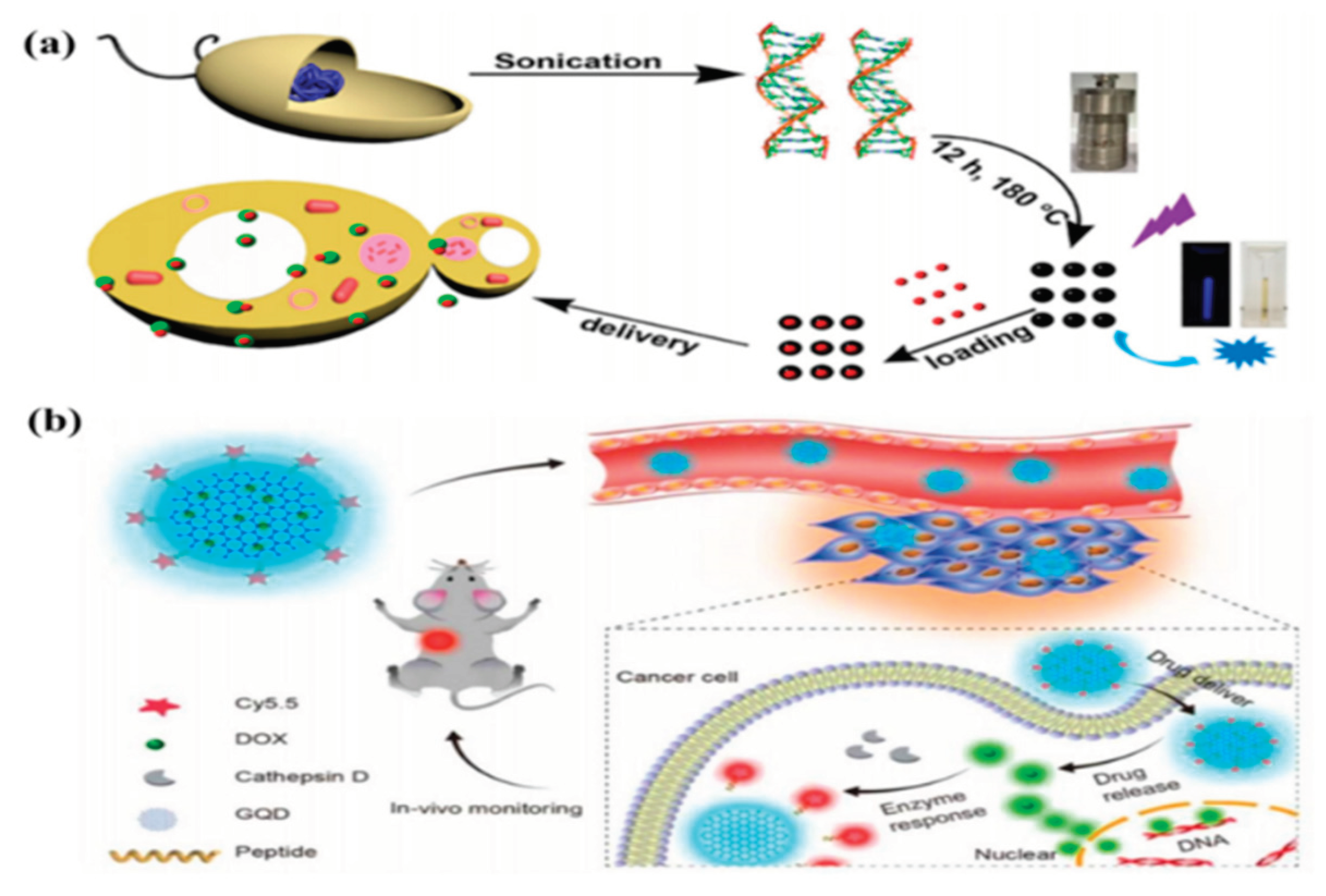
Because of their strong fluorescence, minimal toxicity, chemical inertness, and exceptional biocompatibility, CDs were thought to be multipurpose drug delivery devices. For therapeutic purposes, a certain medication was transported and removed at a specified location. Because of their better qualities, CDs have recently attracted attention in the medicine delivery space. The drug-loaded CDs aggregated and traveled to the cancer cells' nucleus and cytoplasm. Irradiation was used to release the medication at the designated locations. In order to examine medication delivery (seen in Figure 19 (a) and (b)), Ding and colleagues produced BCDs utilizing genomic DNA as a carbon source, as illustrated in Figure 8a. Additionally, due to their effectiveness, CDs with micro/nanopore architectures have drawn more interest when it comes to medication delivery. Micro/nanopore CDs were loaded with the common medication doxorubicin. By adjusting pH, the CD-DOX was liberated in cells. A DOX-loaded CD-based theranostic agent for medication administration was created by Ding et al. [167]. As seen in Figure 8b, the internalization of the nanoagent was monitored by the PL emission signal from CDs.
Figure 19. (a) Diagram showing the manufacture of DNA-BCD and its use in medication delivery (b) theranostic agent strategy based on CD for in vivo monitoring[158]. Copyright2020, Springer Nature.
Preprints 102893 g019

5. Challenges and Future Perspectives

Carbon dots have emerged as a promising class of nanomaterials in various fields due to their unique and remarkable properties[29,159]. While multiple methods exist for their synthesis, including physical, chemical, and biological approaches, priority should be given to the biological route for its cost-effectiveness and environmental friendliness[160,161]. Furthermore, biologically synthesized carbon dots exhibit biocompatibility, enhancing their potential application in the field of biomedicine, particularly in drug delivery[93,162]. In recent years, carbon dots have garnered significant attention for their potential applications in optoelectronics, bioimaging, sensing, energy storage, and catalysis[29,163]. However, there are still several challenges and future perspectives associated with carbon dots[50]. One such challenge is the synthesis techniques employed[164]. To enable their widespread use, it is crucial to develop efficient and scalable synthesis methods for carbon dots[50]. Although various techniques, such as hydrothermal/solvothermal methods, microwave-assisted synthesis, and pyrolysis, have been utilized, precise control over the size, shape, and surface chemistry of carbon dotsremains a necessity[86,165]. The diverse properties exhibited by carbon dots are contingent upon their synthesis conditions and surface functionalization. To ensure reproducibility and reliability, the standardization of synthesis protocols and characterization methods, including transmission electron microscopy (TEM), spectroscopy (UV-Vis, fluorescence), and elemental analysis, is crucial[166,167]. The photoluminescence mechanism of carbon dots is not yet fully understood. Their emission properties stem from the quantum confinement effect, surface states, and functional groups on their surface. Additional research is vital to elucidate the underlying mechanisms and optimize the photoluminescence properties for specific applications. Carbon dots often encounter stability issues, especially when exposed to environmental factors such as heat, light, and moisture. Enhancing the stability and photostability of carbon dots is essential for prolonged performance and practical applications. Although carbon dots exhibit promise for biomedical applications, such as bioimaging and drug delivery, a comprehensive investigation into their potential toxicity and biocompatibility is necessary to ensure their safe implementation in biological systems. To translate the potential of carbon dots into practical applications, their integration into devices and the scaling up of their production are imperative. This endeavor involves addressing challenges related to device fabrication, compatibility with existing technologies, and cost-effective large-scale synthesis. Regarding future perspectives, carbon dots hold tremendous potential for advanced applications, including light-emitting diodes (LEDs), solar cells, sensors, and catalysts. Continued research and development will facilitate the exploration of new applications and the optimization of carbon dots for specific functionalities.

6. Conclusions

The review from 2019 to 2023 significantly advanced our understanding of Carbon dots (CDs), an emerging nanomaterial. Research during this period led to developments in synthesis, characterizations, and applications of CDs. They find use in optoelectronics, bioimaging, sensing, antibacterial, biomedical, energy storage, and catalysis. CD synthesis saw progress through methods like hydrothermal, solvothermal, microwave-assisted, and electrochemical techniques. These allowed precise control over size, morphology, surface functionalization, and optical properties. Environmentally friendly routes using renewable precursors and non-toxic reagents increased their appeal.Characterization techniques played a crucial role in understanding CDs. Spectroscopic, microscopic, and analytical tools provided insights into structure, composition, surface chemistry, and photophysical properties. This knowledge guided synthesis optimization and application development. CDs showcased a wide range of applications. They excelled in optoelectronic devices like LEDs, photovoltaics, and displays due to their strong fluorescence, tunable emission, and high photostability. Future research aims for better control over CDs' properties and scalable synthesis. Understanding photophysical properties and mechanisms through spectroscopic and theoretical studies is crucial. Further research is needed to bridge the gap between lab-scale demonstrations and practical implementation. Integrating CDs into efficient devices like LEDs, solar cells, and displays is important. Exploring their potential in imaging, drug delivery, and theranostics can revolutionize medicine. Enhancing sensitivity, selectivity, and stability of CDs-based sensors for real-time monitoring is crucial. Optimizing CDs' performance and commercial viability in energy storage and catalysis is promising. Finnaly, the review highlights significant progress in CDs from 2019 to 2023. They show versatility and immense potential in various fields. With further research, CDs can become key components in next-gen technologies, advancing electronics, healthcare, environmental monitoring, and energy storage.

Author Contributions

HFE, AB, and FB D have equally contributed for the grand success of this article.

Funding

This research did not receive any specific grant from funding agencies in the public. This work is supported by the WSU.

Data Availability Statement

Unavailable due to its Review paper

Acknowledgments

The authors are gratitude Walter Sisulu University (WSU) for the support

Conflicts of Interest

The authors declare no conflicts of interest.

References

  1. Malik, S.; Muhammad, K.; Waheed, Y. Nanotechnology: A revolution in modern industry. Molecules 2023, 28, 661. [Google Scholar] [CrossRef]
  2. Bobokulova, M. THE ROLE OF NANOTECHNOLOGY IN MODERN PHYSICS. Development and innovations in science 2024, 3, 145–153. [Google Scholar]
  3. Asmatulu, E.; Andalib, M.N.; Subeshan, B.; Abedin, F. Impact of nanomaterials on human health: a review. Environ. Chem. Lett. 2022, 20, 2509–2529. [Google Scholar] [CrossRef]
  4. El-Kady, M.M.; Ansari, I.; Arora, C.; Rai, N.; Soni, S.; Verma, D.K.; Singh, P.; Mahmoud, A.E.D. Nanomaterials: A comprehensive review of applications, toxicity, impact, and fate to environment. J. Mol. Liq. 2023, 370, 121046. [Google Scholar] [CrossRef]
  5. Pokrajac, L.; Abbas, A.; Chrzanowski, W.; Dias, G.M.; Eggleton, B.J.; Maguire, S.; Maine, E.; Malloy, T.; Nathwani, J.; Nazar, L.; et al. Nanotechnology for a Sustainable Future: Addressing Global Challenges with the International Network4Sustainable Nanotechnology. ACS Nano 2022, 15, 18608–18623. [Google Scholar] [CrossRef]
  6. El-Shafey, A.M. Carbon dots: Discovery, structure, fluorescent properties, and applications. Green Processing and Synthesis 2021, 10, 134–156. [Google Scholar] [CrossRef]
  7. Kurian, M.; Paul, A. Recent trends in the use of green sources for carbon dot synthesis–A short review. Carbon Trends 2021, 3, 100032. [Google Scholar] [CrossRef]
  8. Das, S.; Ngashangva, L.; Goswami, P. Carbon Dots: An Emerging Smart Material for Analytical Applications. Micromachines 2021, 12, 84. [Google Scholar] [CrossRef]
  9. Chahal, S.; Macairan, J.-R.; Yousefi, N.; Tufenkji, N.; Naccache, R. Green synthesis of carbon dots and their applications. RSC Adv. 2021, 11, 25354–25363. [Google Scholar] [CrossRef]
  10. Mansuriya, B.D.; Altintas, Z. Carbon Dots: Classification, properties, synthesis, characterization, and applications in health care—An updated review (2018–2021). Nanomaterials 2021, 11, 2525. [Google Scholar] [CrossRef]
  11. Azam, N.; Ali, M.N.; Khan, T.J. Carbon Quantum Dots for Biomedical Applications: Review and Analysis. Front. Mater. 2021, 8, 272. [Google Scholar] [CrossRef]
  12. Nguyen, V.; et al. Double-pulse femtosecond laser ablation for synthesis of ultrasmall carbon nanodots. Materials Research Express 2020, 7, 015606. [Google Scholar] [CrossRef]
  13. Jin, J.; et al. Progress in the Application of Nanozymes Compositing Carbon Dots. Frontiers in Chemistry 2021, 9, 800. [Google Scholar] [CrossRef]
  14. Etefa, H.F.; Kumar, V.; Dejene, F.B.; Efa, M.T.; Jule, L.T. Modification of Flexible Electrodes for P-Type (Nickel Oxide) Dye-Sensitized Solar Cell Performance Based on the Cellulose Nanofiber Film. ACS Omega 2023, 8, 15249–15258. [Google Scholar] [CrossRef]
  15. Armano, A.; Buscarino, G.; Messina, F.; Sciortino, A.; Cannas, M.; Gelardi, F.M.; Giannazzo, F.; Schilirò, E.; Agnello, S. Dynamic modification of Fermi energy in single-layer graphene by photoinduced electron transfer from carbon dots. Nanomaterials 2020, 10, 528. [Google Scholar] [CrossRef]
  16. Hui, S. Carbon dots (CDs): basics, recent potential biomedical applications, challenges, and future perspectives. J. Nanoparticle Res. 2023, 25, 68. [Google Scholar] [CrossRef]
  17. Mkhari, O.; Ntuli, T.D.; Coville, N.J.; Nxumalo, E.N.; Maubane-Nkadimeng, M.S. Supported carbon-dots: A review. J. Lumin. 2023, 255. [Google Scholar] [CrossRef]
  18. Kaczmarek, A.; Hoffman, J.; Morgiel, J.; Mościcki, T.; Stobiński, L.; Szymański, Z.; Małolepszy, A. Luminescent carbon dots synthesized by the laser ablation of graphite in polyethylenimine and ethylenediamine. Materials 2021, 14, 729. [Google Scholar] [CrossRef]
  19. Wang, F.-T.; Wang, L.-N.; Xu, J.; Huang, K.-J.; Wu, X. Synthesis and modification of carbon dots for advanced biosensing application. Anal. 2021, 146, 4418–4435. [Google Scholar] [CrossRef]
  20. Rasal, A.S.; Dehvari, K.; Getachew, G.; Korupalli, C.; Ghule, A.V.; Chang, J.-Y. Efficient quantum dot-sensitized solar cells through sulfur-rich carbon nitride modified electrolytes. Nanoscale 2021, 13, 5730–5743. [Google Scholar] [CrossRef]
  21. Han, B.; Hu, X.; Zhang, X.; Huang, X.; An, M.; Chen, X.; Zhao, D.; Li, J. The fluorescence mechanism of carbon dots based on the separation and identification of small molecular fluorophores. RSC Adv. 2022, 12, 11640–11648. [Google Scholar] [CrossRef]
  22. Kim, A.; Dash, J.K.; Kumar, P.; Patel, R. Carbon-based quantum dots for photovoltaic devices: a review. ACS Appl. Electron. Mater. 2021, 4, 27–58. [Google Scholar] [CrossRef]
  23. Geleta, T.A.; Imae, T. Nanocomposite photoanodes consisting of p-NiO/n-ZnO heterojunction and carbon quantum dot additive for dye-sensitized solar cells. ACS Applied Nano Materials 2021, 4, 236–249. [Google Scholar] [CrossRef]
  24. Kumar, V.; et al. Hybrid 1D Semiconducting ZnO and GaN Nanostructures for Light-Emitting Devices. 1D Semiconducting Hybrid Nanostructures: Synthesis and Applications in Gas Sensing and Optoelectronics 2023, 205–216. [Google Scholar] [CrossRef]
  25. Kumar, V.; et al. Mechanism, properties and applications of phosphors. In Phosphor Handbook; Elsevier, 2023; pp. 33–45. [Google Scholar] [CrossRef]
  26. Kim, A.; Dash, J.K.; Kumar, P.; Patel, R. Carbon-Based Quantum Dots for Photovoltaic Devices: A Review. ACS Appl. Electron. Mater. 2021, 4, 27–58. [Google Scholar] [CrossRef]
  27. Das, S.; Ngashangva, L.; Goswami, P. Carbon Dots: An Emerging Smart Material for Analytical Applications. Micromachines 2021, 12, 84, Note: MDPI stays neutral with regard to jurisdictional claims in published ….. [Google Scholar] [CrossRef]
  28. Abbasi, B.A.; Iqbal, J.; Khan, Z.; Ahmad, R.; Uddin, S.; Shahbaz, A.; Zahra, S.A.; Shaukat, M.; Kiran, F.; Kanwal, S.; et al. Phytofabrication of cobalt oxide nanoparticles from Rhamnus virgata leaves extract and investigation of different bioactivities. Microsc. Res. Tech. 2021, 84, 192–201. [Google Scholar] [CrossRef]
  29. Liu, J.; Li, R.; Yang, B. Carbon dots: a new type of carbon-based nanomaterial with wide applications. ACS Central Sci. 2020, 6, 2179–2195. [Google Scholar] [CrossRef]
  30. Zeng, Q.; Feng, T.; Tao, S.; Zhu, S.; Yang, B. Precursor-dependent structural diversity in luminescent carbonized polymer dots (CPDs): the nomenclature. Light. Sci. Appl. 2021, 10, 1–13. [Google Scholar] [CrossRef]
  31. Zhao, B.; Tan, Z.A. Fluorescent Carbon Dots: Fantastic Electroluminescent Materials for Light-Emitting Diodes. Adv. Sci. 2021, 8, 2001977. [Google Scholar] [CrossRef]
  32. Cai, H.; Abbas, K.; Yang, Y.; Li, Z.; Bi, H. The application of carbon dots in tumor immunotherapy: Researches and prospects. Appl. Res. 2023, 2, e202300001. [Google Scholar] [CrossRef]
  33. Li, S.; Li, L.; Tu, H.; Zhang, H.; Silvester, D.S.; Banks, C.E.; Zou, G.; Hou, H.; Ji, X. The development of carbon dots: From the perspective of materials chemistry. Mater. Today 2021, 51, 188–207. [Google Scholar] [CrossRef]
  34. Pandey, S.; Karakoti, M.; Bhardwaj, D.; Tatrari, G.; Sharma, R.; Pandey, L.; Lee, M.-J.; Sahoo, N.G. Recent advances in carbon-based materials for high-performance perovskite solar cells: gaps, challenges and fulfillment. Nanoscale Adv. 2023, 5, 1492–1526. [Google Scholar] [CrossRef]
  35. Ratre, P.; Nazeer, N.; Kumari, R.; Thareja, S.; Jain, B.; Tiwari, R.; Kamthan, A.; Srivastava, R.K.; Mishra, P.K. Carbon-Based Fluorescent Nano-Biosensors for the Detection of Cell-Free Circulating MicroRNAs. Biosensors 2023, 13, 226. [Google Scholar] [CrossRef]
  36. Bartolomei, B.; Dosso, J.; Prato, M. New trends in nonconventional carbon dot synthesis. Trends Chem. 2021, 3, 943–953. [Google Scholar] [CrossRef]
  37. Lin, H.K.; et al. Synthesis of nanomaterials using bottom-up methods. Advanced Nanomaterials and Their Applications in Renewable Energy 2022, 61–110. [Google Scholar] [CrossRef]
  38. Jorns, M.; Pappas, D. A review of fluorescent carbon dots, their synthesis, physical and chemical characteristics, and applications. Nanomaterials 2021, 11, 1448. [Google Scholar] [CrossRef]
  39. Saleem, M.; Naz, M.Y.; Shukrullah, S.; Shujah, M.A.; Akhtar, M.; Ullah, S.; Ali, S. One-pot sonochemical preparation of carbon dots, influence of process parameters and potential applications: a review. Carbon Lett. 2021, 32, 39–55. [Google Scholar] [CrossRef]
  40. Bian, Z.; et al. Properties of Carbon Dots versus Small Molecules from “Bottom-up” Synthesis. ACS nano 2023, 17, 22788–22799. [Google Scholar] [CrossRef]
  41. Modi, P.D.; et al. Bottom-up approaches for the preparation of carbon dots. In Carbon Dots in Analytical Chemistry; Elsevier, 2023; pp. 15–29. [Google Scholar] [CrossRef]
  42. Ahmed, H.B.; El-Shahat, M.; Allayeh, A.K.; Emam, H.E. Maillard reaction for nucleation of polymer quantum dots from chitosan-glucose conjugate: Antagonistic for cancer and viral diseases. Int. J. Biol. Macromol. 2023, 224, 858–870. [Google Scholar] [CrossRef]
  43. Bazaka, K.; Jacob, M.V.; Ostrikov, K. Sustainable life cycles of natural-precursor-derived nanocarbons. Chem. Rev. 2016, 116, 163–214. [Google Scholar] [CrossRef]
  44. Shi, L.; Wang, B.; Lu, S. Efficient bottom-up synthesis of graphene quantum dots at an atomically precise level. Matter 2023, 6, 728–760. [Google Scholar] [CrossRef]
  45. Kumar, N.; Salehiyan, R.; Chauke, V.; Botlhoko, O.J.; Setshedi, K.; Scriba, M.; Masukume, M.; Ray, S.S. Top-down synthesis of graphene: A comprehensive review. FlatChem 2021, 27, 100224. [Google Scholar] [CrossRef]
  46. Chen, L.; Wang, C.; Liu, C.; Chen, S. Facile Access to Fabricate Carbon Dots and Perspective of Large-Scale Applications. Small 2023, 19, e2206671. [Google Scholar] [CrossRef]
  47. Crista, D.M.; Esteves da Silva, J.C.; Pinto da Silva, L. Evaluation of different bottom-up routes for the fabrication of carbon dots. Nanomaterials 2020, 10, 1316. [Google Scholar] [CrossRef]
  48. Nagarajan, D.; Gangadharan, D.; Venkatanarasimhan, S. Synthetic strategies toward developing carbon dots via top-down approach. In Carbon Dots in Analytical Chemistry; Elsevier, 2023; pp. 1–13. [Google Scholar] [CrossRef]
  49. Alafeef, M.; Srivastava, I.; Aditya, T.; Pan, D. Carbon Dots: From Synthesis to Unraveling the Fluorescence Mechanism. Small 2023, 20, e2303937. [Google Scholar] [CrossRef]
  50. Wareing, T.C.; Gentile, P.; Phan, A.N. Biomass-based carbon dots: current development and future perspectives. ACS Nano 2021, 15, 15471–15501. [Google Scholar] [CrossRef]
  51. Namdari, P.; Negahdari, B.; Eatemadi, A. Synthesis, properties and biomedical applications of carbon-based quantum dots: An updated review. Biomed. Pharmacother. 2017, 87, 209–222. [Google Scholar] [CrossRef]
  52. Yuan, F.; Li, S.; Fan, Z.; Meng, X.; Fan, L.; Yang, S. Shining carbon dots: Synthesis and biomedical and optoelectronic applications. Nano Today 2016, 11, 565–586. [Google Scholar] [CrossRef]
  53. Ryu, J.; Bok, T.; Kim, S.; Park, S. Fundamental understanding of nanostructured Si electrodes: preparation and characterization. Chemnanomat 2018, 4, 319–337. [Google Scholar] [CrossRef]
  54. Sharma, A.; Das, J.J.J.o.n. Small molecules derived carbon dots: synthesis and applications in sensing, catalysis, imaging, and biomedicine. 2019, 17, 1–24. [Google Scholar] [CrossRef]
  55. Lin, X.; Xiong, M.; Zhang, J.; He, C.; Ma, X.; Zhang, H.; Kuang, Y.; Yang, M.; Huang, Q. Carbon dots based on natural resources: Synthesis and applications in sensors. Microchem. J. 2021, 160, 105604. [Google Scholar] [CrossRef]
  56. Behi, M.; Gholami, L.; Naficy, S.; Palomba, S.; Dehghani, F. Carbon dots: a novel platform for biomedical applications. Nanoscale Adv. 2022, 4, 353–376. [Google Scholar] [CrossRef] [PubMed]
  57. Kumar, R.; Singh, D.P.; Muñoz, R.; Amami, M.; Singh, R.K.; Singh, S.; Kumar, V. Graphene-based materials for biotechnological and biomedical applications: Drug delivery, bioimaging and biosensing. Mater. Today Chem. 2023, 33, 101750. [Google Scholar] [CrossRef]
  58. Ozyurt, D.; Al Kobaisi, M.; Hocking, R.K.; Fox, B. Properties, Synthesis, and Applications of Carbon Dots: A Review. Carbon Trends 2023, 12. [Google Scholar] [CrossRef]
  59. Ensminger, D.; Bond, L.J. Ultrasonics: fundamentals, technologies, and applications; CRC press, 2024. [Google Scholar]
  60. Jing, H.H.; et al. Green carbon dots: Synthesis, characterization, properties and biomedical applications. Journal of Functional Biomaterials 2023, 14, 27. [Google Scholar] [CrossRef] [PubMed]
  61. Khayal, A.; Dawane, V.; Amin, M.A.; Tirth, V.; Yadav, V.K.; Algahtani, A.; Khan, S.H.; Islam, S.; Yadav, K.K.; Jeon, B.-H. Advances in the methods for the synthesis of carbon dots and their emerging applications. Polymers 2021, 13, 3190. [Google Scholar] [CrossRef] [PubMed]
  62. Li, X.; et al. Preparation of carbon dots-metal nanoparticles nanocomposites and their application in heterogeneous catalysis. Nano Futures 2023, 7, 042001. [Google Scholar] [CrossRef]
  63. Moradi, M.; Molaei, R.; Kousheh, S.A.; Guimarães, J.T.; McClements, D.J. Carbon dots synthesized from microorganisms and food by-products: active and smart food packaging applications. Crit. Rev. Food Sci. Nutr. 2023, 63, 1943–1959. [Google Scholar] [CrossRef] [PubMed]
  64. Zou, L.; Luo, J.; Xu, W.; Zhai, Y.; Li, J.; Qu, T.; Fu, G. Experimental study on influence of particle shape on shockwave from collapse of cavitation bubble. Ultrason. Sonochemistry 2023, 101, 106693. [Google Scholar] [CrossRef]
  65. Cui, L.; et al. Carbon dots: Synthesis, properties and applications. Nanomaterials 2021, 11, 3419. [Google Scholar] [CrossRef]
  66. Han, Y.; Liccardo, L.; Moretti, E.; Zhao, H.; Vomiero, A. Synthesis, optical properties and applications of red/near-infrared carbon dots. J. Mater. Chem. C 2022, 10, 11827–11847. [Google Scholar] [CrossRef]
  67. Huo, F.; Karmaker, P.G.; Liu, Y.; Zhao, B.; Yang, X. Preparation and biomedical applications of multicolor carbon dots: recent advances and future challenges. Part. Part. Syst. Charact. 2020, 37. [Google Scholar] [CrossRef]
  68. Pundi, A.; Chang, C.-J. Recent advances in synthesis, modification, characterization, and applications of carbon dots. Polymers 2022, 14, 2153. [Google Scholar] [CrossRef] [PubMed]
  69. Ansari, L.; Hallaj, S.; Hallaj, T.; Amjadi, M. Doped-carbon dots: Recent advances in their biosensing, bioimaging and therapy applications. Colloids Surfaces B: Biointerfaces 2021, 203, 111743. [Google Scholar] [CrossRef] [PubMed]
  70. Jiang, Z.; et al. Progress in laser ablation and biological synthesis processes:“Top-Down” and “Bottom-Up” approaches for the green synthesis of Au/Ag nanoparticles. 2022, 23, 14658. [Google Scholar] [CrossRef] [PubMed]
  71. Kaczmarek, A.; Hoffman, J.; Morgiel, J.; Mościcki, T.; Stobiński, L.; Szymański, Z.; Małolepszy, A. Luminescent carbon dots synthesized by the laser ablation of graphite in polyethylenimine and ethylenediamine. Materials 2021, 14, 729. [Google Scholar] [CrossRef] [PubMed]
  72. Khairol Anuar, N.K.; et al. A review on multifunctional carbon-dots synthesized from biomass waste: Design/fabrication, characterization and applications. 2021, 9, 67. [Google Scholar] [CrossRef]
  73. Jumardin, J.; Maddu, A.; Santoso, K.; Isnaeni, I. Synthesis of Fluorescent Carbon Dots (CDs) Using Laser Ablation Method for Bioimaging Application. J. ILMU Fis. | Univ. Andalas 2023, 15, 91–105. [Google Scholar] [CrossRef]
  74. Ahirwar, S.; Mallick, S.; Bahadur, D. Electrochemical method to prepare graphene quantum dots and graphene oxide quantum dots. ACS Omega 2017, 2, 8343–8353. [Google Scholar] [CrossRef]
  75. Chaudhary, M. Colloidal nanocrystals for optoelectronic devices optically controlled at the nanometric scale; Université Paris sciences et lettres, 2022. [Google Scholar]
  76. Wang, B.; Cai, H.; Waterhouse, G.I.N.; Qu, X.; Yang, B.; Lu, S. Carbon dots in bioimaging, biosensing and therapeutics: A comprehensive review. Small Sci. 2022, 2. [Google Scholar] [CrossRef]
  77. Saha, A.; Bhattacharjee, L.; Bhattacharjee, R.R. Synthesis of carbon quantum dots, in Carbon Quantum Dots for Sustainable Energy and Optoelectronics; Elsevier, 2023; pp. 39–54. [Google Scholar]
  78. Kurian, M.; Paul, A. Recent trends in the use of green sources for carbon dot synthesis–A short review. Carbon Trends 2021, 3, 100032. [Google Scholar] [CrossRef]
  79. Paul, A.; Mohan, A. Photoluminescent Carbon Dots: A New Generation Nanocarbon Material. In Handbook of Porous Carbon Materials; Springer, 2023; pp. 231–256. [Google Scholar] [CrossRef]
  80. Pandey, V.K.; Tripathi, A.; Taufeeq, A.; Dar, A.H.; Samrot, A.V.; Rustagi, S.; Malik, S.; Bhattacharya, T.; Kovacs, B.; Shaikh, A.M. Significance and applications of carbon dots in anti cancerous nanodrug conjugate development: A review. Appl. Surf. Sci. Adv. 2024, 19, 100550. [Google Scholar] [CrossRef]
  81. Wang, X.; Wei, Y.; Zhou, H.; Liu, Q.; Zhu, L. Synthesis of graphene nanosheets by the electrical explosion of graphite powder confined in a tube. Ceram. Int. 2021, 47, 21934–21942. [Google Scholar] [CrossRef]
  82. Rigodanza, F.; Burian, M.; Arcudi, F.; Đorđević, L.; Amenitsch, H.; Prato, M. Snapshots into carbon dots formation through a combined spectroscopic approach. Nat. Commun. 2021, 12, 2640. [Google Scholar] [CrossRef]
  83. Ding, H.; Li, X.-H.; Chen, X.-B.; Wei, J.-S.; Li, X.-B.; Xiong, H.-M. Surface states of carbon dots and their influences on luminescence. J. Appl. Phys. 2020, 127, 231101. [Google Scholar] [CrossRef]
  84. Ullal, N.; Mehta, R.; Sunil, D. Separation and purification of fluorescent carbon dots – an unmet challenge. Anal. 2024, 149, 1680–1700. [Google Scholar] [CrossRef] [PubMed]
  85. de Andres, F.; Rios, A. Carbon dots–separative techniques: tools-objective towards green analytical nanometrology focused on bioanalysis. Microchemical Journal 2021, 161, 105773. [Google Scholar] [CrossRef]
  86. Liu, H.; Zhong, X.; Pan, Q.; Zhang, Y.; Deng, W.; Zou, G.; Hou, H.; Ji, X. A review of carbon dots in synthesis strategy. Co-ord. Chem. Rev. 2024, 498, 215468. [Google Scholar] [CrossRef]
  87. Naik, V.M.; Bhosale, S.V.; Kolekar, G.B. A brief review on the synthesis, characterisation and analytical applications of nitrogen doped carbon dots. Anal. Methods 2022, 14, 877–891. [Google Scholar] [CrossRef]
  88. Chao-Mujica, F.J.; Garcia-Hernández, L.; Camacho-López, S.; Camacho-López, M.A.; Contreras, D.R.; Pérez-Rodríguez, A.; Peña-Caravaca, J.P.; Darias-Gonzalez, J.G.; Hernandez-Tabares, L.; de Fuentes, O.A.; et al. Carbon quantum dots by submerged arc discharge in water: Synthesis, characterization, and mechanism of formation. J. Appl. Phys. 2021, 129, 163301. [Google Scholar] [CrossRef]
  89. Rasal, A.S.; Yadav, S.; Yadav, A.; Kashale, A.A.; Manjunatha, S.T.; Altaee, A.; Chang, J.-Y. Carbon quantum dots for energy applications: A review. ACS Appl. Nano Mater. 2021, 4, 6515–6541. [Google Scholar] [CrossRef]
  90. Gutiérrez-Cruz, A.; Ruiz-Hernández, A.R.; Vega-Clemente, J.F.; Luna-Gazcón, D.G.; Campos-Delgado, J. A review of top-down and bottom-up synthesis methods for the production of graphene, graphene oxide and reduced graphene oxide. J. Mater. Sci. 2022, 57, 14543–14578. [Google Scholar] [CrossRef]
  91. Manoj, B. Synthesis of Coal-based Nanocarbon Advances and Applications. Pure and Functionalized Carbon Based Nanomaterials 2020, 52–79. [Google Scholar]
  92. Fatima, T.; Mushtaq, A. Efficacy and challenges of carbon-based nanomaterials in water treatment: A review. Int. J. Chem. Biochem. Sci. 2023, 23, 232–248. [Google Scholar]
  93. Ge, G.; et al. Carbon dots: Synthesis, properties and biomedical applications. Journal of Materials Chemistry B 2021, 9, 6553–6575. [Google Scholar] [CrossRef] [PubMed]
  94. Chahal, S.; Macairan, J.-R.; Yousefi, N.; Tufenkji, N.; Naccache, R. Green synthesis of carbon dots and their applications. RSC Adv. 2021, 11, 25354–25363. [Google Scholar] [CrossRef] [PubMed]
  95. Wang, R.; Lu, K.-Q.; Tang, Z.-R.; Xu, Y.-J. Recent progress in carbon quantum dots: synthesis, properties and applications in photocatalysis. J. Mater. Chem. A 2017, 5, 3717–3734. [Google Scholar] [CrossRef]
  96. Li, W.; Liu, Y.; Wang, B.; Song, H.; Liu, Z.; Lu, S.; Yang, B. Kilogram-scale synthesis of carbon quantum dots for hydrogen evolution, sensing and bioimaging. Chin. Chem. Lett. 2019, 30, 2323–2327. [Google Scholar] [CrossRef]
  97. Qu, S.; Zhou, D.; Li, D.; Ji, W.; Jing, P.; Han, D.; Liu, L.; Zeng, H.; Shen, D. Toward efficient orange emissive carbon nanodots through conjugated sp2-domain controlling and surface charges engineering. Adv. Mater. 2016, 28, 3516–3521. [Google Scholar] [CrossRef]
  98. Chen, X.; Wu, J.; Zhang, J.; Zhang, Z. Synthesis of trichromatic carbon dots from a single precursor by solvent effect and its versatile applications. Arab. J. Chem. 2023, 16, 104576. [Google Scholar] [CrossRef]
  99. Chen, X.; Wu, J.; Zhang, J.; Zhang, Z. Synthesis of trichromatic carbon dots from a single precursor by solvent effect and its versatile applications. Arab. J. Chem. 2023, 16, 104576. [Google Scholar] [CrossRef]
  100. Sarwer, A.; Hamed, S.M.; Osman, A.I.; Jamil, F.; Al-Muhtaseb, A.H.; Alhajeri, N.S.; Rooney, D.W. Algal biomass valorization for biofuel production and carbon sequestration: a review. Environ. Chem. Lett. 2022, 20, 2797–2851. [Google Scholar] [CrossRef]
  101. Velvizhi, G.; Goswami, C.; Shetti, N.P.; Ahmad, E.; Pant, K.K.; Aminabhavi, T.M. Valorisation of lignocellulosic biomass to value-added products: Paving the pathway towards low-carbon footprint. Fuel 2022, 313, 122678. [Google Scholar] [CrossRef]
  102. Tiwari, S.K.; Bystrzejewski, M.; De Adhikari, A.; Huczko, A.; Wang, N. Methods for the conversion of biomass waste into value-added carbon nanomaterials: Recent progress and applications. Prog. Energy Combust. Sci. 2022, 92, 101023. [Google Scholar] [CrossRef]
  103. Khalifa, S.A.M.; Elashal, M.H.; Yosri, N.; Du, M.; Musharraf, S.G.; Nahar, L.; Sarker, S.D.; Guo, Z.; Cao, W.; Zou, X.; et al. Bee pollen: Current status and therapeutic potential. Nutrients 2021, 13, 1876. [Google Scholar] [CrossRef] [PubMed]
  104. Anpalagan, K.K. Carbon quantum dots for bioimaging cells and tissues; Victoria University, 2023. [Google Scholar]
  105. Kaur, K.; et al. Biomedical Applications of Biogenic Carbon-Based Fluorescent Nanoparticles. Biogenic Nanomaterial for Health and Environment 2024, 183–201. [Google Scholar]
  106. Meng, W.; Bai, X.; Wang, B.; Liu, Z.; Lu, S.; Yang, B. Biomass-derived carbon dots and their applications. Energy Environ. Mater. 2019, 2, 172–192. [Google Scholar] [CrossRef]
  107. Madhavikutti, A.S.; et al. Advances in the synthesis approaches of carbon and graphene quantum dots. In Zero-Dimensional Carbon Nanomaterials; Elsevier, 2024; pp. 17–59. [Google Scholar]
  108. Ghasemlou, M.; Pn, N.; Alexander, K.; Zavabeti, A.; Sherrell, P.C.; Ivanova, E.P.; Adhikari, B.; Naebe, M.; Bhargava, S.K. Fluorescent Nanocarbons: From Synthesis and Structure to Cancer Imaging and Therapy. Adv. Mater. 2024, e2312474. [Google Scholar] [CrossRef] [PubMed]
  109. Sun, L.; Zhang, R.; Zhang, T.; Liu, X.; Zhao, Y.; Yang, M.; Cheng, H.; Zhang, Q.; Zhang, Y.; Wu, X.; et al. Synthesis, applications and biosafety evaluation of carbon dots derived from herbal medicine. Biomed. Mater. 2023, 18, 042004. [Google Scholar] [CrossRef]
  110. Pagidi, S.; et al. One-Pot Synthesis of Deep Blue Hydrophobic Carbon Dots with Room Temperature Phosphorescence, White Light Emission, and Explosive Sensor. Advanced Electronic Materials 2022, 8, 2100969. [Google Scholar] [CrossRef]
  111. Das, M.; Thakkar, H.; Patel, D.; Thakore, S. Repurposing the domestic organic waste into green emissive carbon dots and carbonized adsorbent: A sustainable zero waste process for metal sensing and dye sequestration. J. Environ. Chem. Eng. 2021, 9, 106312. [Google Scholar] [CrossRef]
  112. Mansi, M.; Gaurav, S. Synthesis and applications of carbon dots from waste biomass. In Carbon Dots in Analytical Chemistry; Elsevier, 2023; pp. 319–328. [Google Scholar] [CrossRef]
  113. Choi, Y.; et al. Simple microwave-assisted synthesis of amphiphilic carbon quantum dots from A3/B2 polyamidation monomer set. 2017, 9, 27883–27893. [Google Scholar] [CrossRef] [PubMed]
  114. Rai, S.; Singh, B.K.; Bhartiya, P.; Singh, A.; Kumar, H.; Dutta, P.; Mehrotra, G. Lignin derived reduced fluorescence carbon dots with theranostic approaches: Nano-drug-carrier and bioimaging. J. Lumin- 2017, 190, 492–503. [Google Scholar] [CrossRef]
  115. Xu, X.; Ray, R.; Gu, Y.; Ploehn, H.J.; Gearheart, L.; Raker, K.; Scrivens, W.A. Electrophoretic analysis and purification of fluorescent single-walled carbon nanotube fragments. J. Am. Chem. Soc. 2004, 126, 12736–12737. [Google Scholar] [CrossRef]
  116. Speight, J.G. Environmental Organic Chemistry for Engineers; Butterworth-Heinemann, 2016. [Google Scholar]
  117. Khayal, A.; Dawane, V.; Amin, M.A.; Tirth, V.; Yadav, V.K.; Algahtani, A.; Khan, S.H.; Islam, S.; Yadav, K.K.; Jeon, B.-H. Advances in the methods for the synthesis of carbon dots and their emerging applications. Polymers 2021, 13, 3190. [Google Scholar] [CrossRef] [PubMed]
  118. Liu, J.; Li, L.; Niu, W.; Wang, N.; Zhao, D.; Zeng, S.; Chen, S. A Hydrogen-Bonded Organic-Framework-Derived Mesoporous N-Doped Carbon for Efficient Electroreduction of Oxygen. ChemElectroChem 2016, 3, 1116–1123. [Google Scholar] [CrossRef]
  119. Xu, M.; Huang, Q.; Sun, R.; Wang, X. Simultaneously obtaining fluorescent carbon dots and porous active carbon for supercapacitors from biomass. RSC Adv. 2016, 6, 88674–88682. [Google Scholar] [CrossRef]
  120. Paul, S.; Banerjee, S.L.; Khamrai, M.; Samanta, S.; Singh, S.; Kundu, P.P.; Ghosh, A.K. Hydrothermal synthesis of gelatin quantum dots for high-performance biological imaging applications. J. Photochem. Photobiol. B: Biol. 2020, 212, 112014. [Google Scholar] [CrossRef] [PubMed]
  121. Chai, Y.; Feng, Y.; Zhang, K.; Li, J. Preparation of fluorescent carbon dots composites and their potential applications in biomedicine and drug delivery—A review. Pharmaceutics 2022, 14, 2482. [Google Scholar] [CrossRef]
  122. Harish, V.; Ansari, M.; Tewari, D.; Yadav, A.B.; Sharma, N.; Bawarig, S.; García-Betancourt, M.-L.; Karatutlu, A.; Bechelany, M.; Barhoum, A. Cutting-edge advances in tailoring size, shape, and functionality of nanoparticles and nanostructures: A review. J. Taiwan Inst. Chem. Eng. 2023, 149. [Google Scholar] [CrossRef]
  123. Khan, M.E.; Mohammad, A.; Yoon, T. State-of-the-art developments in carbon quantum dots (CQDs): Photo-catalysis, bio-imaging, and bio-sensing applications. Chemosphere 2022, 302, 134815. [Google Scholar] [CrossRef] [PubMed]
  124. Jumardin, J.; Maddu, A.; Santoso, K.; Isnaeni, I. Synthesis of Fluorescent Carbon Dots (CDs) Using Laser Ablation Method for Bioimaging Application. J. ILMU Fis. | Univ. Andalas 2023, 15, 91–105. [Google Scholar] [CrossRef]
  125. Mandal, S.; Das, P. Are carbon dots worth the tremendous attention it is getting: Challenges and opportunities. Appl. Mater. Today 2021, 26, 101331. [Google Scholar] [CrossRef]
  126. Liu, Y.; Roy, S.; Sarkar, S.; Xu, J.; Zhao, Y.; Zhang, J. A review of carbon dots and their composite materials for electrochemical energy technologies. Carbon Energy 2021, 3, 795–826. [Google Scholar] [CrossRef]
  127. Du, Y.; Guo, S. Chemically doped fluorescent carbon and graphene quantum dots for bioimaging, sensor, catalytic and photoelectronic applications. Nanoscale 2016, 8, 2532–2543. [Google Scholar] [CrossRef]
  128. Palma, V.; Barba, D.; Cortese, M.; Martino, M.; Renda, S.; Meloni, E. Microwaves and heterogeneous catalysis: A review on selected catalytic processes. Catalysts 2020, 10, 246. [Google Scholar] [CrossRef]
  129. Cavalloro, V.; et al. Microwave-assisted solid extraction from natural matrices, in Microwave Heating-Electromagnetic Fields Causing Thermal and Non-Thermal Effects; IntechOpen, 2021. [Google Scholar]
  130. Feng, T.; Tao, S.; Yue, D.; Zeng, Q.; Chen, W.; Yang, B. Recent advances in energy conversion applications of carbon dots: from optoelectronic devices to electrocatalysis. Small 2020, 16, e2001295. [Google Scholar] [CrossRef]
  131. Dolai, S.; Bhunia, S.K.; Rajendran, S.; UshaVipinachandran, V.; Ray, S.C.; Kluson, P. Tunable fluorescent carbon dots: synthesis progress, fluorescence origin, selective and sensitive volatile organic compounds detection. Crit. Rev. Solid State Mater. Sci. 2021, 46, 349–370. [Google Scholar] [CrossRef]
  132. Sharma, A.; Das, J. Small molecules derived carbon dots: synthesis and applications in sensing, catalysis, imaging, and biomedicine. J. Nanobiotechnol. 2019, 17, 92. [Google Scholar] [CrossRef]
  133. Li, J.; Li, P.; Wang, D.; Dong, C. One-pot synthesis of aqueous soluble and organic soluble carbon dots and their multi-functional applications. Talanta 2019, 202, 375–383. [Google Scholar] [CrossRef]
  134. Qin, Z.; Wang, W.; Zhan, X.; Du, X.; Zhang, Q.; Zhang, R.; Li, K.; Li, J.; Xu, W. One-pot synthesis of dual carbon dots using only an N and S co-existed dopant for fluorescence detection of Ag+. Spectrochim. Acta Part A: Mol. Biomol. Spectrosc. 2019, 208, 162–171. [Google Scholar] [CrossRef] [PubMed]
  135. Alafeef, M.; Srivastava, I.; Aditya, T.; Pan, D. Carbon dots: from synthesis to unraveling the fluorescence mechanism. Small 2023, 20, e2303937. [Google Scholar] [CrossRef] [PubMed]
  136. Sanmartín-Matalobos, J.; Bermejo-Barrera, P.; Aboal-Somoza, M.; Fondo, M.; García-Deibe, A.M.; Corredoira-Vázquez, J.; Alves-Iglesias, Y. Semiconductor Quantum Dots as Target Analytes: Properties, Surface Chemistry and Detection. Nanomaterials 2022, 12, 2501. [Google Scholar] [CrossRef] [PubMed]
  137. Langer, M.; Paloncýová, M.; Medveď, M.; Pykal, M.; Nachtigallová, D.; Shi, B.; Aquino, A.J.; Lischka, H.; Otyepka, M. Progress and challenges in understanding of photoluminescence properties of carbon dots based on theoretical computations. Appl. Mater. Today 2021, 22, 100924. [Google Scholar] [CrossRef]
  138. Semeniuk, M.; Yi, Z.; Poursorkhabi, V.; Tjong, J.; Jaffer, S.; Lu, Z.-H.; Sain, M. Future perspectives and review on organic carbon dots in electronic applications. ACS Nano 2019, 13, 6224–6255. [Google Scholar] [CrossRef] [PubMed]
  139. Zhang, Q.; Wang, R.; Feng, B.; Zhong, X.; Ostrikov, K. Photoluminescence mechanism of carbon dots: triggering high-color-purity red fluorescence emission through edge amino protonation. Nat. Commun. 2021, 12, 6856. [Google Scholar] [CrossRef] [PubMed]
  140. Yang, M.; et al. Reversible “off–on” fluorescence of Zn2+-passivated carbon dots: mechanism and potential for the detection of EDTA and Zn2+. 2018, 34, 7767–7775. [Google Scholar] [CrossRef] [PubMed]
  141. Liu, J.; Li, R.; Yang, B. Carbon dots: A new type of carbon-based nanomaterial with wide applications. ACS Central Sci. 2020, 6, 2179–2195. [Google Scholar] [CrossRef]
  142. Farshbaf, M.; Davaran, S.; Rahimi, F.; Annabi, N.; Salehi, R.; Akbarzadeh, A. Carbon quantum dots: recent progresses on synthesis, surface modification and applications. Artif. Cells Nanomed. Biotechnol. 2018, 46, 1331–1348. [Google Scholar] [CrossRef]
  143. Jelinek, R.J.C.q.d.S.I.P., Cham, Carbon quantum dots. 2017: p. 29-46.
  144. Zhang, Q.; Sun, X.; Ruan, H.; Yin, K.; Li, H. Production of yellow-emitting carbon quantum dots from fullerene carbon soot. Sci. China Mater. 2017, 60, 141–150. [Google Scholar] [CrossRef]
  145. Ding, H.; Zhou, X.-X.; Zhang, Z.-H.; Zhao, Y.-P.; Wei, J.-S.; Xiong, H.-M. Large scale synthesis of full-color emissive carbon dots from a single carbon source by a solvent-free method. Nano Res. 2022, 15, 3548–3555. [Google Scholar] [CrossRef]
  146. Kumar, P.; Dua, S.; Kaur, R.; Kumar, M.; Bhatt, G. A review on advancements in carbon quantum dots and their application in photovoltaics. RSC Adv. 2022, 12, 4714–4759. [Google Scholar] [CrossRef] [PubMed]
  147. Liu, H.; Xie, Z.; Zheng, M. Unprecedented chiral nanovaccines for significantly enhanced cancer immunotherapy. ACS Appl. Mater. Interfaces 2022, 14, 39858–39865. [Google Scholar] [CrossRef] [PubMed]
  148. Efa, M.T.; Imae, T. Effects of carbon dots on ZnO nanoparticle-based dye-sensitized solar cells. Electrochimica Acta 2019, 303, 204–210. [Google Scholar] [CrossRef]
  149. Etefa, H.F.; Imae, T.; Yanagida, M. Enhanced photosensitization by carbon dots Co-adsorbing with dye on ptype semiconductor (Nickel Oxide) solar cells. ACS Appl. Mater. Interfaces 2020, 12, 18596–18608. [Google Scholar] [CrossRef] [PubMed]
  150. Chu, K.-W.; Lee, S.L.; Chang, C.-J.; Liu, L. Recent progress of carbon dot precursors and photocatalysis applications. Polymers 2019, 11, 689. [Google Scholar] [CrossRef] [PubMed]
  151. Yao, Y.; Zhang, H.; Hu, K.; Nie, G.; Yang, Y.; Wang, Y.; Duan, X.; Wang, S. Carbon dots based photocatalysis for environmental applications. J. Environ. Chem. Eng. 2022, 10, 107336. [Google Scholar] [CrossRef]
  152. Li, T.-X.; Zhao, D.-F.; Li, L.; Meng, Y.; Xie, Y.-H.; Feng, D.; Wu, F.; Xie, D.; Liu, Y.; Mei, Y. Unraveling fluorescent mechanism of biomass-sourced carbon dots based on three major components: Cellulose, lignin, and protein. Bioresour. Technol. 2024, 394, 130268. [Google Scholar] [CrossRef] [PubMed]
  153. Zhang, L.; Lyu, S.; Zhang, Q.; Chmely, S.C.; Wu, Y.; Melcher, C.; Rajan, K.; Harper, D.P.; Wang, S.; Chen, Z. Recycling hot-water extractions of lignocellulosic biomass in bio-refinery for synthesis of carbon nanoparticles with amplified luminescence and its application in temperature sensing. Ind. Crop. Prod. 2020, 145, 112066. [Google Scholar] [CrossRef]
  154. Azara, A.; Belbessai, S.; Abatzoglou, N. A review of filamentous carbon nanomaterial synthesis via catalytic conversion of waste plastic pyrolysis products. J. Environ. Chem. Eng. 2022, 10, 107049. [Google Scholar] [CrossRef]
  155. Shaku, B. Synthesis and Application of Boron-nitrogen Doped Carbon Nanoonions in Supercapacitors; University of the Witwatersrand, Faculty of Science, School of Chemistry, 2020. [Google Scholar]
  156. Kang, C.; Huang, Y.; Yang, H.; Yan, X.F.; Chen, Z.P. A review of carbon dots produced from biomass wastes. Nanomaterials 2020, 10, 2316. [Google Scholar] [CrossRef] [PubMed]
  157. Kanwal, A.; et al. Recent advances in green carbon dots (2015–2022): synthesis, metal ion sensing, and biological applications. Beilstein Journal of Nanotechnology 2022, 13, 1068–1107. [Google Scholar] [CrossRef] [PubMed]
  158. Zhao, C.; Song, X.; Liu, Y.; Fu, Y.; Ye, L.; Wang, N.; Wang, F.; Li, L.; Mohammadniaei, M.; Zhang, M.; et al. Synthesis of graphene quantum dots and their applications in drug delivery. J. Nanobiotechnology 2020, 18, 1–32. [Google Scholar] [CrossRef] [PubMed]
  159. Liu, Y.; et al. Advances in carbon dots: from the perspective of traditional quantum dots. Materials Chemistry Frontiers 2020, 4, 1586–1613. [Google Scholar] [CrossRef]
  160. Abdullahi, S.S.a.; et al. Natural, synthetic, and composite materials for industrial effluents treatment: a mini review on current practices, cost-effectiveness, and sustainability. Case Studies in Chemical and Environmental Engineering 2023, 100570. [Google Scholar] [CrossRef]
  161. Gupta, D.; Boora, A.; Thakur, A.; Gupta, T.K. Green and sustainable synthesis of nanomaterials: Recent advancements and limitations. Environ. Res. 2023, 231, 116316. [Google Scholar] [CrossRef] [PubMed]
  162. Nair, A.; Haponiuk, J.T.; Thomas, S.; Gopi, S. Natural carbon-based quantum dots and their applications in drug delivery: A review. Biomed. Pharmacother. 2020, 132, 110834–110834. [Google Scholar] [CrossRef] [PubMed]
  163. Kumar, Y.R.; Deshmukh, K.; Sadasivuni, K.K.; Pasha, S.K.K. Graphene quantum dot based materials for sensing, bio-imaging and energy storage applications: a review. RSC Adv. 2020, 10, 23861–23898. [Google Scholar] [CrossRef] [PubMed]
  164. Baig, N.; Kammakakam, I.; Falath, W. Nanomaterials: a review of synthesis methods, properties, recent progress, and challenges. Mater. Adv. 2021, 2, 1821–1871. [Google Scholar] [CrossRef]
  165. He, C.; Xu, P.; Zhang, X.; Long, W. The synthetic strategies, photoluminescence mechanisms and promising applications of carbon dots: Current state and future perspective. Carbon 2022, 186, 91–127. [Google Scholar] [CrossRef]
  166. Al Ragib, A.; Al Amin, A.; Alanazi, Y.M.; Kormoker, T.; Uddin, M.; Siddique, A.B.; Barai, H.R. Multifunctional carbon dots in nanomaterial surface modification: a descriptive review. Carbon Res. 2023, 2, 37. [Google Scholar] [CrossRef]
  167. Chen, J.; Luo, J.B.; Hu, M.Y.; Zhou, J.; Huang, C.Z.; Liu, H. Controlled synthesis of multicolor carbon dots assisted by machine learning. Adv. Funct. Mater. 2022, 33, 2210095. [Google Scholar] [CrossRef]
Table 1. Different approaches in synthesizing CDs advantages, disadvantages and Applications.
Methods Advantages disadvantages Applications Ref.
Hydrothermal Simple and cost-effective synthesis method High reaction temperatures and long reaction times Biomedical imaging, sensors, drug delivery [120,121]
Arc discharge High yield of CDs with good photoluminescence properties Complex setup and requirement of a high-power arc discharge Photovoltaics, optoelectronic devices [50,122]
Laser ablation Precise control over size and excellent photoluminescence Expensive laser equipment and potential contamination Bioimaging, photocatalysis [123,124]
Chemical oxidation Simple and versatile method Harsh reaction conditions and potential toxic byproducts Sensing, energy storage [125,126]
Pyrolysis Good stability and tunable properties High temperatures and lack of control over size distribution Photodetection, bioimaging [56,127]
Microwave Short reaction times and easy control of parameters Potential overheating and non-uniform heating Drug delivery, environmental remediation [128,129]
Solvothermal Controlled synthesis in a solvent medium Requirement of specialized equipment and solvents Optoelectronic devices, catalysis [130,131]
Organic approach Environmentally friendly synthesis Limited control over size and properties Sensing, bioimaging [76,132]
ultrasonication Simple and rapid synthesis method Limited control over size and potential aggregation Biosensing, antibacterial agents [61,76]
One-Pot Synthesis Convenient synthesis without additional purification steps Potential variability in size and properties Bioimaging, drug delivery [133,134]
Disclaimer/Publisher’s Note: The statements, opinions and data contained in all publications are solely those of the individual author(s) and contributor(s) and not of MDPI and/or the editor(s). MDPI and/or the editor(s) disclaim responsibility for any injury to people or property resulting from any ideas, methods, instructions or products referred to in the content.
Copyright: This open access article is published under a Creative Commons CC BY 4.0 license, which permit the free download, distribution, and reuse, provided that the author and preprint are cited in any reuse.
Prerpints.org logo

Preprints.org is a free preprint server supported by MDPI in Basel, Switzerland.

Subscribe

© 2024 MDPI (Basel, Switzerland) unless otherwise stated