
Preprint
Case Report
Restoring Severely Atrophic Edentulous Ridge of Mandible Using Self-Expanding Tissue Expander – A Case Report
Altmetrics
Downloads
96
Views
40
Comments
0
A peer-reviewed article of this preprint also exists.

This version is not peer-reviewed
Submitted:
07 April 2024
Posted:
08 April 2024
You are already at the latest version
Alerts
Abstract
Introduction
The placement of implants is often hindered by a severely atrophic mandibular edentulous ridge. A sufficient volume of bone graft is crucial for successful implant restoration. The required bone graft volume directly correlates with the deficiency present in the edentulous ridge. If the residual crestal bone height is so low that the distance between the crestal ridge and the inferior alveolar nerve is less than 2 mm, a significant bone augmentation procedure is necessary. This procedure must ensure the implants are surrounded by a sufficient margin of bone, aiming for a final height of more than 8 mm and a width of more than 7 mm. Achieving primary closure with adequate soft tissue is vital for the success of the bone graft procedure. The key to successful bone grafting lies in the effective management of the soft tissue covering.
Case Report
This case report details the treatment of a patient with severely atrophic edentulous ridges on both sides, addressed using Self Inflating Tissue Expanders (SITEs) to facilitate bone grafting and implant placement. Following soft tissue expansion, bone grafting and implant placement were successfully performed. The implants supported a fixed restoration, which remained stable and complication-free over a seven-year observation period.
Conclusion
The SITE (Self-Inflating Tissue Expander) proved effective in obtaining a substantial amount of bone graft needed for implant installation. A seven-year follow-up confirmed the implants were well-functioning with stable bone support, healthy soft tissue coverage, and adequate vestibular depth.
Keywords:
Subject: Medicine and Pharmacology - Dentistry and Oral Surgery
1. Introduction
Bone grafting is a pivotal step in implant restoration, especially when there is insufficient bone quantity [1]. The objectives of bone grafting are threefold: 1) To increase bone volume to accommodate the implant's minimum size initially, 2) To ensure a bony structure that supports biological width for long-term stability, and 3) To meet esthetic requirements by restoring the balance between living tissue and prosthetic teeth [2].
The desired bone volume for grafting is meticulously planned based on specific objectives. To achieve these goals, a range of bone grafting techniques have been developed, taking into account several crucial aspects:
- Safety: Prioritizing the reduction of surgical risks and complications.
- Efficacy: Focusing on maximizing the graft volume and improving the augmentation of living tissue.
- Longevity: Guaranteeing the graft's stability over time, preventing any degradation.
- Efficiency: Simplifying the procedure to save time and effort for both the practitioner and patient.
- Patient Comfort: Striving to minimize pain and discomfort during and after the procedure.
- Surgical Sites: Adapting the grafting approach to suit the specific anatomical location.
- Graft Materials: Choosing the most suitable materials for the graft based on the clinical requirements and outcomes desired.
These considerations are essential for the development and selection of bone grafting techniques, ensuring that each approach is tailored to meet the patient's needs while adhering to basic surgical principles [3].
To achieve these objectives, a variety of techniques are available. These include the Ridge Split technique, Guided Bone Regeneration (GBR) technique, Lateral Sinus Lift and Graft, Transcrestal Approach, Tunnel Technique, Titanium Mesh Technique, Block Bone Graft Technique, Alveolar distraction osteogenesis as well as Simultaneous Grafting with Implant Placement, and Staged Grafting prior to Implant Placement. Additional methods vary according to the graft material used.
It is crucial to employ the most predictable and efficient surgical technique that ensures safety and meets the patient's needs among the many available options.
Regardless of the chosen bone graft technique, it is imperative to increase soft tissue coverage to achieve tension-free primary closure of the surgical site. This is vital to prevent wound dehiscence, which is a significant risk factor for graft failure or compromised results [4].
Methods to expand soft tissue prior to bone grafting can significantly enhance safety and efficacy. Osmed Company has developed the Self-Inflating Tissue Expander (SITE) [5,6], designed for use in various surgical applications, including plastic, ophthalmic, and oral surgery. Utilizing the fundamental principles of bone grafting, SITE offers added benefits to the bone graft procedure, particularly in severely atrophic cases [7]. The author has applied SITE in the bone grafting of severely atrophic mandibular edentulous ridges, with the goal of achieving fixed restoration using implants. The outcomes have been positive, evidenced by a successful follow-up period of 7 years. This case is now presented for review.
2. Case report
In April 2015, a 65-year-old female patient sought care at Purpose Driven Dental Clinic, reporting discomfort with her old upper and lower removable dentures. Examination revealed severely atrophic upper and lower edentulous ridges, save for three remaining teeth (right upper canine, right upper incisor, and left lower canine). Panoramic X-ray imaging indicated that the residual crestal bone height in the first molar area above the upper border of the inferior mandibular canal was 2.2 mm on the right side and 4.0 mm on the left side. The patient expressed her desire for replacing the edentulous areas with fixed prostheses using dental implants.
While dental implant placement was feasible in some upper posterior molar crestal areas without notable challenges, the lower posterior regions lacked sufficient crestal bone for implantation without significant bone augmentation.
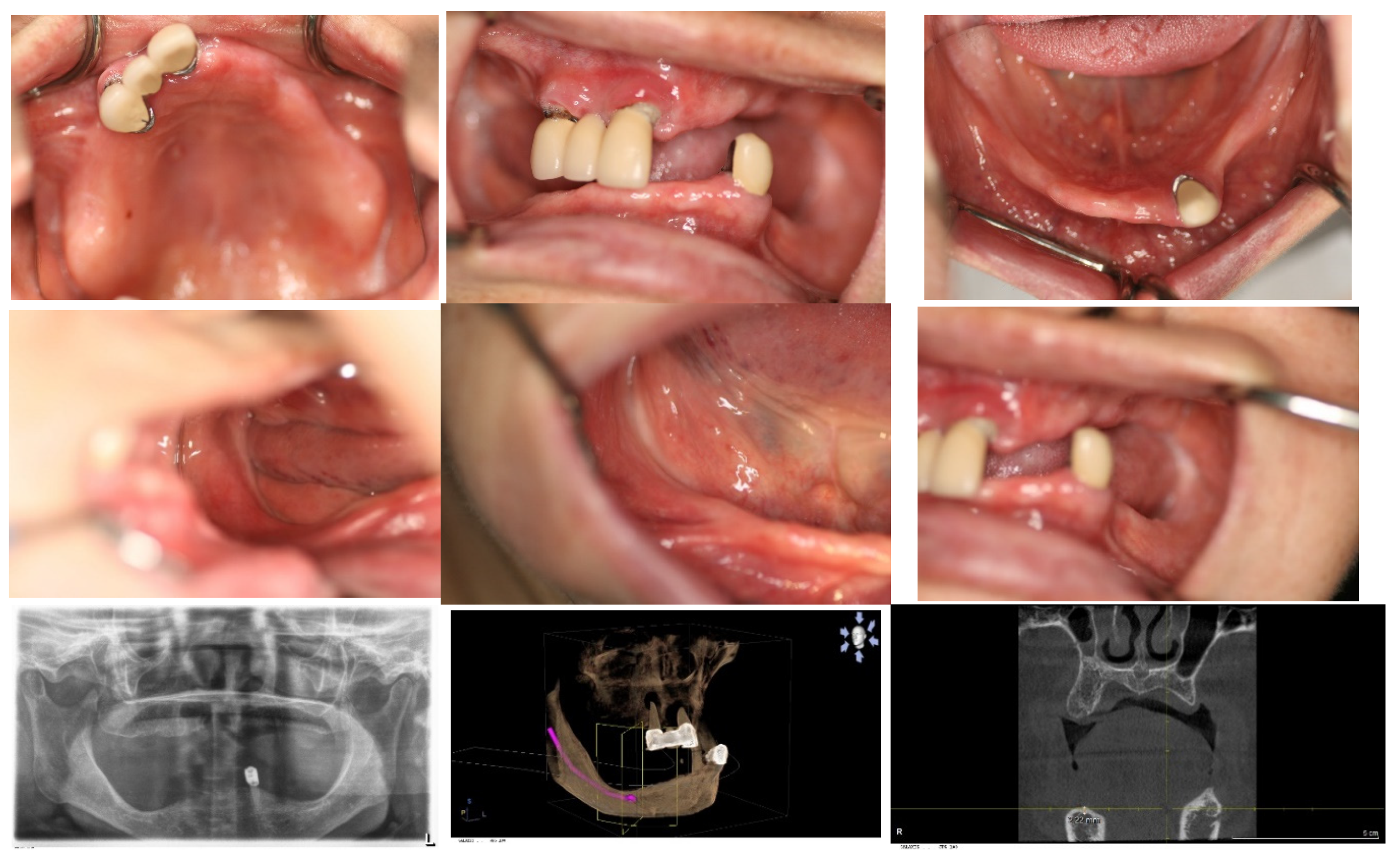
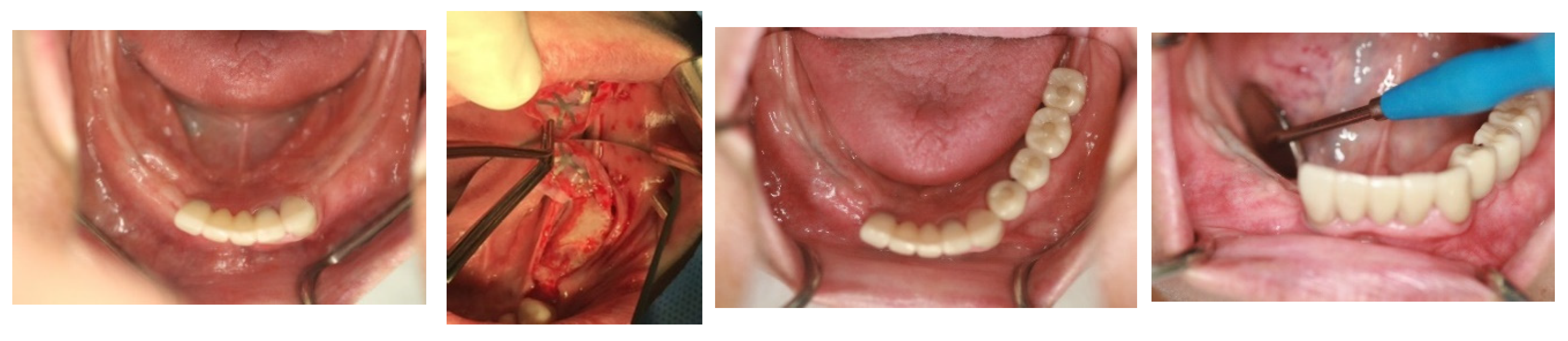
Figure 1.
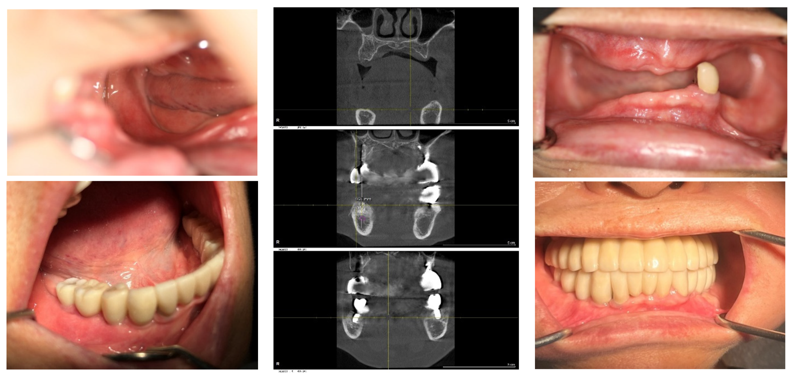
The initial intraoral photos showed that the upper arch had only two teeth remaining, both with root caries and periapical abscesses, while the other teeth were missing. The lower arch displayed severely atrophic mandibular edentulous ridges, with only the left canine remaining. Please note that the residual crest of the right edentulous ridge is flush with the level of the mouth floor.
Figure 1.
The initial intraoral photos showed that the upper arch had only two teeth remaining, both with root caries and periapical abscesses, while the other teeth were missing. The lower arch displayed severely atrophic mandibular edentulous ridges, with only the left canine remaining. Please note that the residual crest of the right edentulous ridge is flush with the level of the mouth floor.
Given this requirement, a preparatory bone augmentation procedure was determined to be necessary. However, the soft tissue covering the crestal bone was too scant for effective vertical bone augmentation. Addressing this issue, a Soft Tissue Expansion procedure was undertaken prior to the bone augmentation using a Self-Inflating Tissue Expander (SITE) (Osmed.com).
Figure 2.
On 28th May 2015, a small incision was made in the right posterior edentulous site of the mandible, through which a Self-Inflating Tissue Expander (SITE) was inserted into a subperiosteal pouch and subsequently sutured.
Figure 2.
On 28th May 2015, a small incision was made in the right posterior edentulous site of the mandible, through which a Self-Inflating Tissue Expander (SITE) was inserted into a subperiosteal pouch and subsequently sutured.
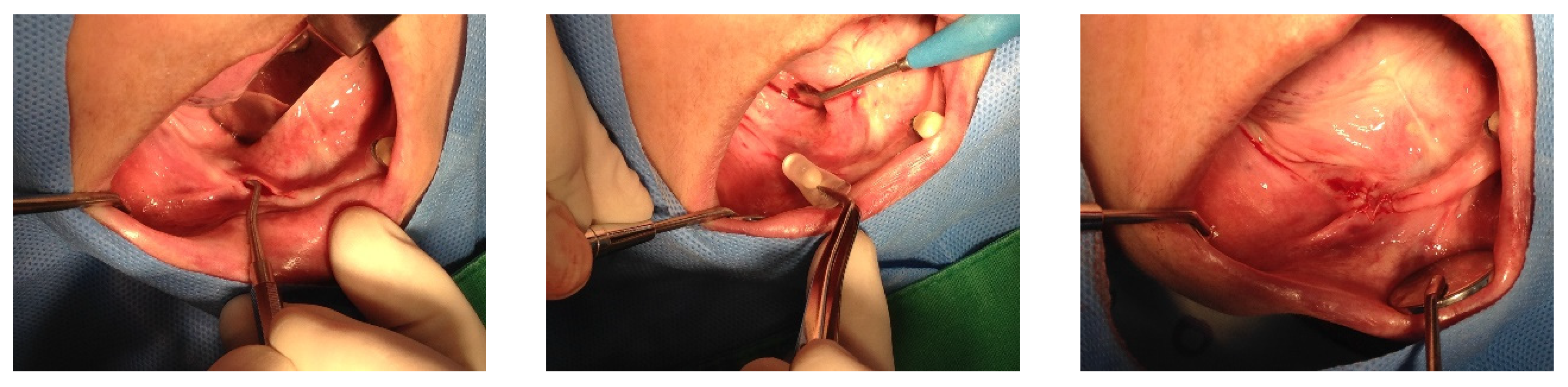
On May 28, 2015, a 1.3 ml SITE (Self-Inflating Tissue Expander, manufactured by Osmed in Germany) device was inserted into the right edentulous ridge through a small pouch created by a 4 mm stab incision. Subsequently, on June 2, 2015, a similar procedure was performed on the left edentulous ridge using the same technique and a 1.3 ml SITE device (manufactured by Osmed in Germany).
On June 2, 2015, wound dehiscence occurred at the insertion site of the SITE on the right side, necessitating the removal of the SITE. Consequently, a new 0.7 ml SITE was inserted on September 9, 2015.
Figure 3.
On 1st June 2015, the same procedure was performed at the left posterior edentulous site of the mandible. Please note the use of a before-and-after template for predicting the final result.
Figure 3.
On 1st June 2015, the same procedure was performed at the left posterior edentulous site of the mandible. Please note the use of a before-and-after template for predicting the final result.
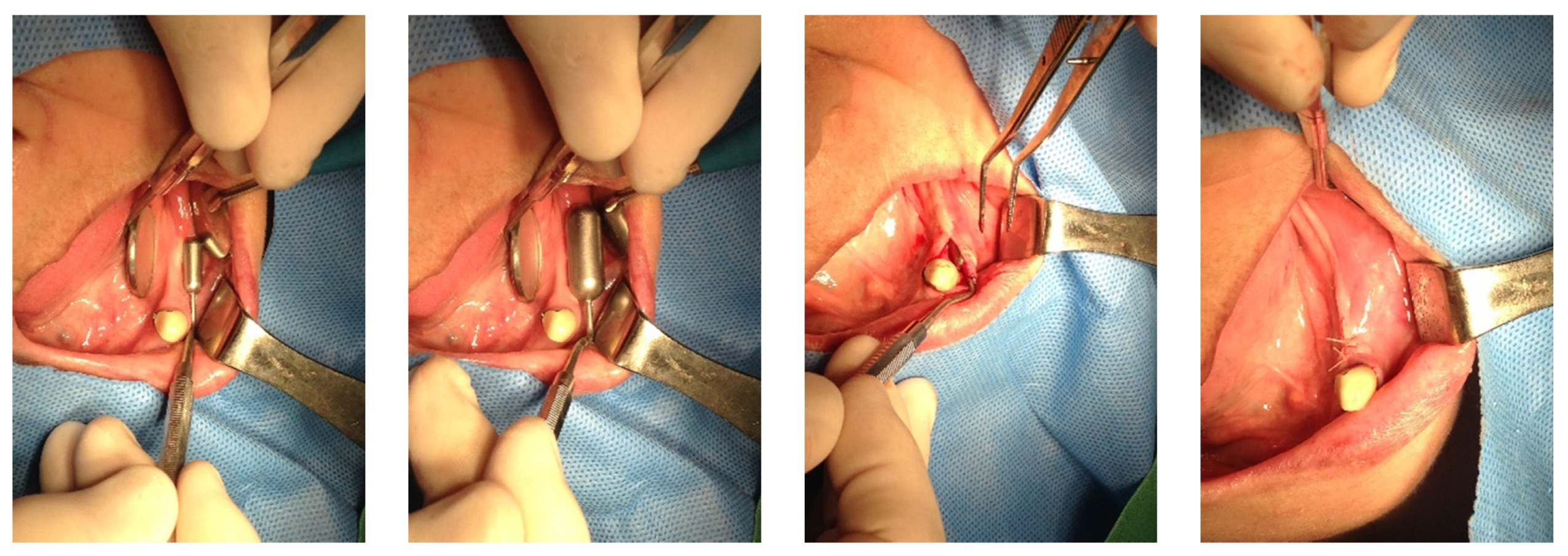
The SITEs were removed after achieving full expansion, in adherence to the manufacturer's recommendations—50 days later on the left side and 37 days later on the right side. The vertical bone augmentation procedures were conducted simultaneously. Xenograft material (Bio-Oss, produced by Geistlich in Switzerland) was utilized for the augmentation and covered with a Goretex titanium-reinforced membrane (made in the USA) through the same small openings used for the SITEs removal. The incisions were sutured with 4-0 Monocryl, and the stitches were removed two weeks later, revealing no significant wound dehiscence.
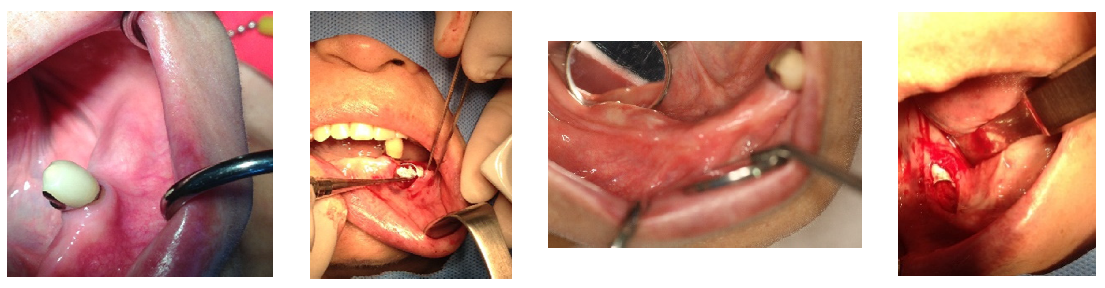
Figure 4.
Bone graft procedures were performed when the SITEs were removed after their full expansion. The GBR technique was used with xenograft (Bio-Oss large granule) and Gore-Tex TR membrane. The same small incisions used for removing SITEs on both sides were sufficient for GBR, thanks to the prior soft tissue expansion. The procedures were carried out on different dates for each side, with the left side being treated on August 25, 2015, and the right side on October 16, 2015.
On June 3, 2015, in the lower anterior area, implants were placed at the sites of both lateral incisors, measuring 3.6 x 10 mm and 3.6 x 11 mm respectively, using Oneplant Warrentec implants manufactured in Korea. Concurrently, a bone graft was performed using the guided bone regeneration (GBR) technique, involving a collagen membrane from ACE Surgical Supply Co., USA, and a xenograft from Bio-Oss, Geistlich, Switzerland, though without the use of a Self-Inflating Tissue Expander (SITE). Importantly, the restoration of this area was completed prior to initiating the implant surgery in the posterior areas.
Following an 8-month period for bone consolidation, implants were then placed in the left lower posterior edentulous areas on April 18, 2016. A 4.5 x 9 mm OsseoSpeed TX implant from Astratec, Germany, was used for the first premolar, and 4.3 x 7 mm and 4.8 x 8 mm Oneplant implants from Warrentec, Korea, were used for the second premolar and the first molar, respectively. Additionally, a 4.0 x 6.0 mm OsseoSpeed TX implant, also from Astratec, Germany, was placed.
Seven months following the first bone graft, performed on the day the SITE was removed from the right posterior area of the mandible, 5 x 9 mm OsseoSpeed TX implants (Astratec, manufactured in Germany) were placed for the first premolar and the canine. However, the procedure to place implants in the molar area was deferred; instead, a second bone graft utilizing the GBR technique was conducted to augment the bone volume, ensuring it could accommodate implants of the appropriate size.
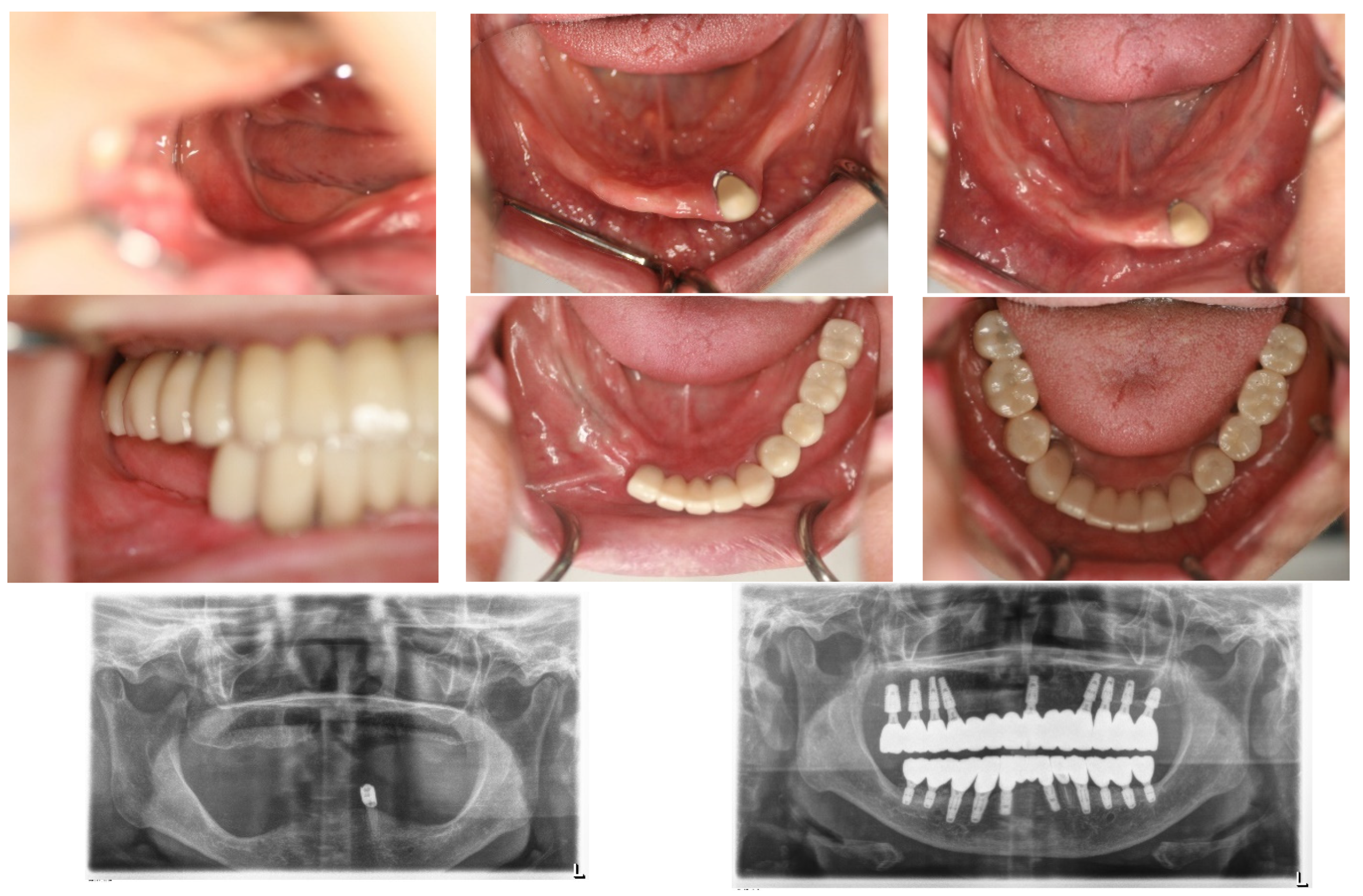
Figure 5.
After an 8-month waiting period for bone consolidation, implants were placed in the left posterior area following the removal of the Gore-Tex membrane. It is noteworthy to mention the well-matured bone achieved from the bone graft procedure. Photographs documenting this progression include one from the left side on April 1, 2016, the day the left natural canine crown was set; April 18, 2016, the day of the operation on the left posterior area of the mandible; and two images from the right side on October 26, 2016, the day of implant placements for the right second premolar and the right first molar, 6 months after the second bone graft.
In the upper jaw, implants (Oneplant Warrentec, manufactured in Korea) were inserted using standard techniques, selecting the most advantageous sites for implant placement for the patient's convenience. The final fixed prosthodontics consisted of five pieces: an anterior piece spanning from the right premolar to the left premolar, both second premolars, and a two-unit posterior molar bridge on each side. All implant prostheses were cemented with ready-made stock abutments extraorally and secured by screw tightening, completing all procedures on January 27, 2016.
Figure 6.
The fixed restorations with implants for the upper arch were completed more simply than those for the lower arch because the existing bone had sufficient quantity to support the implants. The prostheses were divided into five pieces: one anterior bridge, two premolar single crowns, and two bridges for the molar areas. To simplify the procedure and facilitate ease, the most favorable sites were selected for anterior implant placement. Please note the path for inserting the anterior implants, which includes one path extraorally after cementation.
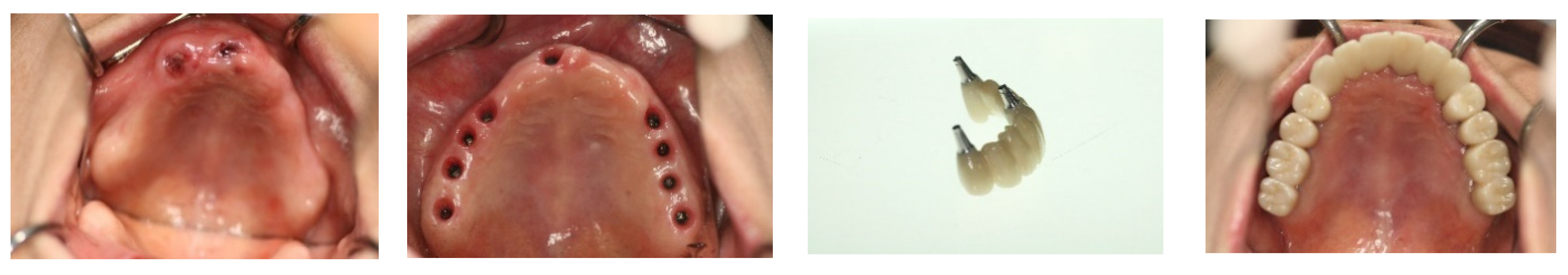
The implant restorations for the lower left posterior area were finalized on September 28, 2016, five months following the implant placements. Subsequently, the restorations for the lower right canine and first premolar were completed on December 23, 2016, seven months post-implantation. The final restorations for the lower right first molar and second molar were accomplished on May 2, 2017, six months after their implantation. This marked the conclusion of all fixed prosthesis work, spanning a total of 23 months from the beginning to the end of the treatment process.
Figure 7.
On May 4, 2017, the fixed restorations with implants for the lower arch were completed, 23 months after the commencement of the procedure, through vertical bone grafting via SITE. The completion on the right side was delayed compared to the left side due to the need for more bone regeneration, although both sides underwent the same procedure. Please compare the before-and-after treatment pictures.
Throughout all procedures, the patient tolerated the treatments well, experiencing no surgical complications such as nerve injury or wound infection. She used temporary removable dentures; however, during the critical postoperative period, she was advised against wearing them to avoid surgical complications from potential trauma. The patient's chewing ability was effectively restored, with occlusion being verified using Shimstock. The graft bone demonstrated stable results, showing no signs of inflammation or resorption. The implants were functioning well, with no mobility or pain reported. The patient expressed satisfaction with the outcome.
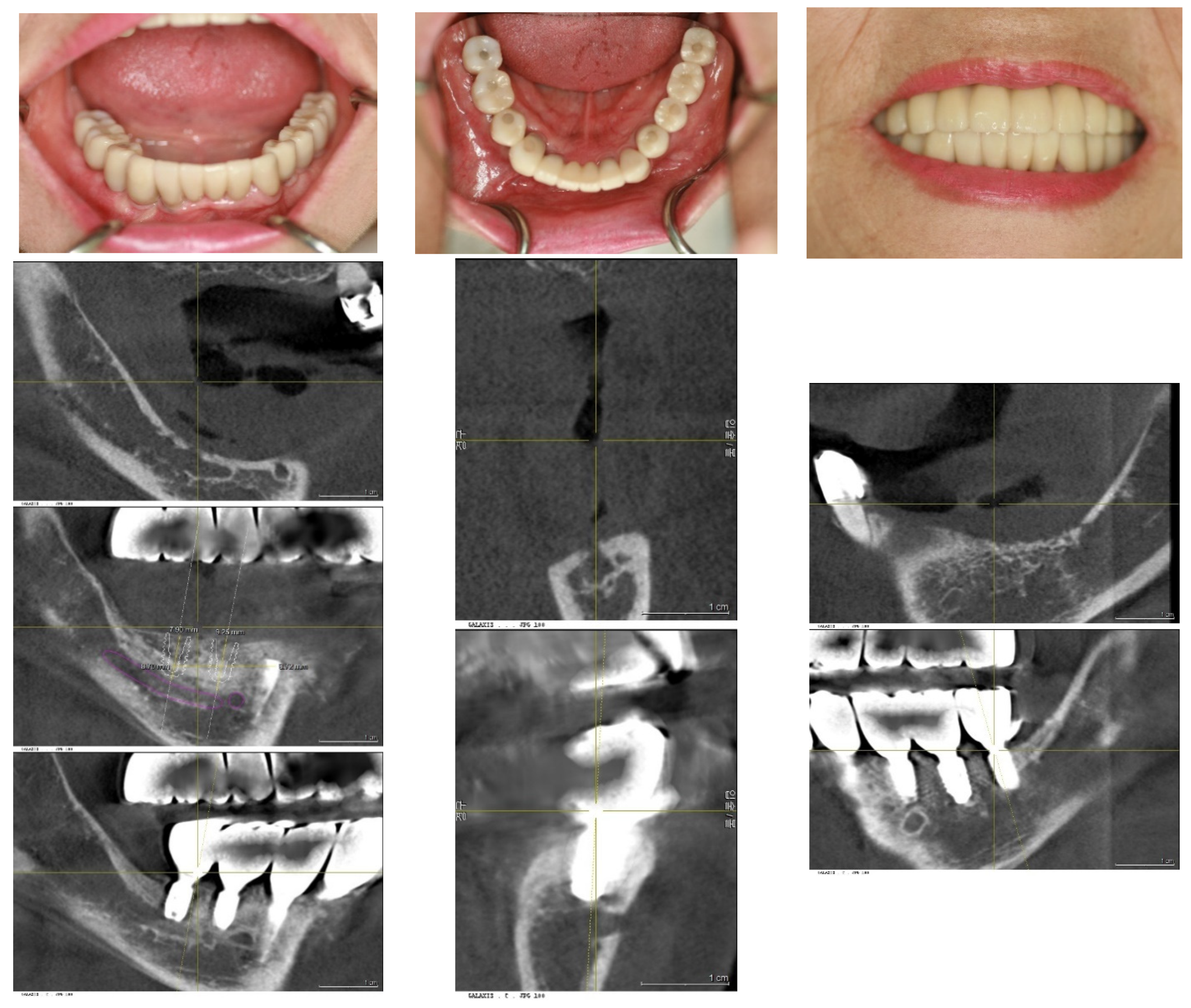
Figure 8.
After 5 months, on October 24th, 2017, a follow-up visit confirmed the success of the treatments. The soft tissue displayed healthy architecture without compromising the vestibular depth. The proportion between the crown height and the mucosal portion appeared natural. Notably, there were no soft tissue complications, such as bleeding, swelling, or erythema. From the frontal view, the overall appearance was deemed acceptable.
During the follow-up period extending to 2024, a minor prosthetic complication occurred: the upper anterior bridge became detached due to the failure of the dental cement, resulting in a loss of retention between the bridge and the implant abutment. Additionally, a small fracture was noted in a portion of the marginal area, characterized by chipping. The patient opted against the fabrication of a new bridge, preferring instead to have the existing anterior bridge re-cemented. This repair was straightforward and did not necessitate the creation of a new prosthetic.
Except for this event, there have been no other complications in both biological and mechanical aspects. This means there have been neither peri-implant mucositis nor peri-implantitis, and there have been no prosthodontic complications.
The patient's masticatory function has been almost fully restored, without any discomfort or difficulties during eating. Her appearance looks natural, as though she had her natural teeth, even though she does not.
Figure 9.
The lower pictures were taken on May 2, 2023, and January 9, 2024, each six years and seven years after the final finishing, respectively. Please compare them with the initial pictures taken before treatment. The rehabilitation using SITE has shown stable results throughout the follow-up period.
3. Results
Vertical bone changes at the right operation site were evaluated before surgery and then annually post-surgery for comparison. Measurements were taken from the superior border of the inferior alveolar canal to the crest of the ridge at two designated points: Point A, the lowest part of the edentulous ridge at baseline, and Point B, positioned above the mental foramen. To ensure measurement consistency, the length of the right upper second molar implant was used as a calibration standard. Prior to these measurements, to ascertain the repeatability of the points A and B assessments, the rectangular distance from the inferior border of the inferior alveolar canal to the bottom border of the mandible was verified.
Initially, in April 2015, the heights at Points A and B were recorded as 1.3 mm and 2.5 mm, respectively. Subsequent to the first bone graft, facilitated by SITE (Self Inflating Tissue Expander), the heights increased to 7.1 mm at Point A and 7.6 mm at Point B by October 2015, resulting in vertical increments of 5.8 mm at Point A and 5.1 mm at Point B. Following the second bone graft, minor increases were observed in May 2016, with heights reaching 8.9 mm at Point A and 9.7 mm at Point B. By the day of implant prosthesis completion in May 2017, both Points A and B recorded a crestal height of 8.3 mm. These heights were then tracked annually, showing a marginal decrease of 0.1 mm from the prosthesis completion in May 2017 to 2023. Over a span of six years, the overall decreases were 1.1 mm at Point A and 0.7 mm at Point B, deeming these results indicative of stable crestal bone changes.
Figure 10.
This graph illustrates the vertical bone changes at the right operation site, detailing measurements from Point A—the baseline's lowest part of the edentulous ridge—and Point B, which is located above the mental foramen.
Figure 10.
This graph illustrates the vertical bone changes at the right operation site, detailing measurements from Point A—the baseline's lowest part of the edentulous ridge—and Point B, which is located above the mental foramen.
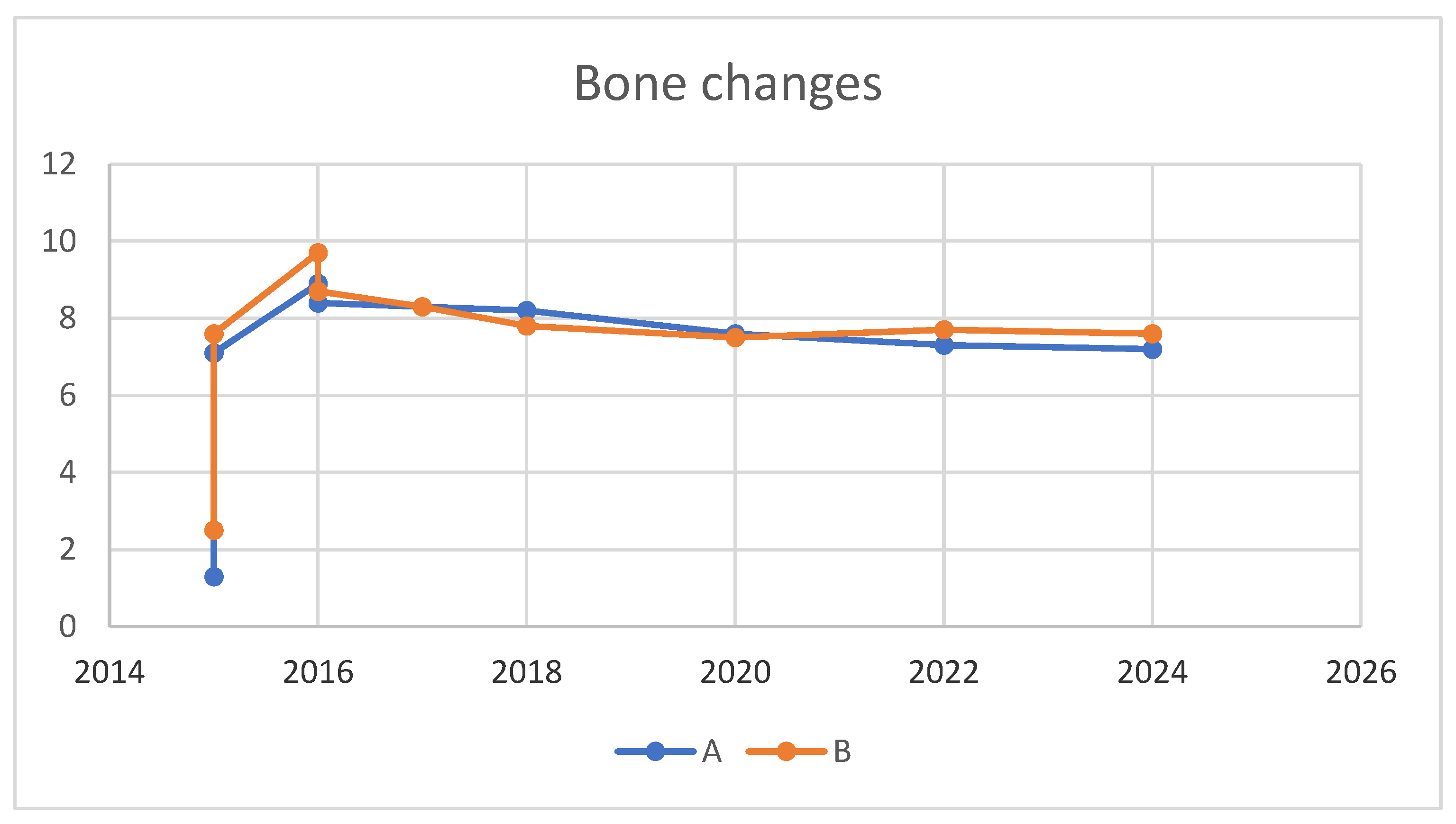

Table 2.
showing a comprehensive overview of the crestal height changes at Points A and B from the initial measurement in April 2015, through the interventions and up to the most recent check in June 2023. The notes column provides additional context to the measurements, highlighting the significant gains after the grafts and the slight annual decreases post-implant completion, which have been deemed stable.
| Time | Point A Height (mm) | Point B Height (mm) | Notes |
|---|---|---|---|
| Apr 2015 | 1.3 | 2.5 | Initial Measurements |
| Oct 2015 | 7.1 | 7.6 | After 1st Bone Graft (Vertical Gains: 5.8mm & 5.1mm for A & B) |
| May 2016 | 8.9 | 9.7 | After 2nd Bone Graft (Increases: 1.8mm & 2.1mm for A & B) |
| Oct 2016 | 8.4 | 8.7 | Adjustment Post-2nd Graft |
| May 2017 | 8.3 | 8.3 | At Completion of Implant Prostheses |
| Apr 2018 | 8.2 | 7.8 | Annual Check |
| June 2020 | 7.6 | 7.5 | Annual Check |
| Dec 2022 | 7.3 | 7.7 | Annual Check |
| June 2024 | 7.2 | 7.6 | Latest Measurement (Total Reductions: 1.1mm & 0.7mm for A & B over 6 years) |
Figure 11.
Panoramic X-ray images displaying the perioperative state along with the annual changes postoperatively.
Figure 11.
Panoramic X-ray images displaying the perioperative state along with the annual changes postoperatively.
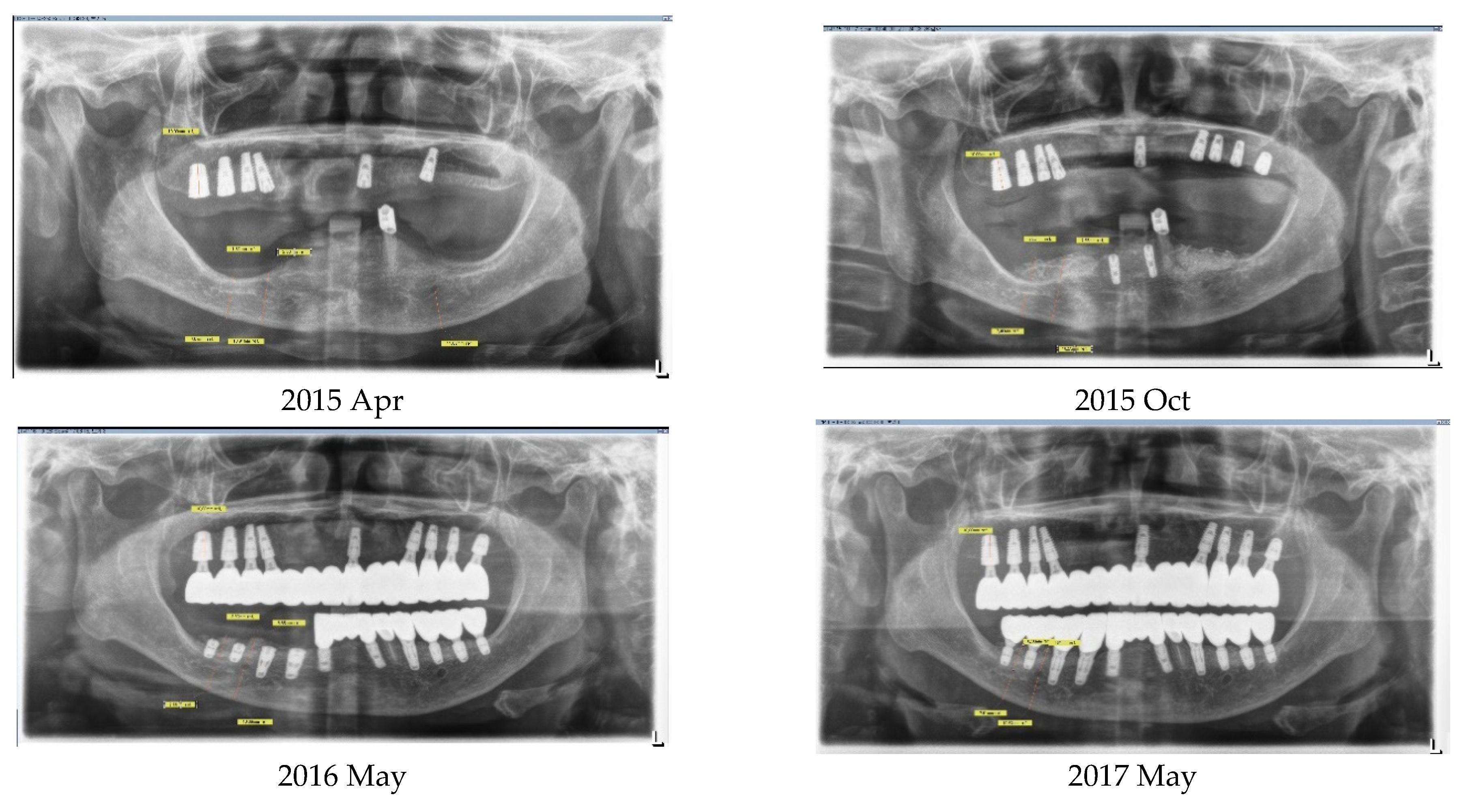
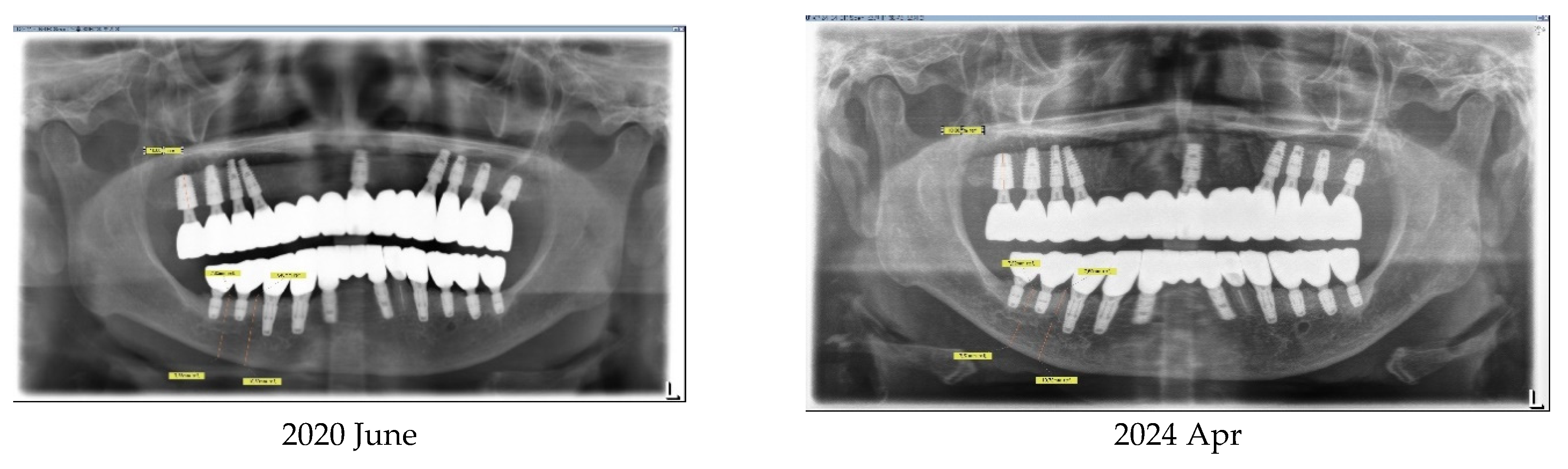
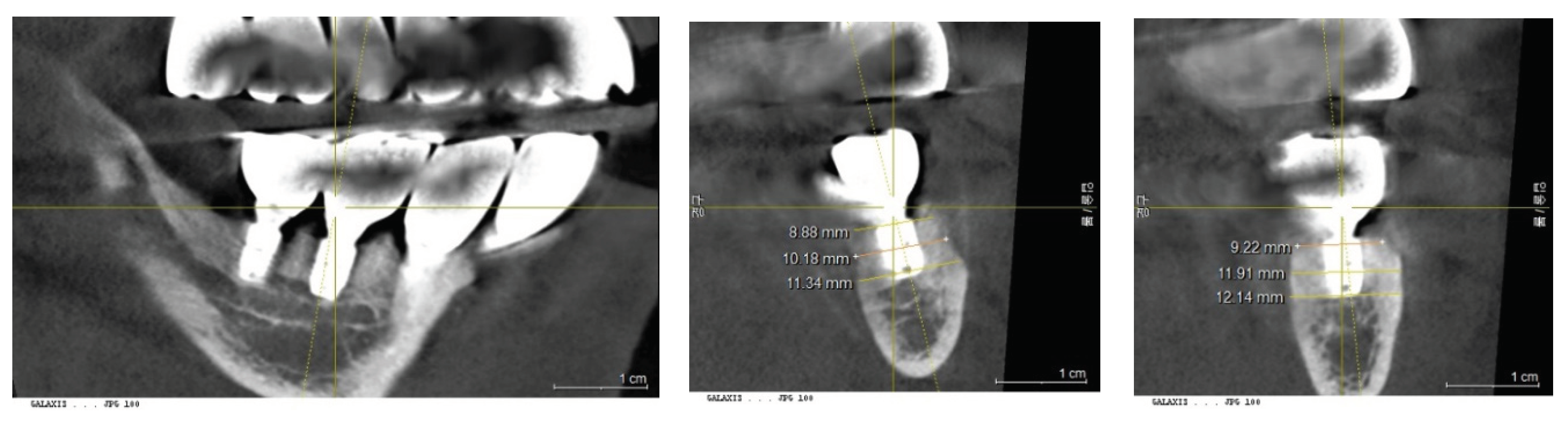
Figure 12.
illustrating the horizontal width measurements at the crestal level, showing 8.9 mm at Point A and 9.2 mm at Point B. Further down, at the middle level, the measurements increased to 10.1 mm and 11.9 mm, respectively, demonstrating a sufficient bone width necessary for implant support.
Figure 12.
illustrating the horizontal width measurements at the crestal level, showing 8.9 mm at Point A and 9.2 mm at Point B. Further down, at the middle level, the measurements increased to 10.1 mm and 11.9 mm, respectively, demonstrating a sufficient bone width necessary for implant support.
4. Discussion
Bone grafting has emerged as a cornerstone in the rehabilitation of oral tissues lost to various causes, including teeth, bone, and the surrounding soft tissues, through dental implantation [8,9]. The loss of teeth invariably leads to the concurrent loss of interdependent tissues, underscoring the complexity of oral rehabilitation [10]. Although implant dentistry primarily aims to restore chewing function, it also encompasses the regeneration of all missing oral tissues, regardless of their immediate contribution to functional improvement.
Bone grafting is crucial for implantation under two primary circumstances. Firstly, it is necessary when the available sites for implantation lack sufficient volume to support the smallest implant sizes, rendering the restoration unfeasible without further augmentation [3]. Secondly, aesthetic considerations and the aim for lasting stability come into play, notably in restoring anterior teeth. This approach is equally important for posterior teeth restorations [9]. For example, a clinical crown that is excessively long compared to adjacent teeth may look unnatural. Additionally, an optimal bone phenotype around the implants, characterized by a proper bone thickness, plays a vital role in maintaining their biological stability over time. Both circumstances, alongside the established understanding that tooth loss inevitably leads to the simultaneous loss of bone and soft tissues, highlight the fundamental importance of bone grafting in implant dentistry.
A broad spectrum of techniques is employed in bone grafting for dental implants, designed to tackle the varied clinical scenarios encountered in patient care. These procedures can be categorized based on the recipient site's topography into two main types: grafts for internal contained defects and grafts for external augmentation. The former includes methods such as socket preservation and sinus grafts, which are directed at repairing deficits within the bone structure. Meanwhile, external augmentation often incorporates a soft tissue releasing procedure integral to the bone grafting process itself, underlining the diversity and complexity of approaches needed to satisfy the comprehensive range of anatomical and aesthetic requirements. However, the pivotal element in external augmentation lies in flap management, which includes soft tissue expansion to ensure primary closure and prevent wound dehiscence, further emphasizing the critical role of meticulous soft tissue handling in the success of these procedures [11].
From this perspective, the soft tissue expander was conceived. Throughout the development process for the right device, Osmed company introduced the Self-Inflating Tissue Expander (SITE), a notable innovation in this field. The Osmed tissue expander is made of a hydrogel material. Hydrogels are highly absorbent polymers that can retain a significant amount of water or biological fluids, swelling to several times their original size. This property allows the Osmed tissue expanders to gradually expand as they absorb bodily fluids, thereby stretching the surrounding tissues over time. The specific composition of the hydrogel used in Osmed tissue expanders is designed to ensure biocompatibility, optimal expansion rates, and sufficient mechanical strength to meet the requirements of the intended medical or dental applications [5,6,7].
If the required volume for external bone augmentation for implant placement is moderate or less, the bone grafting can be performed with conventional techniques, accompanied by soft tissue releasing. However, cases that demand a significantly larger volume for augmentation pose considerably greater challenges [11,12].
While other alternative techniques could have been considered [13], the author opted to utilize the Self-Inflating Tissue Expander (SITE) for the case presented.
The upper arch (full edentulous arch) was successfully restored using standard dental implant procedures without any significant issues. However, a specific treatment plan was devised for the upper side, focusing on selecting the optimal locations for implant placement to achieve the desired outcomes more easily and efficiently. Thus, for the anterior part of the upper arch, the sites for both first premolars and the left incisor were chosen for an 8-unit implant bridge. The implants were positioned in parallel to ensure that the prosthesis could be securely attached by screwing it in place after extraoral cementation, with the margins positioned subgingivally to fulfill aesthetic requirements.
The lower arch, which was fully edentulous except for the left canine, was segmented into four parts to facilitate the surgical process in stages.
The right and left posterior edentulous ridges were severely atrophic, resulting in a minimal distance between the ridge crest and the inferior alveolar canal, alongside a very narrow crest width. Consequently, the majority of the alveolar bone needed for implant placement had to be regenerated through bone grafting to ensure successful outcomes. This was particularly critical for the right posterior ridge, where virtually no bone was available for implant treatment. Initially, the distance measured 1.5 mm at Point A and 2.3 mm at Point B, with Point A representing the lowest part of the edentulous ridge at baseline and Point B located above the mental foramen. Ultimately, as detailed earlier in this report's case study, the bone grafting procedure using the Self-Inflating Tissue Expander (SITE) proved successful for implant placement. The maximum amount of vertical bone gain achieved was 7.6 mm at Point A and 7.2 mm at Point B.
The horizontal width measurements at the crestal level showed 8.9 mm at Point A and 9.2 mm at Point B. Deeper, at the middle level, the measurements increased to 10.1 mm and 11.9 mm, respectively, indicating a substantial growth in bone width crucial for implant support. The peak horizontal bone augmentation reached was 11.9 mm, signifying notable enhancements in the dimensions vital for successful implant placement.
The final outcome with the implant prostheses was realized in May 2017, after nearly two years of treatment from the onset. The patient has undergone annual follow-ups since completion. Crestal bone loss has been recorded at a rate of 0.1 mm per year, which is deemed stable [14]. Throughout this period, there have been no specific events or complications related to the implants, either surgically or prosthodontically. For the left side, similarly excellent results have been observed, with no more than 0.5 mm of bone loss recorded up to 2024 since the treatment's completion.
Upon deeming the bone graft procedure successful, the following claim is substantiated: After achieving pre-surgical objectives and ensuring stable outcomes from bone grafts, it's vital to recognize that the determinants of implants' long-term success do not differ, regardless of bone grafting. These determinants, affecting both the biological and mechanical integrity of implants, can lead to issues like peri-implantitis and mechanical problems such as abutment fractures, impacting implant performance equally in grafted and non-grafted situations. Thus, the use of a bone graft does not modify the risk factors for peri-implantitis [15,16].
Concerning the selection of graft material, Bio-Oss xenograft from Geistlich Pharma was exclusively utilized [17,18,19]. The outcomes of this report confirm that the graft material’s function, acting solely as a scaffold, is effectively achieved, facilitating the regeneration of new bone as long as space is maintained for the bone to form [20]. Notably, this space was established using the SITE method, avoiding typical surgical complications such as wound dehiscence.
5. Conclusion
The SITE played a pivotal role in the successful restoration of an extremely atrophic mandibular edentulous case with fixed dental implants. The space created by SITE enabled successful regeneration without the need for an autogenous bone graft. This technique facilitated a maximum vertical gain of 7.6 mm. Follow-up results over seven years have demonstrated stability in the outcomes, with minimal crestal bone loss recorded at 0.1 mm per year, underscoring the effectiveness and durability of the SITE approach in challenging dental restorations.
Funding
This research has neither fund nor sponsor.
Ethical Approval and Consent to participate
Not applicable.
Consent for publication
Not applicable.
Competing interests/Authors’ contributions
The author declares to have no competing interests.
References
- Rozalia Dimitriou, Elena Jones, Dennis McGonagle, Peter V Giannoudis Bone regeneration: current concepts and future directions BMC Med 2011 May 31:9:66. https://doi.org/10.1186/1741-7015-9-66. [CrossRef]
- Ching Izzie Wang, Shayan Barootchi, Lorenzo Tavelli, Hom-Lay Wang The peri-implant phenotype and implant esthetic complications. Contemporary overview J Esthet Restor Dent. 2021 Jan;33(1):212-223. [CrossRef]
- Daniel Buser, Istvan Urban, Alberto Monje, Marcel F Kunrath, Christer Dahlin Guided bone regeneration in implant dentistry: Basic principle, progress over 35 years, and recent research activities Periodontol 2000. 2023 Oct;93(1):9-25. [CrossRef]
- Jeffrey Garcia, Austin Dodge, Paul Luepke, Hom-Lay Wang, Yvonne Kapila, Guo-Hao Lin Effect of membrane exposure on guided bone regeneration: A systematic review and meta-analysis Clin Oral Implants Res. 2018 Mar;29(3):328-338. [CrossRef]
- Miryam C Obdeijn, Jean-Philippe A Nicolai, Paul M N Werker The osmotic tissue expander: a three-year clinical experience J Plast Reconstr Aesthet Surg. 2009 Sep;62(9):1219-22. [CrossRef]
- Miryam C Obdeijn, Jean-Philippe A Nicolai, Paul M N Werker The osmotic tissue expander: a three-year clinical experience J Plast Reconstr Aesthet Surg. 2009 Sep;62(9):1219-22. [CrossRef]
- Christian Mertens, Oliver Thiele, Michael Engel, Robin Seeberger, Jürgen Hoffmann, Kolja Freier The use of self-inflating soft tissue expanders prior to bone augmentation of atrophied alveolar ridges Clin Implant Dent Relat Res. 2015 Feb;17(1):44-51. [CrossRef]
- Hyun-Suk Cha, Ji-Wan Kim, Jong-Hyun Hwang, Kang-Min Ahn Frequency of bone graft in implant surgery Maxillofac Plast Reconstr Surg. 2016 Mar 31;38(1):19. [CrossRef]
- Bach Le, Naoki Hayashi The Aesthetic Contour Graft - Enhancing peri-implant soft tissue contours and pontic sites with guided bone regeneration J Esthet Restor Dent. 2022 Jan;34(1):188-202. [CrossRef]
- François Bodic, Luc Hamel, Emmanuelle Lerouxel, Michel Félix Baslé, Daniel Chappard Bone loss and teeth Joint Bone Spine. 2005 May;72(3):215-21. [CrossRef]
- Istvan A Urban, Alberto Monje, Jaime Lozada, Hom-Lay Wang Principles for Vertical Ridge Augmentation in the Atrophic Posterior Mandible: A Technical Review Int J Periodontics Restorative Dent. 2017 Sep/Oct;37(5):639-645. [CrossRef]
- Istvan A Urban, Eduardo Montero, Ettore Amerio, David Palombo, Alberto Monje Techniques on vertical ridge augmentation: Indications and effectiveness Periodontol 2000. 2023 Oct;93(1):153-182. [CrossRef]
- Amanda Andre, Orrett E Ogle Vertical and Horizontal Augmentation of Deficient Maxilla and Mandible for Implant Placement Dent Clin North Am. 2021 Jan;65(1):103-123. [CrossRef]
- Vladyslav Demenko, Igor Linetskiy, Larysa Linetska, Vitalij Nesvit, Andrii Shevchenko, Oleg Yefremov, Hans-Werner Weisskircher Prognosis of implant longevity in terms of annual bone loss: a methodological finite element study Comput Methods Biomech Biomed Engin. 2016;19(2):180-7. [CrossRef]
- Khadijeh Al-Abedalla, Jesus Torres, Arthur Rodriguez Gonzalez Cortes, Xixi Wu, Samer Abi Nader, Nach Daniel, Faleh Tamimi Bone Augmented With Allograft Onlays for Implant Placement Could Be Comparable With Native Bone J Oral Maxillofac Surg. 2015 Nov;73(11):2108-22. [CrossRef]
- Goran I Benić 1, Ronald E Jung, David W Siegenthaler, Christoph H F Hämmerle Clinical and radiographic comparison of implants in regenerated or native bone: 5-year results Clin Oral Implants Res. 2009 May;20(5):507-13. [CrossRef]
- Sakshi Goyal, Mina Masood, Curtis Le, Yoga Rajendran, Samir Nanjapa, Ram Vaderhobli Comparative Bone Graft Evaluation for Dental Implant Success: An Evidence-Based Review J Long Term Eff Med Implants. 2021;31(3):33-44. [CrossRef]
- Zeeshan Sheikh, Nader Hamdan, Yuichi Ikeda, Marc Grynpas, Bernhard Ganss, Michael Glogauer Natural graft tissues and synthetic biomaterials for periodontal and alveolar bone reconstructive applications: a review Biomater Res. 2017 Jun 5:21:9. [CrossRef]
- Jad Majzoub, Andrea Ravida, Thomas Starch-Jensen, Mustafa Tattan, Fernando Suárez-López Del Amo The Influence of Different Grafting Materials on Alveolar Ridge Preservation: a Systematic Review J Oral Maxillofac Res. 2019 Sep 5;10(3):e6. [CrossRef]
- Nedal A Abu-Mostafa, Yasser N Alotaibi, Rose N Alkahtani, Farah K Almutairi, Amjad A Alfaifi, Osama D Alshahrani The Outcomes of Vertical Alveolar Bone Augmentation by Guided Bone Regeneration with Titanium Mesh: A Systematic Review J Contemp Dent Pract. 2022 Dec 1;23(12):1280-1288. [CrossRef]
Disclaimer/Publisher’s Note: The statements, opinions and data contained in all publications are solely those of the individual author(s) and contributor(s) and not of MDPI and/or the editor(s). MDPI and/or the editor(s) disclaim responsibility for any injury to people or property resulting from any ideas, methods, instructions or products referred to in the content. |
© 2024 by the authors. Licensee MDPI, Basel, Switzerland. This article is an open access article distributed under the terms and conditions of the Creative Commons Attribution (CC BY) license (http://creativecommons.org/licenses/by/4.0/).
Copyright: This open access article is published under a Creative Commons CC BY 4.0 license, which permit the free download, distribution, and reuse, provided that the author and preprint are cited in any reuse.


MDPI Initiatives
Important Links
© 2024 MDPI (Basel, Switzerland) unless otherwise stated