Preprint
Article

Fracture Resistance Analysis of 3D-Printed and Prefabricated Artificial Teeth for Removable Dentures

Altmetrics

Downloads

158

Views

74

Comments

0

Submitted:

17 April 2024

Posted:

17 April 2024

You are already at the latest version

Alerts
Abstract
The integration of three-dimensional (3D) printed resin denture teeth represents a significant ad-vancement in digital dentistry. This study aimed to assess their ability to withstand chipping and indirect tensile fractures, comparing them with conventionally manufactured resin denture teeth. Four groups, each comprising 30 specimens, were examined: Group 1 featured 3D printed denture teeth (NextDent, 3D Systems, Soesterberg, The Netherlands), while the others included commer-cially obtained Ivostar Shade, SpofaDent Plus, and Major Super Lux teeth. Stereolithography 3D printing was utilized to produce methacrylate-based photopolymerized resin teeth models for Group 1, while the remaining groups were commercially sourced. Chipping and indirect tensile fracture tests were performed at a speed of 1 mm/min until fracture occurred. Statistical analysis was conducted using one-way analysis of variance (ANOVA) with Tukey's honestly significant difference multiple comparisons test (p < 0.05). Results indicated that 3D-printed teeth exhibited higher indirect tensile fracture resistance than Major Super Lux and Ivostar Shade but lower than SpofaDent Plus. Additionally, in the chipping test, buccal chipping occurred in 3D-printed resin teeth without distortion. In the indirect tensile test, the fracture line emerged near the loading point, extending cervically from the inner incline of both cusps. Overall, denture teeth produced via 3D printing with printable resin materials demonstrated sufficient fracture resistance, making them suitable for prosthetic applications.
Keywords: 
Subject: Medicine and Pharmacology  -   Dentistry and Oral Surgery

1. Introduction

Computer-aided design/computer-aided manufacturing (CAD/CAM) systems are widely used in dentistry, particularly for crafting inlays, crowns, fixed partial dentures, and implant prostheses [1]. More recently, CAD/CAM technology has been integrated into the fabrication of full dentures, offering numerous advantages to both dentists and patients compared to traditional complete dentures [2]. These benefits include fewer necessary appointments, enhanced access to spare dentures through the preservation of digital data, and greater convenience and cost-effectiveness in laboratory work compared to traditional methods [3,4].
One of the pioneering developments in the realm of computer-aided design/computer-aided manufacturing (CAD/CAM) for dentistry is the creation of 3D-printed dentures for edentulous patients [5]. This innovative approach incorporates digital modeling, computational optimization, and 3D printing techniques to revolutionize denture design and production [6]. These 3D-printed dentures are composed of methacrylate-based photopolymerized resin, which is processed and cured exclusively through 3D printing. In this method, denture teeth and the denture base are printed separately and then fused using a light-cured bonding agent [7]. The introduction of 3D printing for denture manufacturing represents a novel approach that requires further exploration. If thoroughly studied and developed, 3D-printed dentures could offer more efficient clinical adaptation, reducing patient discomfort and potentially mitigating long-term bone resorption issues [8,9].
Despite the numerous advantages of CAD/CAM dentures, inadequately designed dentures can exhibit concerns like insufficient denture base borders and reduced tissue contact, which negatively impact retention [10]. Additionally, the mechanical properties of 3D-printed denture teeth produced via CAD/CAM methods remain largely unknown. Historically, denture teeth have primarily been crafted from resin materials, and advances in dental resin materials have led to successful clinical applications [11,12]. These advancements have involved the development of new monomers, filler technologies, and self-healing capabilities. Notably, improvements in filler systems have enhanced the mechanical properties of these materials [13].
The susceptibility of denture teeth to fractures or chipping is a recurrent issue, particularly in scenarios where a single complete denture is in opposition to natural teeth or implant-supported over-dentures [14]. The prevalence of these challenges is underscored by research focusing on implant-retained complete dentures, which has identified fractures of denture teeth as a prominent and persistent concern. This concern is further compounded by the evolving landscape of removable prosthetics, characterized by a rising inclination toward utilizing multiple implants to support prostheses [15,16].
The shift toward implant-supported solutions reflects an ongoing quest for improved stability and functionality in dental prosthetics. However, this evolution also brings to the forefront the pressing need for denture teeth endowed with enhanced fracture resistance to withstand the increased biomechanical demands imposed by implant-supported frameworks [11]. Addressing this demand necessitates advancements in material science and manufacturing techniques to develop denture teeth capable of withstanding the rigors of long-term clinical use in diverse oral environments [3]. Moreover, comprehensive approaches encompassing both material selection and prosthetic design are imperative to mitigate the risk of fractures and ensure the longevity and efficacy of implant-supported removable prostheses. As such, ongoing research and innovation in this domain are crucial to meeting the evolving needs and expectations of patients and clinicians alike [9].
In response to this demand, numerous manufacturers have embarked on initiatives to develop denture teeth with superior mechanical properties, notably emphasizing greater fracture resistance [17]. Through advancements in material science and engineering, these manufacturers are exploring innovative formulations and manufacturing techniques to enhance the durability and longevity of denture teeth [18]. The goal is to offer dental professionals and patients alike a reliable solution that can withstand the rigors of everyday use, thereby minimizing the incidence of fractures and enhancing overall prosthetic performance [19].
By continually refining the design and composition of denture teeth, manufacturers aim to address the persistent challenges associated with prosthodontic treatment, ultimately improving patient satisfaction and quality of life [20]. As research and technology progress, the evolution of denture teeth holds promise for revolutionizing the field of prosthodontics, paving the way for more resilient and functional dental prostheses. Resin materials have been a primary area of focus for improving fracture resistance and mechanical properties through increased cross-linking between polymers and the utilization of specialized pre-polymers [21,22]. Moreover, incorporating inorganic fillers into the polymer matrix has been employed to enhance mechanical properties and wear resistance. Despite these efforts, fractures in denture teeth remain a challenge, necessitating the exploration of novel approaches to withstand high loads and forces [23].
Numerous studies have delved into the fracture characteristics of dental restorative materials, with a particular focus on mode I fracture toughness and mixed mode bond strength, recognizing the intricate forces at play during chewing [24]. Traditional fracture criteria, while informative, have demonstrated shortcomings in reliably predicting dental material fractures. Consequently, researchers have turned to alternative methodologies, such as the extended maximum tangential strain criterion, to provide more precise and dependable data predictions [25,26].
While significant strides have been made in utilizing digital technology for creating denture bases, there remains a notable gap in comprehending the durability of 3D-printed teeth [27]. This lack of exploration into the practical functionality of 3D-printed teeth in clinical contexts highlights the urgent need for further investigation. Therefore, it is crucial to conduct additional studies to evaluate the performance and reliability of these dental components under real-world circumstances [12].
Such research endeavors are crucial for several reasons. Firstly, they play a vital role in confirming the effectiveness and dependability of 3D-printed teeth as viable alternatives to traditional options [5]. By subjecting these prosthetic elements to rigorous examination in clinical environments, researchers can gather invaluable insights into their performance attributes, longevity, and ability to withstand wear and tear. Additionally, a comprehensive assessment of 3D-printed teeth enhances understanding of their biomechanical characteristics and compatibility with existing dental prosthetic materials and methodologies [8,21].
This study aimed to assess and contrast the resistance to fractures and wear of various 3D-printed denture teeth from different brands, in comparison to pre-made denture teeth. The initial assumption was that there would be no notable differences in wear and fracture resistance between 3D-printed resin teeth and prefabricated alternatives.

2. Materials and Methods

2.1. Preparing Denture Tooth Specimens
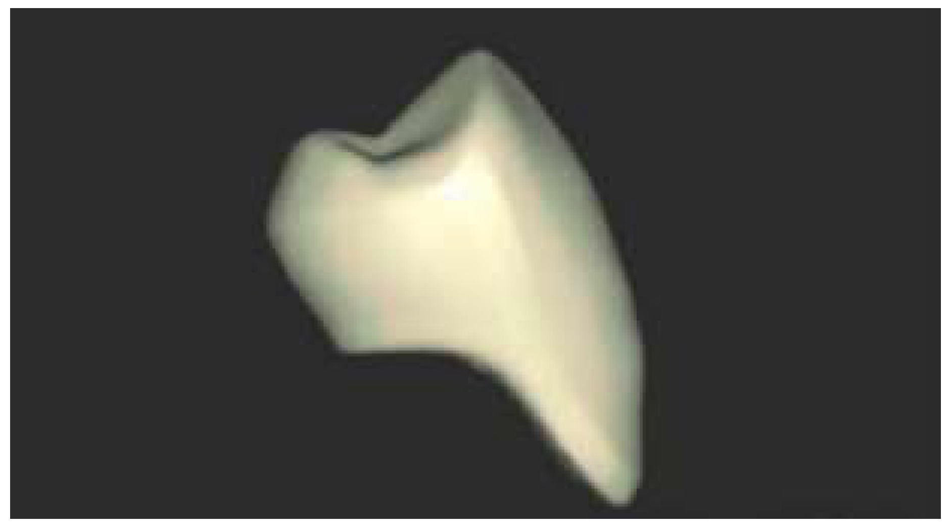
In this study, 3D-printed denture teeth were investigated as well as three different ready-made denture teeth for our experimentation. The 3D-printed resin teeth were crafted layer by layer using methacrylate-based photopolymerized resin NextDent C&B MFH (NextDent, 3D Systems, Soesterberg, The Netherlands) (NextDent, 3D Systems, The Netherlands). They were modeled to match the size and shape of a prefabricated denture maxillary first premolar tooth (Table 1).
Once the digital file of the tooth was inputted into the software of the 3D printer, the intricate procedure of converting virtual data into tangible dental prostheses commenced. Within the controlled environment of the 3D printer chamber, precise commands were executed to meticulously fabricate each tooth according to the specifications outlined in the digital blueprint. Utilizing state-of-the-art technology, such as the Next Dent 5100 manufactured by NextDent, a subsidiary of 3D Systems located in Soesterberg, The Netherlands, the printer adeptly operated to convert the resin into finely detailed dental components.
By employing a methodical layer-by-layer approach, the printer carefully laid down thin, successive layers of resin, progressively constructing the structure of each tooth with remarkable precision and accuracy. This additive manufacturing technique, recognized as stereolithography (SLA), is renowned for its capability to generate objects of high resolution and intricate detail, boasting exceptional surface quality and dimensional precision.
Throughout the printing process, the dental team vigilantly oversaw each stage, ensuring meticulous quality control and adherence to the prescribed design specifications. The outcome was a succession of precisely crafted denture teeth, each reflecting the exact morphology and characteristics specified in the digital tooth file.
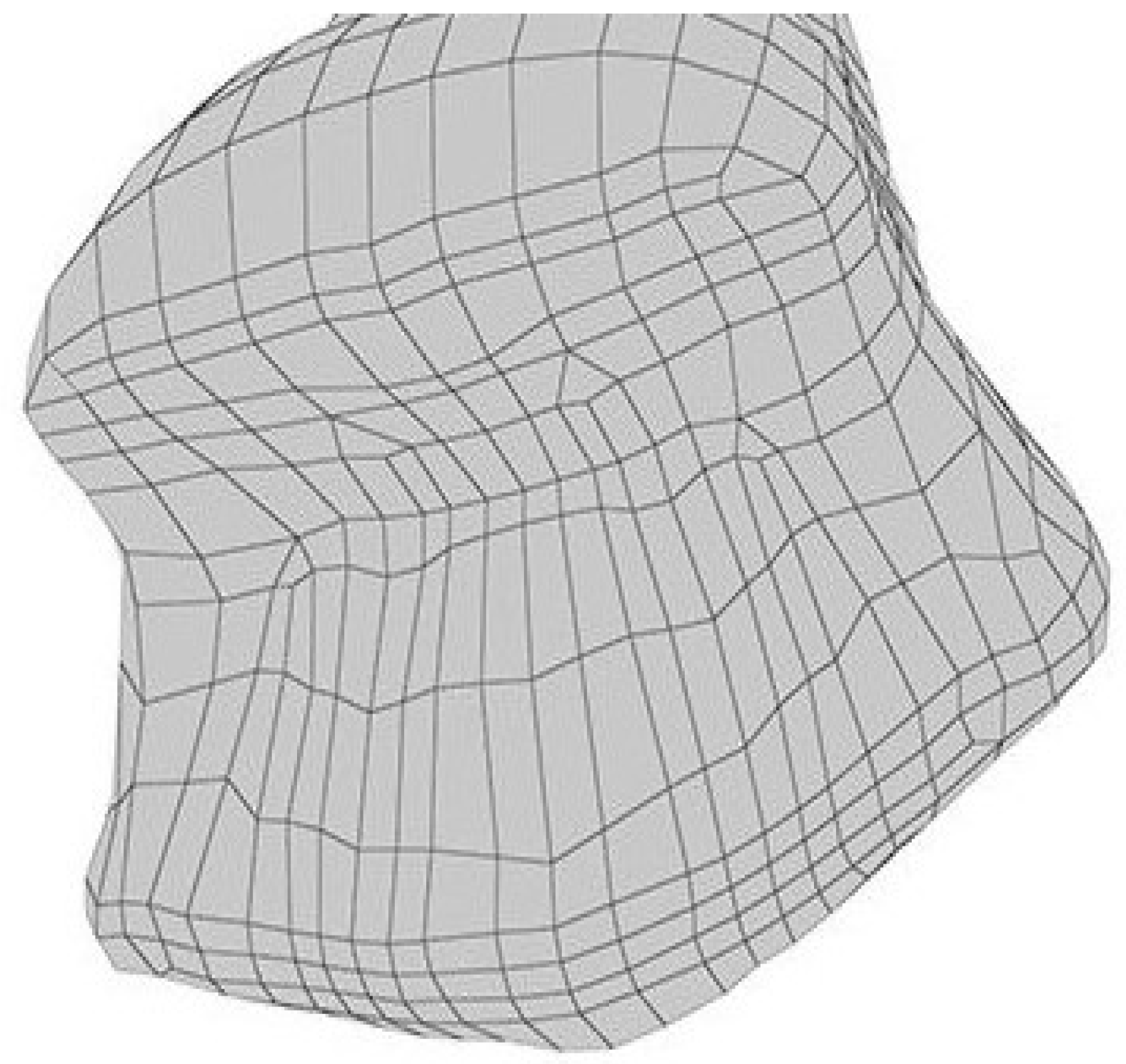
Figure 1. The image of the scanned prefabricated denture tooth.
Preprints 104157 g001
Figure 2. The STL image was uploaded to the 3D-printing software device (occlusal view).
Preprints 104157 g002
The artificial teeth were fabricated with a layer thickness of 50 µm and then cleansed using isopropanol. Isopropyl alcohol (IPA), also known as rubbing alcohol, is a transparent and potent cleaning solution widely employed for various 3D-printed materials. IPA effectively cleans the 3D printer's build platform without leaving any traces or residues. Typically, the cleaning process for 3D-printed components lasts around six minutes, during which IPA is diluted with distilled water at a ratio of 70% isopropyl alcohol to 30% distilled water. Following the cleaning procedure, the specimens underwent a further 45-minute curing process by immersion in glycerin within the post-curing oven to complete the reaction of any remaining monomers.

2.2. Performance of Chipping and Indirect Tensile Fracture Test

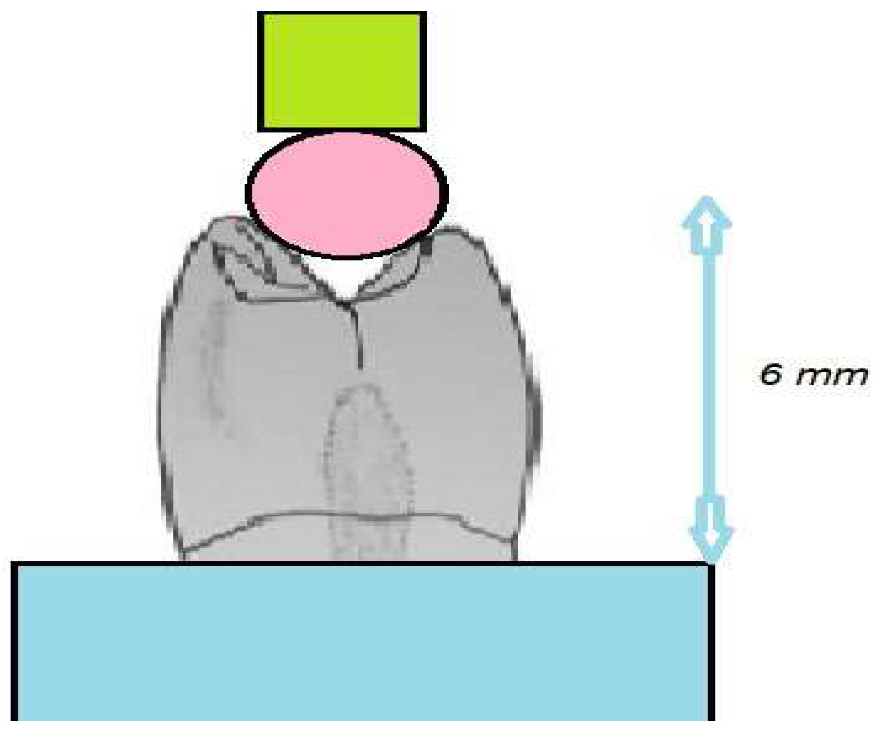
To evaluate the strength of the prepared tooth specimens from a clinical perspective, chipping and indirect tensile fracture tests were conducted, following established methodologies from previous research [18,27]. For the chipping test, specific equipment was designed to secure the denture tooth specimen in a fixed position, preventing any movement when subjected to chipping force. This equipment was initially prototyped using 3D printing and later constructed in metal. A preliminary experiment confirmed the successful immobilization of the tooth specimen during the chipping test. The equipment included a loading rod with a hemispherical end, enabling point-to-point contact at the buccal cusp tip of the tooth. To ensure that the force was applied exclusively to the buccal cusp and to prevent contact with the palatal cusp, the bottom of the denture teeth was ground to maintain a height of 7 mm from the tooth's base to the buccal cusp and 6 mm to the palatal cusp (Figure 3).
The denture tooth specimen was affixed to the equipment, which was then mounted on a universal testing machine (Model 4465, Instron in Canton, MA, USA). The specimen was loaded at a rate of 1 mm per minute, and the test recorded the point at which chipping occurred. This test was repeated 10 times for each type of denture tooth. The technique for measuring the indirect tensile fracture strength is illustrated in Figure 3. The processed denture teeth were set within cylindrical plastic molds using a self-polymerizing resin. All the teeth encased within the specimens were adjusted to have a uniform height of 6 mm from the tooth's base to both the buccal and palatal cusps. This standardization ensured that the applied pressing force acted on surfaces at an identical level.
The specimens were arranged and firmly fastened to the setup of a universal testing machine (Model 4465, Instron, MA, USA). A 4 mm diameter circular metal bar was affixed to the end of the machine's loading rod and kept in place during the testing procedure. The position of this round bar was such that it made contact with both cusp slopes of the denture tooth (Figure 4).
A load was then applied at a rate of 1 mm per minute until the fracture occurred. The magnitude of the load at the point of fracture was recorded. Each type of denture tooth underwent ten such tests.

2.3. Statistical Analysis

Following the statistical analysis, the data underwent rigorous scrutiny to unveil patterns, trends, and significant differences among the diverse groups examined. Employing a one-way analysis of variance (ANOVA) alongside Tukey's honestly significant difference (HSD) multiple comparisons test provided a robust framework for assessing variations across the datasets. Through the sophisticated functionalities of SPSS (IBM Corp., New York, NY, USA), intricate relationships within the data were elucidated, allowing for comprehensive insights into the studied phenomena. With a predetermined significance threshold of p < 0.05, only results surpassing this stringent criterion were deemed statistically significant, ensuring the reliability and validity of the findings. This meticulous approach not only affirmed the credibility of the analysis but also facilitated informed decision-making and further exploration of the research domain.

3. Results

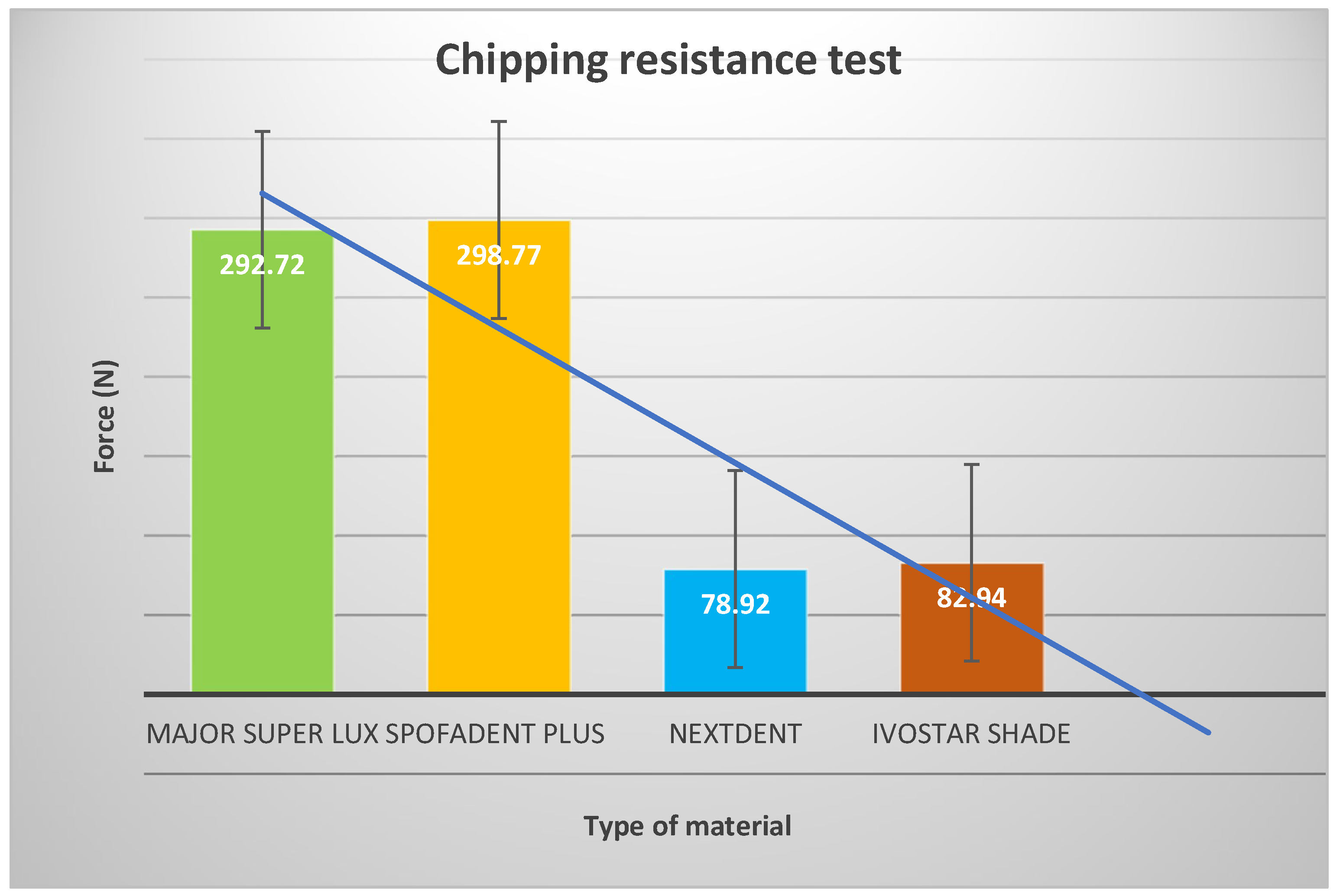
The outcomes of the chipping test are presented in Figure 5. The values for load-to-chipping fractures in Major Super Lux (292.72 ± 46.52 N) and SpofaDent Plus (298.77 ± 45.64 N) denture teeth were notably higher in comparison to the 3D printed resin teeth (78.92 ± 16.77 N), and Ivostar Shade (82.94 ± 23.89 N) teeth (p < 0.05). Furthermore, SpofaDent Plus teeth exhibited significantly greater resistance to chipping fractures compared to Major Super Lux (p < 0.05). The 3D-printed resin teeth and Ivostar Shade teeth did not exhibit significant differences.
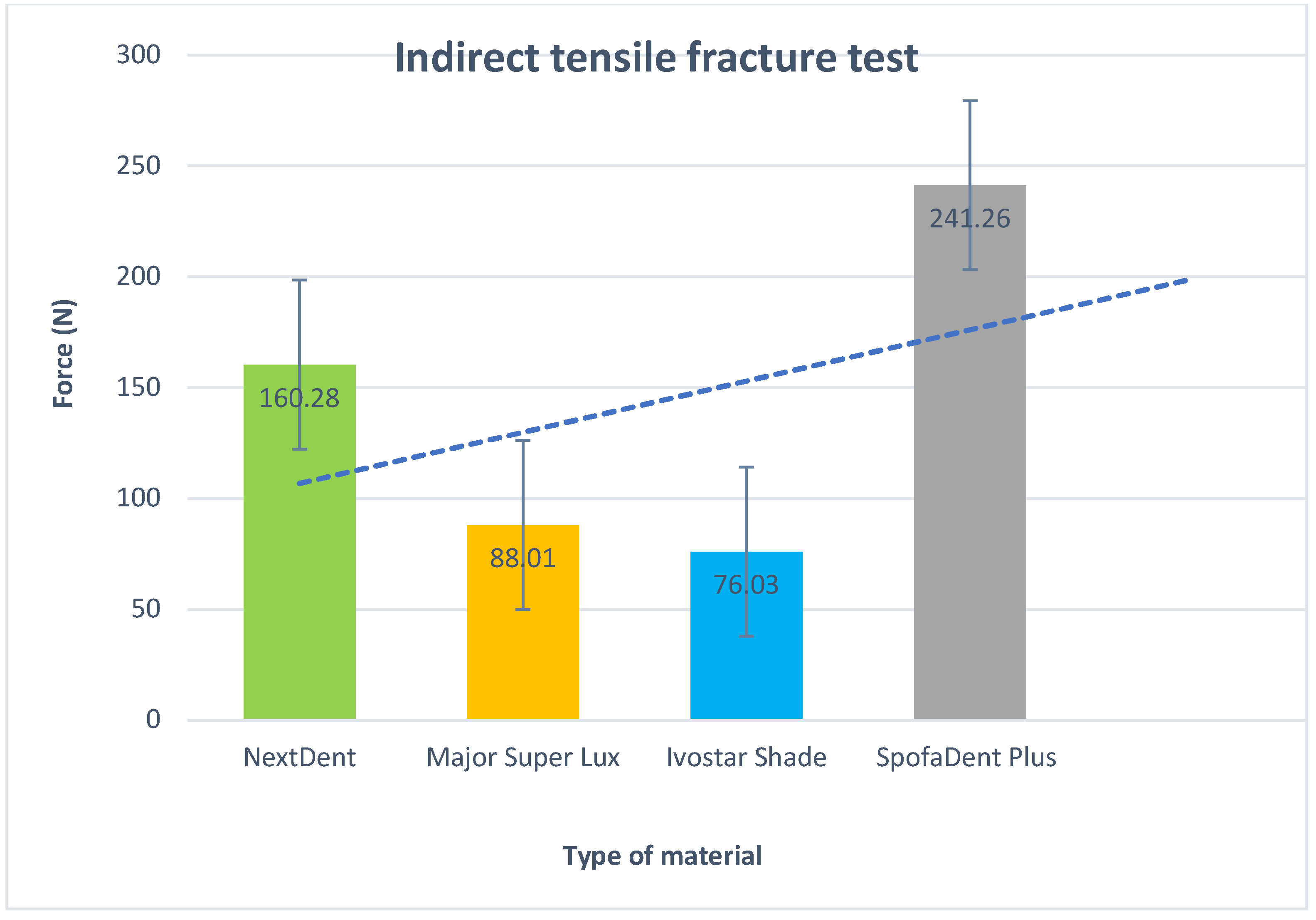
The results of the indirect tensile fracture test can be found in Figure 6. The load-to-tensile fracture values were as follows: 160.28 ± 8.83 N for the 3D printed resin, 88.01 ± 29.05 N for Major Super Lux, 76.03 ± 13.38 N, for Ivostar Shade, and 241.26 ± 26.34 N for SpofaDent Plus teeth. Notably, the load-to-tensile fracture values were significantly higher in the 3D-printed resin and SpofaDent Plus teeth when compared to Major Super Lux, and Ivostar Shade teeth (p < 0.05). Furthermore, SpofaDent Plus teeth displayed significantly higher tensile fracture values than the 3D-printed resin teeth (p < 0.05). However, there were no statistically significant differences in tensile fracture values among Major Super Lux and Ivostar Shade teeth.
A Tukey's Honest Significant Difference post hoc examination was carried out to contrast each type of material with the rest. Table 2 showcases findings from a comparative evaluation of mean values, standard deviations, p-values, and 95% confidence intervals across distinct groups and timeframes. Every cell in the table illustrates a comparison between two distinct types of artificial teeth, which have been investigated in this study.
Based on the analysis of mean differences and confidence intervals, it's evident that NextDent exhibits significantly higher mean values compared to both Ivostar Shade and Major Super Lux, while SpofaDent Plus consistently shows significantly lower mean values compared to NextDent and Major Super Lux. There's no statistically significant difference in mean values between Major Super Lux and Ivostar Shade, indicating their comparable performance. Additionally, SpofaDent Plus tends to have higher mean values compared to Ivostar Shade.

4. Discussion

The null hypothesis had to be rejected as the results exhibited significant differences based on the type of denture teeth. Furthermore, the fracture modes varied among the different denture tooth types and had a substantial impact on their fracture resistance. The study identified two distinct modes of damage: fractures that occurred without noticeable deformation and fractures that transpired following deformation. Various material microstructures can lead to a range of responses to applied loads, spanning from what is essentially a "brittle mode" characterized by cracks to a more "quasi-plastic mode" marked by deformation dominance [28].
The chipping test revealed notable differences in fracture behavior among various denture teeth types. Notably, SpofaDent and Major Super Lux teeth showcased remarkable resilience, displaying robust fracture strength in the face of applied stress. However, their performance was not without indication of deformation. Prior to reaching the critical fracture point, both SpofaDent and Major Super Lux teeth exhibited observable signs of strain, characterized by the formation of a distinctive cone-shaped depression. This pre-fracture deformation suggests a degree of material plasticity, wherein the denture teeth undergo localized structural changes in response to external forces. Such behavior underscores the complex interplay between material composition, mechanical properties, and the inherent ability of denture teeth to withstand mechanical stressors. Understanding these nuances is essential for optimizing denture design and material selection to enhance durability and longevity in clinical applications [29].
In the indirect tensile test, 3D-printed resin and SpofaDent teeth exhibited substantial fracture strength, with SpofaDent specimens showing quasi-plastic deformation prior to fracturing. However, 3D-printed resin, Major Super Lux, and Ivostar Shade teeth did not display quasi-plastic deformation. In the case of the indirect tensile test, the fracture pattern for 3D-printed resin teeth differed from that observed in other denture tooth types. These teeth showed simultaneous fractures in both the buccal and lingual cusps, rather than a central line fracture, as seen in other denture tooth types. There was also no evidence of a quasi-plastic depression pattern preceding fracture in the indirect tensile test. Nevertheless, the fracture strength of 3D-printed resin teeth was as high as that of the teeth with quasi-plastic deformation. This could be attributed to the fact that other denture teeth are composed of a combination of different materials, such as enamel material, dentin material, etc., whereas 3D printed resin teeth are made entirely of the same material through 3D printing.
This study revealed that denture teeth with a quasi-plastic deformation pattern before fracturing exhibited higher fracture resistance compared to those lacking this pattern. This can be attributed to the deformed shape of the teeth, which enabled them to withstand the load for a longer duration. Previous research on conventional denture tooth materials during chipping fracture tests noted significant deformations in soft materials [30]. In that study, the material with the highest hardness displayed the weakest resistance to edge chipping. It also exhibited bulging deformation and initial tooth splitting before the main piece chipped off, findings that align with those of the present study [31]. Typically, rigid materials showed a more abrupt loss of strength compared to quasi-plastic materials [32,33].
The challenges surrounding chipping and fracturing in dental prosthetics persist despite the declining utilization of ceramic denture teeth [34]. These issues transcend traditional removable prostheses and extend into the domain of modern implant-supported solutions, highlighting the universal nature of the problem. The durability and resilience of denture prostheses are intricately linked to the chemical composition of their components, particularly the denture teeth and base materials [35].
In this intricate interplay of materials, fractures predominantly occur within the denture teeth themselves, often referred to as cohesive fractures, rather than at the junction between the teeth and the denture base, termed adhesive fractures [10]. This distinction underscores the critical importance of denture tooth strength in preventing such complications [36]. Understanding the underlying mechanisms driving these fractures is essential for devising effective strategies to enhance the longevity and performance of denture prostheses.
Moreover, advancements in material science and manufacturing technologies hold promise for addressing these challenges. By developing denture teeth with improved fracture resistance and optimizing the interface between the teeth and the denture base, clinicians can better meet the evolving needs and expectations of patients [37]. Additionally, interdisciplinary collaboration between dentists, prosthodontists, materials scientists, and engineers is vital for driving innovation and fostering progress in this critical area of dental care. Ultimately, by leveraging scientific insights and technological innovations, the dental community can strive to provide patients with durable, functional, and aesthetically pleasing prosthetic solutions [38].
Denture teeth typically comprise polymethylmethacrylate (PMMA) or urethane dimethacrylate (UDMA) resins, each exhibiting distinct variations in minor components, filler sizes, and quantities [39]. These nuanced differences in composition profoundly influence the mechanical properties and fracture resistance of denture teeth, thereby impacting the overall durability and longevity of prosthetic devices. As such, understanding the intricacies of these materials and their formulations is paramount for optimizing the performance and reliability of denture prostheses in clinical practice [40]. Efforts to enhance material design and manufacturing processes hold promise for addressing these challenges and advancing the field of dental prosthodontics toward more resilient and long-lasting solutions [41].
Limited studies have focused on the biomechanical aspects of 3D-printed resin teeth [42,43]. The NextDent 3D-printing denture teeth resin is a specially developed material for additive manufacturing. As 3D printing technology continues to advance, and multi-layer artificial teeth become additively manufacturable in the future, a more comprehensive study would be necessary to assess fracture energy. Previous studies have compared the mechanical properties of various types of conventional denture teeth [44,45]. However, these various tests have not provided a complete characterization of denture tooth fractures. The chipping fracture and indirect tensile tests employed in this study were previously used to examine the mechanical strengths of denture teeth.
The limitations of the current study include:
  • Limited Sample Variation: Only four specific types of denture teeth were examined, potentially limiting the representation of diverse materials and manufacturing methods in dental practice. Including more types of denture teeth could offer a more comprehensive understanding of fracture resistance.
  • Lack of Long-Term Durability Assessment: The study assessed fracture resistance over a short period, without considering long-term durability or the effects of repeated stress and wear. Future research should explore the performance of 3D-printed denture teeth over extended periods to assess their longevity.
  • Simplified Testing Conditions: The study conducted tests under controlled laboratory conditions, which may not fully mimic the complex forces experienced by denture teeth during mastication. More realistic conditions, such as simulated chewing forces, could provide deeper insights into the performance of 3D-printed denture teeth.
  • Potential Bias in Sample Selection: There is a possibility of bias in the selection of denture teeth for each group, as certain brands or types may have been chosen based on availability or researcher preference. Randomized selection or blinding methods could help mitigate bias in future studies.
  • Limited Generalizability: The results might not apply universally to all categories of 3D-printed denture teeth or resin materials. Variations in factors like printing parameters, resin compositions, and post-processing methods could impact fracture resistance. Therefore, additional research encompassing a wider array of materials and manufacturing approaches is necessary for broader applicability.

5. Conclusions

The incorporation of three-dimensional (3D) printed resin denture teeth represents a significant leap forward in digital dentistry. This study aimed to evaluate their resilience to chipping and indirect tensile fractures, comparing them to conventionally manufactured resin denture teeth. Results revealed that while the 3D-printed teeth displayed superior resistance to indirect tensile fractures compared to traditional counterparts, they fell short of specific commercially available brands. Nevertheless, the 3D-printed resin teeth demonstrated resilience against buccal chipping and exhibited distinct fracture patterns, indicating structural integrity. Overall, this research suggests that denture teeth produced via 3D printing with printable resin materials offer adequate fracture resistance, endorsing their suitability for prosthetic use in dental practice. This study underscores the potential of 3D printing technology to elevate the quality and longevity of dental prostheses, paving the way for further innovations in digital dentistry.

Author Contributions

Conceptualization, M.D.; methodology, M.D. and R.K.; software, A.V.; validation, M.D., A.V.; formal analysis, R.K.; investigation, M.D.; resources, A.V.; data curation, R.K.; writing—original draft preparation, M.D.; writing—review and editing, M.D.; visualization, M.D.; supervision, A.V.; project administration, R.K; funding acquisition, A.V. All authors have read and agreed to the published version of the manuscript.

Funding

This research was funded by Medical University of Plovdiv, Bulgaria.

Institutional Review Board Statement

Not applicable.

Data Availability Statement

The data presented in this study are available within the article.

Conflicts of Interest

The authors declare no conflict of interest.

References

  1. Gad, M.M.; Al-Thobity, A.M.; Rahoma, A.; Abualsaud, R.; Al-Harbi, F.A.; Akhtar, S. Reinforcement of PMMA Denture Base Material with a Mixture of ZrO2 Nanoparticles and Glass Fibers. Int. J. Dent. 2019, 2019. [Google Scholar] [CrossRef] [PubMed]
  2. Gutteridge, D.L. The effect of including ultra-high-modulus polyethylene fiber on the impact strength of acrylic resin. Br. Dent. J. 1988, 164. [Google Scholar] [CrossRef] [PubMed]
  3. Nakamura, M.; Takahashi, H.; Hayakawa, I. Reinforcement of denture base resin with short-rod glass fiber. Dent. Mater. J. 2007, 26, 733–738. [Google Scholar] [CrossRef] [PubMed]
  4. Karacaer, O.; Polat, T.N.; Tezvergil, A.; Lassila, L.V.; Vallittu, P.K. The effect of length and concentration of glass fibers on the mechanical properties of an injection- and a compression-molded denture base polymer. J. Prosthet. Dent. 2003, 90, 385–393. [Google Scholar] [CrossRef] [PubMed]
  5. Ellakwa, A.E.; Morsy, M.A.; El-Sheikh, A.M. Effect of Aluminum Oxide Addition on the Flexural Strength and Thermal Diffusivity of Heat-Polymerized Acrylic Resin. J. Prosthodont. 2008, 17, 439–444. [Google Scholar] [CrossRef] [PubMed]
  6. Cevik, P.; Yildirim-Bicer, A.Z. The effect of silica and prepolymer nanoparticles on the mechanical properties of denture base acrylic resin. J. Prosthodont. 2018, 27, 763–770. [Google Scholar] [CrossRef] [PubMed]
  7. Sun, J.; Forster, A.M.; Johnson, P.M.; Eidelman, N.; Quinn, G.; Schumacher, G.; Zhang, X.; Wu, W.-L. Improving performance of dental resins by adding titanium dioxide nanoparticles. Dent. Mater. 2011, 27, 972–982. [Google Scholar] [CrossRef] [PubMed]
  8. Nejatian, T.; Nathwani, N.; Taylor, L.; Sefat, F. Denture Base Composites: Effect of Surface Modified Nano- and Micro-Particulates on Mechanical Properties of Polymethyl Methacrylate. Materials 2020, 13, 307. [Google Scholar] [CrossRef] [PubMed]
  9. Dimitrova, M.; Vlahova, A.; Kalachev, Y.; Zlatev, S.; Kazakova, R.; Capodiferro, S. Recent Advances in 3D Printing of Polymers for Application in Prosthodontics. Polymers 2023, 15, 4525. [Google Scholar] [CrossRef]
  10. Wang, R.; Tao, J.; Yu, B.; Dai, L. Characterization of multiwalled carbon nanotube-polymethyl methacrylate composite resins as denture base materials. J. Prosthet. Dent. 2014, 111, 318–326. [Google Scholar] [CrossRef]
  11. Bacali, C.; Badea, M.; Moldovan, M.; Sarosi, C.; Nastase, V.; Baldea, I.; Chiorean, R.S.; Constantiniuc, M. The influence of graphene in the improvement of physico-mechanical properties in PMMA Denture Base Resins. Materials 2019, 12, 2335. [Google Scholar] [CrossRef] [PubMed]
  12. Zafar, M.S. Prosthodontic Applications of Polymethyl Methacrylate (PMMA): An Update. Polymers 2020, 12, 2299. [Google Scholar] [CrossRef] [PubMed]
  13. Gad, M.M.; Al-Thobity, A.M.; Rahoma, A.; Abualsaud, R.; Al-Harbi, F.A.; Akhtar, S. Reinforcement of PMMA Denture Base Material with a Mixture of ZrO2 Nanoparticles and Glass Fibers. Int. J. Dent. 2019, 2019. [Google Scholar] [CrossRef] [PubMed]
  14. Gad, M.M.; Al-Thobity, A.M.; Rahoma, A.; Abualsaud, R.; Al-Harbi, F.A.; Akhtar, S. Reinforcement of PMMA Denture Base Material with a Mixture of ZrO2 Nanoparticles and Glass Fibers. Int. J. Dent. 2019, 2019. [Google Scholar] [CrossRef] [PubMed]
  15. Chuchulska, B.; Dimitrova, M.; Dochev, B. In Vitro Study of the Surface Roughness, Hardness, and Absorption of an Injection-Molded Denture Base Polymer, Manufactured under Insufficient Mold Solidification. Appl. Sci. 2024, 14, 2906. [Google Scholar] [CrossRef]
  16. Dimitrova, M.; Chuchulska, B.; Zlatev, S.; Kazakova, R. Colour Stability of 3D-Printed and Prefabricated Denture Teeth after Immersion in Different Colouring Agents—An In Vitro Study. Polymers 2022, 14, 3125. [Google Scholar] [CrossRef] [PubMed]
  17. Karacaer, O.; Polat, T.N.; Tezvergil, A.; Lassila, L.V.; Vallittu, P.K. The effect of length and concentration of glass fibers on the mechanical properties of an injection-and a compression-molded denture base polymer. J. Prosthet. Dent. 2003, 90, 385–393. [Google Scholar] [CrossRef]
  18. Ellakwa, A.E.; Morsy, M.A.; El-Sheikh, A.M. Effect of Aluminum Oxide Addition on the Flexural Strength and Thermal Diffusivity of Heat-Polymerized Acrylic Resin. J. Prosthodont. 2008, 17, 439–444. [Google Scholar] [CrossRef]
  19. Cevik, P.; Yildirim-Bicer, A.Z. The effect of silica and prepolymer nanoparticles on the mechanical properties of denture base acrylic resin. J. Prosthodont. 2018, 27, 763–770. [Google Scholar] [CrossRef]
  20. Chuchulska, B.; Dimitrova, M.; Vlahova, A.; Hristov, I.; Tomova, Z.; Kazakova, R. Comparative Analysis of the Mechanical Properties and Biocompatibility between CAD/CAM and Conventional Polymers Applied in Prosthetic Dentistry. Polymers 2024, 16, 877. [Google Scholar] [CrossRef]
  21. Nejatian, T.; Nathwani, N.; Taylor, L.; Sefat, F. Denture Base Composites: Effect of Surface Modified Nano- and Micro-Particulates on Mechanical Properties of Polymethyl Methacrylate. Materials 2020, 13, 307. [Google Scholar] [CrossRef]
  22. Zhang, X.-Y.; Zhang, X.-J.; Huang, Z.-L.; Zhu, B.-S.; Chen, R.-R. Hybrid effects of zirconia nanoparticles with aluminum borate whiskers on mechanical properties of denture base resin PMMA. Dent. Mater. J. 2014, 33, 141–146. [Google Scholar] [CrossRef] [PubMed]
  23. Wang, R.; Tao, J.; Yu, B.; Dai, L. Characterization of multiwalled carbon nanotube-polymethyl methacrylate composite resins as denture base materials. J. Prosthet. Dent. 2014, 111, 318–326. [Google Scholar] [CrossRef] [PubMed]
  24. Bacali, C.; Badea, M.; Moldovan, M.; Sarosi, C.; Nastase, V.; Baldea, I.; Chiorean, R.S.; Constantiniuc, M. The influence of graphene in improvement of physico-mechanical properties in PMMA Denture Base Resins. Materials 2019, 12, 2335. [Google Scholar] [CrossRef]
  25. Ravdeep, S.; Mann, N.; Dorin, R. Fracture toughness of conventional, milled and 3D printed denture bases. Dental Materials 2022, 38, 1443–1451. [Google Scholar] [CrossRef]
  26. Srinivasan, M.; Kalberer, N.; Kamnoedboon, P.; Mekki, M.; Durual, S.; Özcan, M.; Müller, F. CAD-CAM Complete Denture Resins: An Evaluation of Biocompatibility, Mechanical Properties, and Surface Characteristics. J. Dent. 2021, 114, 103785. [Google Scholar] [CrossRef]
  27. Nakamura, M.; Takahashi, H.; Hayakawa, I. Reinforcement of denture base resin with short-rod glass fiber. Dent. Mater. J. 2007, 26, 733–738. [Google Scholar] [CrossRef]
  28. Karacaer, O.; Polat, T.N.; Tezvergil, A.; Lassila, L.V.; Vallittu, P.K. The effect of length and concentration of glass fibers on the mechanical properties of an injection-and a compression-molded denture base polymer. J. Prosthet. Dent. 2003, 90, 385–393. [Google Scholar] [CrossRef]
  29. Ellakwa, A.E.; Morsy, M.A.; El-Sheikh, A.M. Effect of Aluminum Oxide Addition on the Flexural Strength and Thermal Diffusivity of Heat-Polymerized Acrylic Resin. J. Prosthodont. 2008, 17, 439–444. [Google Scholar] [CrossRef]
  30. Cevik, P.; Yildirim-Bicer, A.Z. The effect of silica and prepolymer nanoparticles on the mechanical properties of denture base acrylic resin. J. Prosthodont. 2018, 27, 763–770. [Google Scholar] [CrossRef]
  31. Sun, J.; Forster, A.M.; Johnson, P.M.; Eidelman, N.; Quinn, G.; Schumacher, G.; Zhang, X.; Wu, W.-L. Improving performance of dental resins by adding titanium dioxide nanoparticles. Dent. Mater. 2011, 27, 972–982. [Google Scholar] [CrossRef] [PubMed]
  32. Nejatian, T.; Nathwani, N.; Taylor, L.; Sefat, F. Denture Base Composites: Effect of Surface Modified Nano- and Micro-Particulates on Mechanical Properties of Polymethyl Methacrylate. Materials 2020, 13, 307. [Google Scholar] [CrossRef]
  33. Zhang, X.-Y.; Zhang, X.-J.; Huang, Z.-L.; Zhu, B.-S.; Chen, R.-R. Hybrid effects of zirconia nanoparticles with aluminum borate whiskers on mechanical properties of denture base resin PMMA. Dent. Mater. J. 2014, 33, 141–146. [Google Scholar] [CrossRef]
  34. Wang, R.; Tao, J.; Yu, B.; Dai, L. Characterization of multiwalled carbon nanotube-polymethyl methacrylate composite resins as denture base materials. J. Prosthet. Dent. 2014, 111, 318–326. [Google Scholar] [CrossRef]
  35. Bacali, C.; Badea, M.; Moldovan, M.; Sarosi, C.; Nastase, V.; Baldea, I.; Chiorean, R.S.; Constantiniuc, M. The influence of graphene in improvement of physico-mechanical properties in PMMA Denture Base Resins. Materials 2019, 12, 2335. [Google Scholar] [CrossRef]
  36. Dimitrova, M.; Corsalini, M.; Kazakova, R.; Vlahova, A.; Chuchulska, B.; Barile, G.; Capodiferro, S.; Kazakov, S. Comparison between Conventional PMMA and 3D Printed Resins for Denture Bases: A Narrative Review. J. Compos. Sci. 2022, 6, 87. [Google Scholar] [CrossRef]
  37. Alshabib, A.; Silikas, N.; Watts, D.C. Hardness and fracture toughness of resin-composite materials with and without fibers. Dent. Mater. 2019, 35, 1194–1203. [Google Scholar] [CrossRef] [PubMed]
  38. Al-Harbi, F.A.; Abdel-Halim, M.S.; Gad, M.M.; Fouda, S.M.; Baba, N.Z.; AlRumaih, H.S.; Akhtar, S. Effect of nanodiamond addition on flexural strength, impact strength, and surface roughness of PMMA denture base. J. Prosthodont. 2018, 28, 417–425. [Google Scholar] [CrossRef] [PubMed]
  39. Nejatian, T.; Johnson, A.; van Noort, R. Reinforcement of denture base resin. Adv. Sci. Technol. 2006, 49, 124–129. [Google Scholar]
  40. Balos, S.; Pilic, B.; Markovic, D.; Pavlicevic, J.; Luzanin, O. Poly (methyl-methacrylate) nanocomposites with low silica addition. J. Prosthet. Dent. 2014, 111, 327–334. [Google Scholar] [CrossRef]
  41. Nobrega, A.S.; Andreotti, A.M.; Moreno, A.; Sinhoreti, M.A.; dos Santos, D.M.; Goiato, M.C. Influence of adding nanoparticles on the hardness, tear strength, and permanent deformation of facial silicone subjected to accelerated aging. J. Prosthet. Dent. 2016, 116, 623–629. [Google Scholar] [CrossRef] [PubMed]
  42. Vallittu, P.K.; Vojtkova, H.; Lassila, V.P. Impact strength of denture polymethyl methacrylate reinforced with continuous glass fibers or metal wire. Acta Odontol. Scand. 1995, 53, 392–396. [Google Scholar] [CrossRef] [PubMed]
  43. Zhang, X.-Y.; Zhang, X.-J.; Huang, Z.-L.; Zhu, B.-S.; Chen, R.-R. Hybrid effects of zirconia nanoparticles with aluminum borate whiskers on mechanical properties of denture base resin PMMA. Dent. Mater. J. 2014, 33, 141–146. [Google Scholar] [CrossRef] [PubMed]
  44. Nakamura, M.; Takahashi, H.; Hayakawa, I. Reinforcement of denture base resin with short-rod glass fiber. Dent. Mater. J. 2007, 26, 733–738. [Google Scholar] [CrossRef]
  45. Sun, J.; Forster, A.M.; Johnson, P.M.; Eidelman, N.; Quinn, G.; Schumacher, G.; Zhang, X.; Wu, W.-L. Improving performance of dental resins by adding titanium dioxide nanoparticles. Dent. Mater. 2011, 27, 972–982. [Google Scholar] [CrossRef]
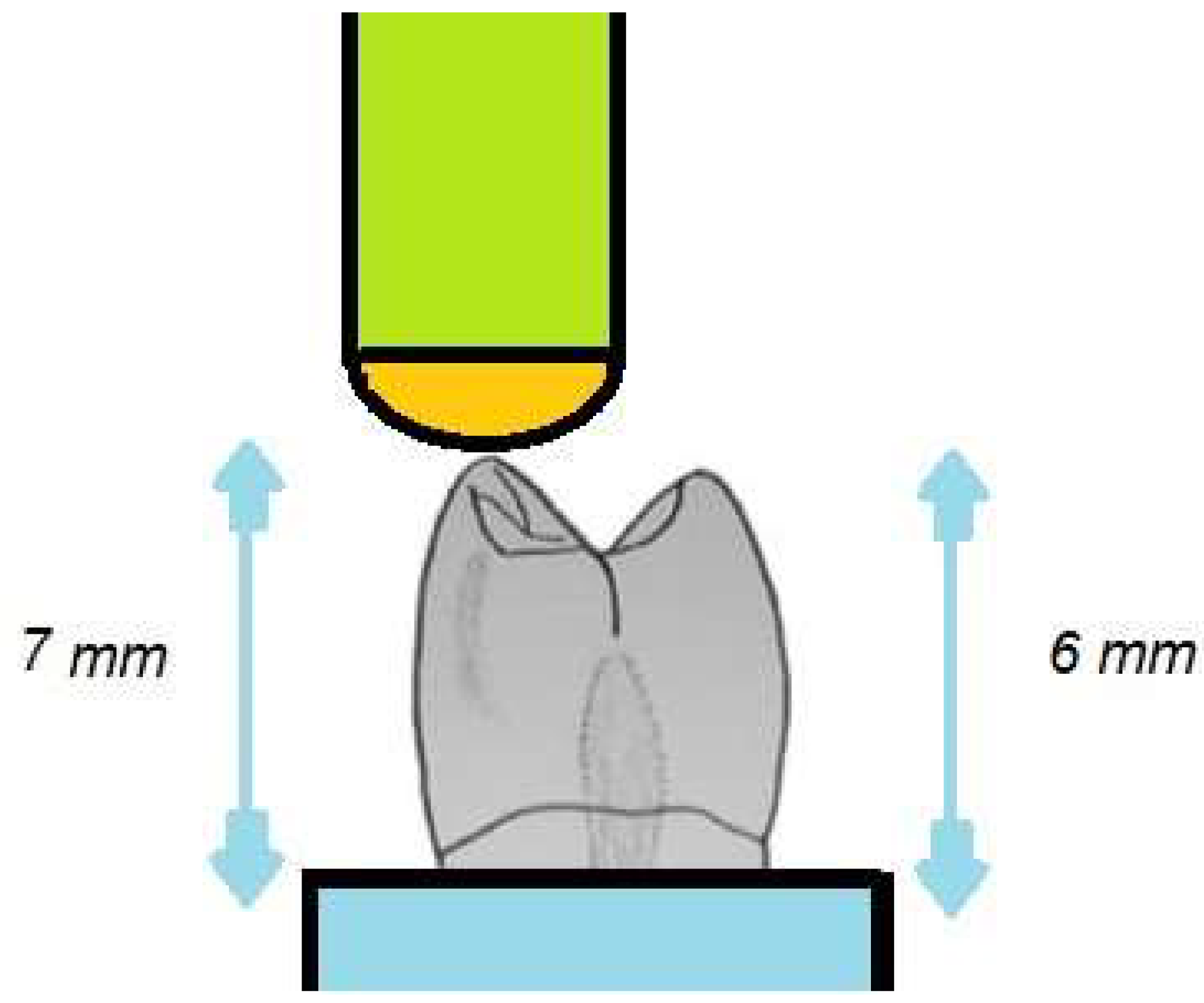
Figure 3. Schematic drawing of the chipping test.
Preprints 104157 g003
Figure 4. Schematic drawing of the indirect tensile fracture test.
Preprints 104157 g004
Figure 5. Interactive plot of the chipping test results.
Preprints 104157 g005
Figure 6. Interactive plot of the indirect tensile fracture test results.
Preprints 104157 g006
Table 1. Types of denture teeth used in this study.
Product Composition Detail Manufacturer
NextDent Methacrylate-based photopolymerized resin 3D printing 3D Systems, Soesterberg, The Netherlands
Ivostar Shade MPM–PMMA * Mold XS Ivoclar Vivadent, Schaan, Liechtenstein
Spofadent™Plus MPM–PMMA * Mold XS SpofaDental, Jičín, Czechia
Major Super Lux MPM–PMMA * Mold XS Major Prodotti Dentari SPA, Moncalieri, TO, Italy
* MPM—multiplex polymer matrix; PMMA—polymethylmethacrylate.
Table 2. Tukey’s Honest Significant Difference post-hoc analysis of fracture toughness of four types of artificial teeth.
(I) V2 (J) V2 Mean Values
(I-J)
Standard Deviation Р 95% - Interval of confidentiality
Lower Upper
NextDent Ivostar Shade -.346413* .022049 <0.01 -.28645 -.17284
Major Super Lux -.325271* .022074 <0.01 -.34831 -.23442
SpofaDent Plus -.478271* .022087 <0.01 -.40421 -.29212
SpofaDent Plus NextDent .429414* .022089 <0.01 .17563 .28646
Major Super Lux -.261857* .022053 .028 -.11876 -.00484
SpofaDent Plus -249657* .022011 <0.01 -.14758 -.06286
Major Super Lux
NextDent .274571* .022049 <0.01 .23852 .34834
Ivostar Shade .141853* .022061 .028 .00487 .11887
SpofaDent Plus -.158002* .022061 .045 -.11508 -.00094
Ivostar Shade NextDent .429277* .022063 <0.01 .29226 .40633
Major Super Lux .314857* .022063 <0.01 .06286 .17687
Ivostar Shade .258300* .022069 .045 .00096 .11508
* The difference in means is significant at the 0.05 significance level.
Disclaimer/Publisher’s Note: The statements, opinions and data contained in all publications are solely those of the individual author(s) and contributor(s) and not of MDPI and/or the editor(s). MDPI and/or the editor(s) disclaim responsibility for any injury to people or property resulting from any ideas, methods, instructions or products referred to in the content.
Copyright: This open access article is published under a Creative Commons CC BY 4.0 license, which permit the free download, distribution, and reuse, provided that the author and preprint are cited in any reuse.
Prerpints.org logo

Preprints.org is a free preprint server supported by MDPI in Basel, Switzerland.

Subscribe

© 2024 MDPI (Basel, Switzerland) unless otherwise stated