
Submitted:
24 April 2024
Posted:
26 April 2024
You are already at the latest version
Abstract
The mortality of birds resulting from collisions and electrocutions with overhead lines, such as power lines, and phone line among others, has been implicated in the decline of various avian species globally. Specifically, overhead line collisions pose a significant threat to the conserva-tion of the Canarian houbara bustard (Chlamydotis undulata fuertaventurae), an endangered sub-species endemic to the Canary Islands.
This study centres on the postmortem findings of Canarian houbara bustards that have collided with overhead lines, providing insights into the post-collision outcomes for these birds. A com-plete standardized necropsy of nine Canarian houbara bustards revealed that polytrauma was the cause of death in all cases. The most notable gross lesions associated with trauma included bone fractures, soft tissue lacerations, haemorrhages, luxations, and hemocoelom, with certain body regions being more frequently affected. Histopathology, immunohistochemistry, and en-tomology analysis confirmed that numerous birds survived the initial trauma, exhibiting varia-ble survival intervals before succumbing to their injuries.
We concluded that when a houbara bustard collide with an overhead line it frequently survives the initial trauma. The histopathology, immunohistochemistry, or entomologic analysis may be helpful to approximate the timing interval between trauma and death.
Keywords:
Houbara bustard
; polytrauma
; cause of death
; collision
; overhead line
; forensic necropsy
; bird population decline
1. Introduction
Mortality of birds with overhead lines (i.e. power lines, phone lines, telegraph wires, etc.) caused by collision and electrocution has been linked to decline of diverse avian species around the world [1,2,3,4]. However, most studies report estimation of mortalities due to power line collisions by documentation of the presence of carcasses along the electric or communication lines, and confirmation of trauma with standardized necropsies are not so commonly performed [2,3,4,5,6]. If initial suspicion of collision is not confirmed with necropsy, other causes of death, such as electrocution, can be overlooked [7].
Although overhead lines constitute a possible hazard for all birds during flight, certain species, such as the bustards, tend to be more predisposed to collision [1,8,9].
The Canarian houbara bustard (Chlamydotis undulata fuertaventurae) is an endemic subspecies of houbara located in Fuerteventura, Lanzarote and La Graciosa in the Canarian Archipelago (Spain) [10] that has been closely monitored during the last years by different scientists [1,10,11]. This subspecies is considered “in danger” on the Spanish Red List [11] and as “vulnerable” by the IUCN (2009) [12], with an estimated population of 525-750 individuals according to different authors [1,11,13,14]. Power line collisions have been recognized as a significant problem for the conservation of these animals [1,6] and recent investigations predict the extinction of the subspecies in 50 years due to anthropogenic mortality [1].
Legislation in different parts of the world may protect endangered species, and when an overhead line is proved to cause repetitive episodes of mortality, corrective measurements must be taken by the owner of problematic overhead lines. Forensic veterinary reports are usually determinant in the judicial process, and to prove a case of collision, there must be clear evidence of blunt force trauma in those reports [15,16]. Moreover, it should be noted that collision with overhead lines is not the only source of trauma for birds, and that an injured bird is more likely to suffered subsequent traumas, such as interactions with other animals (predation) or being hit by a car. These others secondary traumatisms may confuse to the forensic veterinarian who is trying to answer the questions, why and how did this animal die?
In this study, we focus on the lesions observed in houbara bustards that collided with overhead lines and try to determine if the houbara survived the initial trauma.
2. Materials and Methods
This work is part of a more extensive survey of causes of mortality in the Canarian wildlife created by the Canarian Government and known as Red Vigía Canarias (Orden Nº134/2020 de 26 de mayo de 2020).
Inclusions criteria for the study were 1) Animal identified as Canarian houbara bustard; 2) Blunt force trauma as the cause of death; and 3) History suggests collision with an overhead line. Very decomposed animals and animals without known history were excluded.
A complete necropsy was performed in all cases following standardized procedures [17]. Feathers were removed entirely to allow skin observation. Nutritional condition was assessed according to muscle development and the presence of fat. Animals’ age was estimated and included in one of the following categories according to body morphology and skeletal and gonadal development: chick, juvenile or adult.
Samples for histopathological study included adrenal glands, encephalon, oesophagus, eyes, heart, intestine, kidney, liver, lung, pancreas, skeletal muscle, skin, spleen, stomach, testicles, thyroid, tongue, and trachea, which were collected and placed in 4% buffered formalin during 24h and routinely processed. Additionally, when traumatic lesions were observed in any other body location additional tissues, including skin, muscle, and bone, were collected, and processed.
Histochemistry was performed on selected tissue sections, including Periodic Acid Schiff (PAS), phosphotungstic acid hematoxylin (PTAH) and Prussian blue. Chromic acid technique was performed to fix the lipids to the tissues before paraffin embedding, and lung sections were later stained with Oil O Red [18].
Immunohistochemistry against myoglobin and fibrinogen was performed in cases 6, 7 and 9 using previously published methodology [19,20,21]. Briefly, 3 µm thick sections were deparaffined, rehydrated, and labelled with polyclonal anti-myoglobin and anti-fibrinogen anti-bodies (Abcam, Cambridge, United Kingdom), secondary anti-body consisted in polyclonal swine anti-rabbit immunoglobulin (Dako, Glostrup, Denmark) and were visualized using the VECTASTAIN Elite ABC-Peroxidase kit (PK-6100, Vector Laboratories, Peterborough, UK). Negative control followed the same methodology, but primary anti-bodies were omitted. Positive control for myoglobin included the skeletal muscle of each case (internal control) and skeletal muscle from a dolphin with rhabdomyolysis (external control) [20]. Positive control of fibrinogen was the liver of a live-stranded dolphin with extensive hepatocyte vacuolar degeneration [19,21].
Diptera eggs and larvae were collected from case 5 during the necropsy. The eggs were processed for histology and some of them were cultivated inside a sterile container and fed with meat at 25ºC for 5 days [22].
3. Results
Between 2020 and 2023, 98 Canarian houbara bustards were submitted for necropsy. In 15 animals, the cause of death was determined as blunt force trauma. The history of 9 houbaras indicates a collision with an overhead line as the aetiology of trauma (cases 1 to 9, see Table 1). Of these, 8 were submitted from Lanzarote, and 1 was submitted from Fuerteventura. 5 of the animals were male, 4 were female, and in one case, the sex could not be determined due to predation (Case 8). The state of decomposition was established during necropsy, resulting in 7 animals being fresh, and 2 carcasses were incipiently decomposed. 7 of the 9 animals ported a GPS transmitter attached to the back with elastic bands included in another study performed by the Natural Sciences History Museum of Madrid (CSIC) [1].
Two cases were found under a high-voltage power line, whereas the remaining animals were found near medium-voltage electric lines or phone lines (case 2). The distance from the lines ranged from 0 to 400 meters. For more details of the GPS track information, see Alonso et al. (2024) [1].
3.1. Gross Lesions
Lesions associated with blunt force trauma were observed in all the animals and summarized in Table 1. Following, we describe the most significant traumatic lesions and body regions affected:
Bone fractures were observed in most cases (6/9). Fractures in the furcule were noted in 4 animals. Two affected the left side (Cases 2 and 4), and the other were bilateral (Cases 6 and 9). Fractures of the sternum were observed together with the furcule fracture in cases 6 (Figure 1A) and 9. The left humerus was fractured in case 2, whereas in case 1, a multiple fracture of the diaphysis of the left tibiotarsum was noted (Figure 1B). In all cases, the fractures were associated with different degree of haemorrhage, ranging from very mild in the furcule fractures to extensive hematomas in the tibiotarsum fracture. As an incidental finding, Case 3 presented a healed keel fracture. More than one bone was fractured in 44,4% (n=4) of the animals.
Skin and muscle lacerations were observed in 5 of the 9 animals. In Cases 2 and 3, the left pectoral and inguinal regions had skin lacerations with muscle fibres rupture. In Case 5, the skin laceration was in the left ingle (Figure 1C), whereas in Case 4 and 9, the pectoral and axillar regions were involved.
Haemorrhages indicative of recent trauma were detected in all cases. The location of the haemorrhages frequently coincides with areas of bone fractures and skin lacerations. Extensive muscle haemorrhages were associated with bone fractures (Case 1), luxations (Case 5), and GPS elastic bands (Cases 7 and 9).
Luxations were observed in 2 cases. Luxation of the left knee, with severe focally extensive acute hematoma and left inguinal laceration, was noted in Case 5 (Figure 1E). Case 7 presented bilateral coracoid luxation with avulsive fracture of the body of the sternum.
Fatal internal bleeding was noted in 4 cases. Three of the animals developed hemocoelom (Cases 4, 6 and 9) (Figure 1F), whereas case 7 had a severe hematoma between muscle pectoralis and muscle supracoracoideus of the left side.
Attending the affected body region, in most cases, the trauma was located at the ingle (7/9), followed by the chest (4/9), the wings (3/9), the shoulder (3/9), and the neck (3/9). Head trauma was not reported in any case.
Other non-traumatic gross findings included moderate to severe intestinal cestode parasitation in 6 animals (Cases 3, 4, 5, 7, 8 and 9) and moderate generalized muscle and fat atrophy (indicative of poor nutritional condition) in cases 1 and 4.
3.2. Histopathology
Histopathology was performed in 8 of the 9 animals. The most common lesioned tissue was skeletal muscle, and the most representative traumatic lesions were acute skeletal muscle degeneration and segmental necrosis (Figure 2A). These lesions were noted in 6 cases in the pectoral muscle (Cases 1, 2, 3, 4, 7 and 9) and in the quadriceps femoris in Case 5. Muscle haemorrhages were also reported in the pectoral muscle of 3 cases (Case 2, 7 and 9) and the quadriceps femoris of Case 5. Case 7 presented macrophages associated with necrotic muscle fibres, which indicate some chronicity.
Acute myocardial degeneration and necrosis were observed in 4 cases (Cases 1, 2, 5 and 9). Degeneration consisted of nuclear pyknosis with hyperacidophilia (Figure 2B), hypercontraction bands (Figure 2C and 2D) and vacuolization of the sarcoplasm.
Severe pulmonary haemorrhages and oedema were reported in 5 animals. Other lung changes included marked congestion (Cases 5, 7 and 9) and atelectasis (Case 9). Additionally, fat embolism was investigated in three animals (Cases 1, 2 and 3) using Oil O Red, but the lung sections resulted negative for the presence of fat in the lumen of blood vessels in all the sections analyzed.
3.3. Immunohistochemistry
Fibrinogen immunohistochemistry revealed immunopositivity in the intracytoplasmic vacuoles of the hepatocytes of case 9 (Figure 3A), as well as strong intrasarcoplasmic and sarcolemma immunostaining in degenerated myocytes of the skeletal muscle of cases 6 and 7 (Figure 3B). Myoglobin immunohistochemistry revealed intrasarcoplasmic fibrillar staining of cardiac and skeletal myocytes (internal positive control), but degenerated skeletal myocytes showed segmental depletion of myoglobin and extracellular deposition of myoglobin globules (Figure 3C). Renal tissue was negative to myoglobin in all cases (internal negative control). External positive and negative controls stained as expected.
3.4. Entomology
Cases 2 and 5 presented subcutaneous myiasis, presenting numerous eggs and larvae of diptera from 3 to 13 mm (Figure 4A). The histology of the diptera eggs of case 5 showed that most embryos had development of the proctodeo and estomatodeo (Figure 4B), whereas few of them presented full development, which is consistent with embryos between 12 and 24 hours of development [22]. 3 mm larvae hatched after 24 h of egg culturing. After 48 hours, only 13 mm larvae remained in the container, and larger larvae were not observed after additional 72 hours. These results indicate that 3, 8 and 13 mm larvae correspond to instar I, II and III and that III instar develops after 72h.
The entomology, in association with the GPS information and fresh status of the carcass, confirms that the animal collided with the overhead line and survived at least 24 hours [23]. By that time, numerous flies had deposited eggs, causing the myiasis. When the animal finally died, diptera colonization progressed in the carcass until its preservation in refrigeration and packaging for necropsy.
4. Discussion
Based on the postmortem examination, all the animals included in this study died because of blunt-force trauma. That, together with the animal’s history (found near or under overhead lines) and the traumatic pattern, is consistent with a collision with the line as the origin of the trauma.
Different patterns of trauma have been described and related to various sources of trauma. Lesions due to collisions with aircraft, road kills, and collisions with windows are reported in the literature [24,25]. Still, there are scarce reports detailing the traumatic pattern of collisions with overhead lines. Early reports described wind fractures and head trauma in Passeriformes, as well as extremities amputations in small Charadriiforms who collide with the lines [26]. Although we only observed postmortem extremity amputation in one predated animal (Case 8), the lack of extremity amputations may be explained by the houbaras robust skeleton and slower fly compared to Charadriiforms.
In one study of cranes from Japan [5], it was observed that birds who suffer overhead line collisions suffered more commonly fractures of legs and winds, but sternal, costal, coracoidal and vertebral fractures were also reported. The 29% of the animals suspected to die due to overhead collision experienced more than one bone fracture. Large wings and long, thin legs of cranes were noted to be the most susceptible body parts for this type of traumatism. Miller et al. (2010) [27] reported different “leg problems” in whooping cranes (Grus americana), which included legs fracture and hip luxation related to collision. The houbara bustard had shorter and more muscular pelvic limbs, which may explain why we only reported one leg fracture and a knee luxation. Moreover, no head trauma was reported in our study, nor by Takase et al.
Collision with overhead lines was determined to be a relevant cause of mortality in California condors (Gymnogyps californianus) [28]. The authors diagnose collision with power lines in cases where animals presented compatible traumatic injuries and a history of being found in the immediate vicinity of power lines. In another study about causes of mortality in raptors, the authors reported a 1.1% incidence of trauma with power lines as the cause of death but not detailed information about the traumatic lesions [29]. Fanke et al. (2011) [30], reported an incidence of 23.4% of mortality due to power line collision on Eurasian cranes (Grus grus), with trauma along the shoulder girdle, chest, or long bones.
Seven animals in this study ported a GPS transmitter attached to the back with elastic bands. Chronic lesions associated with the transmitter, or the harnesses could not be detected in any of them. However, acute lesions, including haemorrhages, bruising, and lacerations, were noted to coincide with the elastic bands of the transmitter in two cases, which opens the question: could these animals collide because of the GPS? It should be mentioned that other studies reported a relationship between birds carrying transmitters and those who collide [27,31,32]. But Alonso et al. (2024) monitored with GPS a total of 51 Canarian houbara bustards of which only 7 collided. To us, it is uncertain if the transmitter predisposes these animals to collision with overhead lines or if the corpses were more easily found, submitted to necropsy, and linked with the line because of the transmitter information.
Fibrinogen is produced by hepatocytes and resealed in the plasma during stress response and can be detected in degenerated myocytes. In the dog and the mice, the immunohistochemical expression of fibrinogen is greater at more than 60 minutes after trauma [33]. We reported fibrinogen in the hepatocytes forming intracytoplasmic vacuoles in case 9. Also, we observed intra and extracellular globular accumulation of myoglobin and sarcoplasmic fibrinogen deposition, which confirm severe acute muscle degeneration in cases 6 and 7. These results prove that the animals survived initial trauma for some time (minutes to hours). There are many studies about the formation of fibrinogen in avian species. Still, most of them are focused on the inflammatory response of different infectious agents [34]. In contrast, information about the timing of fibrinogen formation after traumatic injuries in avian species is limited and merits further studies.
When traumatic mortalities are investigated in avian species, most of the studies focus on gross lesions, and little is mentioned about complementary studies that may provide information about survival after the initial trauma [2,3,4,5,6,26,27]. In one study, the authors determined that 36% of whooping cranes (Grus americana) that collide with overhead lines survived the trauma [27], whereas Bech et al. (2012) [35] demonstrated that rock ptarmigan hen (Lagopus muta) can fly up to 600 m after collision. In our cases, the histopathology, immunohistochemistry, and entomology support that 44,4% (n=4) of animals survived the trauma but died shortly after due to internal bleeding, following an uncertain period ranging from few minutes to hours (even days, such as in case 5). It has been reported that birds who collide may survive and have another collision [27]. In Case 3, a healed keel fracture was reported, suggesting that this animal could previously collide and survive the trauma.
It has been reported that “poor” fliers and birds with nocturnal flight behaviour are more prone to overhead line collisions [8,9]. Moreover, two species of bustards from Spain, the great bustard (Otis tarda) and little bustard (Tetrax tetrax) had the highest record for collision casualties in a study that included 52 different species [9]. It should be highlighted that the houbara bustard has crepuscular behaviour and is heavier than other birds, which surely predisposes them to collide with overhead lines. To explain the lesions observed, we hypothesize that the houbara may collide with the line, as depicted in Figure 5. The animal can hit the line while flying with the wing extended dorsally (Figure 5A) or ventrally (Figure 5B). In that case, the trauma may affect the wings, as observed in cases 2, 7, and 9. More frequently, the houbara could detect the line at the last moment and try to avoid it, resulting in chest, inguinal or leg trauma during the skipping manoeuvre (Figure 5C), as we detected in cases 1, 3, 5, 6 and 8.
It should be noted that secondary traumatism may occur, produced by the impact against the floor after the collision with the line, so coexistence of various patterns of trauma may be expected. The main reported lesions in waterfowls that collide against the floor were liver rupture, hemocoelom and pulmonary haemorrhages, and occasional sternum and ribs fractures [36]. We observed hemocoelom in cases 4, 6 and 9, but we cannot confirm if the internal haemorrhages debuted from the trauma with the line or subsequent traumatisms after landing or even predation of an injured bird.
5. Conclusions
In conclusion, when a houbara bustard collide with an overhead line it frequently survives the initial trauma. The histopathology, immunohistochemistry, or entomologic analysis may be helpful to approximate the timing interval between trauma and death.
Author Contributions
“Conceptualization, C.S.S. and A.F.; methodology, C.S.S, L.M.P. J.N.S, C.R.S., R.G.G., A.C.R.; validation, E.S., and R.G.G.; investigation, C.S.S.; resources, C.S.S.; data curation, C.S.S. and R.G.G..; writing—original draft preparation, C.S:S. writing—review and editing, O.Q.C.; supervision, A.F. and E.S.; funding acquisition, A.F:. All authors have read and agreed to the published version of the manuscript.”
Funding
This work has been performed with the economic and logistical support from the “Dirección General de Lucha Contra el Cambio Climático y Medio Ambiente” under the creation of the Canarian Network for the Surveillance of the Wildlife Health (Orden Nº134/2020 de 26 de mayo de 2020).
Data Availability Statement
The data presented in this study are available on request from the corresponding author due to legal reasons.
Acknowledgments
We want to thanks to Juan C. Alonso and the team from the CSIC who dedicates great efforts to the study of the Canarian houbara bustard, as well as to the technicians and Ambiental Police officers that collaborate in the collection and shipping of the carcass.
Conflicts of Interest
The authors declare no conflicts of interest.
References
- Alonso, J. C.; Abril-Colón, I.; Ucero, A.; Palacín, C. Anthropogenic mortality threatens the survival of Canarian houbara bustards. Scientific Reports 2024, 14(1), 2056. [Google Scholar] [CrossRef]
- Shaw, J. M.; Reid, T. A.; Schutgens, M.; Jenkins, A. R.; Ryan, P. G. High power line collision mortality of threatened bustards at a regional scale in the Karoo, South Africa. Ibis 2018, 160, 431–446. [Google Scholar] [CrossRef]
- Loss, S. R.; Will, T.; Marra, P. P. Direct Mortality of Birds from Anthropogenic Causes. Annual Review of Ecology, Evolution, and Systematics. 2015, 46(Volume 46, 2015), 99–120. [CrossRef]
- Bevanger, K. Estimates and Population Consequences of Tetraonid Mortality Caused by Collisions with High Tension Power Lines in Norway. Journal of Applied Ecology 1995, 32, 745–753. [Google Scholar] [CrossRef]
- Takase, K.; Haraguchi, Y.; Suzuki, A.; Obi, T. Fracture status of wild cranes (Grus monacha and G. vipio) found dead or in a weak condition at Izumi Plain in Japan. Journal of Veterinary Medical Science 2020, 82, 823–826. [Google Scholar] [CrossRef]
- Garcia-del-Rey, E.; Rodriguez-Lorenzo, J. A. Avian mortality due to power lines in the Canary Islands with special reference to the steppe-land birds. Journal of Natural History 2011, 45, 2159–2169. [Google Scholar] [CrossRef]
- Loss, S. R.; Will, T.; Marra, P. P. Refining Estimates of Bird Collision and Electrocution Mortality at Power Lines in the United States. PLOS ONE 2014, 9(7), e101565. [Google Scholar] [CrossRef]
- Deng, J.; Frederick, P. Nocturnal Flight Behavior of Waterbirds in Close Proximity to a Transmission Powerline in the Florida Everglades. Waterbirds: The International Journal of Waterbird Biology 2001, 24, 419–424. [Google Scholar] [CrossRef]
- Janss, G. F. E. Avian mortality from power lines: a morphologic approach of a species-specific mortality. Biological Conservation 2000, 95, 353–359. [Google Scholar] [CrossRef]
- Martín, A.; Lorenzo, J. A.; Hernández, M. A.; Nogales, M.; Medina, F. M.; Delgado, J. D.; et al. Distribution, status and conservation of the Houbara Bustard Chlamydotis undulata fuertaventurae Rothschild & Hartert, 1894, in the Canary Islands, November-December 1994. Ardeola 1997, 44, 61–69. [Google Scholar]
- Lorenzo, J. A. Avutarda Hubara (Canaria) Chlamydotis undulata fuertaventurae. Libro Rojo de las Aves de España. Dirección General para la Biodiversidad-SEO/BirdLife.Madrid. 2004. [Google Scholar]
- The IUCN Red List of Threatened Species. IUCN Red List of Threatened Species. <https://www.iucnredlist.org/en> Accessed 24.04.18.
- Schuster, C.; Iglesias-Lebrija, J. J.; Carrascal, L. M. Recent population trends of the houbara bustard in the Canary Islands. Methods and conservation status. Animal Biodiversity and Conservation 2012, 35, 125–139. [Google Scholar] [CrossRef]
- Goriup, P. D. The world status of the Houbara Bustard Chlamydotis undulata. Bird Conservation International 1997, 7, 373–397. [Google Scholar] [CrossRef]
- Brownlie, H. W. B.; Munro, R. The Veterinary Forensic Necropsy: A Review of Procedures and Protocols. Veterinary Pathology 2016, 53, 919–928. [Google Scholar] [CrossRef]
- Ressel, L.; Hetzel, U.; Ricci, E. Blunt Force Trauma in Veterinary Forensic Pathology. Veterinary Pathology 2016, 53, 941–961. [Google Scholar] [CrossRef]
- Rae, M. Practical avian necropsy. Seminars in Avian and Exotic Pet Medicine 2003, 12, 62–70. [Google Scholar] [CrossRef]
- Arregui, M.; Fernández, A.; Paz-Sánchez, Y.; Santana, Á.; Sacchini, S.; Sierra, E.; et al. Comparison of Three Histological Techniques for Fat Emboli Detection in Lung Cetacean’s Tissue. Scientific Reports 2020, 10 1, 8251. [Google Scholar] [CrossRef]
- Fernández, A.; Câmara, N.; Sierra, E.; Arbelo, M.; Bernaldo de Quirós, Y.; Jepson, P. D.; et al. Cetacean Intracytoplasmic Eosinophilic Globules: A Cytomorphological, Histological, Histochemical, Immunohistochemical, and Proteomic Characterization. Animals 2023, 13(13), 2130. [Google Scholar] [CrossRef]
- Câmara, N.; Sierra, E.; Fernández, A.; Suárez-Santana, C. M.; Puig-Lozano, R.; Arbelo, M.; et al. Skeletal and Cardiac Rhabdomyolysis in a Live-Stranded Neonatal Bryde’s Whale With Fetal Distress. Frontiers in Veterinary Science 2019, 6. [Google Scholar] [CrossRef]
- Arbelo, M.; Monteros, A. E. de los; Herráez, P.; Andrada, M.; Sierra, E.; Rodríguez, F.; et al. Pathology and causes of death of stranded cetaceans in the Canary Islands (1999−2005). Diseases of Aquatic Organisms 2013, 103, 87–99. [Google Scholar] [CrossRef]
- Pais, M.; Archer, M. S. Histological age estimation of the eggs of Calliphora vicina Robineau Desvoidy (Diptera: Calliphoridae). Forensic Sciences Research 2018, 3, 40–51. [Google Scholar] [CrossRef]
- Brundage, A.; Byrd, J. H. Forensic Entomology in Animal Cruelty Cases. Veterinary Pathology 2016, 53, 898–909. [Google Scholar] [CrossRef]
- Sheehy, S.; Kelly, T. c.; Bourke, P.; O’Callaghan, M.; Fennessy, G.; Bolger, R. A comparison of the injury syndromes associated with different sources of avian mortality. International Bird Strike Committee, Warsaw, 2003. [Google Scholar]
- Lyne, K.; Gassner, I.; Bolger, R.; Kelly, T. Is there a bird strike syndrome?: preliminary results from autopsy findings. Proceedings of the International Bird Strike Committee 1998, 24, 97–124. [Google Scholar]
- Coues, E. The Destruction of Birds by Telegraph Wire. The American Naturalist 1876, 10, 734–736. [Google Scholar] [CrossRef]
- MILLER, J. L.; SPALDING, M. G.; FOLK, M. J. LEG PROBLEMS AND POWER LINE INTERACTIONS IN THE FLORIDA RESIDENT FLOCK OF WHOOPING CRANES. Proceedings of the Eleventh North American Crane Workshop, Sep 23-27, 2008, Wisconsin Dells, Wisconsin, 2010, 156–165.
- Rideout, B. A.; Stalis, I.; Papendick, R.; Pessier, A.; Puschner, B.; Finkelstein, M. E.; et al. PATTERNS OF MORTALITY IN FREE-RANGING CALIFORNIA CONDORS (GYMNOGYPS CALIFORNIANUS). Journal of Wildlife Diseases 2012, 48, 95–112. [Google Scholar] [CrossRef]
- Martínez, J. E.; Zuberogoitia, I.; Jiménez-Franco, M. V.; Mañosa, S.; Calvo, J. F. Spatio-temporal variations in mortality causes of two migratory forest raptors in Spain. European Journal of Wildlife Research 2016, 62, 109–118. [Google Scholar] [CrossRef]
- Fanke, J.; Wibbelt, G.; Krone, O. MORTALITY FACTORS AND DISEASES IN FREE-RANGING EURASIAN CRANES (GRUS GRUS) IN GERMANY. Journal of Wildlife Diseases 2011, 47, 627–637. [Google Scholar] [CrossRef]
- Michael, S.; Gartrell, B.; Hunter, S. HUMERAL REMODELING AND SOFT TISSUE INJURY OF THE WINGS CAUSED BY BACKPACK HARNESSES FOR RADIO TRANSMITTERS IN NEW ZEALAND TAKAHĒ (PORPHYRIO HOCHSTETTERI). Journal of Wildlife Diseases 2013, 49, 552–559. [Google Scholar] [CrossRef]
- Peniche, G.; Vaughan-Higgins, R.; Carter, I.; Pocknell, A.; Simpson, D.; Sainsbury, A. Long-term health effects of harness-mounted radio transmitters in red kites (Milvus milvus) in England. Veterinary Record 2011, 169, 311–311. [Google Scholar] [CrossRef]
- Fechner, G.; Bajanowski, Th.; Brinkmann, B. Immuuohistochemical alterations after muscle trauma. International Journal of Legal Medicine 1993, 105, 203–207. [Google Scholar] [CrossRef]
- O’Reilly, E. L.; Eckersall, P. D. Acute phase proteins: a review of their function, behaviour and measurement in chickens. World’s Poultry Science Journal 2014, 70, 27–44. [Google Scholar] [CrossRef]
- Bech, N.; Beltran, S.; Boissier, J.; Allienne, J. F.; Resseguier, J.; Novoa, C. Bird mortality related to collisions with ski–lift cables: do we estimate just the tip of the iceberg? Animal Biodiversity and Conservation 2012, 35, 95–98. [Google Scholar] [CrossRef]
- Wobeser, G.; Gillespie, M.; Wyatt, T. Mortality of Geese as a Result of Collision with the Ground. Journal of Wildlife Diseases 2005, 41, 463–466. [Google Scholar] [CrossRef]
Figure 1.
Macroscopic findings. A) Case 9. Avulsive fracture of the sternum. B) Case 1. Fracture of the tibiotarsum. C) Case 5. Inguinal skin abrasion and laceration. D) Case 9. Focally extensive hematoma in the left forearm. E) Case 5. Knee luxation (X-rays). Inset: Knee hemartrosis. F) Case 9. Hemocoelom.
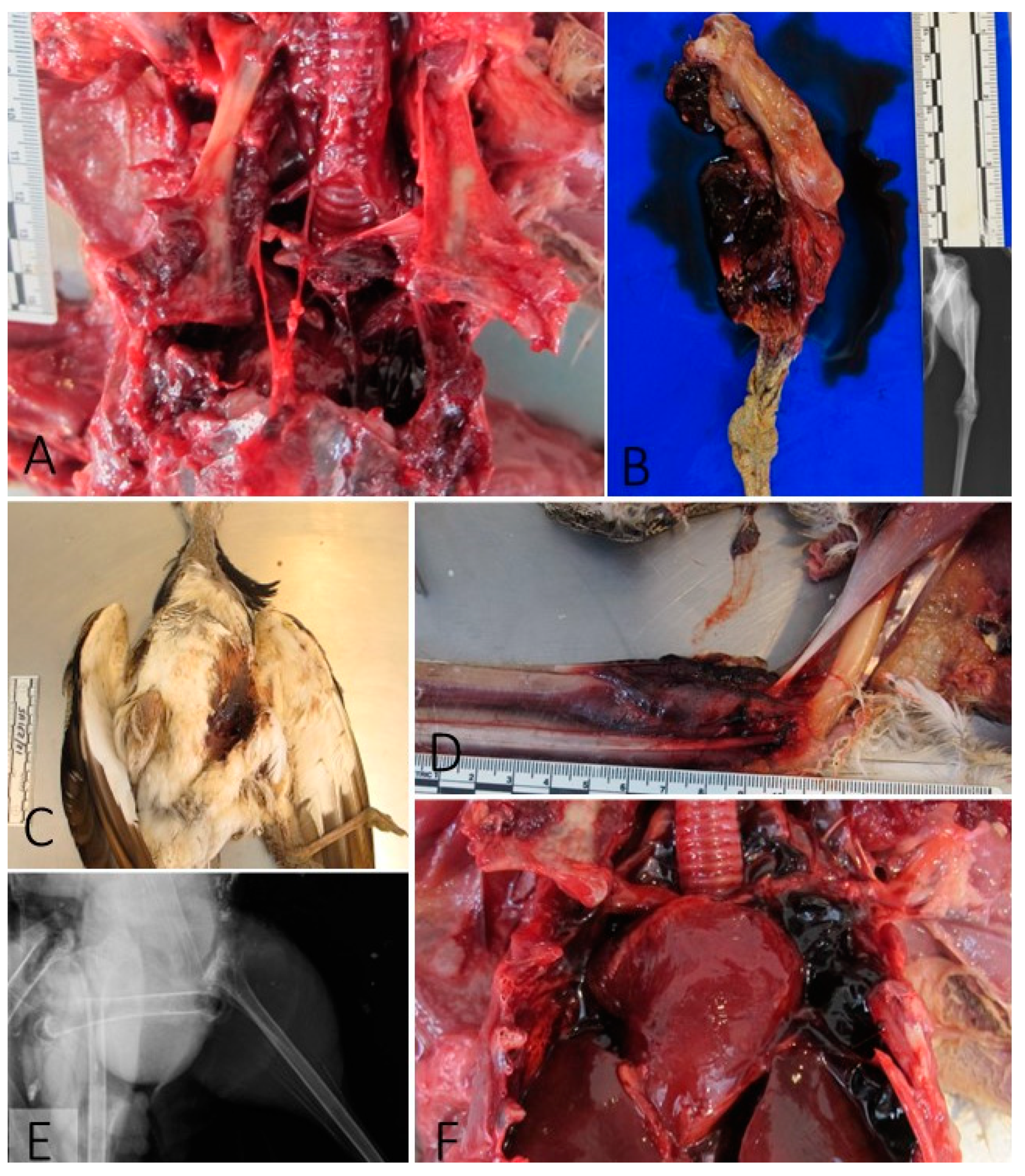
Figure 2.
Histopathological findings. A) Case 9. Segmental necrosis of skeletal muscle (pectoral muscle). B-D) Case 5. Acute myocardial degeneration (B and C, HE). (D, PTAH).
Figure 2.
Histopathological findings. A) Case 9. Segmental necrosis of skeletal muscle (pectoral muscle). B-D) Case 5. Acute myocardial degeneration (B and C, HE). (D, PTAH).
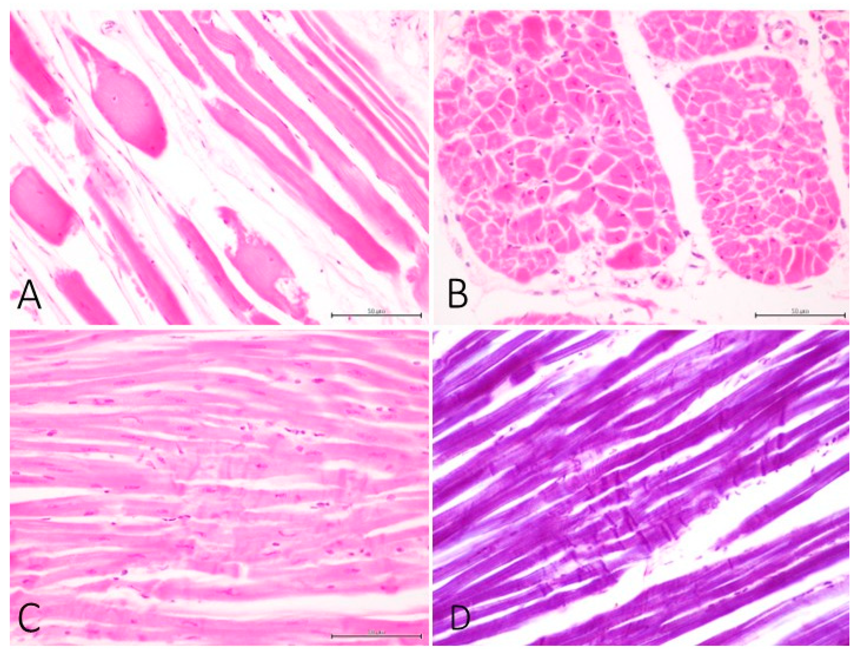
Figure 3.
Immunohistochemistry for fibrinogen (A and B) and myoglobin (C). A) Case 9. Hepatocytes present intracytoplasmic vacuoles that are labelled with anti-fibrinogen Ab. B) Case 6. Skeletal muscle (pectoral) with degenerated myocytes positive to anti-fibrinogen Ab. C) Case 9. Skeletal muscle (pectoral) with segmental depletion of myoglobin and extracellular deposition of myoglobin.
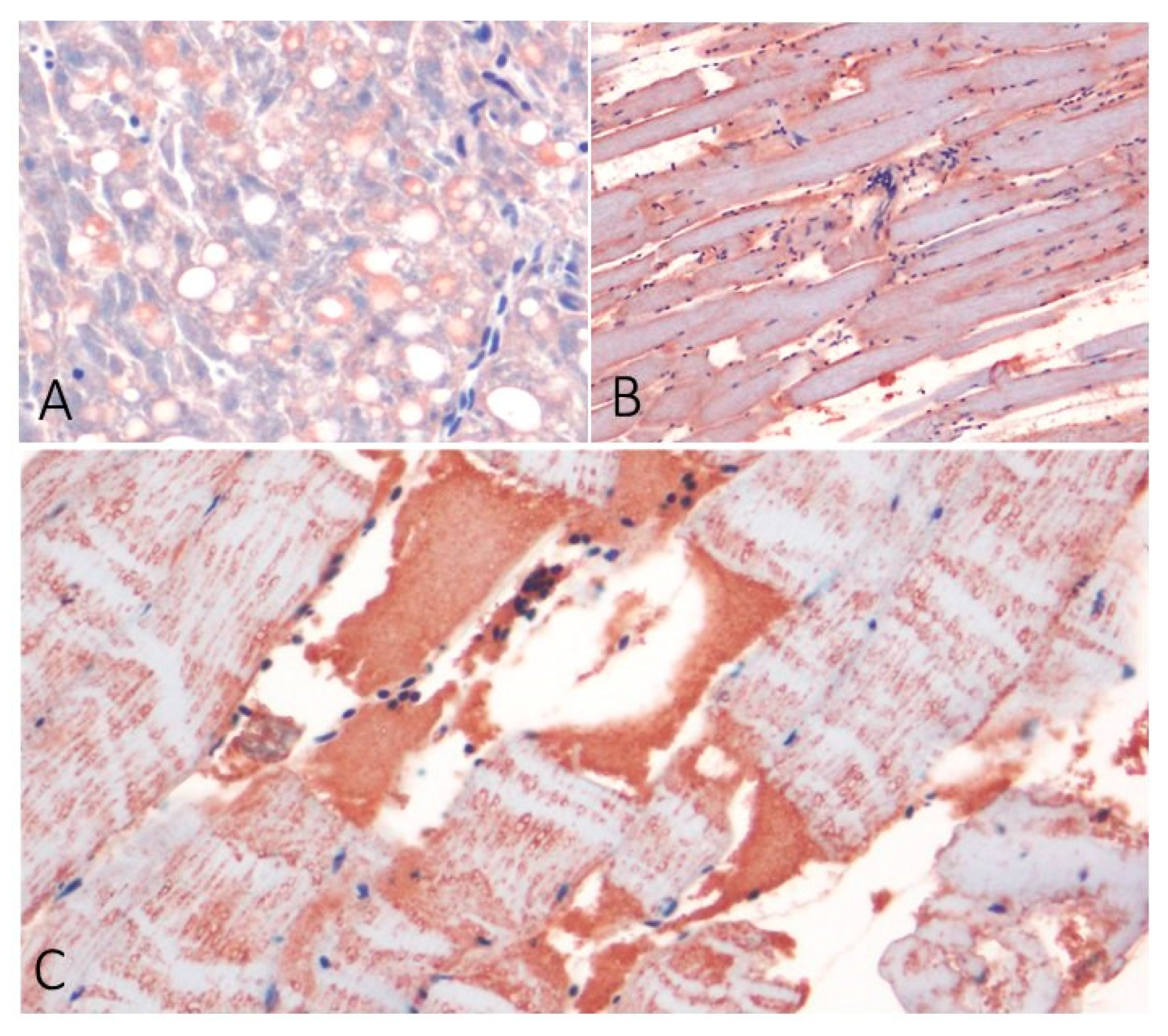
Figure 4.
Entomology. A) Case 2. Presence of larvae of diptera of various stages over a lacerated skin area. B) Case 5. Diptera egg processed for histopathology presented an embryo with early development of digestive structures (HE).
Figure 4.
Entomology. A) Case 2. Presence of larvae of diptera of various stages over a lacerated skin area. B) Case 5. Diptera egg processed for histopathology presented an embryo with early development of digestive structures (HE).
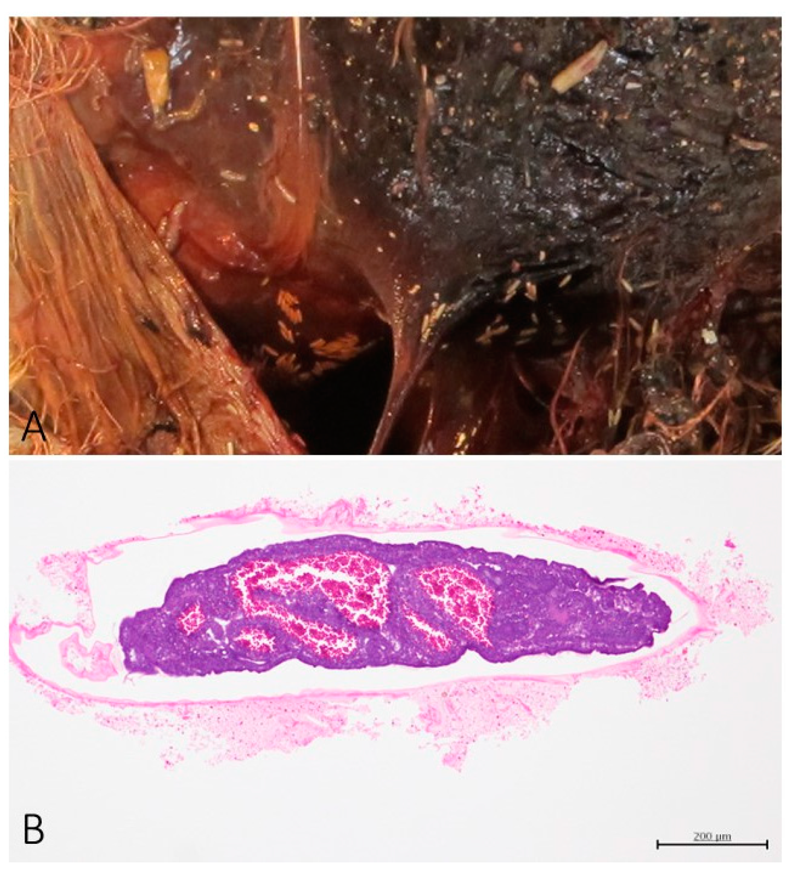
Figure 5.
Suggested possibilities for the collisions of the houbara with the lines.
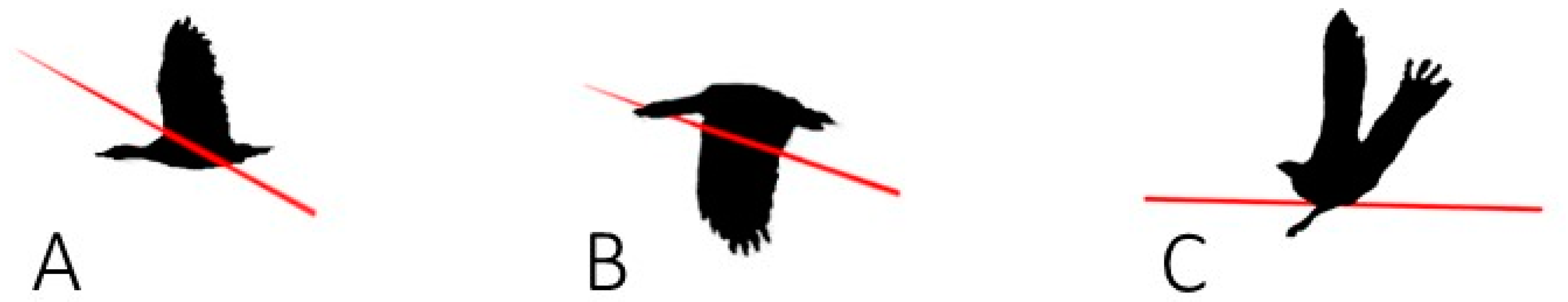

Table 1.
Location and frequency of the traumatic lesions.
| Case Nº | Inguinal | Skin laceration | Chest | Wing | Hemocoelom | Shoulder | Neck | Join luxation | Knee/ leg | Head |
| 1 | x | |||||||||
| 2 | x | x | x | x | ||||||
| 3 | x | x | x | |||||||
| 4 | x | x | x | x | ||||||
| 5 | x | x | x | x | ||||||
| 6 | x | x | x | x | x | |||||
| 7 | x | x | x | |||||||
| 8 | x | x | ||||||||
| 9 | x | x | x | x | x | x | ||||
| % | 77.7 | 55.5 | 44.4 | 33.3 | 33.3 | 33.3 | 33.3 | 22.2 | 22.2 | 0 |
Disclaimer/Publisher’s Note: The statements, opinions and data contained in all publications are solely those of the individual author(s) and contributor(s) and not of MDPI and/or the editor(s). MDPI and/or the editor(s) disclaim responsibility for any injury to people or property resulting from any ideas, methods, instructions or products referred to in the content. |
© 2024 by the authors. Licensee MDPI, Basel, Switzerland. This article is an open access article distributed under the terms and conditions of the Creative Commons Attribution (CC BY) license (http://creativecommons.org/licenses/by/4.0/).
Copyright: This open access article is published under a Creative Commons CC BY 4.0 license, which permit the free download, distribution, and reuse, provided that the author and preprint are cited in any reuse.


