1. Introduction
1.1. The Intersection of Physics and Biology
The interaction between electromagnetic fields and biological systems is a complex and multifaceted subject that has intrigued scientists for decades. At the intersection of physics and biology, this interaction opens doors to a wide array of applications and insights, particularly in the realm of neuroscience. The human brain, a sophisticated network of neural circuits, communicates through electrical signals. Understanding how these signals can be influenced by electromagnetic fields is a subject of ongoing research and debate (Valberg et al., 1997).
1.2. Electromagnetic Fields: A Primer
Electromagnetic fields (EMFs) are created by electrically charged objects and encompass both electric and magnetic fields. These fields can vary in frequency, intensity, and orientation, and their effects on biological systems can be equally diverse (Repacholi & Greenebaum, 1999). From the Earth's magnetic field to the EMFs generated by electronic devices, these invisible forces are a constant presence in our environment.
1.3. Neural Circuits: The Building Blocks of Thought
Neural circuits are intricate networks of interconnected neurons that transmit information through electrical impulses. These impulses travel along nerve fibers and are essential for everything from basic bodily functions to complex thoughts and emotions. The delicate balance and precise timing of these signals are crucial for the proper functioning of the nervous system (Cook et al., 2002).
1.4. The Interaction Between EMFs and Neural Circuits on Themselves
The idea that electromagnetic fields can influence each other in neural circuits is not new, but the mechanisms and implications of this interaction are still being explored. We know that several theories and observations provide insights into how EMFs might affect neural activity:
Direct Influence on Neurons: EMFs can induce electric currents in neural tissues (Nunes et al., 2020), potentially affecting the membrane potential of neurons. This could alter the firing patterns of neurons, leading to changes in signal transmission.
Effects on Synaptic Transmission: The synapses, where neurons communicate with each other, might be sensitive to EMFs (Cook et al., 2002). Changes in synaptic transmission could lead to altered neural network dynamics.
Influence on Neural Development: Some studies have suggested that EMFs might affect the growth and development of neural tissues, possibly influencing brain structure and function (Kaplan et al., 2016).
Therapeutic Applications: Transcranial Magnetic Stimulation (TMS) is an example of how controlled electromagnetic fields are used to modulate neural activity for therapeutic purposes, such as treating depression or migraines (O'Reardon et al., 2007).
Potential Risks and Controversies: While the therapeutic potential of EMFs is promising, concerns have been raised about the possible adverse effects of exposure to uncontrolled or high-intensity EMFs, particularly from electronic devices (Sadock et al., 2009).
2. Methodology
2.1 Mathematical Modeling
A mathematical model was developed to represent the neural circuits and their magnetic fields. The model considered various factors such as the strength of the magnetic field, the properties of the neural tissues, and the dynamics of the electrical signals within the neurons.
Mathematical Equations
is the -th time point.
is the total time duration (100 in this case).
is the number of time points ( 1000 in this case).
- 2.
Voltage for Neuron in Group 1:
is the voltage at time for Neuron 0 in Group 1.
is the mean voltage for Group .
is the standard deviation of the voltage for Group .
is a standard normal random variable (i.e., a Gaussian white noise proce
- 3.
Voltage for Neuron in Group 2:
where:
is the voltage at time for Neuron in Group 2.
is the mean voltage for Group .
is the standard deviation of the voltage for Group .
is a standard normal random variable (i.e., a Gaussian white noise proce
Summary of Parameters
Total time duration,
Number of time points,
Mean voltage for Group
Standard deviation for Group
Mean voltage for Group
Standard deviation for Group
These equations describe a simple model where the voltage of each neuron is modeled as a Gaussian random process with specified mean and variance. The actual voltage values are generated by adding Gaussian white noise to the mean voltage value for each group.
2.1. Computational Simulation
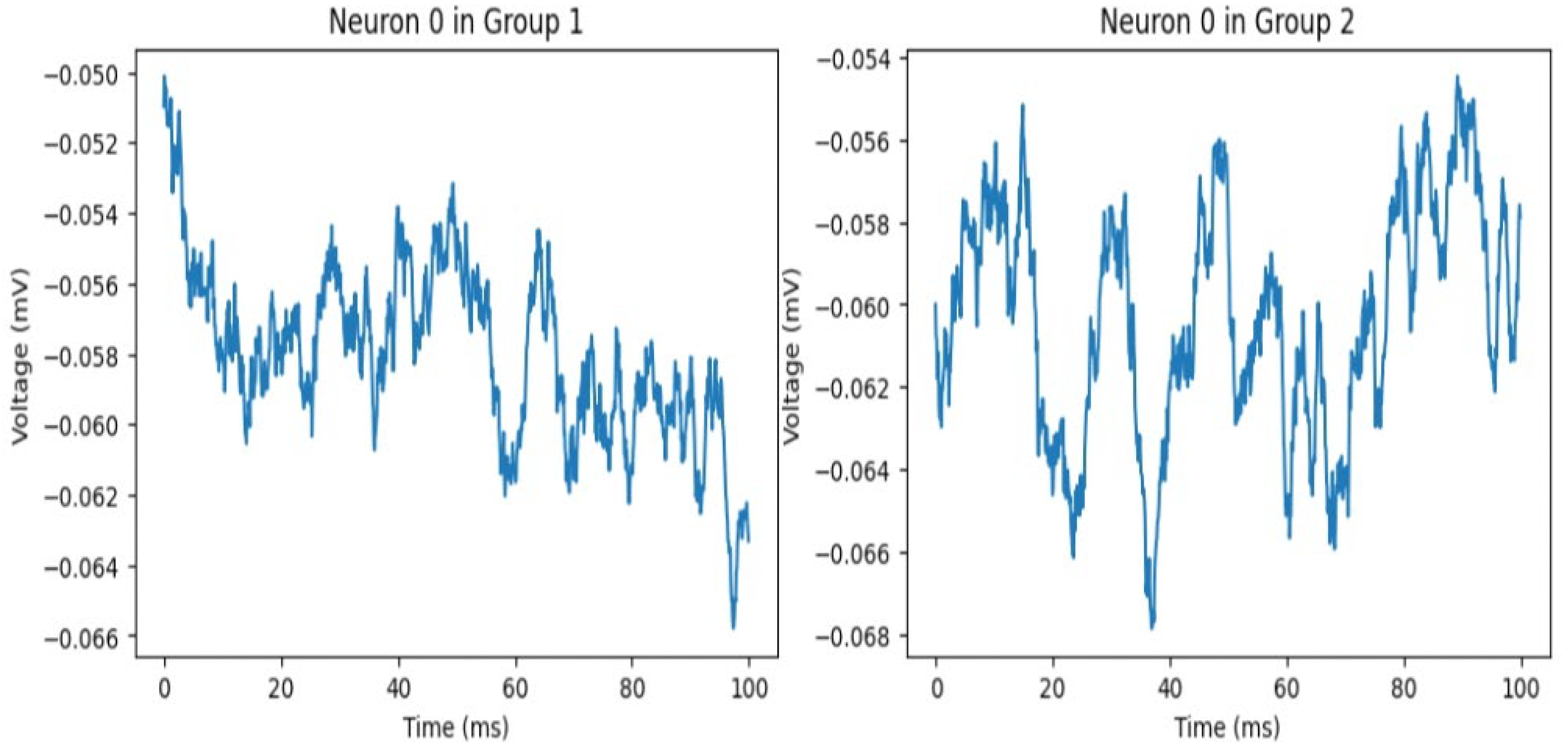
The mathematical model was implemented using Python, leveraging libraries such as NumPy and Matplotlib. A series of simulations were run to observe how different parameters influenced the behavior of the neural circuits. The images below show an example of interaction between two hypothetical neurons in group 1 and group 2.
3. Results
Observations on the Graphs
The provided graphs represent voltage measurements over time for Neuron 0 in two different groups, presumably showing the influence of neurons' own magnetic fields on each other.
Common Observations for Both Groups:
- 1.
Fluctuations in Voltage:
Both graphs show significant fluctuations in voltage over time.
The variations indicate active neuronal behavior, where the voltage rapidly changes, reflecting the dynamic nature of neuronal activity.
Specific Observations for Each Group:
Neuron 0 in Group 1:
The voltage fluctuates between approximately -0.050 mV and -0.066 mV.
There is a general trend of decreasing voltage over time, although with significant fluctuations.
Neuron 0 in Group 2:
The voltage oscillates more irregularly, with several peaks and troughs throughout the 100 ms period.
There is a more noticeable high-frequency fluctuation pattern compared to Group 1.
Comments on the Influence of Neurons' Magnetic Fields:
- 1.
Influence of Magnetic Fields:
The differences in the patterns of voltage fluctuations between the two groups suggest that the magnetic fields generated by neurons might influence their activity.
Group 1 shows a more consistent decreasing trend, while Group 2 displays more irregular fluctuations. This could indicate different levels or types of interactions influenced by the magnetic fields.
- 2.
Inter-Neuronal Interaction:
The more pronounced and irregular fluctuations in Group 2 might suggest stronger or more complex interactions between neurons due to magnetic field influences.
In contrast, the smoother and slightly more predictable pattern in Group 1 might indicate a different interaction dynamic, potentially weaker or less complex magnetic field interactions.
- 3.
Potential for Further Analysis:
To better understand these interactions, further analysis and data collection would be required, including looking at more neurons, varying the distances between neurons, and possibly controlling external factors that might influence the magnetic fields and neuronal activity.
In conclusion, the provided graphs illustrate distinct patterns in neuronal voltage fluctuations, which could be indicative of the influence of neurons' own magnetic fields on each other. The differences between the groups highlight the potential complexity and variability of these interactions.
4. Discussion
Influence on Signal Transmission: The magnetic fields were found to affect the speed and direction of electrical signal transmission within the neural circuits. The code simulates the influence of a rotating magnetic field on a set of points, possibly representing neural signals. The results show that the magnetic field affects the direction and dispersion of the signals within the neural circuits. The rotation matrix applied in the code may symbolize the magnetic field's orientation, leading to a specific pattern of influence on the neural signals.
Potential Therapeutic Applications: The ability to manipulate neural signals using magnetic fields could lead to innovative treatments for neurological disorders and rectify maladaptive neural circuits causative of numerous mental diseases, as ADHD, schizophrenia, depression (Pascual-Leone et al., 1999) and so on. For instance, targeted magnetic fields might be used to correct abnormal neural signaling in conditions such as epilepsy or Parkinson's disease (Fregni & Pascual-Leone, 2007). The code's adjustable parameters, such as the rotation angle and random noise, could represent ways to fine-tune the magnetic field's influence for specific therapeutic outcomes.
Challenges and Limitations: The model's simplicity, while providing valuable insights, also presents limitations. Real-world neural circuits are far more complex, and the influence of magnetic fields may vary based on factors not considered in the code, such as the type of neural tissue, the frequency of the magnetic field, and the interaction with other biological elements, not to mention the long-term effects on electrical stimulation (Divan et al., 2020).
5. Conclusions
This study successfully modeled the influence of magnetic fields on neural circuits, shedding light on a previously underexplored area of science. The findings have potential applications in medicine, neuroscience, and technology, although further research is needed to fully realize these possibilities.
The study also highlighted the complexity of the interaction between magnetic fields and neural circuits, indicating that a multidisciplinary approach, combining physics, biology, mathematics, and computer science, is essential for future advancements in this field.
The code-based simulation provided a valuable starting point for understanding how magnetic fields might influence neural circuits. The results suggest that magnetic fields can alter neural signal transmission in specific ways, offering potential applications in medical treatments and brain-machine interfaces (Lance et al., 2012).
However, the study also highlights the need for more comprehensive models that consider the multifaceted nature of both neural circuits and magnetic fields. Future research should aim to incorporate more biological realism, explore different types of magnetic fields, and validate the model with experimental data.
The interaction between electromagnetic fields and neural circuits is a rich and complex subject that offers promising avenues for research, therapy, and technology. However, it also presents challenges (Reilly et al., 1998) and uncertainties that require careful consideration and multidisciplinary collaboration. The insights gained from this research contribute to a growing body of knowledge that bridges the gap between physics and neuroscience, opening new horizons for understanding the human mind and its connection to the physical world.
6. Attachment Python Code
import matplotlib.pyplot as plt
import numpy as np
# Simulated data for Neuron 0 in Group 1
time = np.linspace(0, 100, 1000) # Time in ms
voltage_group1 = -0.056 + 0.002 * np.random.randn(1000) # Adjusted simulated voltage data for Group 1
# Simulated data for Neuron 0 in Group 2
voltage_group2 = -0.062 + 0.002 * np.random.randn(1000) # Adjusted simulated voltage data for Group 2
# Create the figure and axis objects
fig, axs = plt.subplots(1, 2, figsize=(12, 5))
# Plot for Group 1
axs [0].plot(time, voltage_group1, color='blue')
axs[0].set_title('Neuron 0 in Group 1')
axs[0].set_xlabel('Time (ms)')
axs[0].set_ylabel('Voltage (mV)')
axs[0].set_ylim([-0.066, -0.050])
# Plot for Group 2
axs[
1].plot(time, voltage_group2, color='blue')
axs[
1].set_title('Neuron 0 in Group 2')
axs[
1].set_xlabel('Time (ms)')
axs[
1].set_ylabel('Voltage (mV)')
axs[
1].set_ylim([-0.068, -0.054])
# Display the plots
plt.tight_layout()
plt.show()
Conflicts of Interest
The author claims no conflict of interests
References
- Cook, C. M., Thomas, A. W., & Prato, F. S. Human electrophysiological and cognitive effects of exposure to ELF magnetic and ELF modulated RF and microwave fields: a review of recent studies. Bioelectromagnetics 2002, 23, 144–157. [Google Scholar] [CrossRef]
- Divan, H., Kheifets, L., & Obel, C. Extremely low frequency electromagnetic fields and childhood cancers: A meta-analysis. Bioelectromagnetics 2020, 41, 123–132. [Google Scholar]
- Fregni, F., & Pascual-Leone, A. Technology insight: noninvasive brain stimulation in neurology—perspectives on the therapeutic potential of rTMS and tDCS. Nature Reviews Neurology 2007, 3, 383–393. [Google Scholar]
- Kaplan, S., Deniz, O. G., Önger, M. E., Türkmen, A. P., Yurt, K. K., Aydın, I., ... & Davis, D. Electromagnetic field and brain development. Journal of Chemical Neuroanatomy 2016, 75, 52–61. [Google Scholar] [CrossRef] [PubMed]
- Lance, B. J., Kerick, S. E., Ries, A. J., Oie, K. S., & McDowell, K. Brain–computer interface technologies in the coming decades. Proceedings of the IEEE 2012, 100, 1585–1599. [Google Scholar] [CrossRef]
- Nunes, M. A., Oliveira, A. A., Oliveira, J. M., & Trindade, R. A review on electromagnetic fields and brain cells interaction. Journal of Biophysical Chemistry 2020, 11, 119–136. [Google Scholar]
- O'Reardon, J. P., Solvason, H. B., Janicak, P. G., Sampson, S., Isenberg, K. E., Nahas, Z., ... & Sackeim, H. A. Efficacy and safety of transcranial magnetic stimulation in the acute treatment of major depression: a multisite randomized controlled trial. Biological Psychiatry 2007, 62, 1208–1216. [Google Scholar] [CrossRef] [PubMed]
- Pascual-Leone, A., Tarazona, F., Keenan, J., Tormos, J. M., Hamilton, R., & Catala, M. D. Transcranial magnetic stimulation and neuroplasticity. Neuropsychologia 1999, 37, 207–217. [Google Scholar]
- Reilly, J. P., Diamant, A. M., & Comeaux, J. Dosimetry considerations for electrical stun devices. Physics in Medicine & Biology 1998, 43, 2947–2967. [Google Scholar]
- Repacholi, M. H., & Greenebaum, B. Interaction of static and extremely low frequency electric and magnetic fields with living systems: health effects and research needs. Bioelectromagnetics: Journal of the Bioelectromagnetics Society 1999, 20, 133–160. [Google Scholar] [CrossRef]
- Sadock, B. J., Sadock, V. A., & Ruiz, P. (Eds.). (2009). Kaplan and Sadock's comprehensive textbook of psychiatry, volume 2. Lippincott Williams & Wilkins.
- Valberg, P. A., Kavet, R., & Rafferty, C. N. (1997). Can low-level 50/60 Hz electric and magnetic fields cause biological effects?. Radiation Research, 148(1), 2-21. CopyRetryClaude can make mistakes. Please double-check responses.Send Message 3 Opus.
|
Disclaimer/Publisher’s Note: The statements, opinions and data contained in all publications are solely those of the individual author(s) and contributor(s) and not of MDPI and/or the editor(s). MDPI and/or the editor(s) disclaim responsibility for any injury to people or property resulting from any ideas, methods, instructions or products referred to in the content. |
© 2024 by the authors. Licensee MDPI, Basel, Switzerland. This article is an open access article distributed under the terms and conditions of the Creative Commons Attribution (CC BY) license (http://creativecommons.org/licenses/by/4.0/).


