Preprint
Article

Effect of Crosslinking Agents on Chitosan Hydrogel Carrier for Drug Loading and Releasing for Targeted Drug Delivery

Altmetrics

Downloads

91

Views

41

Comments

0

A peer-reviewed article of this preprint also exists.

This version is not peer-reviewed

Submitted:

23 May 2024

Posted:

24 May 2024

You are already at the latest version

Alerts
Abstract
Numerous study reports on chitosan hydrogels in different forms such as films, porous structures, nanoparticles, and microspheres for biomedical applications, however, this study concentrates on their modifications with different crosslinking agents and observing their effects on drug loading and releasing capacities. Linear chitosan along with crosslinked by two major cross-linkers, i.e., genipin and disulfide have been used to formulate three different hydrogel systems. The cross-linking process is heavily impacted by the temperature and the pH conditions. Three different drugs, i.e., thymoquinone, gefitinib, and erlotinib are loaded in phosphate buffer solutions and infused with three different hydrogel systems, thus, a total of nine combinations are studied and analyzed for drug loading and releasing capabilities with Ultraviolet–visible (UV-Vis) spectroscopy. This study finds thymoquinone shows the lowest loading efficacy compared to two other drugs in all three systems. Gefitinib shows stable loading and releasing regardless of crosslinking systems and genipin crosslinked system shows stable loading and releasing with all three drug molecules. These experimental results agree well with the findings of our previously published results conducted with molecular dynamics simulations.
Keywords: 
Subject: Chemistry and Materials Science  -   Nanotechnology

1. Introduction

Drug delivery is the process of administering medication to the body to achieve a therapeutic effect. Drug delivery is an important field of study in medicine and pharmaceuticals, and ongoing research is focused on developing new drug delivery systems and techniques to improve drug delivery outcomes and patient care. Targeted drug delivery refers to the administration of therapeutic agents or drugs to targeted sites within the body for a specific treatment to maximize the therapeutic benefit of drugs while minimizing their toxic side effects [1,2]. Drug delivery systems are designed to improve the pharmacokinetics (movement of drugs) and pharmacodynamics (biological response of drugs and the corresponding mechanism) of drugs by controlling the rate, duration, and distribution of drug release [3]. Drug delivery systems can be classified into several categories based on their modes of action. Controlled release systems release drugs over an extended period, which improves patient compliance and reduces the frequency of drug administration. Implantable systems are implanted into the body and release drugs over a long period, providing sustained drug release and minimizing the need for repeated drug administration such as the brain or spinal cord. Inhalation systems deliver drugs to the lungs for the treatment of respiratory diseases. Transdermal systems deliver drugs through the skin for the treatment of skin diseases or systemic drug delivery. Drugs are taken by mouth in the form of pills, capsules, or liquids in Oral drug delivery. Topical drug delivery systems are applied to the skin or mucous membranes, such as in the form of creams, ointments, gels, or patches. Drugs are delivered to the lungs through inhalers, nebulizers, or other devices for Inhalation drug delivery. Drugs are delivered directly into the body through injections, including intravenous (into a vein), intramuscular (into a muscle), and subcutaneous (under the skin) injections with injectable drug delivery. Suppository drug delivery inserts drugs into the rectum or vagina in the form of suppositories. Targeted drug delivery is a method of delivering medication to specific cells or tissues in the body, typically using specialized drug delivery systems or carriers that can selectively deliver drugs to the intended site of action. This approach can help minimize side effects and improve the efficacy of the medication which is focused in this study.
Drug delivery systems can be formulated using a variety of materials, including polymers, lipids, and metals. The choice of material such as hydrogel carrier and its synthesis depends on the drug being delivered, the targeted site of action, and the desired mode of action [4]. Hydrogels have been used in drug delivery applications for several decades. The first reported use of hydrogels for drug delivery dates to the 1970s when hydrogel contact lenses were introduced for the extended war. Since then, hydrogels have been widely explored for various biomedical applications, including drug delivery. Over the years, significant advances have been developed for drug delivery with hydrogels, including the design of stimuli-responsive hydrogels that can release drugs in response to specific stimuli, such as changes in pH, temperature, or light [5]. Today, hydrogels are considered one of the most promising drug delivery platforms due to their biocompatibility, tunable properties, high water content, and versatility. Hydrogels are a class of materials that can absorb and retain large amounts of water, and they are commonly used for drug delivery applications due to their unique properties. Some of the different types of hydrogels used for drug delivery include natural hydrogel, synthetic hydrogel, self-healing hydrogels, and stimuli-responsive hydrogels. Natural hydrogels are made from natural polymers such as collagen, gelatin, chitosan, hyaluronic acid, and alginate. Synthetic hydrogels are synthesized from synthetic polymers such as polyethylene glycol (PEG), polyvinyl alcohol (PVA), and poly(N-isopropylacrylamide) (PNIPAAm). Self-healing hydrogels can repair themselves after being damaged, which can help to extend their lifetime as drug delivery systems. Stimuli-responsive hydrogels can respond to a variety of stimuli, including pH, temperature, light, electric fields, and magnetic fields, and can be used for targeted drug delivery. pH-responsive hydrogels change their properties in response to changes in pH, which can be used to trigger drug release in response to changes in the local environment. Temperature-responsive hydrogels change their properties in response to changes in temperature, which can also be used to trigger drug release. In This study, a stimuli-responsive hydrogel is used and modified with different cross-linking agents. Some common hydrogels used in targeted drug delivery include polyethylene glycol (PEG), poly(N-isopropylacrylamide) (PNIPAAm), alginate, hyaluronic acid (HA), Poly(lactic-co-glycolic acid) (PLGA), and chitosan hydrogels. PEG is a widely used hydrogel material due to its biocompatibility and tunable properties. PEG hydrogels can be used for sustained drug release and can be engineered to be responsive to various stimuli. PNIPAAm is a thermoresponsive hydrogel that undergoes a reversible phase transition in response to changes in temperature. PNIPAAm hydrogels have been used for drug delivery applications that require triggered drug release in response to changes in temperature. Alginate is a natural polymer that can form hydrogels through ionic crosslinking. Alginate hydrogels have been used for drug delivery applications due to their biocompatibility and ability to protect drugs from degradation. HA is a natural polymer that is biocompatible and biodegradable. HA hydrogels have been used for targeted drug delivery due to their ability to target specific cell types through interactions with cell surface receptors. PLGA is a biodegradable polymer that has been widely used for drug delivery applications due to its ability to degrade into non-toxic byproducts and its tunable properties. PLGA hydrogels can be engineered for sustained drug release and can be used for targeted drug delivery. Chitosan is a natural polymer that is biocompatible and biodegradable. Chitosan hydrogels have been used for drug delivery applications due to their ability to release drugs in a sustained and controlled manner [6]. Chitosan is made from chitin ‒ a natural polymer that is found in the shells of crustaceans such as crabs, shrimp, and lobsters. Chitosan hydrogels have been widely studied for their potential applications in wound healing, tissue engineering, and drug delivery. Chitosan hydrogels have several properties that make them attractive for these applications. For example, they are biocompatible, biodegradable, and non-toxic, which makes them suitable for use in biological systems. They are also mucoadhesive, which means they can adhere to mucosal surfaces in the body, such as the lining of the gastrointestinal tract or the skin. Chitosan hydrogels have been used to promote wound healing in both animal models and human clinical trials. They have been shown to promote cell proliferation and migration to enhance the formation of new blood vessels, which is important for tissue regeneration. They enhance the absorption of drugs across mucosal surfaces, such as the gastrointestinal tract, due to their mucoadhesive properties. Chitosan hydrogels have been used for the delivery of a wide range of drugs, including small molecules, proteins, and nucleic acids. They can be prepared using a variety of techniques, including physical crosslinking, chemical crosslinking, and ionic gelation. In ionic gelation, chitosan is crosslinked with a polyanion such as sodium tripolyphosphate to form a hydrogel [5]. Further research is needed to optimize the properties of chitosan hydrogels and to explore their full potential in these applications. Therefore, this research focuses on preparing different types of chitosan hydrogels, i.e., linear hydrogels, and crosslinking with two other crosslinkers named genipin and disulfide. Genipin is a small molecule crosslinking agent, that acts as a nontoxic cross-linker and can undergo self-polymerization under neutral conditions. Disulfide is a polymer-polymer crosslinking agent that has acute toxicity popular for in-situ gel formation where the reaction takes place under neutral conditions and provides mucoadhesion. The crosslinking in hydrogels is typically achieved through the formation of covalent or non-covalent bonds between polymer chains. The formation of covalent bonds between polymer chains in hydrogels can be investigated using density functional theory (DFT) calculations. Free radicals play an important role in the crosslinking process, which is generated by the initiator molecules used to initiate polymerization [7]. The reaction between free radicals and polymer chains affects the stability and properties of the resulting crosslinked network. However, the formation of covalent bonds between polymer chains is energetically favorable, and thus the resulting crosslinked network becomes stable and mechanically robust [7]. The non-covalent bonding interactions between polymer chains in hydrogels through hydrogen bonding are affected by different factors, such as temperature and pH, and play an important role in the formation, stability, strength, and other properties of hydrogels [8]. The hydrogen bonding interactions could be tuned by adjusting the environmental conditions. Therefore, different crosslinking agents offer a design space to optimize various properties of hydrogels. Drug loading and releasing in chitosan hydrogel is an important aspect of drug delivery. The drug loading and release characteristics of chitosan hydrogel can be controlled by various factors such as the type of drug, chitosan concentration, pH, temperature, and the presence of other additives [9,10]. The drug loading and release characteristics of chitosan hydrogel using the model drug ibuprofen have been investigated to observe the effect of chitosan concentration on drug loading and releasing, as well as the mechanism of drug release [11]. The drug loading efficiency increased with increasing chitosan concentration and the release of the drug was sustained over 24 hours. It is reported that the release of the drug followed a diffusion-controlled mechanism, which was confirmed by fitting the release data into mathematical models. A similar investigation on chitosan hydrogel with the model drug doxorubicin has been reported to analyze the effect of pH on drug release [12]. The release of the drug was faster at lower pH values and the release followed a combination of diffusion and erosion-controlled mechanisms. Therefore, the drug release kinetics could be controlled by adjusting the chitosan concentration and the pH of the release medium. Overall, these studies demonstrate the potential of chitosan hydrogel as a drug delivery system and provide insights into the factors that control drug loading and release. The results of these studies could be useful in the design and optimization of chitosan-based drug delivery systems for various applications.

2. Results and Discussion

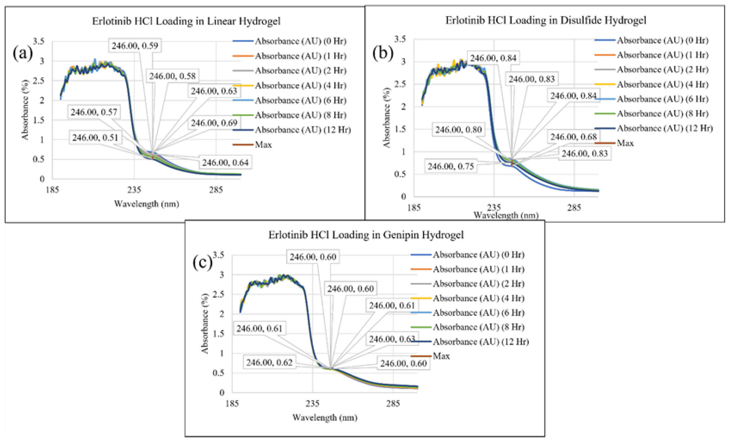
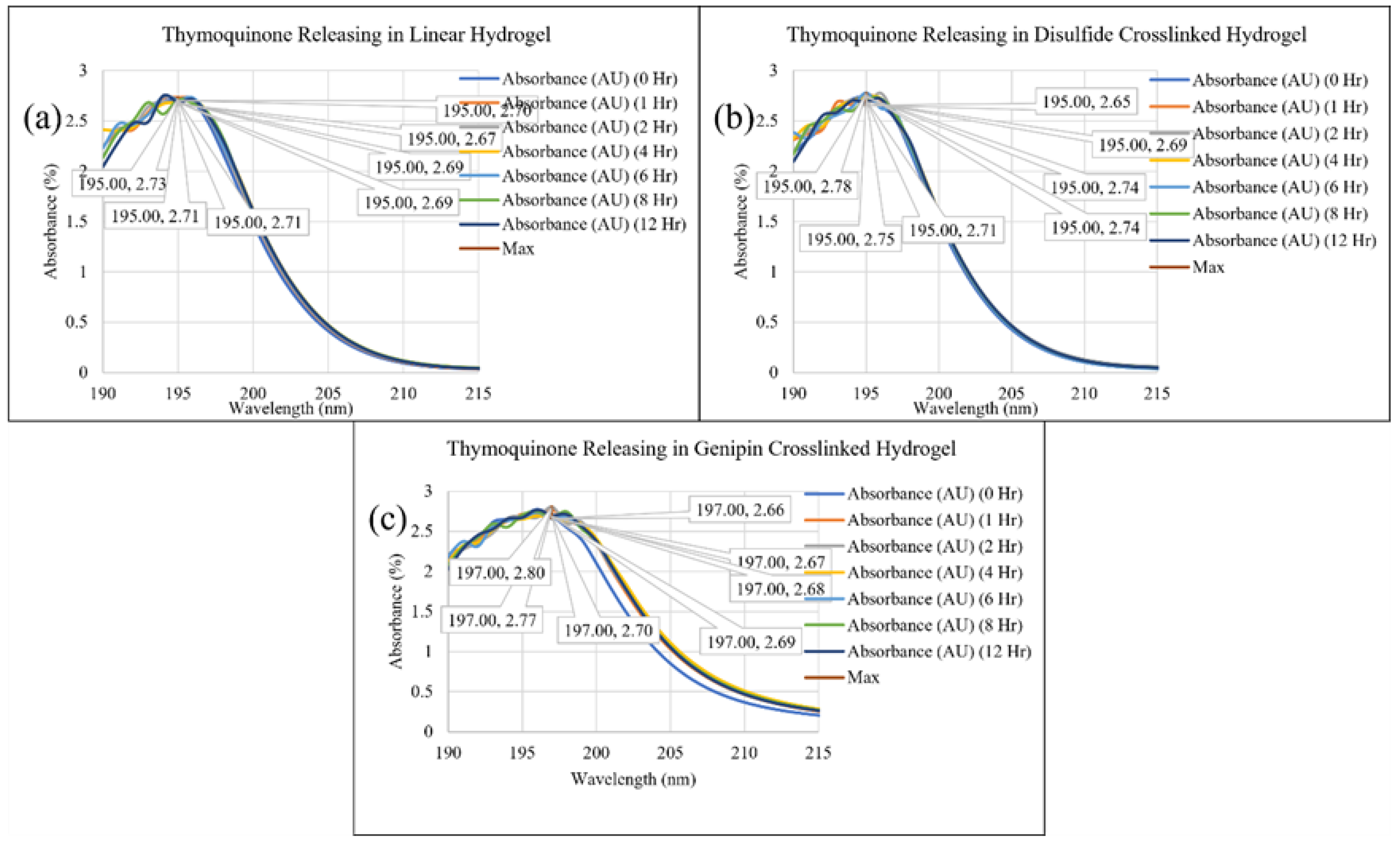
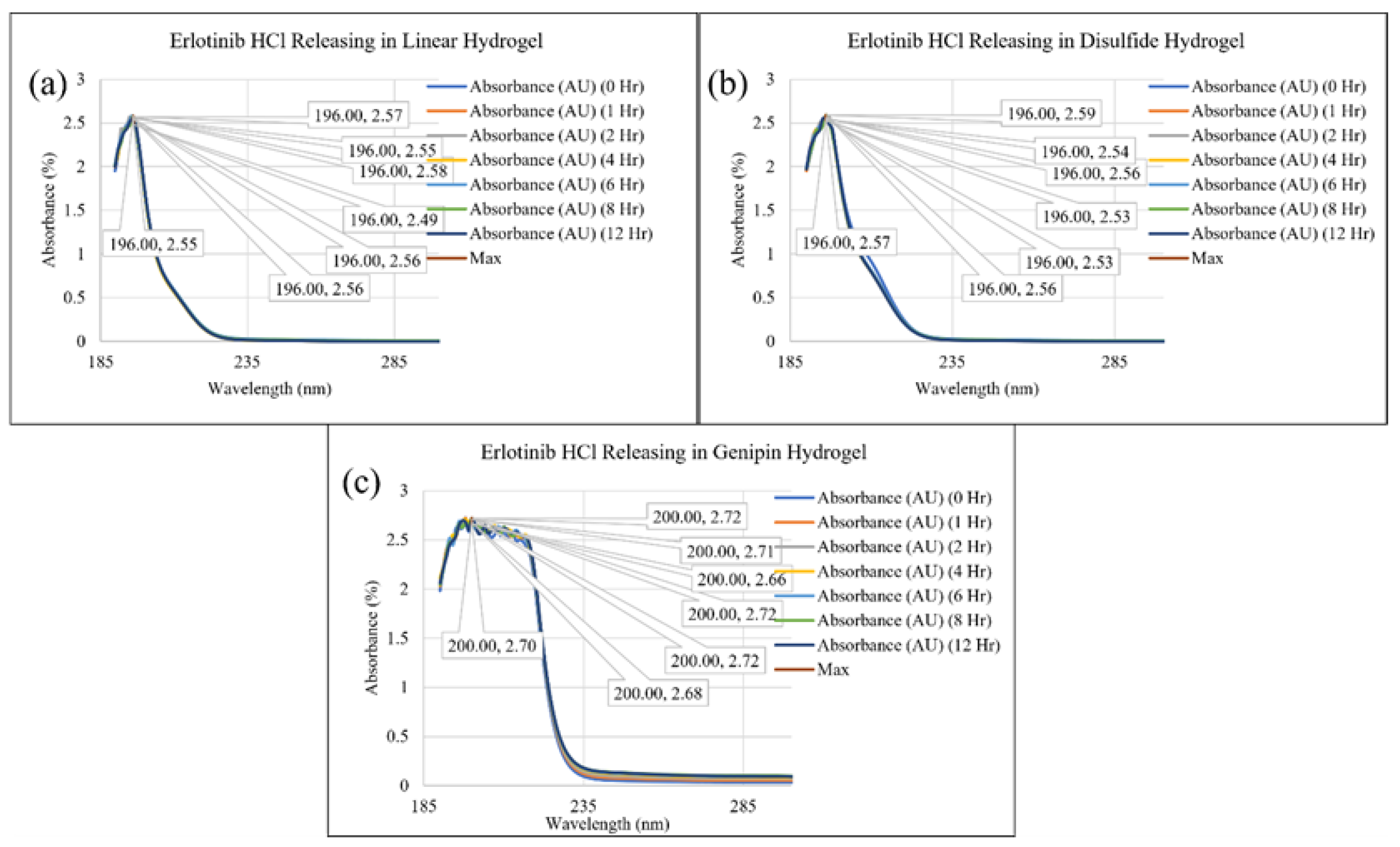
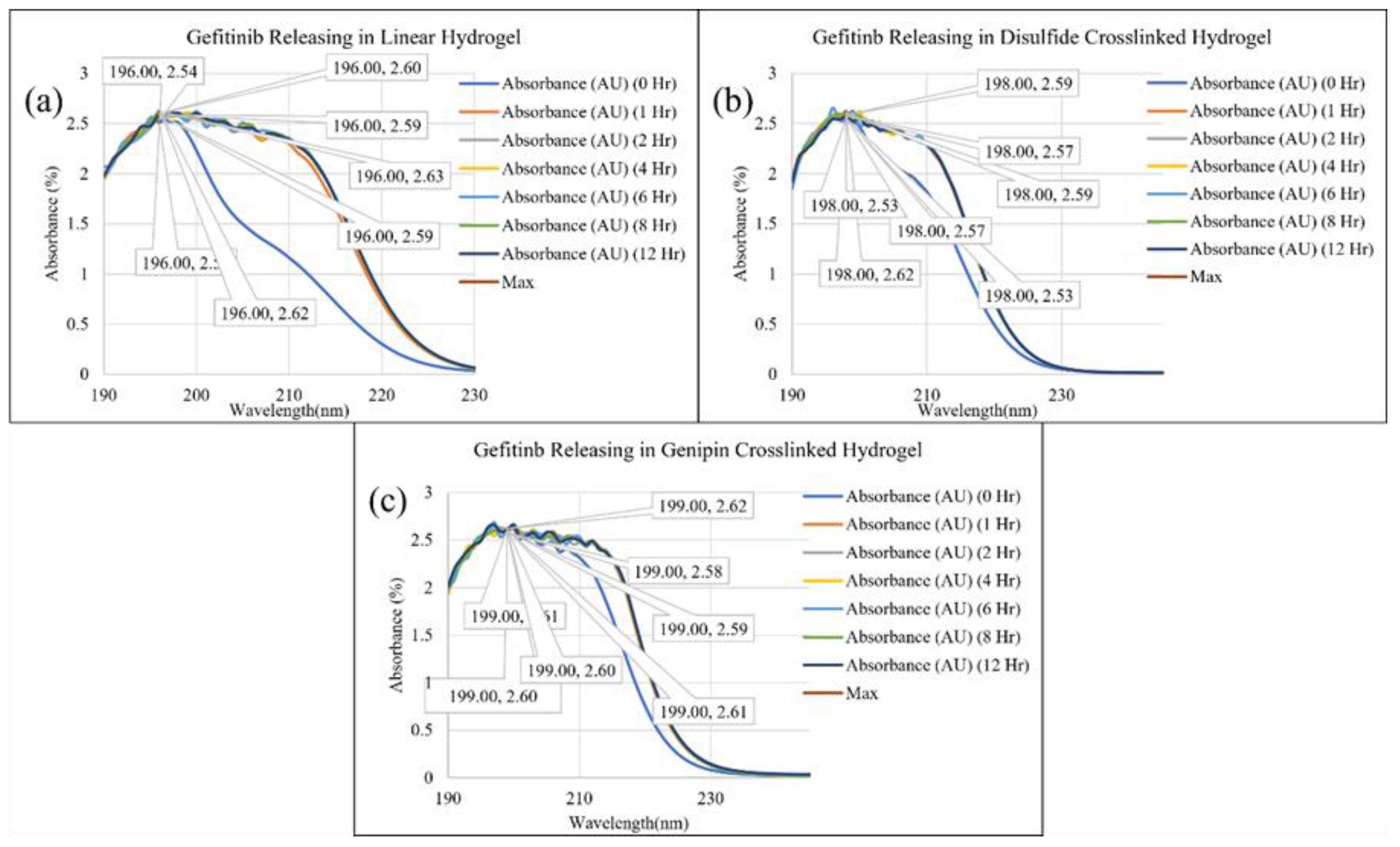
In this research, we experimented with chitosan hydrogels utilizing various crosslinking agents and made predictions about their behavior when cross-linked differently and characterized for their drug loading and releasing capacity. We investigated the distribution of three different drugs in three different hydrogels, resulting in the examination of nine unique systems, for both loading and releasing that makes eighteen different analyses at different time. Figure 1, Figure 2, Figure 3, Figure 4, Figure 5 and Figure 6 show the UV-Vis spectrums of the loading and releasing of different drugs in different systems. Figure 1 (a) to (c) depicts thymoquinone loading in the linear hydrogel, disulfide crosslinked hydrogel, and genipin crosslinked hydrogel, respectively. Similarly, Figure 2 and Figure 3 show Erlotinib HCl loading and gefitinib loading, respectively, in three different hydrogels. Likewise, Figure 4, Figure 5 and Figure 6 depict the spectrum while releasing these drug delivery systems. Beer-Lambert Law is used to calculate the drug absorptivity at different times. The law states that the absorbance of a solution is directly proportional to the concentration of the absorbing material present in the solution and the path length of light. The equation is given as follows:
A = ε lc
Where A is absorbance (obtained from UV-Vis), ɛ is molar absorptivity (calculated in this study), l is path length of light (1 cm), and c is concentration (known based on the amount of drug). The molecular weight of thymoquinone, gefitinib and erlotinib hydrochloride are 164.2g/mol, 446.9g/mol and 429.9 g/mol, respectively. No of moles are calculated by dividing the total weight of the drug (10 mg) by the corresponding molecular weight. Thus, the concentration of thymoquinone, gefitinib, and erlotinib are calculated as 0.061µM/ml, 0.0224µM/ml, and 0.0232µM/ml, respectively. Therefore, molar absorptivity at different hours is calculated using the absorbance value from UV-vis spectroscopy, the path length of light, and the concentration value.
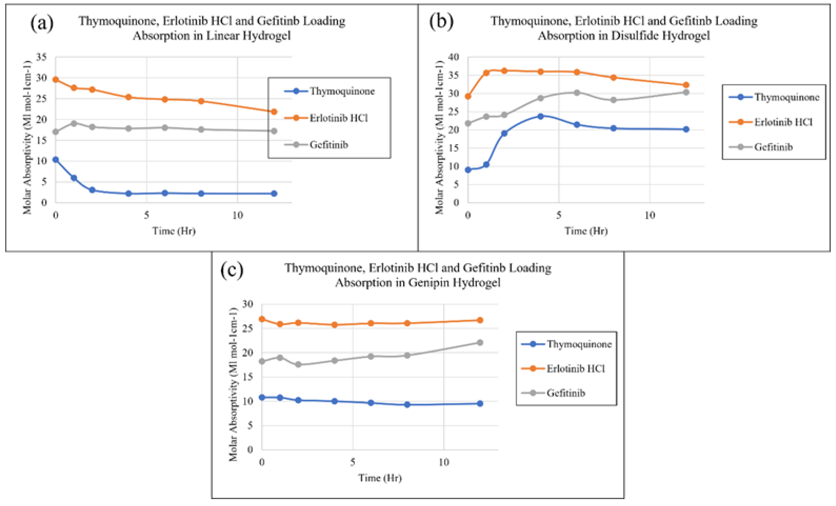
To analyze and compare the performances of different drugs in different systems, the molar absorptivity of three different drugs in linear chitosan hydrogels is plotted in Figure 7 (a). Maximum absorbance for thymoquinone drug in linear hydrogel at different times, i.e., 0 hours, 1 hour, 2 hours, 4 hours, 6 hours, 8 hours, and 12 hours data are taken and compared with the similar data of erlotinib and gefitinib in linear hydrogel as shown in Figure 7 (a). Similar comparisons are made for these three drugs in disulfide and genipin crosslinked, respectively, as shown in Figures 7 (b) and (c). Figure 7 (a) indicates the loading rate of thymoquinone in the linear hydrogel decreases over time, whereas gefitinib shows a steady state loading, however, it is lower than erlotinib. Thymoquinone shows an exponential decrease in the first couple of hours and then becomes stable, which is the lowest among these three drugs. Figure 7 (b) shows that the loading rate of all three drugs increases with time initially and then drops slowly to be stable in disulfide crosslinked hydrogel.
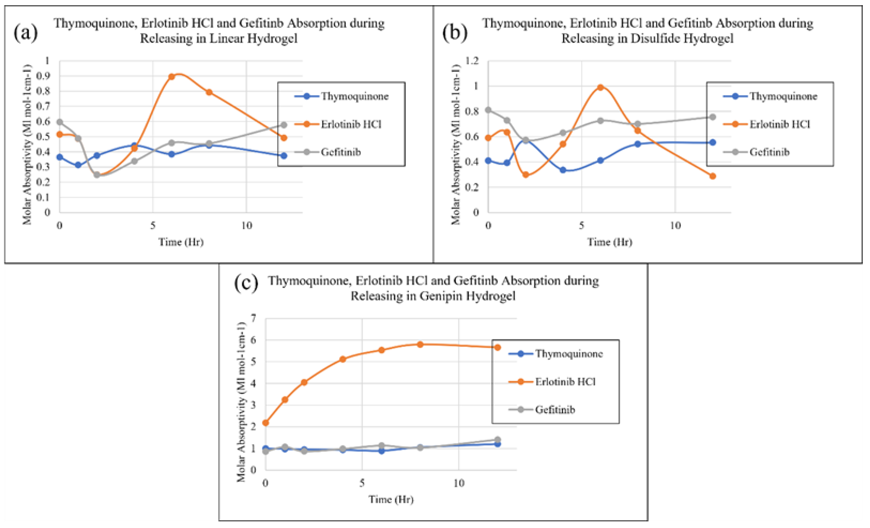
Erlotinib and thymoquinone show almost steady and constant loading, whereas gefitinib shows an increment over time in genipin crosslinked as depicted in Figure 7 (c). Depending on the target site, such as initial slower/higher or stable over the whole period, the loading rate can be controlled during the effective time. However, all three figures show that erlotinib has the highest loading capacity in all three types of gels, whereas, thymoquinone shows the lowest, and gefitinib lies between these two. This indicates thymoquinone is hard to load compared to two other drugs. Figures 8 (a) to (c) show the UV-Vis data while releasing. Figures 8 (a) and (b) indicate that the release rate of these drugs fluctuates in linear and disulfide cross-linked systems and the fluctuation of erlotinib is higher compared to two other drugs. The peak of these fluctuations also suggests that the erlotinib release rate is faster than the other two whereas thymoquinone and gefitinib are competitive. However, Figure 8 (c) depicts an exponential increment of erlotinib release over time whereas two other drugs show steady state release over time in genipin crosslinked system. Depending on the drug activity required in the diseased site, the release rate can be tuned and these figures indicate that erlotinib shows a higher release rate, however, geninpin crosslinked system shows a stable release rate regardless of drugs and exhibited the same for loading rate. In addition, gefitinib shows both stable loading and release in all hydrogel systems meaning it would minimize the patient’s reaction or side effects.
Therefore, this suggests that gefitinib may be an optimized drug to balance between loading and releasing rate and stability which agrees well with our previous study using molecular dynamics simulations [13]. Molecular weight, diffusivity, morphology of carrier, and surface charge play important roles in the sustained rate of loading and releasing as we have discussed in our previous study. To compare the performance of one drug in different systems, the loading and release of each drug are plotted in three different systems.
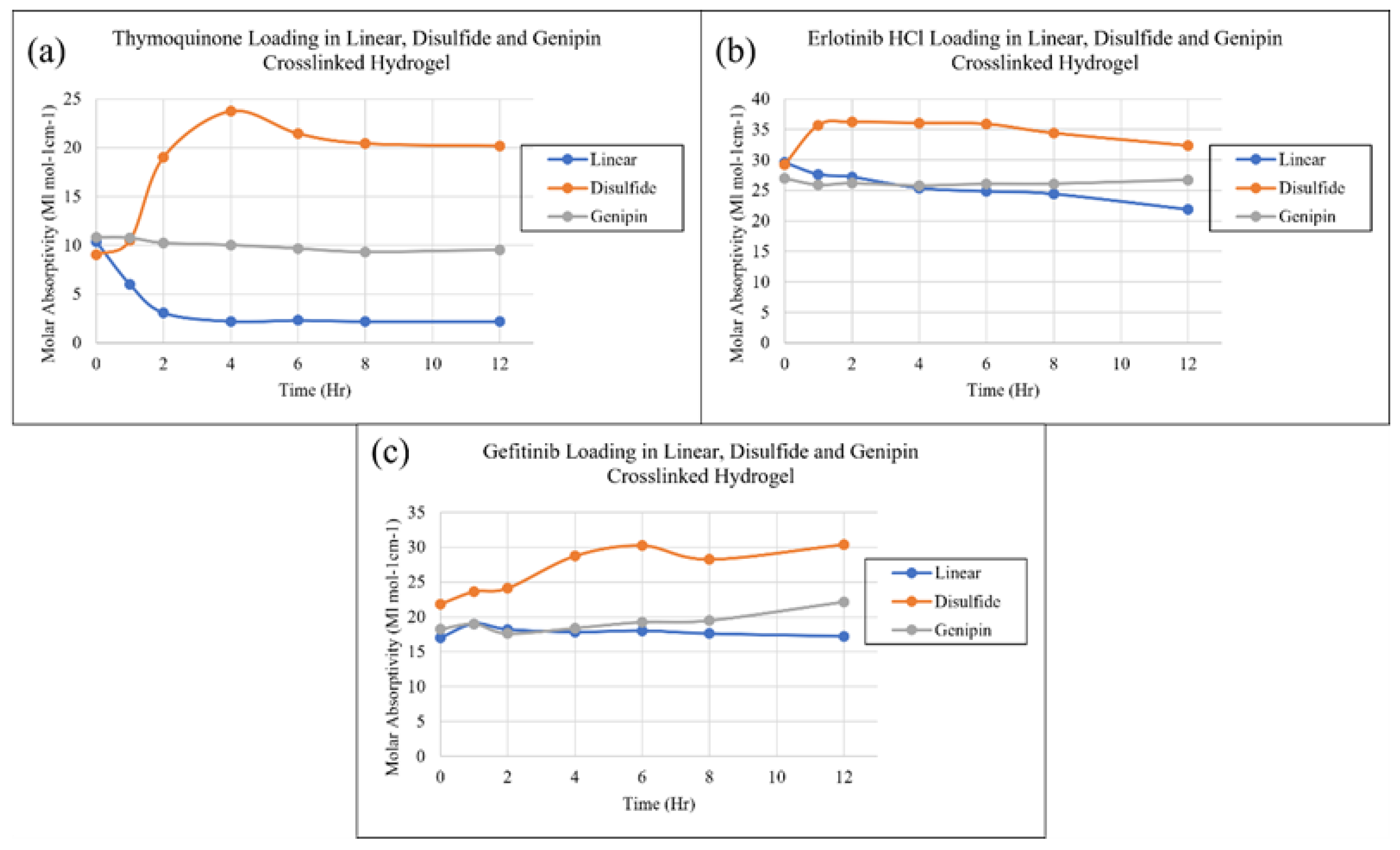
Figure 9 (a) shows the loading data of thymoquinone in three different types of gels which indicates that linear chitosan has the lowest loading capacity, disulfide crosslinked shows the highest, and genipin lies in between these two for thymoquinone drug. Thymoquinone loading increases initially in the disulfide crosslinked system and decreases in linear chitosan gel. Figure 9 (b) shows that both disulfide and linear systems show an overall decreasing loading capacity for erlotinib and Figure 9 (c) shows both these two systems an increasing loading rate for gefitinib. However, genipin shows a stable loading rate in all three systems. In addition, disulfide crosslinked systems show the highest loading capacity and linear gel shows the lowest, whereas genipin shows the middle between these two.
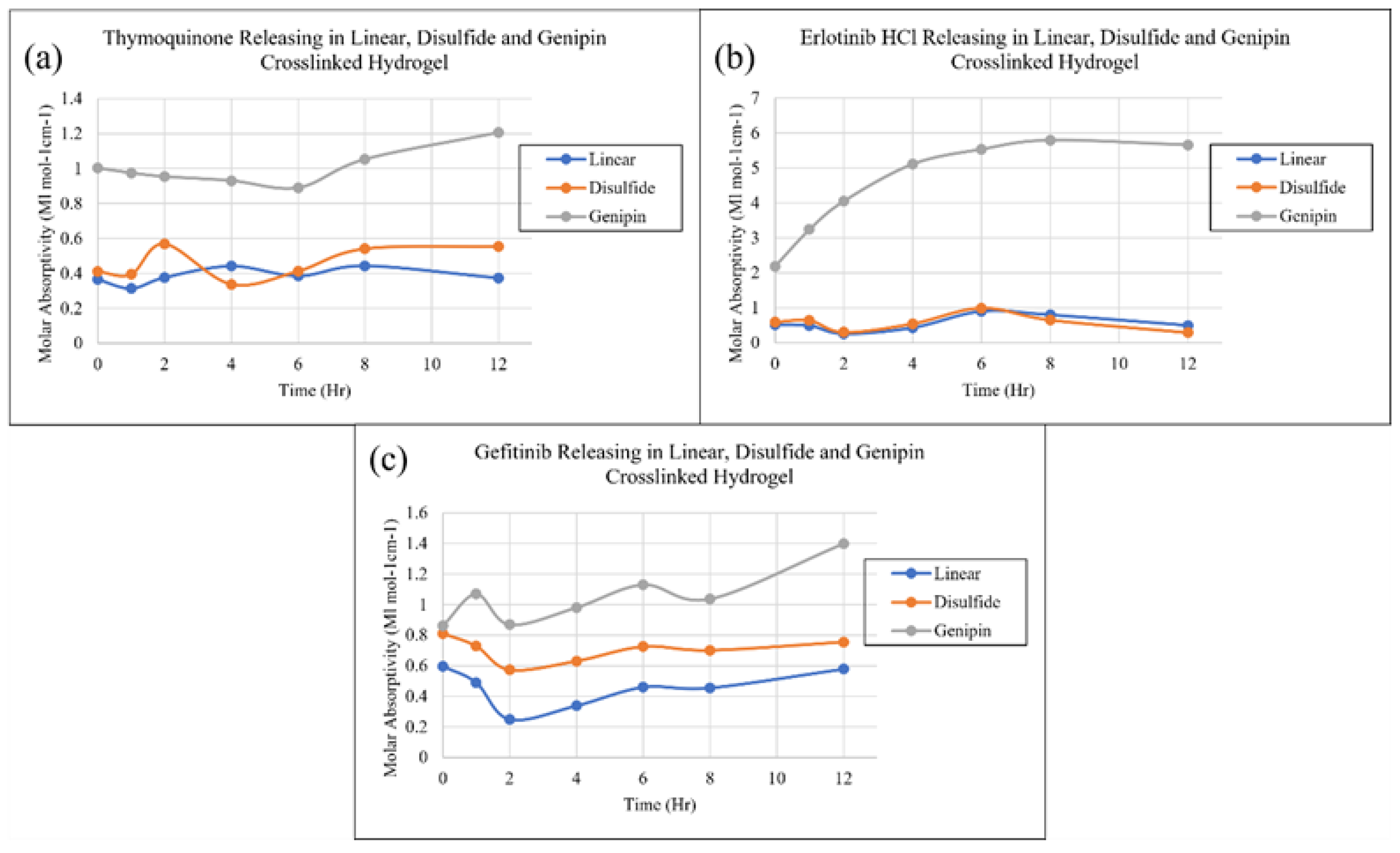
Figure 10 (a) to (c) show the drug release data of three drugs in three systems. Genipin crosslinked systems show a higher release rate regardless of drugs. Linear gel shows the lowest release rate for gefitinib, however, it is competitive with two other drugs. This experimental study demonstrates that the customization of chitosan structure with crosslinking agents improves therapeutic results and that can minimize side effects by regulating drug release characteristics sensitive to bodily situations [14]. In addition to the study of biocompatibility, formulations, several other factors such as cytotoxicity, cell viability, and the potential application of hydrogel in living tissues, efficacy, safety for long-term stability and degradation behavior, and clinical trials are necessary to investigate before administering it to the human body [15,16,17,18,19].

3. Conclusions

Thymoquinone shows the lowest loading efficacy compared to two other drugs in all three systems. Thymoquinone consistently exhibited higher diffusivity meaning it was hard to load in the gel. Gefitinib shows stable loading and releasing regardless of crosslinking systems. Erlotinib shows higher loading and releasing capabilities, however, its release is very fluctuating in linear and disulfide crosslinked systems (it shows stable release in the genipin crosslinked system). Disulfide crosslinked systems show the highest loading capacity and linear gel shows the lowest, whereas genipin shows the middle between these two. However, the genipin crosslinked system shows a stable loading and releasing rate regardless of drugs, whereas two other systems show varying loading and releasing tendencies depending on drugs. Genipin crosslinked systems show the highest release rate regardless of drugs. Gefitinib appeared to perform better in all systems, whereas the genipin crosslinked system was compatible with all the drug molecules showing balanced loading and releasing as found in the previous study. The chitosan hydrogel modified with different cross linkers in the study have a huge prospect in tissue engineering as well [20].

4. Materials and Methods

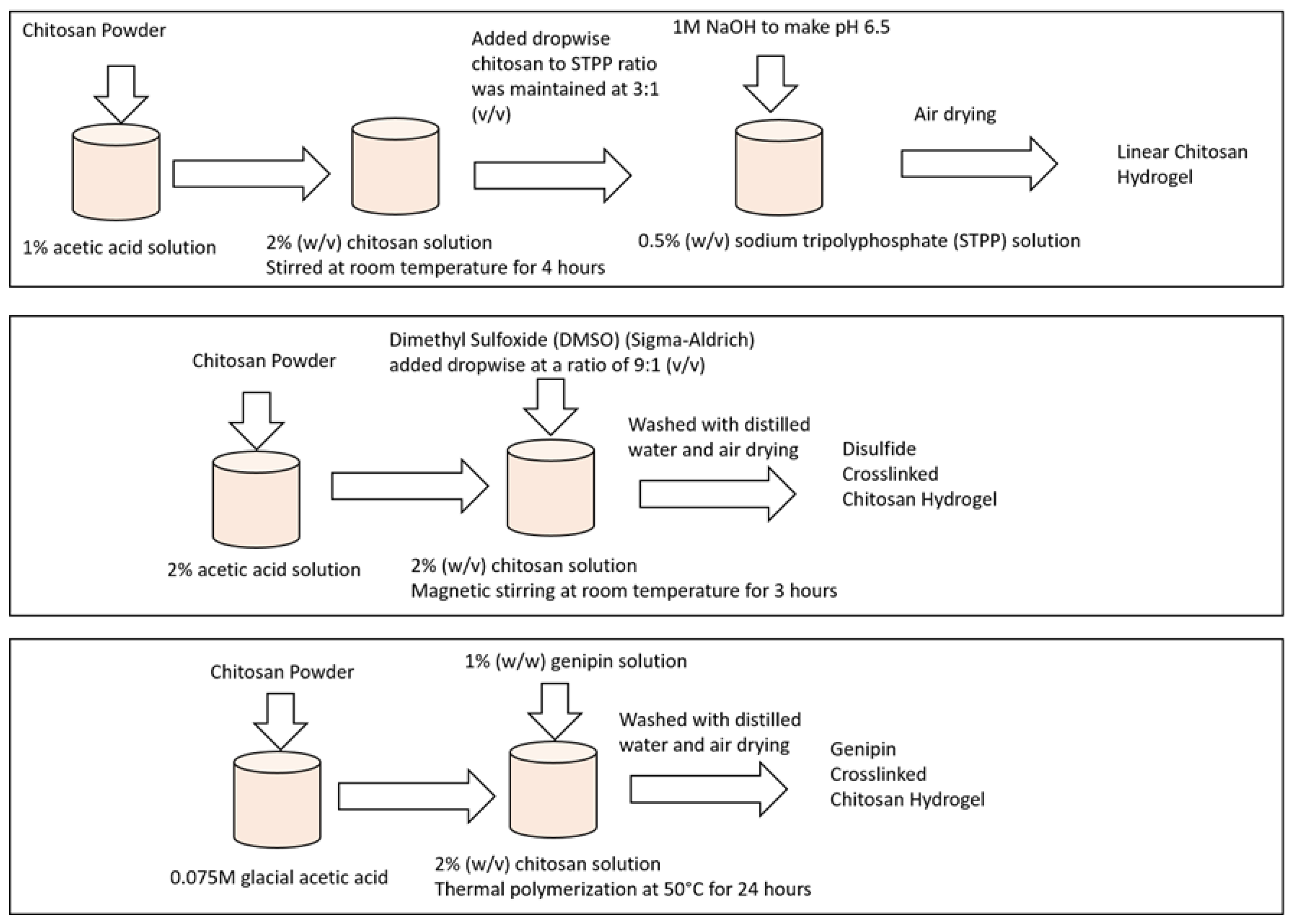
Figure 11 depicts the sample preparation method.
A chitosan-based hydrogel with various crosslinkers is typically prepared in a multi-step procedure for targeted drug delivery. The proper quantity of chitosan powder was weighed and added to a container for chitosan dissolution. The Chitosan was stirred in a suitable solvent, such as glacial acetic acid until completely dissolved. Sodium hydroxide (NaOH) was used to adapt the pH of the chitosan solution to the required range. The appropriate cross-linking agent was added to the chitosan solution depending on the cross-linking mechanism desired such as covalent or ionic. A calcium chloride (CaCl2) or sodium tripolyphosphate (STPP) suspension was used for ionic cross-linking. The crosslinker was then added to the chitosan solution dropwise while swirling until gelation occurred. Mixtures of glutaraldehyde, genipin, or EDC/NHS (N-ethyl-N′-(3-(dimethylamino)propyl) carbodiimide/N-hydroxysuccinimide) were used for ionic cross-linking. Dropwise while stirring, the cross-linking solution was added to the chitosan solution, which was prepared individually. The cross-linked chitosan solution was given time to rest for gelation and hydrogel formation. The gelation time depends on the type and amount of the crosslinker utilized. The drug components were dissolved in a suitable solvent such as Dimethyl sulfoxide (DMSO) to prepare a drug solution. The chitosan hydrogel was submerged in the medication solution to facilitate drug loading into the hydrogel network. The amount of drug loaded in the hydrogel is dictated by the loading circumstances, hydrogel type, and time. After adding drugs, the hydrogel was washed with deionized water or a suitable buffer solution to eliminate any excess or unbound drug molecules. The hydrogels loaded with drugs are now immersed in the solution again to release the drug to analyze releasing efficacy. Drug loading and release tests were performed in physiologically realistic environments such as phosphate-buffered saline (PBS) to assess the controlled loading and release capabilities.

4.1. Sample Preparation

4.1.1. Preparation of Linear Chitosan Hydrogel Sample

Initially, chitosan powder with a molecular weight of 190,000 Da (Sigma-Aldrich) was dissolved in a 1% acetic acid solution, resulting in a 2% (w/v) chitosan solution to prepare linear chitosan hydrogel. This solution was stirred at room temperature for 4 hours until achieving a homogeneous mixture. Simultaneously, a sodium tripolyphosphate (STPP) solution was prepared by dissolving STPP (Sigma-Aldrich) in distilled water, yielding a concentration of 0.5% (w/v). The pH of the STPP solution was adjusted to 6.5 using 1M NaOH. Next, the chitosan solution was gradually added dropwise to the STPP solution while continuously stirring at room temperature. The chitosan to STPP ratio was maintained at 3:1 (v/v). The resulting mixture was allowed to react for 30 minutes at room temperature, leading to the formation of a complex and the subsequent hydrogel formation through the interaction between chitosan and STPP. Following the reaction, the hydrogel was washed with distilled water to eliminate any unreacted STPP and acetic acid. Finally, the hydrogel was air-dried to obtain the chitosan hydrogel in solid form [21].

4.1.2. Preparation of Disulfide Crosslinked Chitosan Hydrogel Sample

The preparation process involved several steps. Firstly, chitosan solution was prepared by dissolving chitosan powder (MW 190,000 Da, Sigma-Aldrich) in a 2% acetic acid solution with a concentration of 2% (w/v). The mixture was stirred using a magnetic stirrer for 3 hours until it became clear. Simultaneously, a disulfide crosslinker solution Dimethyl Sulfoxide (DMSO) (Sigma-Aldrich) was added dropwise at a ratio of 9:1 (v/v) and stirred for 30 minutes at room temperature to facilitate crosslinking. The resulting mixture was then poured into a mold and left to gel for 2 hours at room temperature. Afterward, the chitosan hydrogel was washed with distilled water to remove any unreacted crosslinker. Following this, the chitosan hydrogel was air-dried in a vacuum chamber for 24 hours. Finally, the dried chitosan hydrogel was subjected to characterization to assess its physical and chemical properties.

4.1.3. Preparation of Genipin Crosslinked Chitosan Hydrogel Sample

Genipin crosslinked chitosan hydrogels were synthesized via thermal free radical polymerization. To initiate the process, chitosan was dissolved in 0.075M glacial acetic acid, reaching a final concentration of 2% (w/v). Subsequently, a 1% (w/w) genipin solution was used as a chemical crosslinker for thermal polymerization, which occurred at 50°C for 24 hours. The chitosan hydrogel beads were then washed with distilled water to remove any unreacted genipin. The chitosan hydrogel beads were air-dried for over 24 hours. The dried chitosan hydrogel beads were then characterized for their physical and chemical properties [9,10].
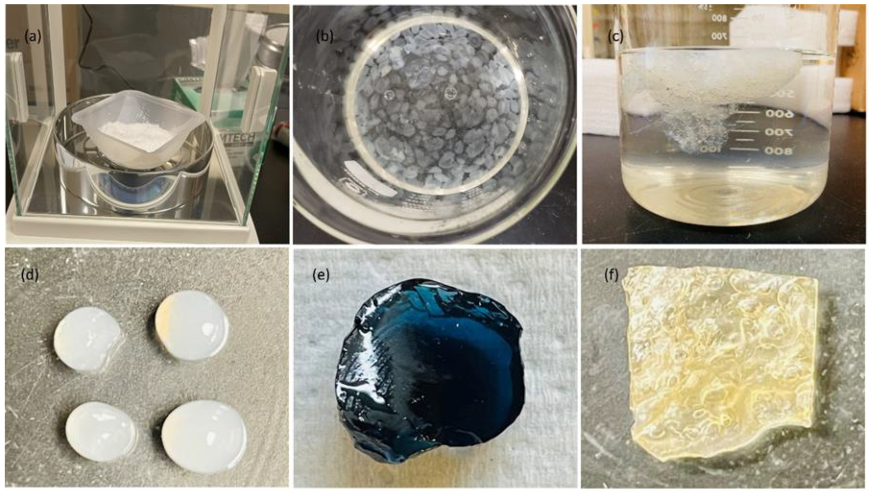
Figure 12 shows the final samples prepared for drug loading and release characterizations.

4.2. Drug Selection

4.2.1. Thymoquinone

In recent years, there has been an increasing interest in naturally occurring phytochemical compounds (found in plants that protect plants against bacteria, viruses, and fungi) that possess potential anti-cancer properties. This interest stems from their relative non-toxicity, cost-effectiveness, and availability in ingestible forms. It is noteworthy that more than 25% of drugs utilized in the past two decades have been directly derived from plants, and an additional 25% are chemically modified natural products. Among these natural compounds, the black seed (Nigella sativa, from the Ranunculaceae family) has gained particular attention. This annual herb is known by various names, including Black Caraway Seed and "the Blessed Seed," and it thrives in countries neighboring the Mediterranean Sea, as well as Bangladesh, India, and Pakistan [22,23]. Thymoquinone is a phytochemical compound found in the seeds of the Nigella sativa (black cumin) plant. It has been studied for its potential medicinal properties, including anti-inflammatory, antioxidant, and anticancer effects. Thymoquinone has been shown to have cytotoxic effects on a variety of cancer cell lines, including breast cancer, prostate cancer, and pancreatic cancer.

4.2.2. Erlotinib Hydrochloride

Erlotinib hydrochloride is an important medication used in cancer treatment, specifically for certain types of lung cancer and pancreatic cancer. It is an epidermal growth factor receptor (EGFR) tyrosine kinase inhibitor (TKI) that inhibits the signaling pathways involved in cancer cell growth and proliferation. In non-small cell lung cancer (NSCLC), erlotinib hydrochloride has shown efficacy, particularly in patients with specific EGFR mutations. Studies have demonstrated improved progression-free survival and overall survival in NSCLC patients treated with erlotinib hydrochloride compared to conventional chemotherapy. It has become an important targeted therapy option, particularly for patients with EGFR mutation-positive NSCLC. Additionally, erlotinib hydrochloride has also shown clinical benefit in the treatment of pancreatic cancer. Clinical trials have demonstrated improved overall survival in patients receiving erlotinib hydrochloride in combination with gemcitabine, a standard chemotherapy drug for pancreatic cancer [24,25]. The use of erlotinib hydrochloride in cancer treatment is supported by various clinical studies and has been approved by regulatory authorities for specific indications. However, it is important to note that the efficacy and suitability of erlotinib hydrochloride may vary depending on individual patient characteristics and cancer type. Therefore, it is crucial to consult with a healthcare professional for personalized treatment recommendations.

4.2.3. Gefitinib

Gefitinib is a small-molecule tyrosine kinase inhibitor that is used in cancer treatment, particularly for non-small cell lung cancer (NSCLC). It targets and inhibits the epidermal growth factor receptor (EGFR) tyrosine kinase, which is overexpressed in many cancer cells. Gefitinib has been studied for its potential use in targeted drug delivery using chitosan hydrogel as a carrier [26,27]. Li et al. (2016) prepared a chitosan hydrogel loaded with gefitinib for sustained release in cancer treatment [26]. The chitosan hydrogel was prepared using a method that involved in situ crosslinking of chitosan and β-glycerophosphate. The gefitinib was then incorporated into the hydrogel by simple mixing. The hydrogel was then characterized for its drug release-properties and the results showed sustained release of gefitinib over 24 hours.

4.3. Ultraviolet–Visible (Uv-Vis) Spectroscopy

We used ultraviolet-visible (UV-Vis) technology to analyze drug loading and releasing tendencies in chitosan hydrogels in this study. UV-Vis is a spectroscopic technique that measures the absorption of ultraviolet and visible light by a sample. UV-Vis spectroscopy is based on the Beer-Lambert law, which states that the absorbance of a solution is proportional to the concentration of the absorbing species and the path length of the light through the solution. By measuring the absorbance of a solution containing the drug and chitosan hydrogel at different time intervals, the amount of drug released from the hydrogel can be determined. To study drug loading, the drug and chitosan hydrogel are mixed and allowed to equilibrate. The mixture is then centrifuged to separate the hydrogel from the solution. The supernatant can be analyzed using UV-Vis spectroscopy to determine the presence of the drug in the solution. To study drug release, the drug-loaded chitosan hydrogel can be immersed in a buffer solution and kept at a constant temperature. The buffer solution can be periodically sampled and analyzed using UV-Vis spectroscopy at different time points. Charting the quantity of drugs given over time will reveal the drug's release kinetics [28,29]. Therefore, the UV-Vis spectroscopy absorbance data of the supernatant samples from nine different samples as investigated in this study for loading and releasing are used to compare the drug loading and releasing efficacy.

Author Contributions

Conceptualization, M. U.; methodology, S.K; validation, M.U., C.D.; formal analysis, M.U. C.D.; investigation, S.K.; resources, M.U., C.D.; data curation, S.K.; writing—original draft preparation, M.U., S.K.; writing—review and editing, M.U., S.K.; visualization, M.U..; supervision, M.U.; project administration, M.U.; funding acquisition, M.U.. All authors have read and agreed to the published version of the manuscript.

Funding

The authors would like to thank the University of Texas (UT) Systems STARs grant and the University of Texas Permian Basin (UTPB) College of Engineering Graduate Student scholarship to support this study partially. S.K. received a tuition waiver to complete his thesis from the UTPB College of Engineering. M.U. would like to appreciate the course releases from UTPB as a matching fund with the UT System STARs grant to continue research on targeted drug delivery. C.D. acknowledges his UT Systems STARs grant to collaborate in this research for experimental characterizations. All authors certify that they have no affiliations with or involvement in any organization or entity with any financial interest or non-financial interest in the subject matter or materials discussed in this manuscript.

Institutional Review Board Statement

Not applicable.

Informed Consent Statement

Not applicable.

Data Availability Statement

Not applicable.

Acknowledgments

The authors would like to acknowledge the Office of Research and Sponsored Programs at UTPB for developing this collaboration between the Department of Mechanical Engineering and the Department of Chemistry.

Conflicts of Interest

The authors declare no conflicts of interest.

References

  1. Torchilin, V. P. (2000). Drug targeting. European Journal of Pharmaceutical Sciences, 11, S81-S91.
  2. Torchilin, V. P. (1995). Handbook of targeted delivery of imaging agents (Vol. 26). CRC Press.
  3. Buxton, I. L. (2006). Pharmacokinetics and pharmacodynamics. Goodman and Gilman’s the pharmacologic basis of therapeutics, 11th Ed. New York: McGraw-Hill, 1-52.
  4. Sonker, M. , Bajpai, S., Khan, M. A., Yu, X., Tiwary, S. K., & Shreyash, N. (2021). Review of Recent Advances and Their Improvement in the Effectiveness of Hydrogel-Based Targeted Drug Delivery: A Hope for Treating Cancer. ACS Applied Bio Materials, 4(12), 8080-8109.
  5. Madhumathi, K. , & Tamura, H. (2015). Chitosan-based hydrogels for drug delivery and tissue engineering. International Journal of Biological Macromolecules, 80, 737–749. [CrossRef]
  6. Dash, M. , Chiellini, F., Ottenbrite, R.M., & Chiellini, E. (2011). Chitosan—a versatile semi-synthetic polymer in biomedical applications. Progress in Polymer Science, 36(8), 981-1014. [CrossRef]
  7. Roy, D., & Gupta, M. N. (2017). Modeling the Formation of Hydrogels Using Density Functional Theory. Journal of Physical Chemistry B, 121(47), 10609–10616. [CrossRef]
  8. Zhao, Y. , Yang, J., Zhang, C., & Cheng, R. (2017). Theoretical investigation of non-covalent interactions in hydrogels. Journal of Materials Chemistry B, 5(29), 5739–5746. [CrossRef]
  9. Abitha M. Heimbuck, Tyler R. Priddy-Arrington, Benjamin J. Sawyer, Mary E. Caldorera-Moore, Effects of post-processing methods on chitosan-genipin hydrogel properties, Materials Science and Engineering: C, Volume 98,2019, Pages 612-618, ISSN 0928-4931. [CrossRef]
  10. Yew, M. Y. , Basri, M., Masoumi, H. R. F., Ahmad, M. B., Ismail, M., & Abdul Rahman, M. B. (2016). Fabrication of chitosan hydrogel beads cross-linked with genipin for controlled release of vitamin C. International journal of biological macromolecules, 93, 1478-1486. [CrossRef]
  11. Abdel-Bar, H. M. , El-Magd, A. A., & Al-Mahallawi, A. M. (2018). Effect of chitosan concentration on the performance of ibuprofen-loaded chitosan hydrogels. International Journal of Biological Macromolecules, 117, 41–49. [CrossRef]
  12. Hu, Q. , Zhu, Y., Zhang, X., & Liu, Y. (2014). pH-sensitive chitosan-based hydrogel microspheres for doxorubicin delivery. Carbohydrate Polymers, 101, 1235–1243. [CrossRef]
  13. Uddin, M. S., & Ju, J. (2020). Effect of crosslinking agents on drug distribution in chitosan hydrogel for targeted drug delivery to treat cancer. Journal of Polymer Research, 27, 1-10.
  14. E. S. De Alvarenga, “Characterization and Properties of Chitosan,” in Biotechnology of Biopolymers, InTech, 2011. doi 10.5772/17020.
  15. F. Ahmadi, Z. Oveisi, S. M. Samani, and Z. Amoozgar, "Chitosan-based hydrogels: characteristics and pharmaceutical applications.," Res. Pharm. Sci., vol. 10, no. 1, pp. 1–16, 2015, [Online]. Available: https://www.ncbi.nlm.nih.gov/pmc/articles/PMC4578208/.
  16. K. Miller, “Chitosan,” WebMD, 2023. https://www.webmd.com/vitamins-and- supplements/chitosan-uses-and-risks.
  17. L. L. Palmese, M. Fan, R. A. Scott, H. Tan, and K. L. Kiick, “Multi-stimuli-responsive, liposome-crosslinked poly (ethylene glycol) hydrogels for drug delivery,” J. Biomater. Sci. Polym. Ed., vol. 32, no. 5, pp. 635–656, Mar. 2021. [CrossRef]
  18. M. Prabaharan and J. F. Mano, “Chitosan-Based Particles as Controlled Drug Delivery Systems,” Drug Deliv., vol. 12, no. 1, pp. 41–57, Jan. 2004. [CrossRef]
  19. L. Upadhyaya, J. Singh, V. Agarwal, and R. P. Tewari, "The implications of recent advances in carboxymethyl chitosan-based targeted drug delivery and tissue engineering applications," J. Control. Release, vol. 186, pp. 54–87, Jul. 2014. [CrossRef]
  20. Wang, Q., Wang, X., & Feng, Y. (2023). Chitosan hydrogel as tissue engineering scaffolds for vascular regeneration applications. Gels, 9(5), 373.
  21. Qu C, Li X, Zhao Y, Han X, Liu Y, Du Y. Preparation and characterization of chitosan hydrogel containing silver nanoparticles for wound dressing application. J Biomater Appl. 2016;31(4):524-534.
  22. Gali-Muhtasib, H. , Roessner, A., & Schneider-Stock, R. (2006). Thymoquinone: a promising anti-cancer drug from natural sources. International journal of biochemistry and cell biology, 38(8), 1249-1253. [CrossRef]
  23. https://pubchem.ncbi.nlm.nih.gov/compound/123631#section=Structures.
  24. Moore, M. J. , Goldstein, D., Hamm, J., et al. (2007). Erlotinib plus gemcitabine compared with gemcitabine alone in patients with advanced pancreatic cancer: a phase III trial of the National Cancer Institute of Canada Clinical Trials Group. Journal of Clinical Oncology, 25(15), 1960-1966.
  25. https://pubchem.ncbi.nlm.nih.gov/compound/176871#section=Structures.
  26. Li, W., Ma, G., & Zhou, L. (2016). In situ crosslinked chitosan hydrogel for the controlled release of gefitinib. Carbohydrate Polymers, 138, 194-202. [CrossRef]
  27. National Center for Biotechnology Information. "PubChem Compound Summary for CID 123631, Gefitinib" PubChem, https://pubchem.ncbi.nlm.nih.gov/compound/Gefitinib. Accessed 10 March, 2022.
  28. Kim YJ, Yoon JJ, Park JH. Drug release from electro spun fibers - a review. J Ind Eng Chem. 2015; 29:1-11.
  29. La WG, Kang SW. Effect of crosslinking degree and pH on chitosan hydrogel for drug release. Korean J Chem Eng. 2014;31(8):1373-1379.
  30. Yao, F., & Zhang, L. (2011). Swelling behaviors of chitosan hydrogels in different pH and salt solutions. Journal of Applied Polymer Science, 119(4), 2244-2251.
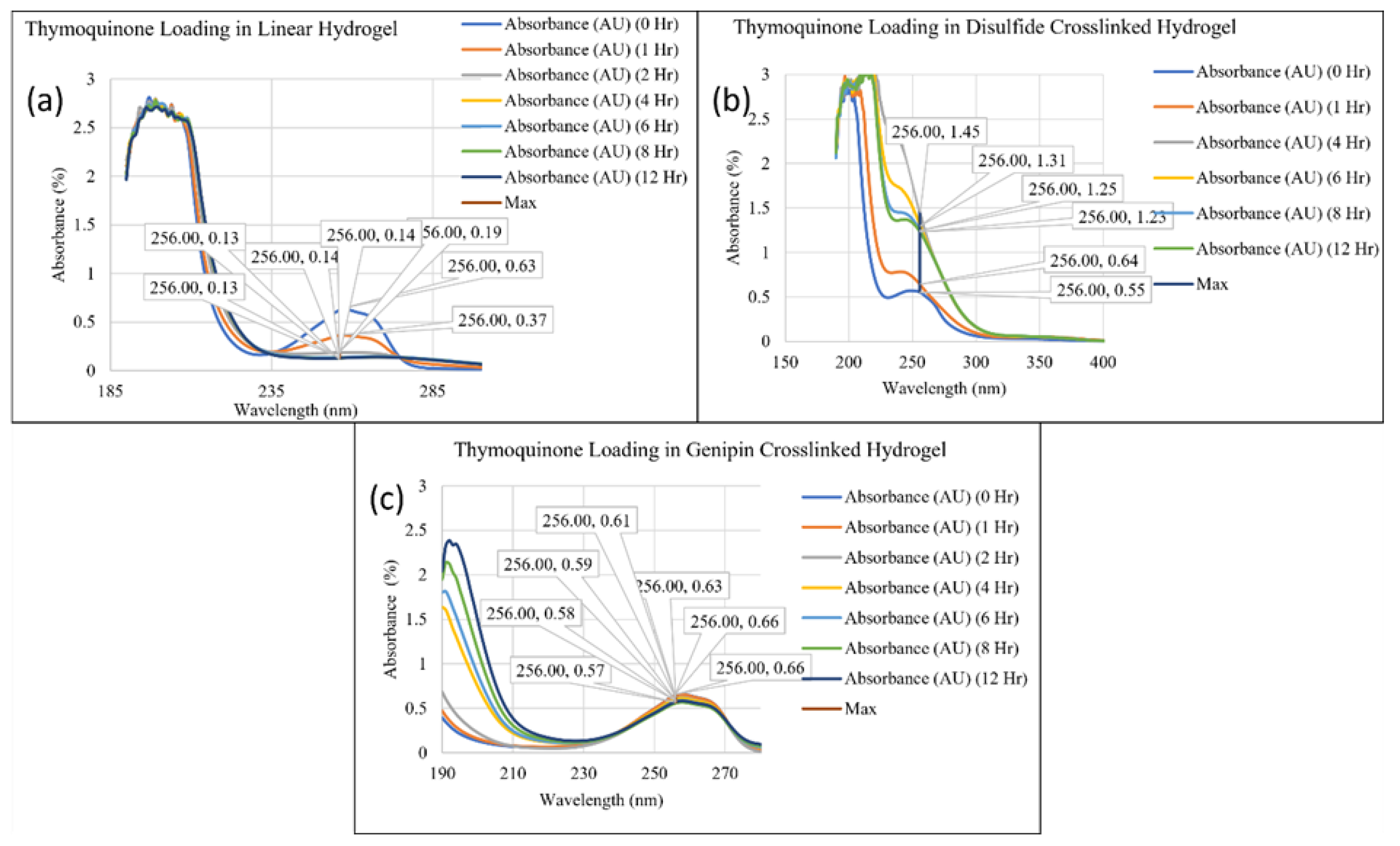
Figure 1. UV-Vis spectra of thymoquinone loading in (a) linear hydrogel, (b) disulfide crosslinked hydrogel, and (c) genipin crosslinked hydrogel.
Preprints 107340 g001
Figure 2. UV-Vis spectra of erlotinib HCl loading in (a) linear hydrogel, (b) disulfide crosslinked hydrogel, and (c) genipin crosslinked hydrogel.
Preprints 107340 g002
Figure 3. UV-Vis spectra of gefitinib loading in (a) linear hydrogel, (b) disulfide crosslinked hydrogel, and (c) genipin crosslinked hydrogel.
Preprints 107340 g003
Figure 4. UV-Vis spectra of thymoquinone releasing in (a) linear hydrogel, (b) disulfide crosslinked hydrogel, and (c) genipin crosslinked hydrogel.
Preprints 107340 g004
Figure 5. UV-Vis spectra of erlotinib HCl releasing in (a) linear hydrogel, (b) disulfide crosslinked hydrogel, and (c) genipin crosslinked hydrogel.
Preprints 107340 g005
Figure 6. UV-Vis spectra of gefitinib releasing in (a) linear hydrogel, (b) disulfide crosslinked hydrogel, and (c) genipin crosslinked hydrogel.
Preprints 107340 g006
Figure 7. Comparison of thymoquinone, Erlotinib HCl, and Gefitinib Loading in (a) linear hydrogel, (b) disulfide crosslinked hydrogel, and (c) genipin crosslinked hydrogel.
Preprints 107340 g007
Figure 8. Comparison of thymoquinone, Erlotinib HCl, and Gefitinib Releasing in (a) linear hydrogel, (b) disulfide crosslinked hydrogel, and (c) genipin crosslinked hydrogel.
Preprints 107340 g008
Figure 9. Comparison of a drug loading in three different hydrogels, i.e., linear hydrogel, disulfide crosslinked hydrogel, and genipin crosslinked hydrogel: (a) thymoquinone, (b) Erlotinib HCl, and (c) Gefitinib.
Preprints 107340 g009
Figure 10. Comparison of a drug releasing in three different hydrogels, i.e., linear hydrogel, disulfide crosslinked hydrogel, and genipin crosslinked hydrogel: (a) thymoquinone, (b) Erlotinib HCl, and (c) Gefitinib.
Preprints 107340 g010
Figure 11. Sample preparation methodology.
Preprints 107340 g011
Figure 12. (a) chitosan powder, (b) formation of beads in linear chitosan, (c) spongy gel with disulfide crosslinker, (d) final sample of linear chitosan, (e) final sample of genipin crosslinked chitosan, and (f) final sample of disulfide crosslinked chitosan.
Preprints 107340 g012
Disclaimer/Publisher’s Note: The statements, opinions and data contained in all publications are solely those of the individual author(s) and contributor(s) and not of MDPI and/or the editor(s). MDPI and/or the editor(s) disclaim responsibility for any injury to people or property resulting from any ideas, methods, instructions or products referred to in the content.
Copyright: This open access article is published under a Creative Commons CC BY 4.0 license, which permit the free download, distribution, and reuse, provided that the author and preprint are cited in any reuse.
Prerpints.org logo

Preprints.org is a free preprint server supported by MDPI in Basel, Switzerland.

Subscribe

© 2024 MDPI (Basel, Switzerland) unless otherwise stated