1. Introduction
Neuroprosthesis are external devices integrated with neuromodulation which reinforce the input or output of a neural system, replacing or enhancing sensory, motor, and cognitive functions, aiming at improving the quality of life of impaired patients [
1,
2]. The technological development of the last years has moved towards the enhancement of the connection of such external devices to the human nervous systems to enable a safer, more effective and selective electrical stimulation.
As a part of our nervous system, peripheral nerves lie out from the brain and spinal cord and are characterized by different anatomical structures. They act as a conduit, bidirectionally exchanging information between different areas of the body and the central nervous system, communicating signals for both sensations and motor commands. Peripheral nerves are made of mixed nerve fibers: axons of motor or sensory fibers, surrounded by an insulating myelin called endoneurium, are bundled into groups named fascicles.
In the context of restoring the functions of the peripheral nervous system, there is the need of neural interfaces which are highly selective and minimal invasive, leading to an unavoidable trade-off between selectivity and invasiveness. Various technologies for neural interfaces have been researched for many years, such as extraneural, intrafascicular, penetrating, and regenerative interfaces. However, some limitations concern these solutions; to mention some, the stiffness of the penetrating electrodes, the difficulty for the implantation technique for the intrafascicular electrodes or the low signal-to-noise ratio of the extraneural electrodes [
3,
4,
5].
In this context, nanotechnology has emerged as a potential novel approach to enhance the coupling between external devices and neuron system, since the dimensions of nanoengineered materials could allow a non-invasive and highly focal neuromodulation.
Particularly, the Magneto-Electric NanoParticles (MENPs) has demonstrated as a cutting-edge technology to be used for a single-neuron level stimulation, both at the central and peripheral sites [
6,
7,
8,
9]. Their unique innovation potential is due to the magnetoelectric (ME) effect, which consists in a linear coupling between the external applied magnetic field and the generated local electric field, and vice versa. The direct effect occurs when the ME material is placed inside an external magnetic field: the magnetostrictive phase experiences a non-zero strain which is mechanically transferred to the piezoelectric phase, and, in turn, it transduces the mechanical deformation into electrical charges. This property, specific of magnetoelectric composites and of multiferroic solids that simultaneously possess both the ferromagnetic and ferroelectric ordering, has been lately proven also at room temperature, allowing their use in biomedical applications. The magnitude of the two phases’ interaction is defined by the ME coefficient α
E= δE/δH [V/cmOe]. The properties of ME composites depend on the properties of the selected phase materials of the nanostructure, the geometry of the components, such as the phase volume ratio and the particle and sample sizes, the frequency of the external field used to elicit the ME effect, and the quality of the mechanical contact between the two phases [
6,
10]. Among all the MENPs described in literature, the core-shell cobalt ferrite (CoFe
2O
4) barium titanate (BaTiO
3) nanoparticles showed promising properties for biomedical applications, as proven by previous in vivo and in vitro studies [
7,
11,
12,
13], and a good in vitro and in vivo biocompatibility [
7,
14].
MENPs can be administered to the desired site using different strategies, e.g., intranasally or intravenously, in the case of the central nervous system, and navigated via the application of a magnetic field gradient [
7,
15], or directly placed into the target region, e.g., by stereotactic injection into a specific region [
7,
14]. Subsequently, MENPs can be activated through the application of a proper DC or, as in case of neural stimulation, an AC magnetic field at a specific low frequency, eliciting a localized electric field at a frequency equal to twice the frequency of the stimulating AC magnetic field [
6,
9]. Finally, post-treatment, MENPs can be pulled away from the localized area by the application of a reversed magnetic field gradient [
9] or can be independently cleared from the body. In fact, as shown in a mouse model, nanoparticles are excreted from the body within a two-month period, depending on their size and on the specific organ in which they are [
16].
This study aims to describe and characterize the use of MENPs for the electrical stimulation of the peripheral nervous system. The electromagnetic physical quantities generated by the presence of MENPs and their interactions with the dynamics of the nervous system were quantified by a computational approach. More in detail, the study focused on the quantification of the electric field distribution induced by MENPs in the human body, the thresholds needed to elicit action potentials, the impact of pulse shape and the selectivity of axons’ activation inside the neuronal fibers, and, finally, the complex interplay between the two [
17,
18]. To this purpose, finite element method (FEM) and axons dynamics model are combined.
This study underlined the ability to use MENPs as stimulating tools and, thorough a quantitative analysis enabled the investigation of the influence of MENPs concentration and orientation on the effectiveness of MENPs’ stimulation.
3. Results
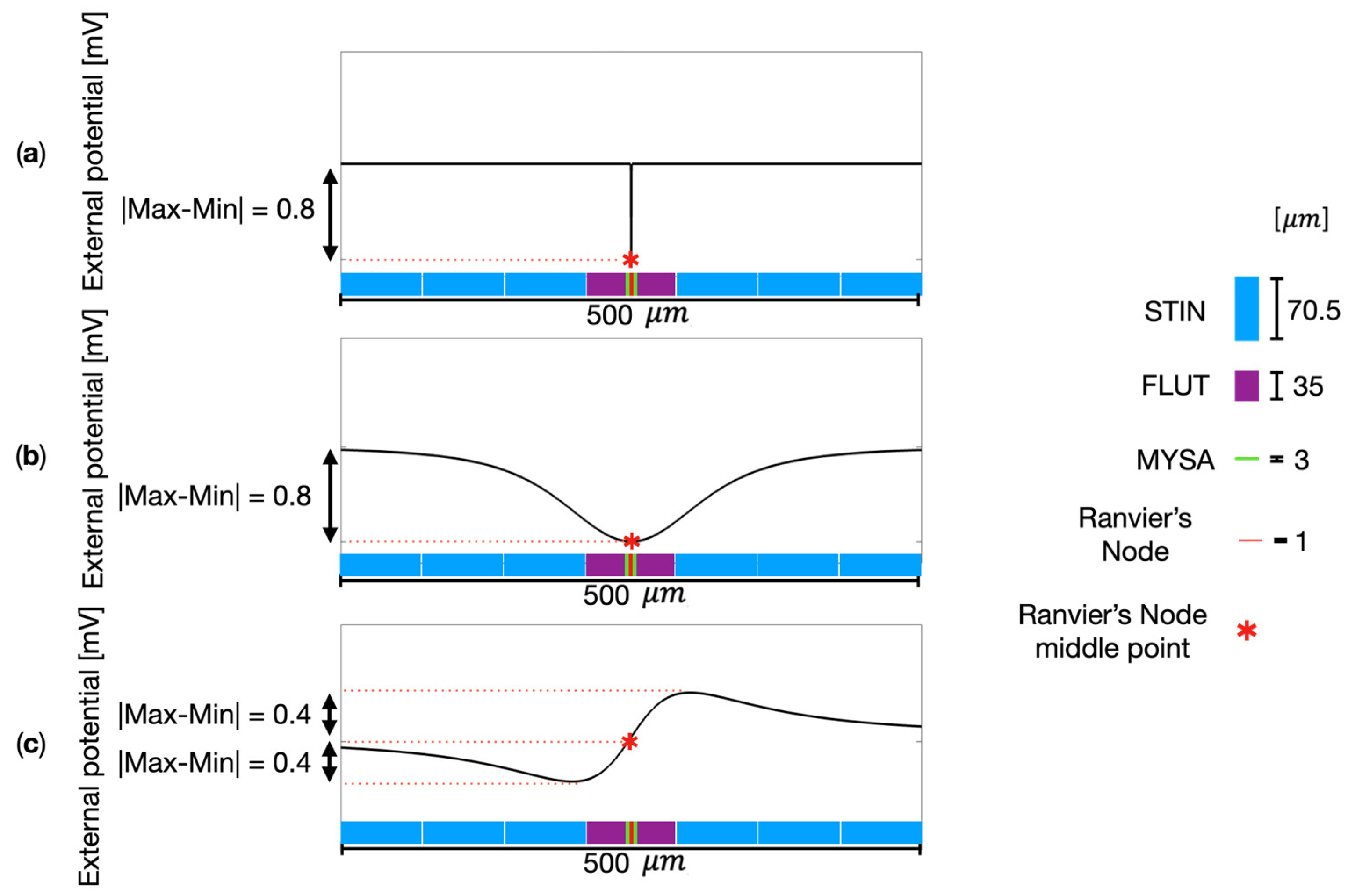
Figure 4 shows some examples of the electric potential
elicited by the single MENP or MENPs cluster along the neural fiber. For the sake of readability, the images are focused on a segment of 500-
length along the fiber, centered on the Ranvier’s node closer to the MENP or MENPs cluster.
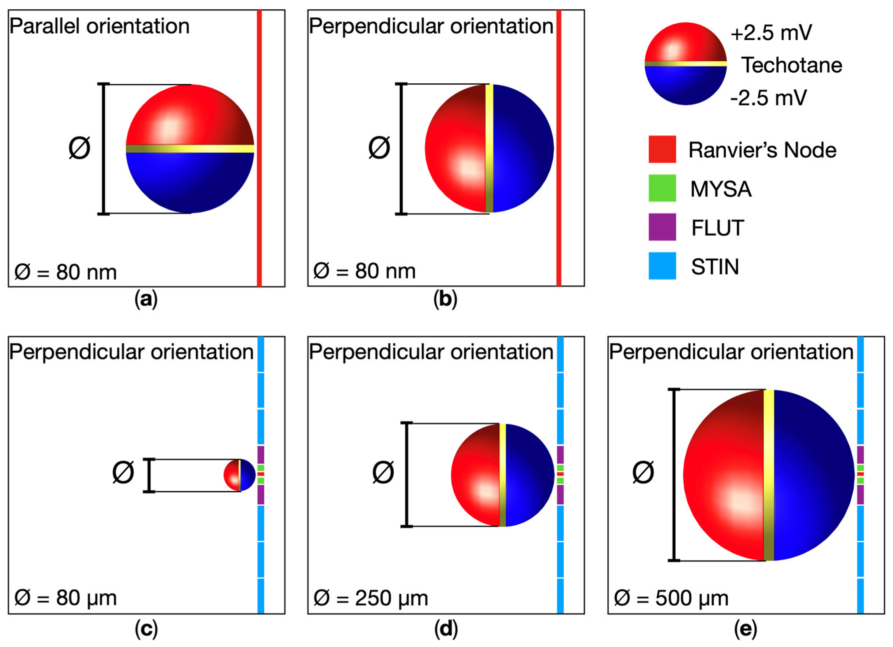
Figure 4a shows the case study of a single MENP positioned at 2R from the neural fiber, perpendicularly oriented. As well expected, slightly negative values, i.e., excitatory effect, are observed in correspondence of the MENP. Due to the specific dimensions of the MENP (80 nm) and the Ranvier’s node length (1
m), the MENP stimulation solely affects the electric potential of the Ranvier’s node, without affecting any other segments of the fiber model and resulting in a punctual stimulation typical of a nanoelectrode. As a comparison,
Figure 4b and 4c illustrate the potential distribution elicited along the fiber when the stimulating source is a MENPs cluster with diameter of 80
placed at distance 2R and with perpendicular and parallel orientations, respectively. The graphs clearly show that the external potential perceived by the fiber is indeed affected by the orientation of the MENPs cluster. When the MENPs cluster is perpendicularly oriented (
Figure 4 b), the external potential behavior is similar the case of the single MENP stimulation: an excitatory effect is observed along the neural fiber, with the negative peak in correspondence of the Ranvier’s node. From the comparison of
Figure 4a and 4b, it is evident that dissimilarities in the widening of the external potential peak are directly linked to the differences in the dimensions of both the single MENP and MENPs cluster. The influence over the perceived external potential of the fiber is wider by increasing the MENPs diameter. Looking at the single MENP, the fiber is excited over 75% of the peak value at a total length of almost 80 nm, while the 80-
m MENPs cluster causes strong excitation in the micrometer range, i.e., an extension of 80
m along the fiber senses the external potential over 75% of its peak value. The magnitude of the two peaks is 0.8 mV for both the single MENP and the MENPs cluster when perpendicularly oriented. A very different behavior is observed for the MENPs cluster parallelly oriented (
Figure 4c). For this latter, both negative values, i.e., excitatory effect, and positive values, i.e., inhibitory effect, are perceived along the fiber length. The total magnitude of the variation is 0.8 mV.
Table 2 shows the
s values, by discriminating the different MENPs configurations. When considering a single MENP, placed at distance 1R from the fiber, the
value is the same for both the perpendicular and the parallel orientations. Due to the similarity of the results, the following analysis of the influence of the MENP – fiber distance over the
value was performed only for the MENP perpendicularly oriented. The
value increases by increasing the MENP – fiber distance, growing from 4.8 for a 1R distance to 31.1 for a 3R distance. If considering clusters of MENPs whether perpendicularly or parallelly oriented, the
s trend is consistent for each different configuration and mirrors the one observed when stimulating with a single MENP, i.e., the
value increases by increasing the MENP – fiber distance. Exceptionally, the 500-
m MENPs cluster, parallelly oriented shows an opposite trend, decreasing the MF value from 23.5 for a 2R distance to 17.3 for a 3R distance. Moreover, results evidence an influence of the clusters orientations and dimensions over the
values. Typically, lower
s are required to evoke an action potential when cluster of MENPs are perpendicularly rather than parallelly oriented, with the exception of the MENPs cluster of 500
m at distance 3R. For example, when stimulating with a MENP cluster of 80
m, the
value ranges from 5.8 to 34.2 for the perpendicular orientation, whereas ranges from 7.9 to 85.8 for the parallel orientation. Regarding the MENPs dimensions,
generally decreases by increasing the cluster diameter. As an example,
s of 7.9, 6.3, and 3.9 are needed for cluster diameters of 80, 250 and 500
m respectively, parallel oriented. A recurring opposite trend is visible for the MENPs cluster of 500
m at distance 3R for both orientations, and for the same cluster when oriented parallel and placed at distance 2R.
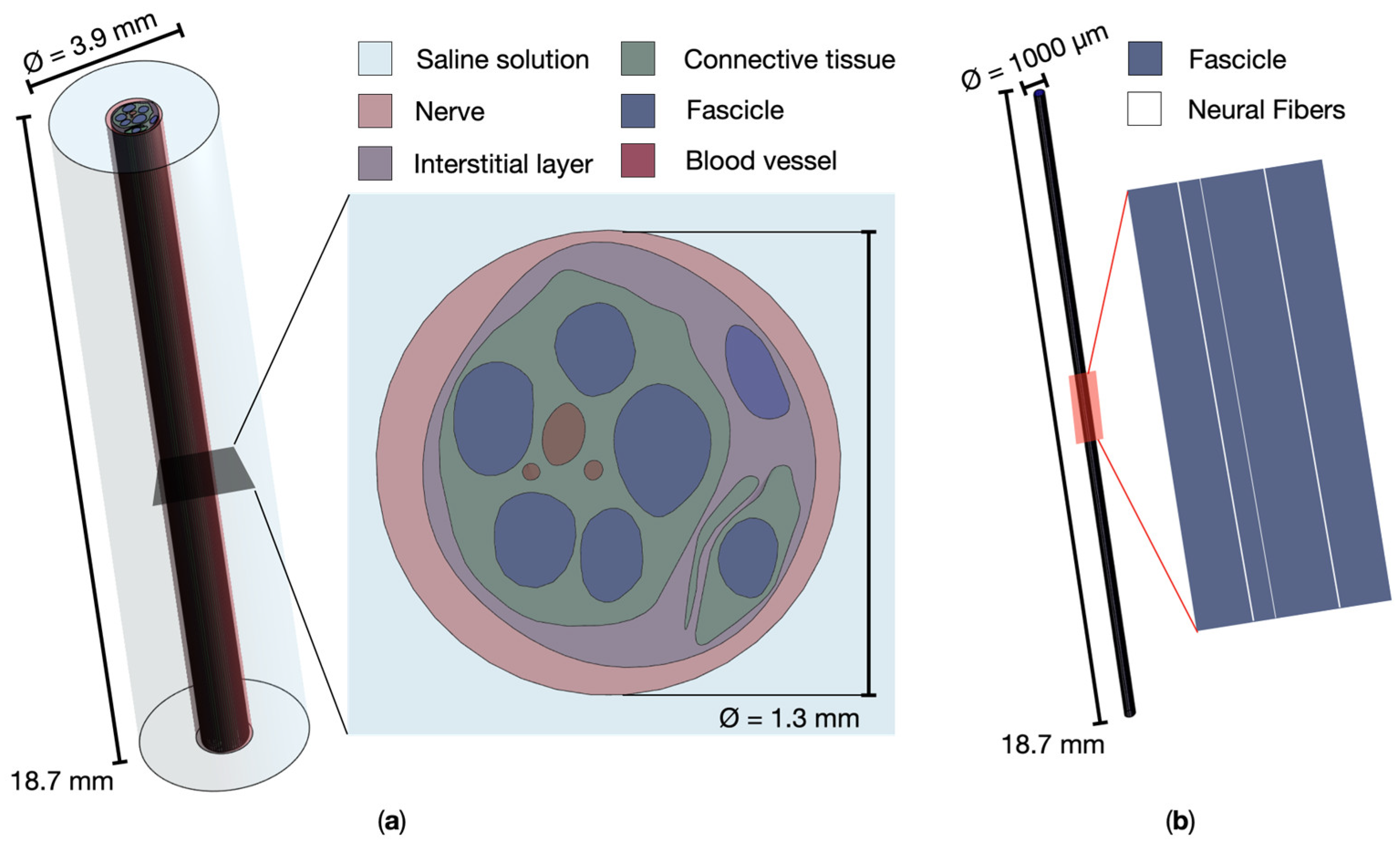
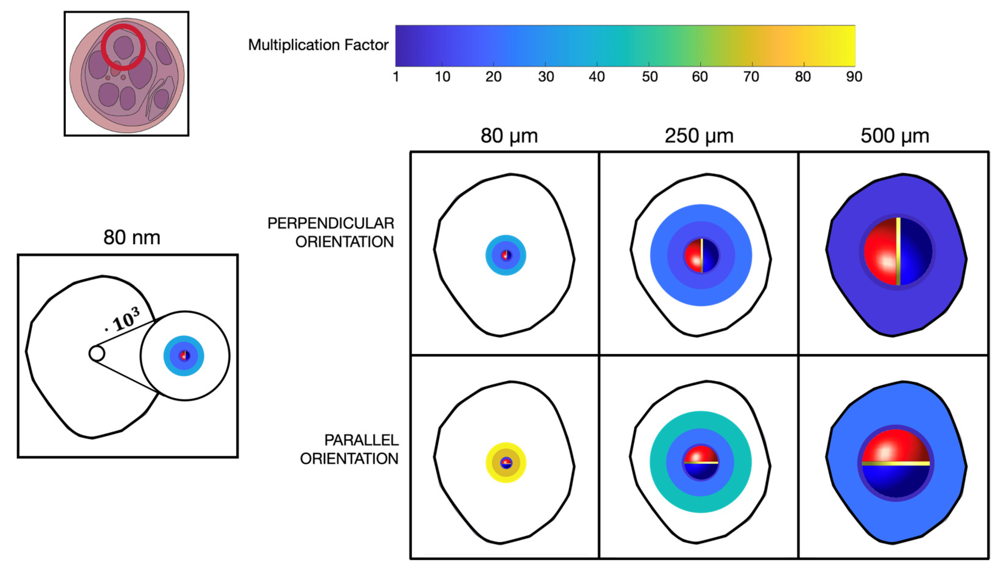
Figure 5 shows a representation of the recruited volume of the fascicle injected with the MENPs, reported on the cross-section view, when considering different MENPs configurations. More specifically, the figure shows the MF values needed to activate the neural fibers in the circular areas of 1R, 2R and 3R around the MENPs. Due to the definition of the MENP-fiber distance, the activated region with radius 1R is barely visible. The fascicle diameter was of 1000
m, comprised within the range of peripheral fascicles diameters [
28]. When considering the single 80 nm nanoparticle placed in the middle of the fascicle, even if using MF equal to 31.1, i.e., multiplying the potential on the surface of the MENP by more than thirty times, the fibers recruited by the stimulations are confined in a very narrow volume around the nanoparticle. If considering clusters of MENPs, the recruitment volumes are larger with lower MF values: for the 80
m cluster in perpendicular configuration about a quarter of the fascicle volume was recruited with MF equal to 34.2, while for the 250
m in perpendicular configuration two third of the volume of the fascicle is recruited with lower MF, equal to 21.9.
The different behavior of equivalent current
injected in the intracellular space along the fiber length varying MENPs dimension is detailed in
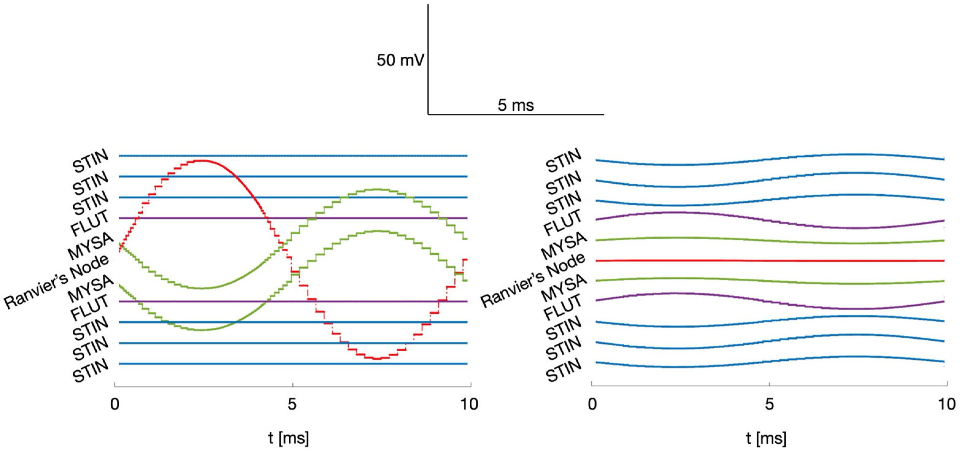
Figure 6, where
Figure 6a and 6b shows the equivalent current when the stimulating source, perpendicularly oriented and placed at a distance 1R from the fiber, is a single MENP and a 500-
m cluster, respectively. For the sake of readability, the extension along the fiber length is the same as in
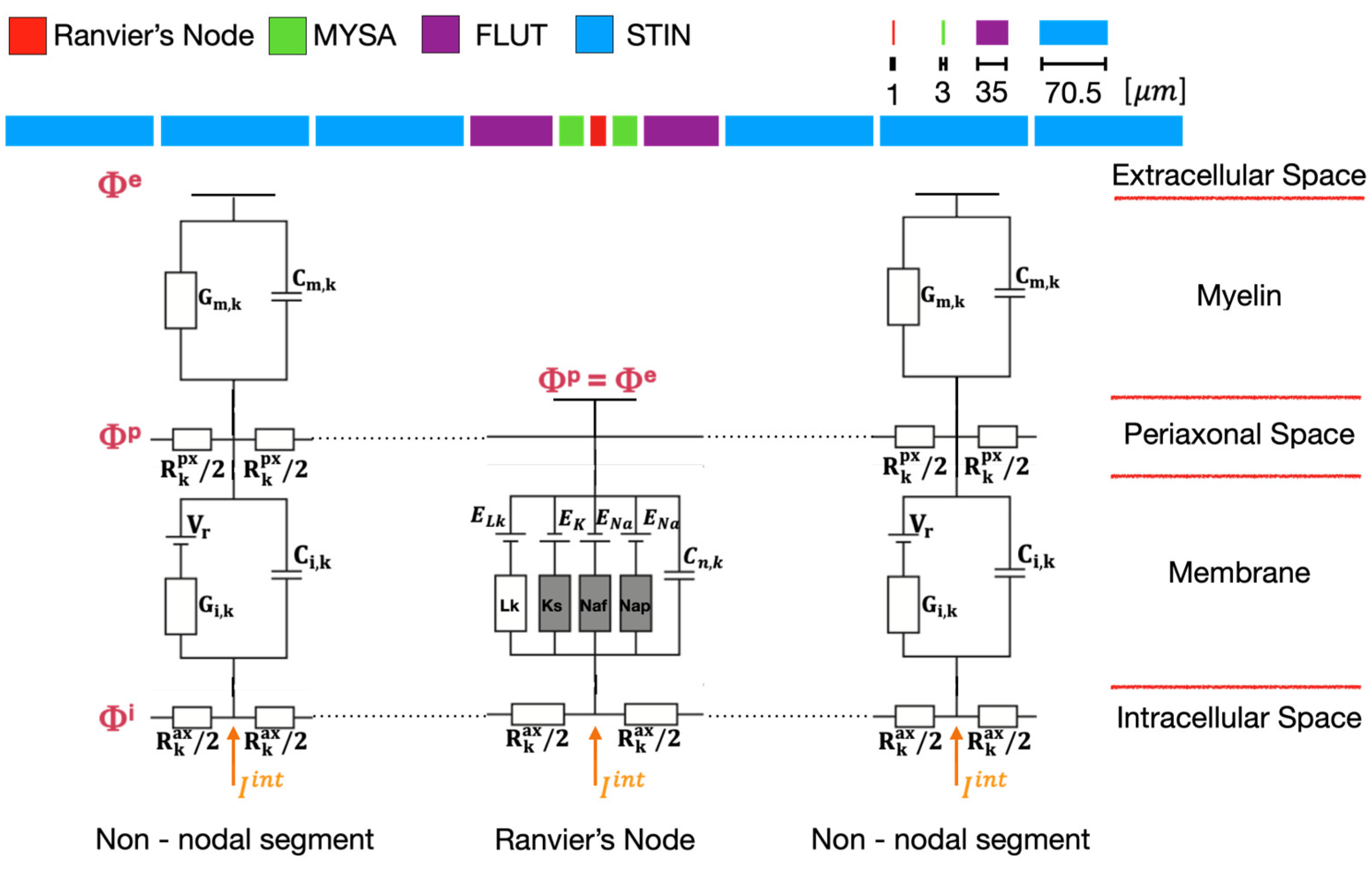
Figure 4, and the timeframe covers the whole stimulus duration. As well expected, when stimulating with a single MENP perpendicularly oriented, the injected current is localized in the Ranvier’s node which comprises the MENP and its two adjacent MYSA segments, whereas it is zero in all the other segments. The maximum amplitudes in absolute value of the current injected in the Ranvier’s node are 47.7 nA, 47.2 and 47.5 nA for distances 1R, 2R and 3R, respectively. Consequently, the amplitude of the current that must be injected in the Ranvier’s node to elicit an action potential when the stimulating source is a single MENP, and the stimulus is a one-period sinusoid, is approximately 47 nA, regardless of the MENP – fiber distance. Consistent with previous results, when the stimulating source is a cluster, its influence over the neural fiber is less localized and widening as the cluster dimensions increase, thereby affecting more fiber compartments. For example, when considering the 500-
m MENPs cluster, the injected current is zero in the Ranvier’s node, while it differs from zero in all the other visible non-nodal segments. Although the amplitudes of the current injected in the non-nodal compartment are small compared to the 47 nA of the first case-study, neural fibers activation remains achievable due to non-linear summation of the injected current values [
26]. In both case studies, the injected current on the sides of the Ranvier’s node is symmetrically distributed. As axonal excitation is dependent on the extracellular potential outside each node, the injected current time course mirrors the sinusoidal temporal profile of the extracellular MENPs stimulation.
4. Discussion
As the demand for safe, effective, and targeted electrical stimulation in the field of neuroprosthesis continues to rise, along with highly selective and minimally invasive neural interfaces, the research is actively seeking and proposing innovative methods, strategies, and materials to meet these needs [
29].
In this context, MENPs could play a significant role. This study aims to characterize the use of MENPs for the electrical stimulation of the peripheral nervous system through a computational assessment of the interaction of MENPs with the nervous system dynamics. The computational approach here used allowed to investigate the influence of the parameters that most affect the electric potential distribution generated by the MENPs in the nervous tissue, such as the MENPs-fiber distance, the MENPS concentration, and orientation. The different electric potential distribution such estimated, were translated into a detailed assessment of the electric response of motor neural fibers. All the analyses performed assumed that the MENPs were positioned near a Ranvier’s node of a single neural fiber.
As expected, the electric potential distribution due to the presence of MENPs appears to be localized around them [
21], and the extent to which that distribution can stimulate the nerve tissue is strictly related to the MENPs concentration. Indeed, the influence of the MENPs increases with increasing size of the agglomerates (
Figure 4). The same trend can be observed for the recruitment capability (
Figure 5 and
Table 2). The volume of the excited tissue increases with the increasing size of the MENPs for similar magnification factors. The ability to excite the fibers becomes broader as the MENPs become larger, up to the possibility of activating an entire nerve fascicle. Larger agglomerates mean a higher probability of stimulating the neural fibers and a higher ability to recruit than less targeted stimulation. As the recruitment ability of the MENPs increases, their efficiency improves, while their selectivity decreases significantly, i.e., individual MENPs show selectivity in the nanometer range, which increases to the micrometer range in agglomerates. For single MENPs and small agglomerates, selectivity is high, but recruitment is lower, as the placement of the MENPs along the neural fiber plays a significant contribution. Consequently, a trade-off between the selectivity and resolution of MENPs is evidenced by our simulations. When activated, it is assumed that all the MENPs are aligned in the same direction, namely that of the external magnetic field. This makes it possible to control the orientation of the MENPs by specifying the orientation of the external magnetic field exerted. In this study, the influence of two different orientations on the nervous tissue around the MENPs were analyzed. The results show how the two different MENPs alignments affects differently the external potential sensed by the fiber length and how controlling the MENPs orientation enables the control of the stimulation of the fibers. More specifically, the orientation of the MENPs influences both the electrical potential perceived along the fiber length, which can be equated to monopolar (in case of perpendicular orientation) or bipolar stimulation (in case of parallel orientation) (
Figure 4), and the amplification of the external potential distribution needed for fibers activation (
Table 2). Bipolar stimulation shows a greater influence of the distance between the MENPs and the fiber as the growth of the amplification demanded to excite the nervous tissue is greater than with monopolar stimulation. When considering larger distances from the MENPs, the required potential gain over the surface of the agglomerates approximately doubled when moving from perpendicular to parallel orientation. This trend is less pronounced when the agglomerates are attached to the fiber. The use of monopolar or bipolar stimulation controls the stimulating signal, which can be inhibitory or excitatory, and, therefore, commands the spiking activity of the neural fibers and their firing time [
30]. This study allowed to estimate the current injected into the intracellular space, as defined by [
26,
27]. To excite an action potential, a minimum value of 47 nA was estimated to be injected into the neural fiber when the stimulation is punctually localized in the node of Ranvier, irrespective of the distance, and the stimulus is a one-period sinusoid. As the concentration of MENPs increases, the stimulation becomes less localized, leading to a variation in the equivalent current injected along the length of the fiber. The non-linear summation [
26] of the currents injected into the fibers’ intracellular space (
Figure 6b) influences the behavior of the neural tissue and potentially triggers an action potential. The set of currents influences the recruitment volume, resulting in a wider stimulation at the increase of MENPs concentration, as visible in
Figure 5. Therefore, the stimulating system can be calibrated and characterized in relation to the injected current. This estimate is consistent with the method based on the use of the activating function, defined as the second derivative of the external potential measured along the fiber of interest, which is widely used in the literature to characterize and calibrate the effect of nerve fiber stimulation [
19,
31,
32].
In this study, the interaction between MENPs and the surrounding nervous tissue was extensively characterized. Specific configurations were analyzed, incorporating simplifications and assumptions regarding the placement and orientation of MENPs. These findings are heavily influenced by the positioning of the MENPs, which were assumed to be near a Ranvier’s node of a neural fiber. The results suggest a first quantitative analysis that supports in vitro experiments by providing effective information for the design of experimental studies. Considering the great variability of the interaction between the MENPs and the surrounding nervous tissue, future research will address possible strategies to optimize this approach. Different morphological structures, different signals delivered to MENPs, a more specific characterization of the environment close to neural membrane and, more in general of the nerve models, would allow to fit and explain the experimental behavior and then exploit the potential of MENPs based electric stimulation.
As discussed in the literature [
6,
10], the magnetoelectric effect is strongly influenced by the structure of the interface, the materials and the frequency of the external fields used to trigger the ME effect. Considering these parameters, it is possible to strongly vary the ME coefficient [
33,
34,
35] and thus better tune and optimize the electric output of the MENPs to the values needed to activate the nerve fibers. However, the characterization of the MENPs was beyond the scope of this study. The results of this work not only confirm previous experimental studies utilizing MENPs for neural tissue stimulation [
7,
11,
12,
13], but also lay the ground for more optimized and targeted future experimental in vitro and in vivo research.
