Preprint
Article

German Chamomile (Matricaria chamomilla L.) Flowers Extract, Its Amino Acids Preparations and 3D-Printed Dosage Forms: Phytochemical, Pharmacological, Technological and Molecular Docking Study

Altmetrics

Downloads

104

Views

55

Comments

0

A peer-reviewed article of this preprint also exists.

Submitted:

21 June 2024

Posted:

24 June 2024

You are already at the latest version

Alerts
Abstract
German chamomile (Matricaria chamomilla L.) is an essential oil-content medicinal plant widely used worldwidely. The aim of this study was (1) to gain knowledge of the phytochemical composition and the analgesic and soporific activity of M. chamomilla flowers extract and its amino acid preparations, (2) to predict the mechanisms of their effects by molecular docking, and (3) to develop aqueous printing gels and novel 3D-printed oral dosage forms for M. chamomilla flowers extracts. In total 22 phenolics and 14 amino acids were identified and quantified in the M. chamomilla extracts. The in-vivo animal studies with rodents showed that the oral administration of such extracts revealed the potential for treating of sleep disorders and diseases accompanied by pain. Amino acids were found to potentiate these effects. Glycine enhanced the analgesic activity the most, while lysine and β-alanine improved the soporific activity. The molecular docking analysis revealed a high probability of GABAA and NMDA receptor antagonism and COX-1,2 inhibition by the extracts. A polyethylene oxide (PEO) based gel composition with the M. chamomilla extracts were proposed for preparing a novel 3D-printed dosage form for the oral administration. These 3D-printed extract preparations can be used for example in dietary supplement applications.
Keywords: 
Subject: Biology and Life Sciences  -   Life Sciences

1. Introduction

German chamomile (Matricaria chamomilla L., Asteraceae family) is an essential oil-content medicinal plant widely used in Europe, America and Asia in folk and official medicine [1,2,3]. M. chamomilla has been traditionally used for the treatment of gastrointestinal disorders [4], neuropsychiatric problems [5], skin and mouth diseases, common cold and respiratory infections [6]. The chamomile remedies are widely used against pain and infections [3].
The essential oil components of M. chamomilla flowers have been intensely studied, and in the previous research works from 22 to over 120 such constituents have been identified [2,3]. In addition to essential oil components, M. chamomilla extracts contain also phenolic substances, such as phenolic and hydrocinnamic acids, coumarins and flavonoids [3,7,8,9,10,11].
In both human patients and animal models, the therapeutic effect of chamomile remedies has been revealed on different diseases, such as gastrointestinal and skin, nervous, and cardiovascular ailments, metabolic disorders, eye dysfunctions and allergies [3]. Among galenic chamomile medicines, the dry extract based on the chamomile tincture has been shown to possess the most significant soporific and analgesic activity [7]. To enhance these effects, the modifications of the extracts by biologically active substances can be used. The well-known strategy is the conjugation of extract constituents with amino acids [12,13]. The present strategy was used for example in the development of antiherpetic drug “Valtrex” by synthesizing acyclovir with valine [14], and in the development of “L-lysine escinate” by combining β-escin (chestnut triterpene saponins) with L-lysine [14,15]. Leonurus cardiaca L. tincture was also combined with amino acids, which caused to the discovery of novel extracts with enhanced anxiolytic activity [16]. Blueberry [17], highbush blueberry [13], bearberry [12,18] and cranberry leaf extracts [19] in combination with arginine have enabled to develop new active ingredients with a promising hypoglycemic and hypolipidemic activity. The abovementioned examples suggest the feasibility of this chamomile extract modification strategy in the development of new active constituents.
Plant-origin medicines characterize with a positive safety profile. In combinatorial drug therapy, plant-origin extract(s) and tincture(s) are combined with synthetic drug(s) to enhance the drug treatment and efficacy. To date, the galenic dosage forms of such plant-origin materials (i.e., tinctures, liquid extracts, teas, decoctions, etc.), however, have low level of compliance and a lack of standardization. In this case the pharmaceutical 3D printing of novel oral dosage forms could be a promising approach to enhance the efficacy of plant-origin remedies and their compliance. The successful development of medicinal 3D-printed dosage forms for plant-origin materials (such as chamomile extracts) requires a multidisciplinary expertise in pharmaceutical technology, polymer chemistry, engineering sciences and pharmacognosy.
The aim of the study was three-fold: (1) to carry out research and gain knowledge of the phytochemical constituents, the analgesic and soporific activity of M. chamomilla flowers extract and its amino acids preparations, (2) to predict the mechanisms of their effects by molecular docking, and (3) to develop aqueous gel formulations loaded with the extracts for semi-solid extrusion (SSE) 3D printing and to prepare novel 3D-printed oral dosage forms for the present chamomile extracts.

2. Results

The M. chamomilla dry extracts were brown powders with a specific smell. The loss-on-drying (LOD) data of the extracts were from 4.1% to 4.7%.

2.1. Phytochemical Study

The main phenolics of the dry M. chamomilla extract and its amino acids preparations were studied by a UPLC-MS/MS method, and the results are gained in Table 1. The quantification of hydrocinnamic acids, flavonoids and total phenolics, was also carried out by European pharmacopeia (Ph.Eur.) methods of spectrophotometry [20]. 22 phenolics (2 phenolic and 7 hydroxycinnamic acids, and 13 flavonoids) were identified and quantified in the dry M. chamomilla extracts and its amino acids preparations.
The amino acids in the dry M. chamomilla extract and its amino acids preparations were studied by a UPLC-MS/MS method and are presented in Table 2. A total of 14 amino acids were identified and quantified in the dry German chamomile extracts.

2.2. Pharmacological Research in Analgesic and Soporific Activit, and Their Molecular Docking Study

Chamomile galenic remedies are used in folk medicine for skin and dental diseases, and insomnia. Therefore, we were interested in investigating and proving the analgetic and soporific activity of the dry chamomile extract and its amino acids preparations as well. This study enabled also to settle the influence of amino acids on the level of these effects.

2.2.1. Analgesic Activity

The analgesic activity of the dry M. chamomilla extract and its amino acid preparations were studied with mice in a hot-plate test, and the results are shown in Table 3. The dry chamomile extracts impacted on the thermal stimulus response. Some amino acids enhanced this extracts’ effect and were compared to acetaminophen.

2.2.2. Soporific Activity

The soporific activity of the M. chamomilla dry extract and its amino acid preparations were studied by fixing the sleep duration and presented in Table 4. The extracts prolonged the sleeping period. The results were compared with the group of rats, which consumed sodium thiopental in a dose of 40 mg/kg, and the animal group given the M. chamomilla dry extract to establish the influence of amino acids on this effect.

2.2.3. Molecular Docking Study

Phenolics are promising alternatives agents with analgesic, anti-inflammatory and sedative-hypnotic effects, particularly in targeting COX-1/COX-2, mu-opioid/kappa-opioid/NMDA/GABAA receptors, prostaglandin E synthase and 5-LOX. Therefore, the molecular docking of the identified phenolics was carried out to predict their soporific and analgesic activity and results are presented in Table 5.

2.3. 3D-printed Oral Dosage forms with the M. chamomilla Dry Extract

The aqueous 12% PEO gels loaded with the German chamomile dry extract (0.5, 1.0 and 1.5 g in 10 g), were yellow-brownish viscous masses with a specific smell.
The viscosity of the abovementioned gels with the German chamomile dry extract was investigated at a speed of 0.05 RPM and a shear rate of 0.100 1/s at a room temperature 22 ± 2 °C (Table 6). The speed of the printing head was 0.5 mm/s, as it was reported previously for PEO gels with plant extracts [19,21]. The other operating parameters of SSE 3D printing were also optimized [22] and implemented in our study for 3D printing of the gels with the M. chamomilla dry extract. Standard-size square-shaped 3D lattices (30 × 30 × 0.5 mm, 6 layers) round-shaped discs (diameter of 20 mm, 5 layers) were printed for the verification of the feasibility of the aqueous PEO gels with the German chamomile dry extract for SSE 3D printing (Figure 1). The surface area of the 3D-printed lattices, average Spractical/S theoretical ratio of the lattices and average mass of the scaffolds were calculated and the results are presented in Table 6.

3. Discussion

Total 22 phenolics were determined in the M. chamomilla extract and its amino acid preparations. These findings are in line with the results reported in previous studies [7]. We found that the native M. chamomilla extract (70% aqueous ethanol) contains hydroxycinnamic acids, such as chlorogenic acid, 3,5-dicaffeoylquinic acid, 4,5-dicaffeoylquinic acid, and 3,4-dicaffeoylquinic acid. This finding differed from the previously known results [8,23,24], where caffeic and ferulic acid were shown to be the main hydroxycinnamic acids in the M. chamomilla extract. Among flavonoids luteolin and quercetin derivatives are predominated, such constituents as luteolin-7-O-glucoside and isoquercitrin are the main ones. In the previous studies, the main flavonoids of M. chamomilla flowers were apigenin and luteolin, patuletin and quercetin [23]. The significant amount of 3,4-dihydroxyphenylacetic acid in the M. chamomilla extracts could provide the analgesic activity of such plant extracts [25]. We found that the content of all identified compounds in the amino acid preparations of M. chamomilla extracts is lower than in the native extract.
14 amino acids were found in the dry M. chamomilla extract, and six (6) of them are essential. Glutamic acid, proline and threonine are predominated. Glutamic acid is the most abundant excitatory neurotransmitter in the vertebrate nervous system. It serves as the precursor for synthesising of the inhibitory gamma-aminobutyric acid (GABA) in GABAergic neurons [26,27]. Therefore, it could affect the soporific activity of the M. chamomilla dry extract. L-Proline is a weak agonist of the glycine receptor and of both NMDA and non-NMDA (AMPA/kainate) ionotropic glutamate receptors [28,29]. Threonine is an essential amino acid, which is used to synthesize glycine during the endogenous production of L-carnitine in the brain and liver [30,31]. It also regulates neurotransmission in the brain and helps fight depression. Lack of threonine contributes to the rapid development of fatigue [32].
Previous studies proved that chamomile extracts have anti-inflammatory and analgesic activity [1,7,33]. The modifications with amino acids provided some potentiation of the M. chamomilla extract analgesic activity. These were typical for the extracts modified with glycine. The extract preparations with phenylalanine, valine and lysine at the doses 50 and 100 mg/kg showed a higher analgetic effect than the control group and the native M. chamomilla extract. However, the most promising one was the extract modified with glycine. At the doses of 50 and 100 mg/kg, the activity of the extract with glycine even exceeded the analgesic effect of acetaminophen.
Chamomile tea has been used for ages in the treatment of sleep disorders [1,33]. In previous studies, we found that the M. chamomilla extracts had a sedative effect [7]. The most effective dose of the German chamomile extract was 50 mg/kg, which increased the sleep duration by 117.3% [7]. We found that β-alanine and lysine exhibit a synergistic soporific effect with the M. chamomilla extract. Thus, at 25 mg/kg and 100 mg/kg doses, the lysine-modified extract increased the duration of sleep by 158.9 and 152.9% compared to the M. chamomilla extract at similar doses. The extract with β-alanine also prolonged the sleep period by 156.4% compared to the native extract at the same dose. Previously, it was shown with dry Motherwort (Leonurus cardiaca L.) extracts that glycine, valine, and arginine potentiate their anxiolytic activity [16]. Still, for the chamomile ones, these amino acids did not similarly manifest themselves; in this case, the modification with lysine and β-alanine turned out to be more promising.
The binding interactions of ligands in the binding pockets of the selected proteins were furtherly analyzed using docking studies. We obtained binding affinity values in kcal/mol, CNN pose estimation, and CNN binding affinity for each of the 8 proteins interacting with the 17 ligands via GNINA. As seen in Table 5, the highest value of affinity (kcal/mol) in relation to the comparison drugs was obtained for the rutin-bound mu-opioid receptor (-11.12 kcal/mol), luteolin 7,3′-diglucoside with 5-lipoxygenase (-8.95 kcal/mol), luteolin 7,3′-diglucoside with GABAA receptor (-7.32 kcal/mol) and 3,5-dicaffeoylquinic acid with NMDA receptor (-10.70 kcal/mol). At the same time, cryptochlorogenic acid showed the highest stabilized interaction as assessed by the CNN pose with 5-lipoxygenase (0.8774). Also, none of the docking scores for 17 flavonoids to Prostaglandin E synthase, COX-1, COX-2, and kappa-opioid receptor showed sufficient affinity to compete with co-crystallized reference ligands.
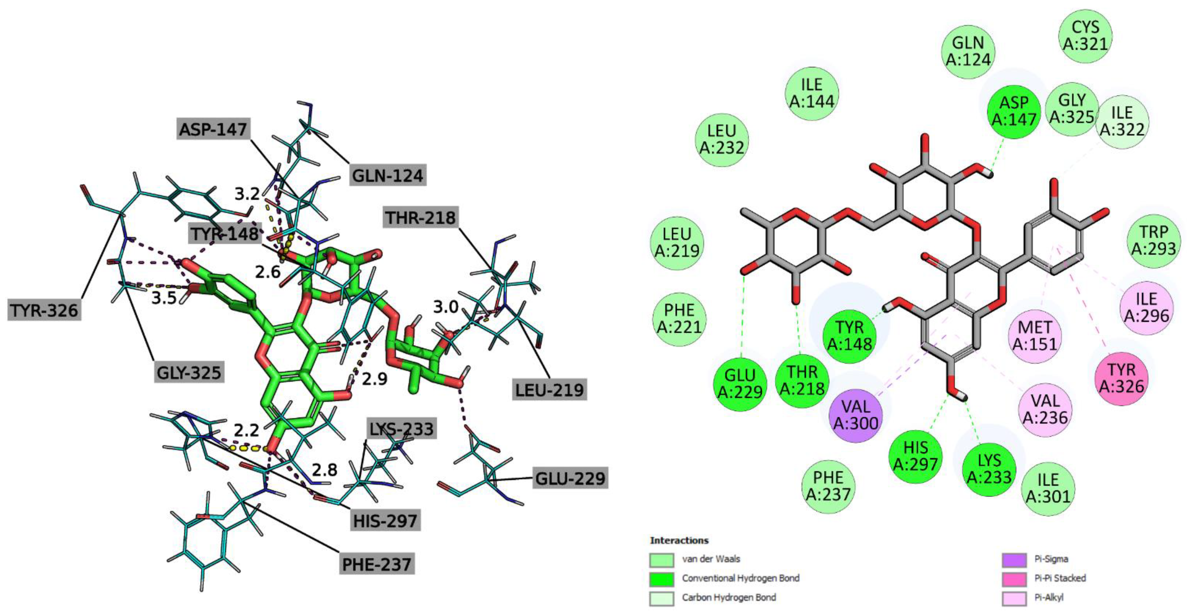
Based on the two- and three-dimensional interaction diagrams illustrated in Figure 2, it was determined that rutin is well integrated into the active site of the mu-opioid receptor (PBD ID 5C1M) due to the formation of hydrogen bonds with ASP147 (2.6 Å), HIS297 (2.2 Å), TYR148 (2.9 Å), LYS233 (2.8 Å) THR218 (3.0 Å), as well as the π-π stacking interaction between the π -electron cloud of the aromatic ring of 3,4-dihydroxyphenyl and 4-hydroxyphenyl TYR326.
The surface of the protein binding site is surrounded by other hydrophobic contacts, mostly π-alkyl interactions: ILE296 and MET151 have a bond with aromatic 3,4-dihydroxyphenyl, and VAL236 and VAL300 binds with 5,7-dihydroxy-4H-chromen-4-one. A stabilizing π-σ interaction with VAL300 was also present in the last fragment.
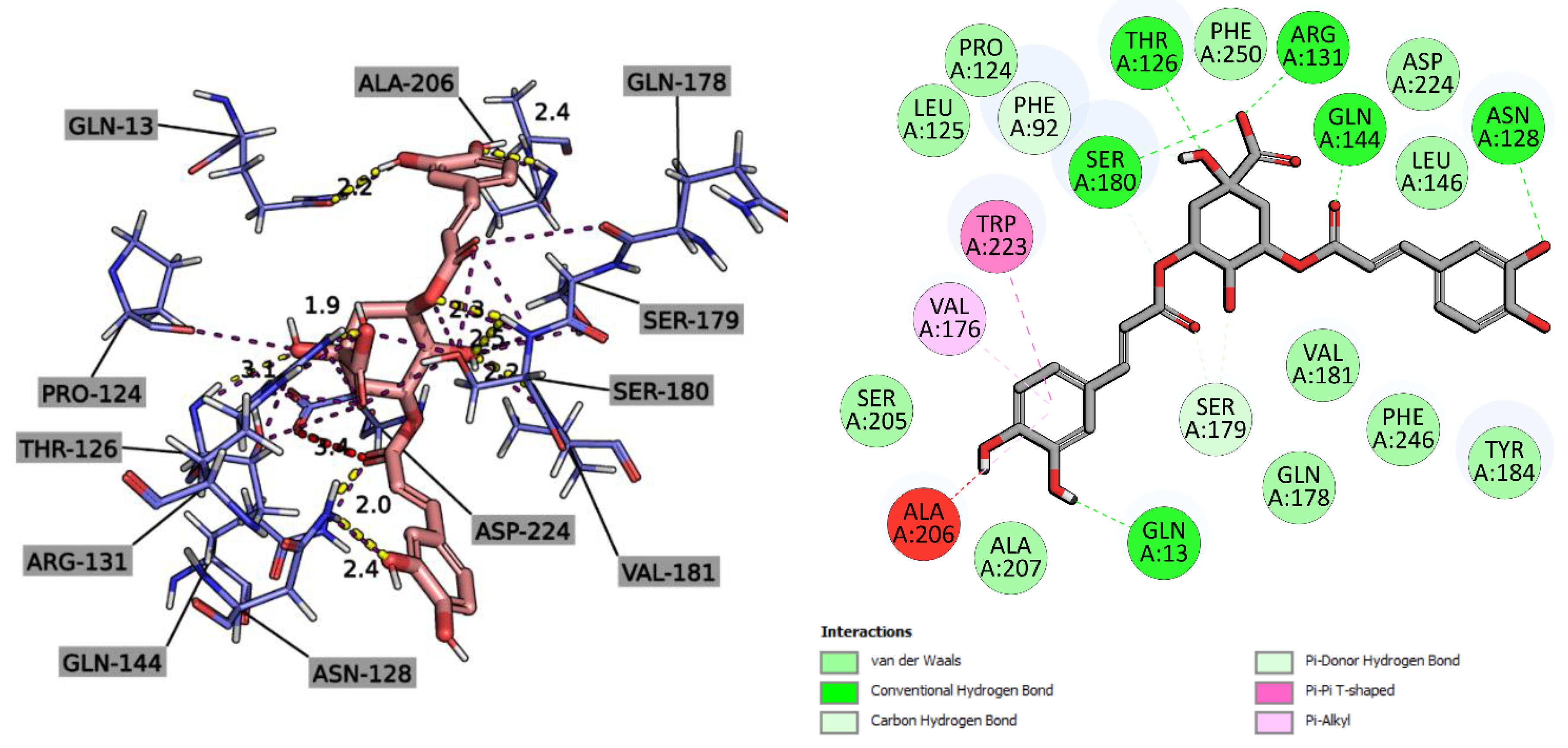
The binding mode is presented in the article [34], demonstrating the formation of a stable complex of the NMDA receptor GluN1 through hydrogen bonds with 6-(3-fluoro-5-(3-(methylamino)prop-1-yn-1-yl)phenethyl)-4-methylpyridin-2-amine: PRO124, THR126, ARG131 and SER296. The PHE92 residue stabilizes the binding of the reference ligand through a π-stacking interaction. In our case (Figure 3), the best effect of 3DWW with 3,5-dicaffeoylquinic acid was achieved through a series of hydrogen contacts of oxo functional groups with GLN13 (2.2 Å), ALA206 (2.4 Å), SER180 (2.3 Å), SER179 (2.5 Å), VAL181 (2.2 Å), THR126 (1.9 Å), ASN128 (2.4, 2.0 Å).
In addition, 3,5-dicaffeoylquinic acid forms through one of the 3,4-dihydroxyphenyls a π-alkyl and π-π T-stacking chemical bond with amino acids VAL176 and TRP223, respectively.
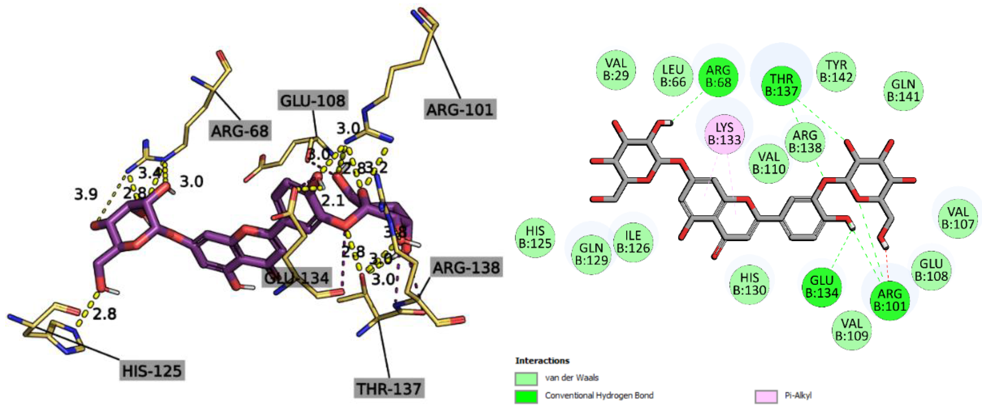
Unlike AKBA, which due to its skeleton forms three hydrogen bonds with AGR156, VAL128 and HIS148 [35], Luteolin 7,3′-diglucoside reaches a higher affinity value of -10.85 kcal/mol when binding to 5-lipoxygenase by forming many hydrogen contacts, but already with other amino acids: ARG101 (3.0, 3.0, 2.8, 3.2 Å) THR137 (3.0, 3.0, 2.8 Å), HIS125 (2.8 Å), ARG68 (2.8, 3.4, 3.0 Å), GLU134 (2.1). This complex had one hydrophobic bond in the form of π-alkyl interaction of the 5-hydroxy-4H-chromen-4-one fragment with the alkyl chain of LYS133 (Figure 4).
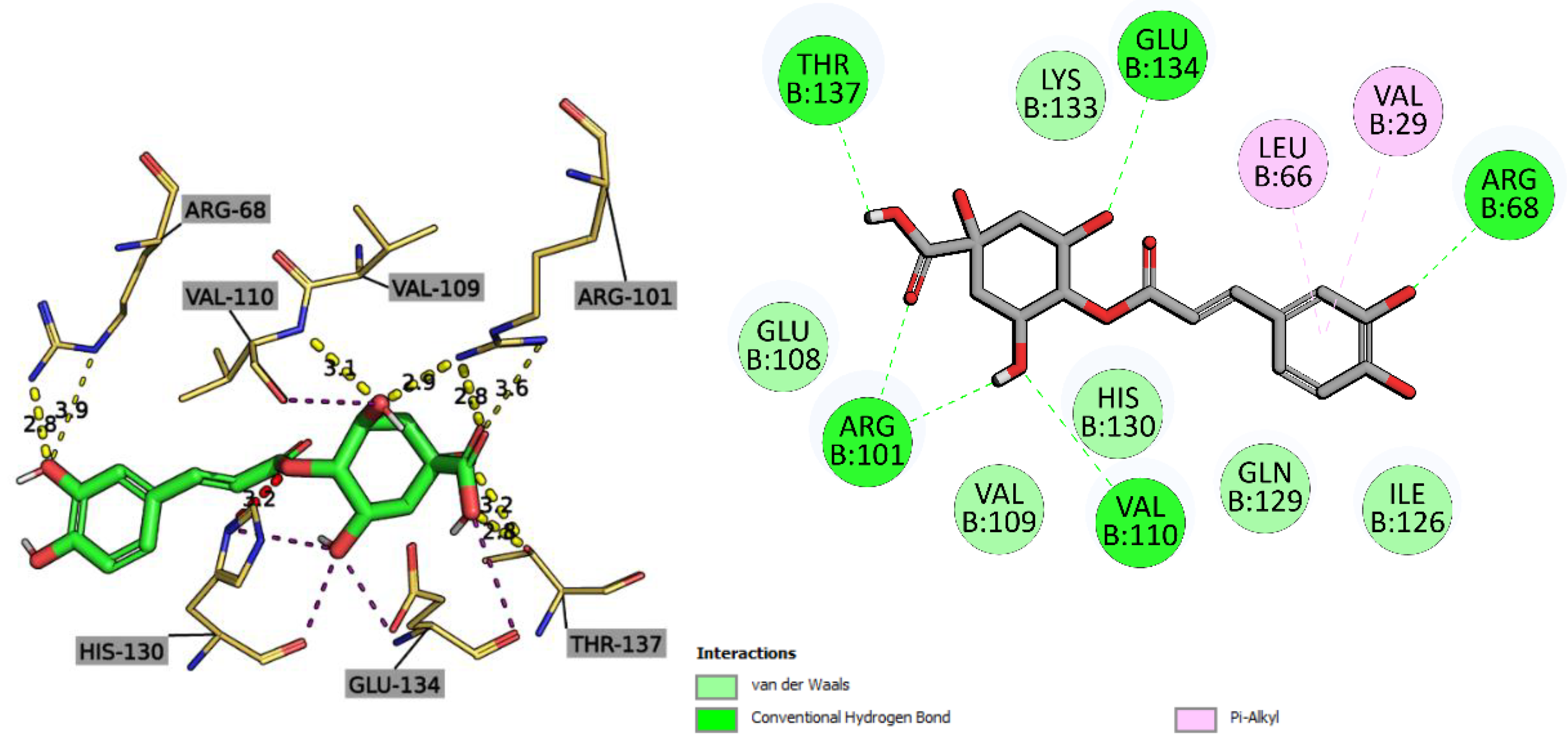
For cryptochlorogenic acid (Figure 5), a similar binding profile was recorded for the Luteolin 7,3′-diglucoside_5-LOX complex, with the best CNN pose score of 0.8774 points. The difference was an additional hydrogen contact between the VAL110 residue and the hydroxyl group of 1,3,5-trihydroxycyclohexane-1-carboxylic acid. Also, amino acid residues VAL29 and LEU66 formed a π-alkyl bond with the polarizable π-electron cloud of the 3,4-dihydroxyphenyl aromatic ring.
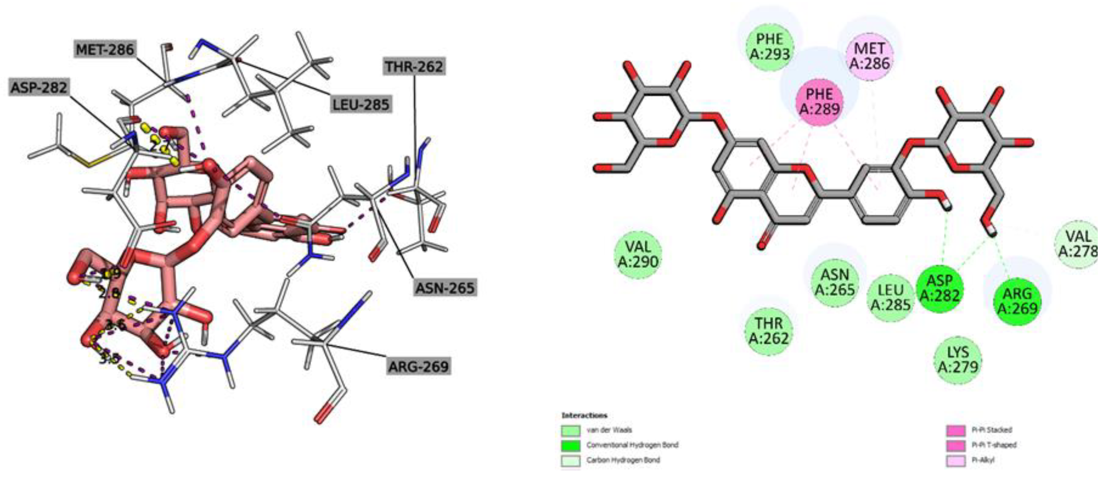
The determined affinity index for Luteolin 7,3′-diglucoside for the GABAA receptor was -7.17 kcal/mol. The ligand exhibited a position characterized by two classical hydrogen bonds with ASP282 (1.9, 2.7 Å) through hydroxyl groups (Figure 6). ARG269 also formed hydrocarbon bonds through the available amino groups. The PHE289 residue stabilizes and orients the ligand through π-π stacking and π-π T-stacking with respect to the aromatic systems of Luteolin 7,3′-diglucoside. The last hydrophobic interaction was π-alkyl type binding for the ligand and the aromatic phenyl of MET286.
Through docking studies, the phytoconstituents of M. chamomilla extract had a distinct binding position with the selected proteins compared to the co-crystallized ligands, where the compounds studied here preferentially formed more hydrogen bonds than hydrophobic and electrostatic ones. This is due to the large number of oxo functional groups, thereby increasing the probability of forming H-bonds between the ligand and amino acid residues. Based on the results of docking and analysis of binding modes, it can be concluded that the present phytochemicals can most likely exert their anti-inflammatory, sedative and analgesic activity by inhibiting the studied enzymes and receptors. However, further studies of molecular dynamics are necessary to finally confirm the stability of the formed complexes. They also should be studied both in vitro and in vivo to assess their safety and efficacy as analgesics.
Therefore, the phytochemicals found in M. chamomilla flowers proved to be promising candidates for the development of analgesic drugs. However, further research work and development are needed to adopt in use such new potent anti-inflammatory agents and hypnotics for therapeutic applications.
The aqueous PEO gels mixed with the M. chamomilla extract showed a homogeneous structure. The aqueous 12% PEO gel was a feasible platform for the printing gels with the M. chamomilla extract at the concentrations ranging from 0.5 to 1.5 g per 10 mL. The corresponding SSE 3D-printed scaffolds were uniform in shape and size (Figure 1). By visual inspection dissolving the printed samples in purified water at a room temperature (22 ± 2 °C) without mixing it was settled that 3D-printed dosage forms were completely disintegrated within 20-25 min, thus suggesting that they could be used as an immediate-release oral delivery system for the present plant extract.

4. Materials and Methods

4.1. Chemicals

Deionized water was obtained by using a Millipore Simplicty UV station (Merck Millipore, Burlington, MA, USA). Ethanol, formic acid, acetonitrile have origin from VWR (Radnor, PA, USA). DL-valine, glycine, phenylalanine (Acros Organics, Geel, Belgium), β-alanine (OstroVit, Zambrow, Poland), L-arginine, L-lysine (Fits OÜ, Tallin, Estonia), Tween-80 (Ferak, Berlin, Germany), and aluminum chloride (Sigma-Aldrich, Sant Louis, MI, USA), rutin, chlorogenic and gallic acids (Carl Roth, Karlsruhe, Germany) were used in our experiments.

4.2. Plant Material

Matricaria chamomilla L. flowers were produced by MK Loodusravi OÜ (Venevere village, Põhja-Sakala municipality, Viljandi County, Estonia (58.598228 N, 25.704950 E) and packed in an airtight plastic bag. The identity of the raw material was confirmed by Prof. Ain Raal, Institute of Pharmacy, University of Tartu, Tartu, Estonia [20,36]. The M. chamomilla flowers raw material met the European Pharmacopeia’s requirements [20]. Its loss on drying was 6.8% [20].

4.3. Preparation of Extracts

The dried M. chamomilla flowers (500.0 g) were extracted with 70% aqueous ethanol solution (3000 mL) by macerating at an ambient room temperature for one day. The extraction was repeated twice with the same solvent (on 1000.0 mL each stage). The liquid extracts were combined, kept for sedimentation during two days, and filtrated. The six amino acids of interest, arginine (2.31 g), phenylalanine (2.19 g), β-alanine (1.17 g), glycine (0.99 g), valine (1.44 g), and lysine (1.92 g), were added in a three-fold equimolar amount to the total phenolic compounds in terms of gallic acid to the six portions of the liquid extract (300 mL each one). The resulting solutions were kept overnight at 22 ± 2 °C.
The native liquid extract and its solutions with amino acids were separately evaporated in a rotary vacuum evaporator (vacuum 150 mbar, rotation 80 rpm, heating bath 80 °C) to form thick extracts, which were then lyophilized in a SCANVAC COOLSAFE 55-4 Pro (LaboGene ApS, Denmark) apparatus. Finally, the 7 dry extracts were obtained: the dried M. chamomilla flowers extract (Gch) and its amino acids preparations with arginine (Gch-Arg), phenylalanine (Gch-Phe), β-alanine (Gch-β-Ala), glycine (Gch-Gly), valine (Gch-Val), and lysine (Gch-Lys).

4.4. Spectrophotometric Assay of Main Groups of Phytochemicals

The assay of flavonoids, hydroxycinnamic acids, and total phenols in the M. chamomilla extract and its amino acid preparations were carried out with a spectrophotometer Shimadzu UV-1800 (Shimadzu Corporation, Tokyo, Japan). Flavonoids were calculated in terms of rutin. Optical density was measured at 417 nm after the reaction with aluminium chloride [20,37]. Hydroxycinnamic acids were estimated after adding sodium molybdate and sodium nitrite and measuring optical density at 525 nm. The calculation was performed in terms of chlorogenic acid [20]. Total phenolics were assayed in terms of gallic acid at 270 nm [38]. The experiments were repeated three times.

4.5. Assay of Phenolics by UPLC-MS/MS

M. chamomilla flowers samples were analysed with a UPLC-MS/MS system to determine quantitative composition of phenolic compounds. The compounds of interest were separated form plant matrix using an Acquity H-class UPLC system (Waters, Milford, MA, USA) coupled with a YMC Triart C18 (100 × 2.0 mm 1.9 µm) column. Column temperature was maintained at 40 °C. Mobile phase was supplied at 0.5 mL/min flow rate consisting of an aqueous solution of formic acid (0.1%) as eluent A, and MS-grade acetonitrile as eluent B. The following linear gradient was applied: from 0 to 1 min, constant flow at 95% of solvent A; 1 to 5 min, linear increase of solvent B to 30%; 5 to 7 min, to 50%; 7.5 to 8 min, wash column with 100% solvent B; 8.1 return to initial conditions for a total analysis time of 10 min. Mass spectrometric data was acquired with a triple-quadrupole tandem mass spectrometer (Xevo, Waters, USA) working in negative electrospray ionization (ESI) mode. The following parameters for the MS/MS acquisition were set: the capillary voltage was set to -2 kV, cone voltage set at 30 V, desolvation gas (nitrogen) was heated to 400 °C and supplied at flow rate of 700 L/h, the curtain gas (nitrogen) flow supplied at 20 L/h. The temperature of ion source was maintained at 150 °C. Analytical grade standards were used to identify phenolic compounds in M. chamomilla flowers by comparing their MS/MS spectral data and retention times. Linear regression fit and standard dilution methods were used for quantitative determination of phenolic compounds [39,40].

4.6. Assay of Amino Acids by UPLC-MS/MS

Analysis of amino acids in M. chamomilla flowers was carried out on Acquity H-class UPLC system (Waters, Milford, MA, USA) equipped with Xevo TQD mass spectrometer (Waters, Milford, MA, USA). One microliter of extracts was injected on a BEH Amide (150 mm × 2.1 mm, 1.7 µm) column (Waters, Milford, MA, USA) with column temperature set at 25 °C. The mobile phase consisting of aqueous solution of 10 mmol ammonium formate with 0.125% formic acid (eluent A) and acetonitrile (eluent B) was delivered at 0.6 mL/min. Gradient elution method with following settings was applied: 0 min to 1 min., 95% B; 1–3.9 min, 70% B; 3.9–5.1 min, 30% B; 5.1–6.4 min, column was flushed with 70% of eluent A; at 6.5 min, gradient was returned to initial composition for a total run time of 10 min. Mass spectrometer conditions were set as follows: positive electrospray ionization was set to +3.5 kV, cone voltage set at 30 V, desolvation gas flow at 800 L/h, and temperature at 400 °C. Ion source temperature was set at 120 °C. Peak assignment and identification of amino acids in M. chamomilla flowers extracts were done while comparing their retention times and MS/MS data with those of analytical grade standards. Linear regression fit models were obtained using the standard dilution method for quantification of amino acids.

4.7. Pharmacological Study

The analgesic and soporific activity were studied with rodents (rats and mice) in compliance with the rules of the “European Convention for the Protection of Vertebrate Animals Used for Experimental and Other Scientific Purposes” (Strasbourg, 1986), Directive 2010/63/EU of the European Parliament and of the Council of the European Union (2010) on the protection of animals used for scientific purposes, the Order of the Ministry of Health of Ukraine No. 944 “On Approval of the Procedure for Preclinical Study of Medicinal Products and Examination of Materials for Preclinical Study of Medicinal Products” (2009), and the Law of Ukraine No. 3447-IV “On the protection of animals from cruel treatment” (2006) [41,42,43,44,45]. The research was approved by the Bioethics Commission of the National University of Pharmacy, Ukraine (protocol №4 from 3 October 2023).

4.7.1. Analgesic Activity

The analgesic activity of the M. chamomilla extract and its amino acid preparations was studied with mice (20–40 g) in comparison with acetaminophen (paracetamol-Zdorovye, 500 mg capsules, pharmaceutical company “Zdorovye”, Kharkiv, Ukraine).
Before the test animals don’t have access to food during 2 hours. The experimental groups (6 mice in each group) were formed randomly: Group 1—intact animals (1 mL 0.9% solution of NaCl per 100 g of body weight); Group 2-4—the animals treated with extract Gch at the doses of 25, 50 and 100 mg/kg; Group 5-7—the animals treated with extract Gch-Arg at the doses of 25, 50 and 100 mg/kg; Group 8-10—the animals treated with extract Gch-Phe at the doses of 25, 50 and 100 mg/kg; Group 11-13—the animals treated with extract Gch-β-Ala at the doses of 25, 50 and 100 mg/kg; Group 14-16—the animals treated with extract Gch-Gly at the doses of 25, 50 and 100 mg/kg; Group 17-19—the animals treated with extract Gch-Val at the doses of 25, 50 and 100 mg/kg; Group 20-22—the animals treated with extract Gch-Lys at the doses of 25, 50 and 100 mg/kg; and control group (CG) 23—the animals treated with acetaminophen at the dose of 50 mg/kg.
The agents were administered intragastrically, and subsequently the animal was carefully placed on a hot plate (55 °C) for 30 min. The duration of the mouse’s stay on the hot plate (in seconds) before the beginning of protective reflexes was chosen as an indicator of pain sensitivity. The time during which the animals were the hot plate did not exceed 60 s. After the agent consumption, the mice were examined at the regular intervals at 0.5, 1, 2, 3, and 4 hours. The analgesic effect was revealed when the latent period of response after the administration of the agent significantly increased the control group [7,46,47].

4.7.2. Soporific Activity

The soporific activity of the M. chamomilla extract and its amino acid preparations was studied with white rats (190–280 g). The reference drugs were sodium thiopental lyophilizate (for injection solution, PLC “Kiivmedpreparat”, Kyiv, Ukraine), and “Valerian syrup AN NATUREL” syrup (LLC Beauty and Health, Kharkiv, Ukraine).
The experimental groups (6 mice in each group) were formed randomly: Group 1—intact animals (1 mL 0.9% solution of NaCl per 100 g of body weight); Group 2-4—the animals treated with extract Gch at the doses of 25, 50 and 100 mg/kg; Group 5-7—the animals treated with extract Gch-Arg at the doses of 25, 50 and 100 mg/kg; Group 8-10—the animals treated with extract Gch-Phe at the doses of 25, 50 and 100 mg/kg; Group 11-13—the animals treated with extract Gch-β-Ala at the doses of 25, 50 and 100 mg/kg; Group 14-16—the animals treated with extract Gch-Gly at the doses of 25, 50 and 100 mg/kg; Group 17-19—the animals treated with extract Gch-Val at the doses of 25, 50 and 100 mg/kg; Group 20-22—the animals treated with extract Gch-Lys at the doses of 25, 50 and 100 mg/kg; and control group (CG) 23—the animals treated with sodium thiopental at the dose of 40 mg/kg; control group (CG valerian) 24—the animals treated with valerian syrup at the dose of 2.14 mg/kg. The duration of sleep was determined by the time period for which the rats were in a lateral position [7,48,49,50].

4.7.3. In Silico Studies

Selection of ligands. The following constituents derived from M. chamomilla were used: 4,5-dicaffeoylquinic acid (PubChem CID: 5281780), 3,4-dicaffeoylquinic acid (PubChem CID: 6474309), luteolin 7,3′-diglucoside (PubChem CID: 44258089), 3,5-dicaffeoylquinic acid (PubChem CID: 13604688), rutin (PubChem CID: 5280805), luteolin 7-O-glucoside (PubChem CID: 5280637), isorhamnetin (PubChem CID: 5281654), luteolin (PubChem CID: 5280445), cryptochlorogenic acid (PubChem CID: 9798666), chlorogenic acid (PubChem CID: 1794427), hyperoside (PubChem CID: 5281643), isorhamnetin-3-glucoside (PubChem CID: 6455477), luteolin-4-O-glucoside (PubChem CID: 12304738), neochlorogenic acid (PubChem CID: 5280633), caffeic ccid (PubChem CID: 689043), 3,4-dihydroxyphenylacetic acid (PubChem CID: 547), and vanillic acid (PubChem CID: 8468). The abovementioned constituents were converted to 2DSDF format and subjected to minimization by using the Open Babel tool for facilitating the determination of their binding affinity in specific targets.
Preparation of targeted proteins. The three-dimensional arrangements of various protein complexes were retrieved from the RCSB Protein Data Bank in pdb-format, including the mu-opioid receptor-Gi protein complex (PDB: 5C1M) [51], SC-558 bound at the COX-2 active site (PDB: 1CX2) [52], COX-1 complexed with ibuprofen (PDB: 1EQG) [53], crystal structure of the NMDA receptor GluN1 (PDB: 4KFQ) [34], nanobody-stabilized active state of the kappa-opioid receptor (PDB: 6B73) [54], human hematopoietic prostaglandin D synthase (PDB: 2CVD) [55], stable-5-lipoxygenase bound to AKBA (PDB: 6NCF) [55], and human GABAA receptor (PDB: 6X3X) [48,56].
Docking Analysis. Docking studies were performed for the most active compounds which had the potential to be a best inhibitor of selected proteins in the binding site of enzymes using GNINA [57], which utilizes an ensemble of convolutional neural networks (CNNs) as a scoring function. The scores include binding affinities (kcal/mol), CNN pose scores (a probability that the pose has a low root mean square deviation to the binding pose), and CNN affinities predicted by GNINA. Ligand molecules had provided ten degrees of freedom. Using an autobox ligand, a grid box with an active site in the center was built. BIOVIA Discovery Studio Visualizer 2020 [58] and Pymol 2.6 (Schrödinger LLC., New York, USA) was eventually accelerated to evaluate the docking sites for the potential linking approaches.

4.8. 3D Printing of M. chamomilla Extract

PEO (MW approx. 900,000, Sigma-Aldrich, USA) at the concentrations of 12% were implemented for preparing aqueous gels with M. chamomilla extract for the SSE 3D printing. For this 1.2 g of PEO was mixed in 10 mL of purified water at least for 13–15 hours at an ambient room temperature [19,21,22]. Tween 80 (Laborat GMBH, Berlin, Germany) was used to create more stable and homogenised system, and enhance the release of M. chamomilla extracts from the 3D-printed scaffolds [19,21]. The M. chamomilla extract (0.5, 1.0, and 1.5 g) and Tween 80 as a surface-active agent (0.5 g) were added to 12% PEO gel. The gels viscosity was measured at room temperature (22 ± 2 °C) with a Physica MCR 101 rheometer (Anton Paar, Austria).
The 12% PEO gels loaded with the M. chamomilla extract were directly printed using a bench-top semi-solid (SSE) 3D printing system (System 30 M, Hyrel 3D, Norcross, GA, USA). The printing control software of a SSE 3D printer was Repetrel, Rev3.083_K, Hyrel 3D, USA. Such parameters were used: printing head speed—0.5 mm/s; blunt needle (Gauge, 21G); without heating of syringe content and a printed plate.
A model 4 × 4 grid lattice (30 × 30 × 0.5 mm) and the round shapes (20 mm in diameter) were designed with Autodesk 3ds Max Design 2017 software (Autodesk Inc., San Francisco, CA, USA) and FreeCAD software (vers. 0.19/release date 2021) [59]. Total six (6) layers were printed for preparing the lattices and five (5) layers for the round-shaped scaffolds.
Lattice weight and area measurements were determined for evaluation of 3D printability. The 3D printed lattice theoretical surface area was 324 mm2. It was compared with the corresponding areas of experimental 3D-printed lattices and their ratio were calculated [21,22]. The photographs were analysed with ImageJ (National Institute of Health, Bethesda, MD, USA) image analysis software (version 1.51k). The weights of the 3D-printed preparations were determined with an analytical scale (Scaltec SBC 33, Scaltec, Germany).

4.9. Statistical Analysis

The average value was calculated at least for three measurements in the phytochemical study and six ones in the pharmacological study. The values of the confidence interval were calculated using the Student’s criterion limit. The data are presented as the mean ± SD [20,60].

5. Conclusions

The present study showed with animal models (rats and mice) the potential of the M. chamomilla extract and its amino acid preparations in treating sleep disorders and diseases accompanied by pain. Total 22 polyphenolic compounds and 14 amino acids were identified and quantified in the extracts. The performance and efficacy of the extract can be additionally augmented by the conjugation with amino acids. Glycine potentiates analgesic activity, while lysine and β-alanine preferably improve soporific activity of the M. chamomilla extract. The molecular docking analysis revealed a high probability of COX-1,2 inhibition, GABAA and NMDA receptor antagonism by the M. chamomilla extract constituents. The formulation of novel 3D-printed oral dosage forms for the extracts is a promising approach for preparing the medicinal or dietary supplement products of such extracts.

Author Contributions

Conceptualization, O.K., V.J., I.K., J.H. and A.R.; methodology, O.K., V.J., I.K., O.V., Ye.K., J.H. and A.R.; software, O.K., V.Z., Ye.K., and J.S.; validation, O.K., A.R.; formal analysis, J.S., Ye.K., V.Z. and O.K.; investigation, J.S., I.K., O.K., V.Z. and Ye.K.; resources, O.K., I.K., J.H. and A.R.; data curation, I.K, O.K., Ye.K., V.O. and A.R.; writing original draft preparation, O.K., J.S., V.J., I.K., Ye.K., V.O., J.H. and A.R.; writing-review and editing, V.J., O.K., J.H. and A.R.; visualization, J.S., Ye.K. and O.Ku.; supervision, O.K., I.K., V.J., J.H. and A.R.; project administration, O.K., J.H. and A.R.; funding acquisition, O.K., J.H. and A.R. All authors have read and agreed to the published version of the manuscript.

Funding

This research was funded by the Estonian Research Council grant (PRG1903) and the European Union in the MSCA4Ukraine project “Design and development of 3D-printed medicines for bioactive materials of Ukrainian and Estonian medicinal plants origin” [ID number 1232466].

Institutional Review Board Statement

The research in the analgesic and soporific activity of the extracts with rodents (rats and mice) is approved by the Bioethics Commission of the National University of Pharmacy, Ukraine (protocol №4 from 3 October 2023).

Informed Consent Statement

Not applicable.

Data Availability Statement

The data supporting the results of this study can be obtained from the corresponding authors upon reasonable request.

Acknowledgments

The authors sincerely thank all the defenders of Ukraine who made the performance of this study possible. The authors sincerely appreciate the support of the Partners who are standing with Ukraine.

Conflicts of Interest

The authors declare no conflicts of interest.

References

  1. Sah, A.; Naseef, P.P.; Kuruniyan, M.S.; Jain, G.K.; Zakir, F.; Aggarwal, G. A Comprehensive Study of Therapeutic Applications of Chamomile. Pharmaceuticals 2022, 15, 1284. [Google Scholar] [CrossRef] [PubMed]
  2. Gupta, V.; Mittal, P.; Bansal, P.; Khokra, S.L.; Kaushik, D. Pharmacological potential of matricaria recutita—A review. International Journal of Pharmaceutical Sciences and Drug Research 2010, 2, 12–16. [Google Scholar]
  3. El Mihyaoui, A.; Esteves Da Silva, J.C.G.; Charfi, S.; Candela Castillo, M.E.; Lamarti, A.; Arnao, M.B. Chamomile (Matricaria chamomilla L.): A Review of Ethnomedicinal Use, Phytochemistry and Pharmacological Uses. Life 2022, 12, 479. [Google Scholar] [CrossRef] [PubMed]
  4. Menale, B.; De Castro, O.; Di Iorio, E.; Ranaldi, M.; Muoio, R. Discovering the Ethnobotanical Traditions of the Island of Procida (Campania, Southern Italy). Plant Biosystems—An International Journal Dealing with all Aspects of Plant Biology 2022, 156, 450–468. [Google Scholar] [CrossRef]
  5. Neves, J.M.; Matos, C.; Moutinho, C.; Queiroz, G.; Gomes, L.R. Ethnopharmacological Notes about Ancient Uses of Medicinal Plants in Trás-Os-Montes (Northern of Portugal). Journal of Ethnopharmacology 2009, 124, 270–283. [Google Scholar] [CrossRef] [PubMed]
  6. Güzel, Y.; Güzelşemme, M.; Miski, M. Ethnobotany of Medicinal Plants Used in Antakya: A Multicultural District in Hatay Province of Turkey. Journal of Ethnopharmacology 2015, 174, 118–152. [Google Scholar] [CrossRef]
  7. Sepp, J.; Koshovyi, O.; Jakstas, V.; Žvikas, V.; Botsula, I.; Kireyev, I.; Tsemenko, K.; Kukhtenko, O.; Kogermann, K.; Heinämäki, J.; et al. Phytochemical, Technological, and Pharmacological Study on the Galenic Dry Extracts Prepared from German Chamomile (Matricaria chamomilla L.) Flowers. Plants 2024, 13, 350. [Google Scholar] [CrossRef] [PubMed]
  8. Raal, A.; Orav, A.; Püssa, T.; Valner, C.; Malmiste, B.; Arak, E. Content of Essential Oil, Terpenoids and Polyphenols in Commercial Chamomile (Chamomilla Recutita L. Rauschert) Teas from Different Countries. Food Chemistry 2012, 131, 632–638. [Google Scholar] [CrossRef]
  9. Orav, A.; Raal, A.; Arak, E. Content and Composition of the Essential Oil of Chamomilla Recutita (L.) Rauschert from Some European Countries. Natural Product Research 2010, 24, 48–55. [Google Scholar] [CrossRef]
  10. Rawat, A.; Gupta, A.; Kholiya, S.; Chauhan, A.; Kumar, D.; Venkatesha, K.T.; Upadhyay, R.K.; Padalia, R.C. Comparative Study of Chemical Composition of Two Cultivars of German Chamomile, Matricaria chamomilla L. Syn Chamomilla recutita L. Rauschert. Journal of Biologically Active Products from Nature 2022, 12, 488–506. [Google Scholar] [CrossRef]
  11. Stanojevic, L.P.; Marjanovic-Balaban, Z.R.; Kalaba, V.D.; Stanojevic, J.S.; Cvetkovic, D.J. Chemical Composition, Antioxidant and Antimicrobial Activity of Chamomile Flowers Essential Oil (Matricaria chamomilla L.). Journal of Essential Oil Bearing Plants 2016, 19, 2017–2028. [Google Scholar] [CrossRef]
  12. Kravchenko, G.; Krasilnikova, O.; Raal, A.; Mazen, M.; Chaika, N.; Kireyev, I.; Grytsyk, A.; Koshovyi, O. Arctostaphylos uva-ursi L. Leaves Extract and Its Modified Cysteine Preparation for the Management of Insulin Resistance: Chemical Analysis and Bioactivity. Nat. Prod. Bioprospect. 2022, 12, 30. [Google Scholar] [CrossRef] [PubMed]
  13. Koshovyi, O.; Granica, S.; Piwowarski, J.P.; Stremoukhov, O.; Kostenko, Y.; Kravchenko, G.; Krasilnikova, O.; Zagayko, A. Highbush Blueberry (Vaccinium corymbosum L.) Leaves Extract and Its Modified Arginine Preparation for the Management of Metabolic Syndrome—Chemical Analysis and Bioactivity in Rat Model. Nutrients 2021, 13, 2870. [Google Scholar] [CrossRef] [PubMed]
  14. Kovalenko, V.N. Compendium 2020. Medicines; MORION: Kyiv, Ukraine, 2020. [Google Scholar]
  15. Parfenov, V.A. Use of L-Lysine Aescinate in Central Nervous System Diseases. Neurology, Neuropsychiatry, Psychosomatics 2011, 0, 99. [Google Scholar] [CrossRef]
  16. Koshovyi, O.; Raal, A.; Kireyev, I.; Tryshchuk, N.; Ilina, T.; Romanenko, Y.; Kovalenko, S.M.; Bunyatyan, N. Phytochemical and Psychotropic Research of Motherwort (Leonurus cardiaca L.) Modified Dry Extracts. Plants 2021, 10, 230. [Google Scholar] [CrossRef]
  17. Koshovyi, O.M.; Zagayko, A.L.; Kolychev, I.O.; Akhmedov, E.Yu.; Komissarenko, A.N. Phytochemical Study of the Dry Extract from Bilberry Leaves. Azerbaijan Pharmaceutical and Pharmacotherapy Journal 2016, 16, 18–23. [Google Scholar]
  18. Chaika, N.; Koshovyi, O.; Ain, R.; Kireyev, I.; Zupanets, A.; Odyntsova, V. Phytochemical Profile and Pharmacological Activity of the Dry Extract from Arctostaphylos Uva-Ursi Leaves Modified with Phenylalanine. ScienceRise: Pharmaceutical Science 2020, 0, 74–84. [Google Scholar] [CrossRef]
  19. Koshovyi, O.; Vlasova, I.; Laur, H.; Kravchenko, G.; Krasilnikova, O.; Granica, S.; Piwowarski, J.P.; Heinämäki, J.; Raal, A. Chemical Composition and Insulin-Resistance Activity of Arginine-Loaded American Cranberry (Vaccinium Macrocarpon Aiton, Ericaceae) Leaf Extracts. Pharmaceutics 2023, 15, 2528. [Google Scholar] [CrossRef] [PubMed]
  20. European Pharmacopoeia, 11th ed.; Council of Europe: Strasbourg, 2022.
  21. Koshovyi, O.; Heinämäki, J.; Raal, A.; Laidmäe, I.; Topelius, N.S.; Komisarenko, M.; Komissarenko, A. Pharmaceutical 3D-Printing of Nanoemulsified Eucalypt Extracts and Their Antimicrobial Activity. European Journal of Pharmaceutical Sciences 2023, 187, 106487. [Google Scholar] [CrossRef]
  22. Viidik, L.; Seera, D.; Antikainen, O.; Kogermann, K.; Heinämäki, J.; Laidmäe, I. 3D-Printability of Aqueous Poly(Ethylene Oxide) Gels. European Polymer Journal 2019, 120, 109206. [Google Scholar] [CrossRef]
  23. Catani, M.V.; Rinaldi, F.; Tullio, V.; Gasperi, V.; Savini, I. Comparative Analysis of Phenolic Composition of Six Commercially Available Chamomile (Matricaria chamomilla L.) Extracts: Potential Biological Implications. IJMS 2021, 22, 10601. [Google Scholar] [CrossRef] [PubMed]
  24. Mulinacci, N.; Romani, A.; Pinelli, P.; Vincieri, F.F.; Prucher, D. Characterization of Matricaria recutita L. Flower Extracts by HPLC-MS and HPLC-DAD Analysis. Chromatographia 2000, 51, 301–307. [Google Scholar] [CrossRef]
  25. Zhao, H.; Jiang, Z.; Chang, X.; Xue, H.; Yahefu, W.; Zhang, X. 4-Hydroxyphenylacetic Acid Prevents Acute APAP-Induced Liver Injury by Increasing Phase II and Antioxidant Enzymes in Mice. Front. Pharmacol. 2018, 9, 653. [Google Scholar] [CrossRef] [PubMed]
  26. Gruenbaum, B.F.; Zlotnik, A.; Oleshko, A.; Matalon, F.; Shiyntum, H.N.; Frenkel, A.; Boyko, M. The Relationship between Post-Traumatic Stress Disorder Due to Brain Injury and Glutamate Intake: A Systematic Review. Nutrients 2024, 16, 901. [Google Scholar] [CrossRef]
  27. Owens, S.L.; Ahmed, S.R.; Lang Harman, R.M.; Stewart, L.E.; Mori, S. Natural Products That Contain Higher Homologated Amino Acids. ChemBioChem 2024, e202300822. [Google Scholar] [CrossRef] [PubMed]
  28. Henzi, V.; Reichling, D.B.; Helm, S.W.; MacDermott, A.B. L-Proline Activates Glutamate and Glycine Receptors in Cultured Rat Dorsal Horn Neurons. Mol Pharmacol 1992, 41, 793–801. [Google Scholar]
  29. Abdelnour, S.A.; Khalil, W.A.; Khalifa, N.E.; Khalil, F.M.A.; Hassan, M.A.E. L-Proline: A Promising Tool for Boosting Cryotolerance and Fertilizing Ability of Cryopreserved Sperm in Animals. Animal Reproduction Science 2024, 263, 107429. [Google Scholar] [CrossRef]
  30. Adeva-Andany, M.; Souto-Adeva, G.; Ameneiros-Rodríguez, E.; Fernández-Fernández, C.; Donapetry-García, C.; Domínguez-Montero, A. Insulin Resistance and Glycine Metabolism in Humans. Amino Acids 2018, 50, 11–27. [Google Scholar] [CrossRef]
  31. Memmott, R.J.; Young, L.A. An Encounter with Homeless Mothers and Children: Gaining an Awareness. Issues Ment Health Nurs 1993, 14, 357–365. [Google Scholar] [CrossRef]
  32. Kozlov, V.A. Proteinogenic Acids; Cheboksary, 2012. [Google Scholar]
  33. Chaves, P.F.P.; Hocayen, P.D.A.S.; Dallazen, J.L.; De Paula Werner, M.F.; Iacomini, M.; Andreatini, R.; Cordeiro, L.M.C. Chamomile Tea: Source of a Glucuronoxylan with Antinociceptive, Sedative and Anxiolytic-like Effects. International Journal of Biological Macromolecules 2020, 164, 1675–1682. [Google Scholar] [CrossRef]
  34. Kvist, T.; Steffensen, T.B.; Greenwood, J.R.; Mehrzad Tabrizi, F.; Hansen, K.B.; Gajhede, M.; Pickering, D.S.; Traynelis, S.F.; Kastrup, J.S.; Bräuner-Osborne, H. Crystal Structure and Pharmacological Characterization of a Novel N-Methyl-d-Aspartate (NMDA) Receptor Antagonist at the GluN1 Glycine Binding Site. Journal of Biological Chemistry 2013, 288, 33124–33135. [Google Scholar] [CrossRef] [PubMed]
  35. Gilbert, N.C.; Gerstmeier, J.; Schexnaydre, E.E.; Börner, F.; Garscha, U.; Neau, D.B.; Werz, O.; Newcomer, M.E. Structural and Mechanistic Insights into 5-Lipoxygenase Inhibition by Natural Products. Nat Chem Biol 2020, 16, 783–790. [Google Scholar] [CrossRef] [PubMed]
  36. Dobrochaeva, D.N.; Kotov, M.I.; Prokudin, Y.N.; Barbarich, A.I. Key to Higher Plants of Ukraine; Naukova Dumka: Kyiv, Ukraine, 1999. [Google Scholar]
  37. Vlasova, I.; Gontova, T.; Grytsyk, L.; Zhumashova, G.; Sayakova, G.; Boshkayeva, A.; Shanaida, M.; Koshovyi, O. Determination of Standardization Parameters of Oxycoccus Macrocarpus (Ait.) Pursh and Oxycoccus Palustris Pers. Leaves. SR: PS 2022, 48–57. [Google Scholar] [CrossRef]
  38. Huzio, N.; Grytsyk, A.; Raal, A.; Grytsyk, L.; Koshovyi, O. Phytochemical and Pharmacological Research in Agrimonia Eupatoria L. Herb Extract with Anti-Inflammatory and Hepatoprotective Properties. Plants 2022, 11, 2371. [Google Scholar] [CrossRef] [PubMed]
  39. Vilkickyte, G.; Raudone, L.; Petrikaite, V. Phenolic Fractions from Vaccinium vitis-idaea L. and Their Antioxidant and Anticancer Activities Assessment. Antioxidants 2020, 9, 1261. [Google Scholar] [CrossRef]
  40. Marzullo, L.; Ochkur, O.; Orlandini, S.; Renai, L.; Gotti, R.; Koshovyi, O.; Furlanetto, S.; Del Bubba, M. Quality by Design in Optimizing the Extraction of (Poly)Phenolic Compounds from Vaccinium Myrtillus Berries. Journal of Chromatography A 2022, 1677, 463329. [Google Scholar] [CrossRef] [PubMed]
  41. European Convention for the Protection of Vertebrate Animals Used for Experimental and Other Scientific Purposes; 1986; Vol. Official Journal L 222, p. P 0031-0037.
  42. Council Directive 2010/63/EU on the Protection of Animals Used for Scientific Purposes 2010.
  43. On the Protection of Animals from Cruel Treatment; 2009.
  44. On Approval of the Procedure for Preclinical Study of Medicinal Products and Examination of Materials of Preclinical Study of Medicinal Products; 2009.
  45. Regulating the Application of Principles of Good Laboratory Practice and the Verification of Their Applications for Tests on Chemical Substances; 1986; Vol. 1, pp. 145–146.
  46. Masocha, W.; Kombian, S.B.; Edafiogho, I.O. Evaluation of the Antinociceptive Activities of Enaminone Compounds on the Formalin and Hot Plate Tests in Mice. Sci Rep 2016, 6, 21582. [Google Scholar] [CrossRef]
  47. Inaltekin, A.; Kivrak, Y. Evaluation of the Effect of Vortioxetine on Pain Threshold by Hot-Plate Test in Mice. Archives of Neuropsychiatry 2021. [Google Scholar] [CrossRef]
  48. Sepp, J.; Koshovyi, O.; Jakštas, V.; Žvikas, V.; Botsula, I.; Kireyev, I.; Severina, H.; Kukhtenko, O.; Põhako-Palu, K.; Kogermann, K.; et al. Phytochemical, Pharmacological, and Molecular Docking Study of Dry Extracts of Matricaria Discoidea DC. with Analgesic and Soporific Activities. Biomolecules 2024, 14, 361. [Google Scholar] [CrossRef]
  49. Hossain, Md.F.; Talukder, B.; Rana, M.N.; Tasnim, R.; Nipun, T.S.; Uddin, S.M.N.; Hossen, S.M.M. In Vivo Sedative Activity of Methanolic Extract of Stericulia Villosa Roxb. Leaves. BMC Complement Altern Med 2016, 16, 398. [Google Scholar] [CrossRef]
  50. Forouzanfar, F.; Ghorbani, A.; Hosseini, M.; Rakhshandeh, H. Hydroalcoholic Extract of Needles of Pinus Eldarica Enhances Pentobarbital-Induced Sleep: Possible Involvement of GABAergic System. Avicenna J Phytomed 2016, 6, 449–457. [Google Scholar] [PubMed]
  51. Huang, W.; Manglik, A.; Venkatakrishnan, A.J.; Laeremans, T.; Feinberg, E.N.; Sanborn, A.L.; Kato, H.E.; Livingston, K.E.; Thorsen, T.S.; Kling, R.C.; et al. Structural Insights into Μ-Opioid Receptor Activation. Nature 2015, 524, 315–321. [Google Scholar] [CrossRef] [PubMed]
  52. Kurumbail, R.G.; Stevens, A.M.; Gierse, J.K.; McDonald, J.J.; Stegeman, R.A.; Pak, J.Y.; Gildehaus, D.; Iyashiro, J.M.; Penning, T.D.; Seibert, K.; et al. Structural Basis for Selective Inhibition of Cyclooxygenase-2 by Anti-Inflammatory Agents. Nature 1996, 384, 644–648. [Google Scholar] [CrossRef] [PubMed]
  53. Selinsky, B.S.; Gupta, K.; Sharkey, C.T.; Loll, P.J. Structural Analysis of NSAID Binding by Prostaglandin H2 Synthase: Time-Dependent and Time-Independent Inhibitors Elicit Identical Enzyme Conformations. Biochemistry 2001, 40, 5172–5180. [Google Scholar] [CrossRef]
  54. Che, T.; Majumdar, S.; Zaidi, S.A.; Ondachi, P.; McCorvy, J.D.; Wang, S.; Mosier, P.D.; Uprety, R.; Vardy, E.; Krumm, B.E.; et al. Structure of the Nanobody-Stabilized Active State of the Kappa Opioid Receptor. Cell 2018, 172, 55–67.e15. [Google Scholar] [CrossRef]
  55. Aritake, K.; Kado, Y.; Inoue, T.; Miyano, M.; Urade, Y. Structural and Functional Characterization of HQL-79, an Orally Selective Inhibitor of Human Hematopoietic Prostaglandin D Synthase. Journal of Biological Chemistry 2006, 281, 15277–15286. [Google Scholar] [CrossRef]
  56. Kim, J.J.; Gharpure, A.; Teng, J.; Zhuang, Y.; Howard, R.J.; Zhu, S.; Noviello, C.M.; Walsh, R.M.; Lindahl, E.; Hibbs, R.E. Shared Structural Mechanisms of General Anaesthetics and Benzodiazepines. Nature 2020, 585, 303–308. [Google Scholar] [CrossRef]
  57. McNutt, A.T.; Francoeur, P.; Aggarwal, R.; Masuda, T.; Meli, R.; Ragoza, M.; Sunseri, J.; Koes, D.R. GNINA 1.0: Molecular Docking with Deep Learning. J Cheminform 2021, 13, 43. [Google Scholar] [CrossRef]
  58. Moazzem Hossen, S.M.; Akramul Hoque Tanim, M.; Shahadat Hossain, M.; Ahmed Sami, S.; Uddin Emon, N. Deciphering the CNS Anti-Depressant, Antioxidant and Cytotoxic Profiling of Methanol and Aqueous Extracts of Trametes Versicolor and Molecular Interactions of Its Phenolic Compounds. Saudi Journal of Biological Sciences 2021, 28, 6375–6383. [Google Scholar] [CrossRef]
  59. Riegel, J.; Mayer, W.; Havre, Y.V. FreeCAD 2001.
  60. Lapach, S.N.; Chubenko, A.V.; Babich, P.N. Statistical Methods in Biomedical Research Using Excel; MORION: Kyiv, 2000. [Google Scholar]
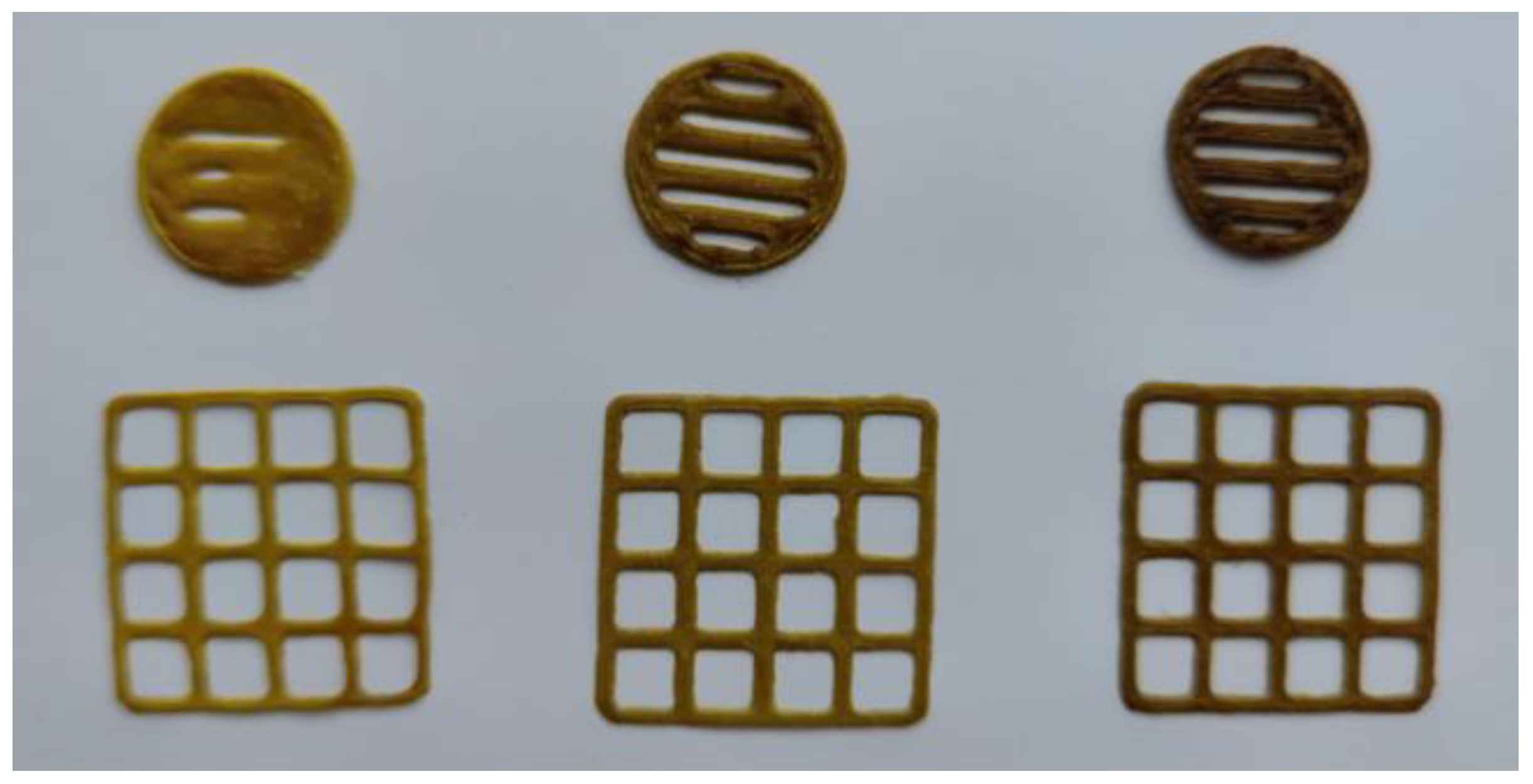
Figure 1. Photographs of the semisolid extrusion (SSE) 3D-printed lattices and round-shape discs. The constructs were printed based on the aqueous 12% polyethylene oxide (PEO) gel with the M. chamomilla dry extract.
Preprints 110048 g001
Figure 2. 3D and 2D representations of the best key interactions of rutin in the mu-opioid receptor binding pocket.
Preprints 110048 g002
Figure 3. 2D and 3D binding modes of 3,5-dicaffeoylquinic acid inside the active site of the NMDA receptor GluN1.
Preprints 110048 g003
Figure 4. 2D and 3D diagrams of the interaction of Luteolin 7,3′-diglucoside and 5-lipoxygenase.
Preprints 110048 g004
Figure 5. 2D interaction of cryptochlorogenic acid and 5-LOX, 3D image of the surface of the active site of the enzyme.
Preprints 110048 g005
Figure 6. 2D and 3D visualization of the interaction between Luteolin 7,3′-diglucoside and the GABAA receptor.
Preprints 110048 g006
Table 1. Content of polyphenolic compounds in the M. chamomilla extracts.
Substance Content in the extract
Gch [7] Gch-Arg Gch-Phe Gch-β-Ala Gch-Gly Gch-Val Gch-Lys
UPLC-MS/MS, µg/g of a dry extract
Neochlorogenic acid 444.94 ± 20.16 329.21 ± 4.47 322.26 ± 8.51 373.81 ± 7.33 385.18 ± 13.11 382.70 ± 27.35 359.65 ± 12.79
Luteolin 310.93 ± 22.73 270.78 ± 15.81 320.87 ± 9.72 283.67 ± 8.20 309.48 ± 12.86 320.93 ± 27.22 301.31 ± 6.67
Isoquercitrin 921.16 ± 85.20 774.55 ± 41.11 850.92 ± 85.24 872.54 ± 34.08 880.89 ± 79.41 894.15 ± 43.80 838.97 ± 25.75
Cryptochlorogenic acid 80.74 ± 13.48 62.26 ± 9.76 70.97 ± 10.16 58.20 ± 5.03 62.47 ± 5.47 75.10 ± 20.95 55.98 ± 12.71
Luteolin-4-O-glucoside 45.11 ± 3.67 37.98 ± 1.88 49.59 ± 4.86 44.06 ± 3.86 41.68 ± 3.82 49.25 ± 6.75 43.68 ± 6.82
Chlorogenic acid 11742.31 ± 376.34 8984.21 ± 397.30 9532.56 ± 179.37 10341.94 ± 211.50 10567.30 ± 220.17 10014.55 ± 167.55 9342.78 ± 355.66
Quercetin 172.15 ± 12.01 138.19 ± 1.54 162.89 ± 8.50 149.96 ± 6.55 156.29 ± 6.19 149.27 ± 17.77 141.29 ± 6.20
Isorhamnetin-3-O-rutinoside 15.40 ± 1.60 12.03 ± 1.51 11.58 ± 1.28 12.78 ± 1.05 15.18 ± 1.55 14.68 ± 0.98 12.94 ± 1.50
Isorhamnetin-3-glucoside 410.75 ± 52.07 355.44 ± 24.71 402.53 ± 13.93 385.13 ± 32.20 448.75 ± 38.82 429.60 ± 16.85 406.41 ± 15.58
Luteolin-3,7-diglucoside 20.72 ± 1.88 15.60 ± 0.98 24.16 ± 0.70 19.03 ± 4.42 21.86 ± 1.36 20.04 ± 2.27 22.53 ± 6.19
Vanilic acid 86.58 ± 5.54 64.91 ± 5.23 69.80 ± 7.89 90.63 ± 2.98 88.66 ± 3.46 79.04 ± 3.04 85.28 ± 12.40
Caffeic acid 43.04 ± 3.22 40.58 ± 3.01 45.80 ± 6.485 47.91 ± 7.11 37.44 ± 4.50 36.52 ± 6.60 45.18 ± 4.35
3,4-Dihydroxyphenylacetic acid 184.05 ± 13.38 149.40 ± 12.77 145.05 ± 5.55 180.78 ± 11.94 154.06 ± 3.07 170.92 ± 4.40 151.33 ± 8.57
Isorhamnetin 125.32 ± 12.71 92.98 ± 5.25 116.50 ± 7.41 106.09 ± 5.58 101.81 ± 1.42 112.95 ± 3.76 102.56 ± 5.78
Apigenin 578.65 ± 63.91 462.21 ± 23.88 537.43 ± 47.49 506.71 ± 11.63 502.22 ± 3.14 549.22 ± 26.09 493.34 ± 29.54
Kaempherol-3-O-glucoside 50.76 ± 2.10 40.41 ± 3.67 49.94 ± 3.18 50.78 ± 5.32 55.22 ± 3.52 58.84 ± 2.27 48.15 ± 2.58
Rutin 126.49 ± 5.73 104.66 ± 3.94 102.88 ± 7.79 113.02 ± 14.86 109.13 ± 10.54 107.17 ± 10.24 99.26 ± 7.71
Hyperoside 366.82 ± 21.21 308.22 ± 21.78 315.79 ± 16.66 357.39 ± 12.20 387.16 ± 16.43 345.88 ± 21.19 347.21 ± 19.20
Luteolin-7-O-glucoside 1061.82 ± 83.68 874.36 ± 62.18 975.10 ± 57.44 1031.03 ± 73.93 1094.50 ± 72.63 1021.56 ± 59.44 1070.74 ± 37.66
4.5-Dicaffeoylquinic acid 4912.17 ± 416.85 3844.32 ± 149.55 4375.54 ± 208.35 4320.08 ± 175.34 4671.16 ± 242.58 4336.20 ± 352.82 4227.72 ± 365.27
3.5-Dicaffeoylquinic acid 2512.69 ± 213.23 1966.47 ± 76.50 2238.19 ± 106.57 2209.83 ± 89.69 2389.41 ± 124.08 2218.07 ± 180.47 2162.58 ± 186.85
3.4-Dicaffeoylquinic acid 5152.17 ± 437.22 4032.151 ± 156.86 4589.32 ± 218.53 4531.15 ± 183.91 4899.39 ± 254.43 4548.06 ± 370.05 4434.28 ± 383.12
Spectrophotometry, %
Phenolic compounds 9.70 ± 0.52 7.87 ± 0.41 8.50 ± 0.34 9.40 ± 0.62 7.43 ± 0.59 8.29 ± 0.53 7.49 ± 0.09
Hydrocinnamic acids 3.47 ± 0.15 2.75 ± 0.29 3.28 ± 0.33 3.20 ± 0.14 2.64 ± 0.21 2.94 ± 0.31 2.50 ± 0.32
Flavonoids 9.92 ± 0.32 7.16 ± 0.38 8.50 ± 0.37 7.95 ± 0.09 7.55 ± 0.32 7.71 ± 0.77 7.26 ± 0.57
Notes: Gch—the dry M. chamomilla extract, obtained with 70% aqueous ethanol solution and its amino acids preparations with arginine (Gch-Arg), phenylalanine (Gch-Phe), β-alanine (Gch-β-Ala), glycine (Gch-Gly), valine (Gch-Val) and lysine (Gch-Lys).
Table 2. Content of amino acids in the M. chamomilla extracts UPLC-MS/MS.
Substance Content in the extract, mg/g
Gch Gch-Arg Gch-Phe Gch-β-Ala Gch-Gly Gch-Val Gch-Lys
Alanine (Ala) 2.90 ± 0.10 2.27 ± 0.10 2.10 ± 0.07 2.59 ± 0.14 2.61 ± 0.23 2.59 ± 0.15 2.31 ± 0.11
Arginine (Arg) 1.34 ± 0.27 105.99 ± 2.03 3.09 ± 0.39 1.33 ± 0.16 1.03 ± 0.49 0.83 ± 0.07 7.48 ± 0.58
Aspartic acid (Asn) 1.44 ± 0.13 1.14 ± 0.04 1.10 ± 0.9 1.31 ± 0.14 0.99 ± 0.05 1.08 ± 0.05 1.20 ± 0.28
Glutamic acid (Glu) 2.13 ± 0.14 1.61 ± 0.06 1.73 ± 0.16 1.94 ± 0.30 2.04 ± 0.15 1.87 ± 0.16 2.02 ± 0.36
Glycine (Gly) 0.38 ± 0.01 0.37 ± 0.01 0.36 ± 0.01 0.32 ± 0.02 107.30 ± 8.80 0.92 ± 0.09 0.46 ± 0.07
Isoleucine (Ile) 1.76 ± 0.08 1.35 ± 0.04 1.43 ± 0.06 1.51 ± 0.07 1.43 ± 0.15 1.51 ± 0.09 1.32 ± 0.22
Leucine (Leu) 1.77 ± 0.11 1.37 ± 0.19 1.35 ± 0.14 1.53 ± 0.37 1.35 ± 0.43 3.07 ± 0.31 1.22 ± 0.09
Lysine (Lys) 1.10 ± 0.02 0.58 ± 0.09 0.69 ± 0.04 0.90 ± 0.01 0.90 ± 0.04 0.81 ± 0.02 298.56 ± 11.13
Phenylalanine (Phe) 1.07 ± 0.07 0.84 ± 0.08 181.68 ± 17.38 1.98 ± 0.69 0.96 ± 0.06 1.05 ± 0.15 0.74 ± 0,08
Proline (Pro) 2.70 ± 0.20 2.21 ± 0.02 2.25 ± 0.18 2.48 ± 0.25 2.56 ± 0.09 3.52 ± 0.19 2.63 ± 0.17
Serine (Ser) 1.96 ± 0.27 1.60 ± 0.04 1.45 ± 0.19 1.66 ± 0.06 1.77 ± 0.09 1.51 ± 0.09 1.61 ± 0.09
Threonine (Thr) 2.19 ± 0.22 1.81 ± 0.30 1.75 ± 0.13 1.94 ± 0.132 2.84 ± 0.09 2.06 ± 0.48 1.96 ± 0.19
Tyrosine (Tyr) 1.13 ± 0.13 0.85 ± 0.06 0.98 ± 0.01 1.02 ± 0.09 1.05 ± 0.05 1.00 ± 0.09 1.06 ± 0.11
Valine (Val) 1.52 ± 0,20 1.29 ± 0.11 1.34 ± 0.20 1.53 ± 0.15 1.47 ± 0.14 140.05 ± 7.33 1.86 ± 0.17
β-Alanine (β-Ala) 0 0 0 138.14 ± 9.77 0 0 0
Sum of amino acids 23.40 123.26 201.29 160.18 128.30 161.86 324.43
Table 3. Analgesic activity of the M. chamomilla extract and it’s amino acid preparations.
Phytosubstance Group Dose (mg/kg) The time of response (s)/Analgesic effect (%) in comparison to (reference drug) and [control]
Before after administration in
before 30 min 60 min 120 min 180 min 240 min
Control 1 6.84 ± 0.47 7.20 ± 0.29 7.10 ± 0.61 7.08 ± 0.27 7.15 ± 0.65 6.73 ± 0.94
Gch 2 25 7.93 ± 0.29 9.63 ± 0.54
[34%]
(-8%) *
9.97 ± 0.60
[40%]
(-4%) *
8.65 ± 0.48
[22%] #
(-18%) *
7.98 ± 0.12
[12%]
(-16%)
8.43 ± 0.21
[25%]
(1%)
3 50 7.68 ± 0.20 11.43 ± 0.85
[59%] #
(9%)
11.83 ± 0.77
[67%] #
(14%)
11.72 ± 0.73
[65%] #
(11%)
11.13 ± 0.73
[56%] #
(18%)
8.67 ± 0.31
[29%] #
(4%)
4 100 8.46 ± 0.42 12.50 ± 0.36
[74%] #
(20%)
12.52 ± 0.31
[76%] #
(21%) *
12.47 ± 0.30
[76%] #
(18%) *
9.63 ± 0.50
[35%] #
(2%)
9.02 ± 0.39
[34%] #
(8%)
Gch-Arg 5 25 7.39 ± 0.31 8.36 ± 0.45
[16%] #
(-20%) *
8.12 ± 0.60
[14%]
(-22%) *
7.86 ± 0.41
[11%]
(-25%) *
8.93 ± 0.33
[25%] #
(-6%)
8.24 ± 0.52
[21%]
(-1%)
6 50 7.52 ± 0.16 8.40 ± 0.11
[17%]
(-20%)
8.52 ± 1.11
[20%]
(-18%)
8.85 ± 0.93
[25%]
(-16%)
9.00 ± 0.99
[26%]
(-5%)
7.82 ± 0.51
[16%]
(-6%)
7 100 7.63 ± 0.41 8.43 ± 0.63
[17%]
(-19%)
8.48 ± 0.77
[19%]
(-18%)
9.20 ± 0.53
[30%] #
(-13%)
9.05 ± 0.80
[27%]
(-4%)
7.45 ± 0.47
[11%]
(-11%)
Gch-Phe 8 25 7.54 ± 0.31 8.04 ± 0.42
[12%]
(-23%) *
8.51 ± 0.49
[20%]
(-18%) *
9.04 ± 0.23
[28%] #
(-14%)
9.10 ± 0.16
[27%] #
(-4%)
8.81 ± 0.60
[31%]
(6%)
9 50 7.35 ± 0.26 9.58 ± 0.86
[33%] #
(-8%)
10.67 ± 0.95
[50%] #
(-3%)
10.37 ± 0.86
[46%] #
(-2%)
9.70 ± 0.68
[36%] #
(-3%)
8.12 ± 0.51
[21%]
(-3%)
10 100 7.43 ± 0.57 8.65 ± 0.68
[20%] #
(-17%)
9.13 ± 0.65
[29%] #
(-12%)
10.00 ± 0.50
[41%] #
(-5%)
9.88 ± 0.51
[38%] #
(5%)
8.03 ± 0.68
[19%]
(-4%)
Gch-β-Ala 11 25 7.47 ± 0.27 8.79 ± 0.53
[22%] #
(-16%) *
9.79 ± 0.72
[38%] #
(-6%)
8.71 ± 0.44
[23%] #
(-17%) *
8.73 ± 0.68
[22%]
(-8%)
7.83 ± 0.41
[16%]
(-6%)
12 50 7.60 ± 0.16 8.33 ± 1.00
[16%]
(-20%)
8.92 ± 1.01
[26%]
(-14%)
8.93 ± 0.87
[26%] #
(-15%)
9.17 ± 0.75
[28%] #
(-3%)
7.98 ± 0.25
[19%]
(-4%)
13 100 7.59 ± 0.39 8.55 ± 0.58
[19%]
(-18%)
8.53 ± 0.68
[20%]
(-18%)
8.55 ± 0.60
[21%]
(-19%)
8.47 ± 0.37
[18%]
(-10%)
8.33 ± 0.29
[24%]
(0%)
Gch-Gly 14 25 7.47 ± 0.27 9.86 ± 0.60
[37%] #
(-6%)
10.46 ± 0.84
[47%] #
(1%)
9.86 ± 0.49
[39%] #
(-6%)
10.11 ± 0.58
[41%] #
(7%)
9.89 ± 0.72
[47%] #
(18%) *
15 50 7.60 ± 0.16 10.02 ± 0.67
[39%] #
(-4%)
10.35 ± 0.67
[46%] #
(-0.3%)
10.38 ± 0.59
[47%] #
(-1%)
10.00 ± 0.59
[40%]
(6%)
9.46 ± 0.16
[40%]
(13%)
16 100 7.59 ± 0.39 11.25 ± 0.59
[56%] #
(8%)
11.40 ± 0.66
[61%] #
(10%)
11.68 ± 0.65
[65%] #
(11%)
11.23 ± 0.64
[57%] #
(19%) *
10.75 ± 0.45
[60%] #
(29%) *
Gch-Val 17 25 7.50 ± 0.28 8.20 ± 0.49
[14%]
(-22%) *
8.45 ± 0.48
[19%]
(-19%) *
8.70 ± 0.53
[23%] #
(-17%)
8.35 ± 0.33
[17%]
(-12%)
7.52 ± 0.41
[12%]
(-10%)
18 50 7.53 ± 0.16 9.23 ± 0.83
[28%] #
(-12%)
9.67 ± 0.83
[36%] #
(-7%)
10.00 ± 0.81
[43%] #
(-4%)
10.23 ± 0.73
[43%] #
(8%)
9.60 ± 0.60
[43%] #
(15%)
19 100 7.56 ± 0.38 8.87 ± 0.57
[23%] #
(-15%)
9.50 ± 0.55
[34%] #
(-9%)
9.93 ± 0.54
[40%] #
(-6%)
10.15 ± 0.40
[42%] #
(7%)
9.33 ± 0.51
[39%] #
(12%)
Gch-Lys 20 25 7.48 ± 0.29 9.83 ± 0.50
[37%] #
(-6%)
10.12 ± 0.47
[42%] #
(-3%)
9.73 ± 0.51
[37%] #
(-8%)
9.52 ± 0.42
[33%] #
(1%)
9.13 ± 0.40
[36%] #
(9%)
21 50 7.57 ± 0.15 9.55 ± 0.79
[33%] #
(-9%)
9.57 ± 0.86
[35%] #
(-8%)
9.48 ± 0.82
[34%] #
(-10%)
9.30 ± 0.79
[30%] #
(-2%)
8.34 ± 0.25
[24%]
(-0.1%)
22 100 7.58 ± 0.38 9.03 ± 0.50
[25%] #
(-14%)
9.45 ± 0.42
[33%] #
(-9%)
9.40 ± 0.43
[33%] #
(-11%)
9.42 ± 0.47
[32%] #
(-0.4%)
9.03 ± 0.48
[34%] #
(-8%)
Acetaminophen 23 50 7.23 ± 0.72 10.54 ± 0.73 10.38 ± 0.62 10.53 ± 0.74 9.45 ± 0.60 8.35 ± 0.36
Notes: * Statistically significant (p < 0.05) to the group consumed sodium thiopental; # Statistically significant (p < 0.05) to the group consumed acetaminophen.
Table 4. Impact of the M. chamomilla dry extract and its amino acids preparations on the duration of thiopental-induced sleep, t ± Δt.
Active ingredient/group (n = 6) Dose (mg/kg) Average duration of sleep (min). Soporific effect (%) in comparison to the group given thiopental Soporific effect (%) in comparison to the group given the chamomile extract
Control group (1) 0 -
Gch 2 25 79.67 ± 7.18 75.99 100
3 50 115.33 ± 12.60 110.02 100
4 100 76.67 ± 9.91 * 73.13 100
Gch-Arg 5 25 74.99 ± 6.51 *# 71.54 -5.87
6 50 40.02 ± 5.93 *# 40.09 -65.30
7 100 50.85 ± 7.55 *# 48.50 -33.68
Gch-Phe 8 25 43.32 ± 8.33 *# 41.32 -45.63
9 50 54.46 ± 11.12 *# 51.95 -52.78
10 100 57.77 ± 8.17 *# 55.10 -24.65
Gch-β-Ala 11 25 132.88 ± 7.91 *# 126.76 66.79
12 50 137.14 ± 3.19 *# 130.82 18.91
13 100 196.61 ± 11.69 *# 187.55 156.44
Gch-Gly 14 25 73.22 ± 6.10 *# 71.54 -8.10
15 50 105.76 ± 15.15 100.89 -8.30
16 100 93.21 ± 12.46 88.91 21.57
Gch-Val 17 25 53.86 ± 5.52 *# 51.37 -32.40
18 50 61.13 ± 5.99 *# 51.95 -47.00
19 100 95.54 ± 4.62 91.13 24.61
Gch-Lys 20 25 206.27 ± 8.52 *# 196.76 158.91
21 50 201.65 ± 6.88 *# 192.35 74.85
22 100 193.90 ± 14.71 *# 184.96 152.90
Valerian extract (23) 2,15 96.83 ± 8.46 92.37
Thiopental (24) 40 104.83 ± 8.76 100
Notes: * Statistically significant (p < 0.05) to the group consumed sodium thiopental; # Statistically significant (p < 0.05) to the group consumed “Valerian syrup AN NATUREL”.
Table 5. The docking score of phytochemical’s binding at the active site of the selected proteins.
Ligand 5C1M/mu-opioid 1CX2/COX-2 1EQG/COX-1 4KFQ/NMDA-receptor 6B73/kappa-opioid 2CVD/PTGES 6NCF/5-LOX 6X3X/GABAa receptor
Affinity (kcal/mol) CNN pose score CNN affinity Affinity (kcal/mol) CNN pose score CNN affinity Affinity (kcal/mol) CNN pose score CNN affinity Affinity (kcal/mol) CNN pose score CNN affinity Affinity (kcal/mol) CNN pose score CNN affinity Affinity (kcal/mol) CNN pose score CNN affinity Affinity (kcal/mol) CNN pose score CNN affinity Affinity (kcal/mol) CNN pose score CNN affinity
4,5-Dicaffeoylquinic acid -10.19 0.4587 5.896 -1.78 0.5513 6.424 -1.38 0.4501 6.242 -9.45 0.4413 6.042 -6.84 0.7377 4.908 -8.20 0.3372 5.217 -8.06 0.7459 5.636 -6.4 0.3781 5.109
3,4-Dicaffeoylquinic acid -10.19 0.4587 5.896 -1.78 0.5513 6.424 -1.38 0.4501 6.242 -9.45 0.4413 6.042 -6.84 0.7377 4.908 -8.20 0.3372 5.217 -8.06 0.7459 5.636 -5.6 0.4765 5.05
Luteolin 7,3′-diglucoside -9.29 0.4652 5.633 0 0.5548 6.591 0 0.4685 6.659 -8.05 0.3440 5.598 -6.53 0.548 4.789 -9.46 0.3781 5.388 -10.85 0.3891 5.219 -7.17 0.5276 4.759
3,5-Dicaffeoyl-quinic acid -9.80 0.5466 5.714 -2.37 0.5267 5.852 -6.4 0.5493 6.401 -10.70 0.7496 6.361 -5.85 0.6750 4.807 -9.54 0.4659 5.364 -10.05 0.4505 5.099 -7.06 0.5644 4.538
Rutin -11.12 0.3228 5.738 0 0.5586 6.675 0 0.5043 6.284 -5.80 0.4154 5.613 -6.83 0.7248 4.935 -7.82 0.4735 4.92 -8.76 0.4502 4.943 -6.56 0.7816 4.815
Luteolin-7-O-glucoside -9.56 0.6739 5.884 0 0.4945 5.261 -0.38 0.5469 6.672 -9.11 0.2525 5.192 -2.26 0.6834 4.345 -8.45 0.3774 5.211 -8.24 0.5702 4.935 -5.87 0.6119 4.747
Isorhamnetin -7.99 0.7924 5.645 -8.15 0.6803 6.101 0 0.6922 6.566 -8.53 0.6732 5.100 -5.18 0.7515 4.517 -9.4 0.646 5.517 -8.52 0.7166 4.927 -6.3 0.8264 4.903
Luteolin -8.21 0.8413 5.526 0 0.5640 5.002 -6.95 0.6118 6.131 -4.88 0.4978 5.136 -5.57 0.7010 4.32 -8.94 0.5414 5.098 -8.58 0.7573 4.886 -6.47 0.761 4.699
Cryptochlorogenic acid -8.41 0.8107 5.056 -3.88 0.5025 5.612 -1.41 0.4915 5.184 -8.07 0.3818 4.514 -5.46 0.6593 4.018 -8.07 0.3780 4.151 -8.65 0.8774 4.860 -6.14 0.3561 3.749
Chlorogenic acid -8.27 0.7236 5.005 -4.97 0.4685 5.577 -5.99 0.5017 5.635 -8.81 0.4930 4.985 -5.39 0.7198 4.143 -7.72 0.4304 5.017 -7.68 0.5863 4.803 -5.91 0.6869 4.286
Hyperoside -9.78 0.3398 5.640 0 0.4976 5.883 0 0.4257 5.864 -4.89 0.4491 5.527 -5.47 0.7574 4.694 -8.1 0.3867 4.833 -8.27 0.4721 4.612 -6.09 0.7415 4.627
Isorhamnetin-3-glucoside -10.28 0.2467 5.633 0 0.501 6.609 0 0.5186 6.516 -10.11 0.5157 5.600 -5.28 0.674 4.764 -7.56 0.3456 4.278 -8.08 0.762 4.563 -6.16 0.7908 4.703
Luteolin-4-O-glucoside -9.50 0.6026 5.424 -4.49 0.5279 6.47 0 0.619 6.331 -9.59 0.6868 5.422 -5.54 0.7177 4.601 -8.33 0.3488 4.772 -7.89 0.619 4.53 -4.98 0.5845 4.984
Neochlorogenic acid -6.94 0.4411 4.648 -5.45 0.4810 5.717 -0.66 0.5154 6.087 -9.00 0.6977 5.053 -5.45 0.6040 4.031 -7.01 0.3980 4.306 -7.39 0.8060 4.485 -5.71 0.5854 4.054
Caffeic acid -5.40 0.8155 4.474 -6.89 0.6681 4.986 -6.64 0.6885 4.797 -7.04 0.7202 4.359 -3.76 0.6726 3.384 -5.61 0.8203 4.69 -5.51 0.7808 3.871 -5.61 0.7254 3.823
3,4-Dihydroxy phenylacetic acid -5.53 0.7237 3.999 -6.04 0.5821 4.396 -6.15 0.7965 4.58 -5.84 0.7776 4.128 -3.48 0.6418 3.225 -5.11 0.7007 3.331 -5.41 0.7654 3.771 -5.36 0.7053 3.632
Vanilic acid -5.67 0.9025 4.038 -5.75 0.6248 4.096 -5.64 0.7333 4.369 -6.34 0.9128 4.316 -3.18 0.6980 2.988 -6.46 0.7923 4.085 -5.09 0.6891 3.456 -4.86 0.77 3.388
Reference ligand -11.10 0.9215 7.847 -11.51 0.9735 7.432 -7.78 0.9496 6.941 -8.33 0.983 6.599 -6.51 0.8109 6.063 -10.28 0.9167 6.396 -10.34 0.8341 6.362 -7.08 0.8793 6.578
Table 6. Physical properties of the aqueous polyethylene oxide (PEO) gels loaded with M. chamomilla dry extract, and the corresponding 3D-printed M. chamomilla dry extract lattices and discs (mean ± SD, n=3).
Gels:
Extract (g)/10 g
Viscosity, cP
(22 ± 2 °C)
Surface area of the 3D lattices, mm2 S practical/S theoretical ratio Mass of lattices, mg Mass of round-shaped discs, mg
0.5 137100 ± 9908 362.7 ± 59.9 1.19 129.9 ± 9.1 117.4 ± 1.2
1.0 179633 ± 9785 456.0 ± 67.4 1.41 149.4 ± 3.4 146.5 ± 7.9
1.5 181767 ± 9887 453.2 ± 68.1 1.40 187.4 ± 7.9 180.4 ± 9.4
Disclaimer/Publisher’s Note: The statements, opinions and data contained in all publications are solely those of the individual author(s) and contributor(s) and not of MDPI and/or the editor(s). MDPI and/or the editor(s) disclaim responsibility for any injury to people or property resulting from any ideas, methods, instructions or products referred to in the content.
Copyright: This open access article is published under a Creative Commons CC BY 4.0 license, which permit the free download, distribution, and reuse, provided that the author and preprint are cited in any reuse.
Prerpints.org logo

Preprints.org is a free preprint server supported by MDPI in Basel, Switzerland.

Subscribe

© 2024 MDPI (Basel, Switzerland) unless otherwise stated