Preprint
Article

Probiotic Potential of Lactobacilli and Bifidobacteria Found in Breast Milk in Gabon: Isolation, Identification, Antibacterial Activity and Perspective for Antibiotherapy.

Altmetrics

Downloads

164

Views

72

Comments

0

Submitted:

01 July 2024

Posted:

02 July 2024

You are already at the latest version

Alerts
Abstract
Mother's milk is the complete food of choice and the best nutritional source for its bio-immunological richness and its microbiota composed of lactic acid bacteria. The aim of this study was to assess the probiotic potential of the lactic acid bacteria present in breast milk. 34 samples (colostrum, transition milk, mature milk) were taken from breastfeeding women. They were enriched and inoculated onto MRS and MRS+cysteine agar. The sensitivity profile of the bacterial strains was assessed using the diffusion method. PCR was used to search for resistance genes specific to the target bacteria after extraction of the genomic DNA from the isolated strains. Their probiotic capacity was explored by assessing their ability to develop under different conditions. Forty-nine bacterial strains were isolated and Lactobacillus acidophilus was the most representative species in the three types of milk. These strains were able to develop both in hostile conditions and over a wide range of temperatures. They also showed antibacterial activity against pathogenic strains of Salmonella typhimurium, multi-resistant Staphylococcus aureus and Escherichia coli (ATCC25299). These results show that breast milk has potential probiotic activity that can be explored further to make the most of it, particularly in terms of antibiotic therapy.
Keywords: 
Subject: Biology and Life Sciences  -   Immunology and Microbiology

1. Introduction

Probiotics are defined as living microorganisms which, when administered in adequate amounts, confer a benefit to the host organism [1,2]. Currently, various microorganisms including Lactobacillus and Bifidobacterium are used as probiotics in humans even if they are not selected for specific applications [3,4].However, as probiotic effects are specific to micro-organisms, it is imperative to select strains more effectively through standardized selection processes [5], with the end-users in mind. These users include newborns, children, pregnant women or the elderly, all of whom have different microbiota [6]. Therefore, the selection of specific microorganism could be a promising approach for wellbeing, particularly in newborns where probiotic applications can have a significant positive impact [7,8,9], for example in case of gastro enteric diseases. The benefit roles of breastfeeding in reduction of the incidence of infectious diseases are well established [10,11,12]. Indeed, breastfeeding is an important source of lactic acid bacteria for the infant, which can also protect the infant from pathogenic microbes [4], particularly in infant’s gut [13,14]. Specific strains of probiotic lactic acid bacteria (LAB), belonging to the genera Staphylococcus, Streptococcus, Lactobacillus et Bifidobacterium spp., isolated from breast milk have demonstrated their ability to inhibit the growth of a wide range of pathogenic bacteria through competitive exclusion or by producing antimicrobial molecules, including bacteriocins and organic acids [15, 16. 17]. . Infants fed with formula milk are not exposed to these potentially beneficial bacteria for health, which is why the provision of Bifidobacterium and Lactobacillus spp. probiotics in infant formulas or milk substitutes remains a priority [18]. The diversity of probiotic lactic acid bacteria isolated from breast milk and their characteristics have been studied and published [19] and their role in preventing gastrointestinal infections is attracting even more interest [20]. In addition to their chemical inhibition capacities, probiotic microbes are capable of forming physical barriers by aggregating and thus preventing colonization by pathogenic microbes. . Probiotic bacteria are endowed with immunomodulatory properties and can also resist to bile salts, low pH, lysozyme, without adhesion capacities to intestinal cells [21,22].
Working on breastmilk implies taking into account a number of factors, such as lactation stage, maternal age, body mass index (BMI), place of residence, smoking, dietary habits, parity, maternal health and other environmental factors [23,24,25,26]. From the above, one wonders what would be the probiotic potential of Lactobacilli and Bifidobacteria isolated from breast milk in Gabon? However, it is important to note that research published on the composition of breast milk microbiota and the factors affecting it are currently limited. In this context, our study was initiated in Gabon to shed light on the probiotic potential of bacteria isolated from breast milk, especially since similar studies have not yet been conducted. The overall objective of this study is to identify lactobacilli and bifidobacteria with probiotic potential in the breast milk of healthy breastfeeding women in Port-Gentil, Gabon.

2. Results

2.1. Socio-Demographic Characteristic of Studied Population

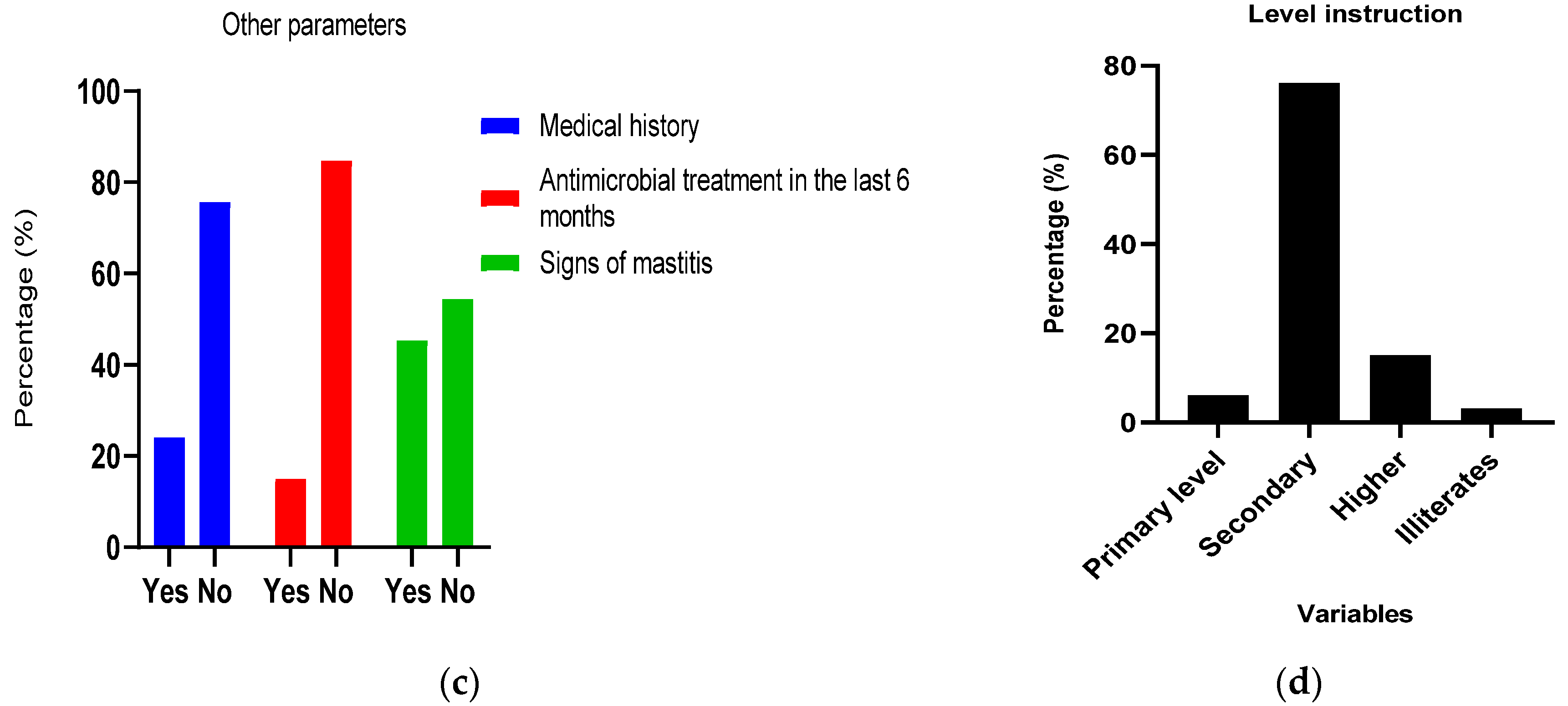
The age group of [26,27,28,29,30] represented 40% of the population, followed by the 21 to 25 age group, with a range from 10 to 50 years. The average age was 14 (Figure 1A). Among the women included in the study, 75.75% had a medical history such as hepatitis or ectopic pregnancy. Regarding antibiotic therapy, only 15.15% had been undergoing it for at least six months. Additionally, 45.45% had experienced signs of mastitis (Figure 1B). From Figure 1C, over 70% of women had medical history such as hepatitis or ectopic pregnancy. However, over 80% had not undergone antibiotic therapy for at least six months. Regarding the educational level of women, 76% have completed secondary education, and these women predominantly live in cohabitation (Figure 1D).
Breastfeeding women's knowledge of breast milk composition and the benefits of breastfeeding was assessed, and the data are presented in Table 1 below. From this table, it emerges that women have a good understanding of breast milk composition and its benefits. It also shows that 66.66% of women are aware of the need for exclusive breastfeeding for at least 6 months, as recommended by the World Health Organization (WHO) while 33.33% have a poor understanding of this guideline.

2.2. Isolation and Biochemical and Molecular Identification of Bacterial Species from Milk Samples

Table 2 shows the distribution of the different bacterial species isolated from the milk samples collected. A total of 49 bacterial strains were isolated, of which 28 (57.14%) were Lactobacilli and 21 (42.86%) Bifidobacteria. Biochemical tests for bacterial identification revealed that all 49 strains were negative for the reaction, all were oxidase-negative, and all bacteria presumed to be bifidobacteria tested positive for the F6KKP reaction. The distribution according to fermentation type was uneven among the strains. However, it is noteworthy that the hetero-fermentative strains accounted for nearly all the isolates. Among the Lactobacilli, Lactobacillus acidophilus was the most representative species in colostrum, transitional breast milk and mature breast milk. As for the Bifidobacteria, only the species Bifidobacterium infantis was isolated from the three types of breast milk, while Bifidobacterium pseudolungum was only isolated from colostrum.
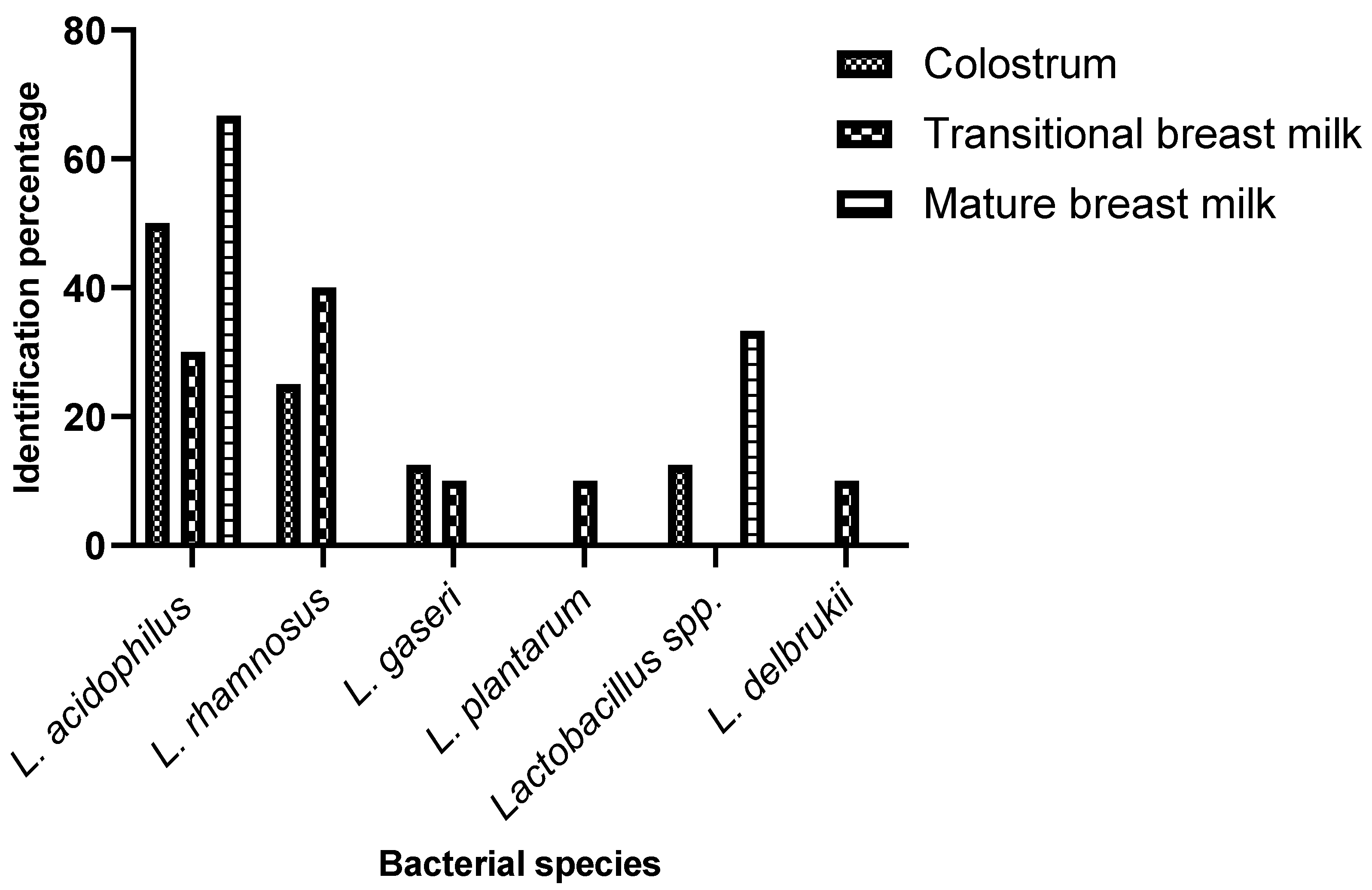
Figure 2 and Figure 3 below present the results of bacterial identification according to the different types of milk, respectively for the genus Lactobacillus and the genus Bifidobacterium. Our data show the highly diversity of bacteria in colostrum. From Figure 2, it appears that colostrum and transitional milk have more bacteria than mature milk. L. acidophilus is the most representative species as it is found in all three types of milk in significant proportions: 30% in transitional milk, 50% in colostrum, and 66.7% in mature milk.
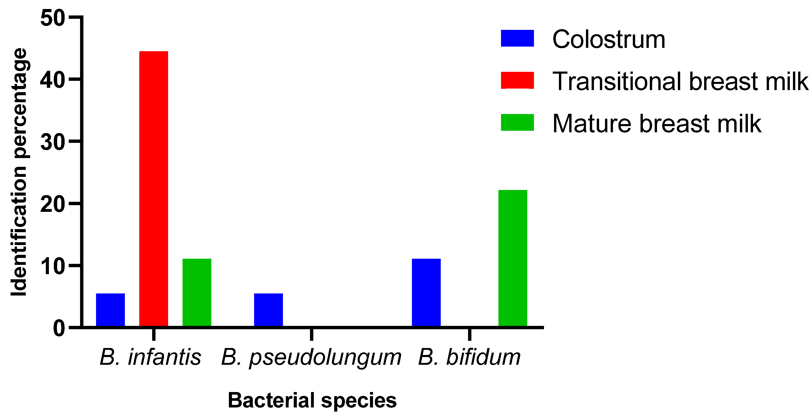
From Figure 3, it is evident that B. infantis is present in all types of milk in highly variable proportions: 44.5% in transitional milk, 5.5% in colostrum, and 11.1% in mature milk. Only three species of Bifidobacterium were isolated from all three types of milk. These are B. infantis, B. pseudolungum, and B. bifidum.

2.3. Antimicrobial Sensitivity Testing of Isolated Bacteria

The sensitivity of Lactobacilli and Bifidobacteria strains to antibiotics was assessed using the disc diffusion method. The resistance or susceptibility of a strain was determined by measuring the zones of inhibition around the discs. From our results it appears that the strains are highly resistant to the tested antibiotics. Lactic acid bacteria are therefore naturally resistant to many antibiotics. But some strains such as Lactobacillus spp, L. plantarum and L.gaseri are sensitive to Streptomycin, Gentamicin and Ampicillin (Table 3).

2.4. Evaluation of Probiotic Capacity In Vitro

2.4.1. Antimicrobial Activity

The antibacterial activity of the Lactobacilli and Bifidobacteria strains was studied using the well technique against Salmonella typhimurium, Staphylococcus aureus isolated in laboratory and a reference strain Escherichia coli ATCC 25922. Only five of all the strains isolated showed good resistance to low pH. The antibacterial activity of L. gaseri (26C), Lactobacillus spp (27C), L. plantarum (21M), B. bifidum (21M) and B. infantis (2C) are active on all indicator strains tested; both Gram-positive and Gram-negative, with zones of inhibition greater than 6 mm, indicating positive or strong antagonistic potential.. The strongest antimicrobial activity was observed with the B. bifidum strain (21 M) against all pathogens with inhibition diameters greater than at least 10mm (Table 4).

2.4.2. Capacity of Bacteria to Grow in a Range of Temperatures

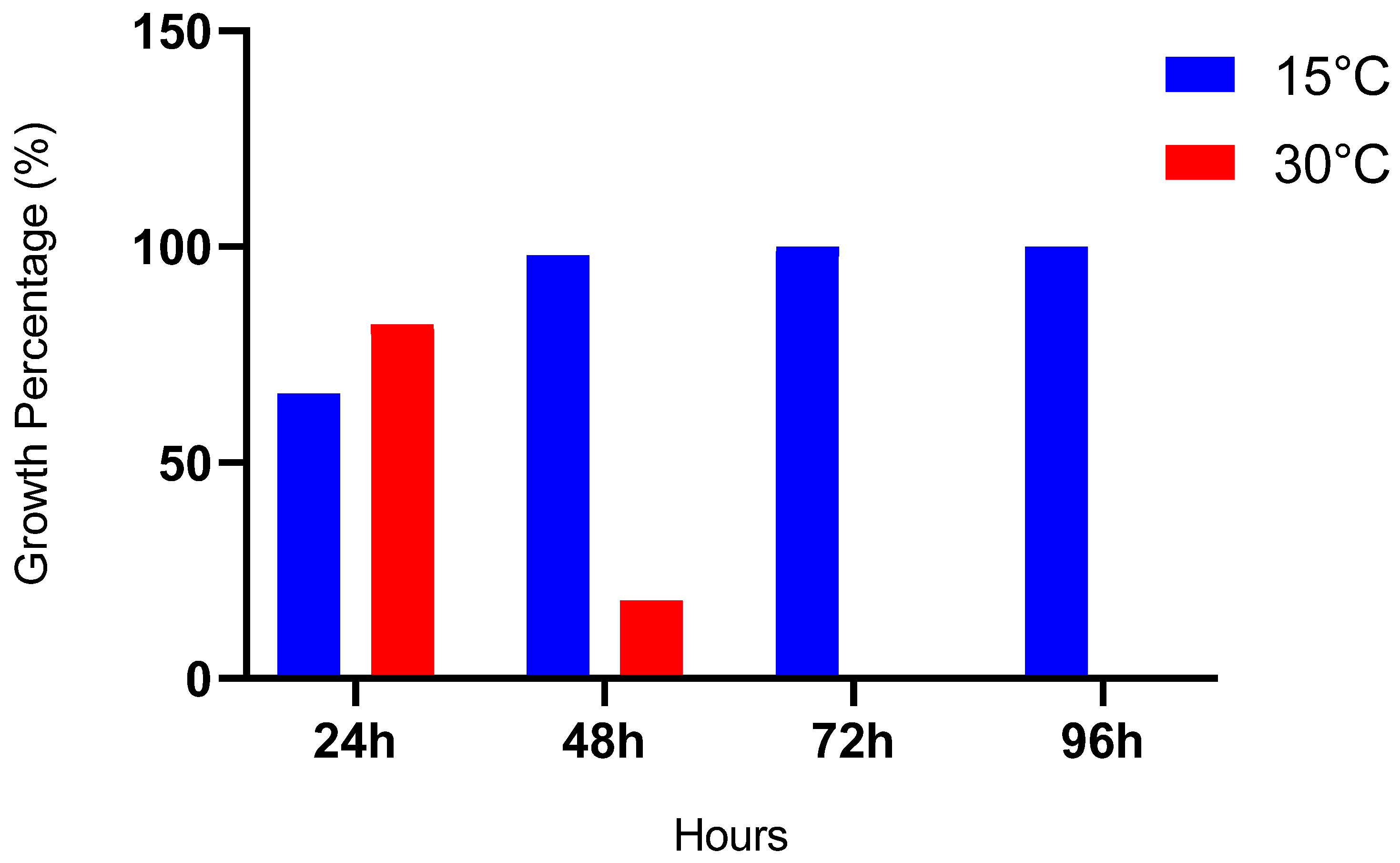
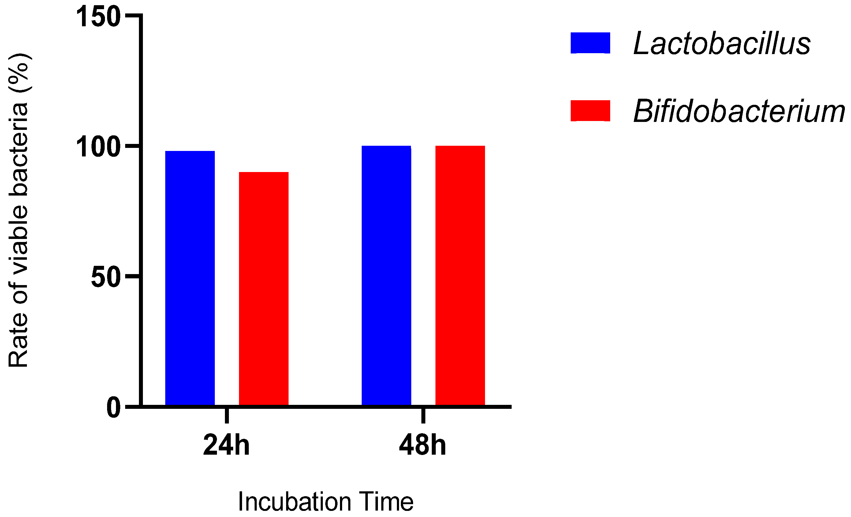
From the figure below (Figure 4), it is evident that the isolated bacteria can grow over a wide range of temperatures. However, after 48 hours of incubation at 30°C, there is a decrease in the number of bacteria. Nevertheless, they can survive at high temperatures. In this study, all tested strains were able to grow at both 15°C and 30°C. Therefore, Lactobacillus and Bifidobacterium genus are both mesophilic and thermophilic.

2.4.3. Bacteria's Ability to Grow in a Hostile Environment

The Figure 5 demonstrates that all the bacteria in the study have the potential to grow in a hostile environment. The strains isolated here were mostly resistant to NaCl at 6.5%.

2.4.4. Ability to Survive in Simulated Intestinal Conditions

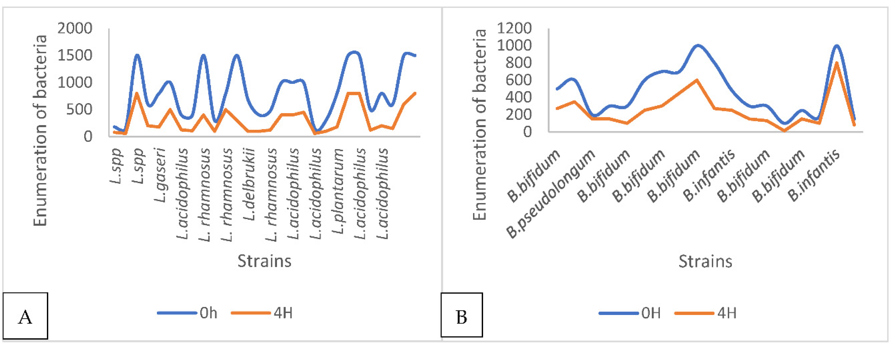
Those most resistant to simulated gastric juice were carefully observed for their ability to survive in simulated human small intestine conditions; at pH 8, in the presence of pancreatin. After 4 hours of exposure, the proportion of viable cells was assessed by counting on MRS and MRS+0.05% cysteine agar in Petri dishes. The results of the study of the ability of the strains to survive in the simulated intestinal conditions are presented in Figure 6. They were tested at pH 8, in the presence of pancreatin, and all strains underwent a drastic reduction in their viability. We were therefore particularly interested in 10 strains with resistance at low pH alone. Our idea is to verify that intestinal conditions had no more influence on these 10 strains. Indeed, these strains resist at low pH, and their sensitivity to intestinal conditions depends on the strain. Figure 6A shows the results for pH 8 and Figure 6B shows the results for pH below 8.

2.4.5. Ability to Survive in Gastric Conditions in the Presence of Bile Salts

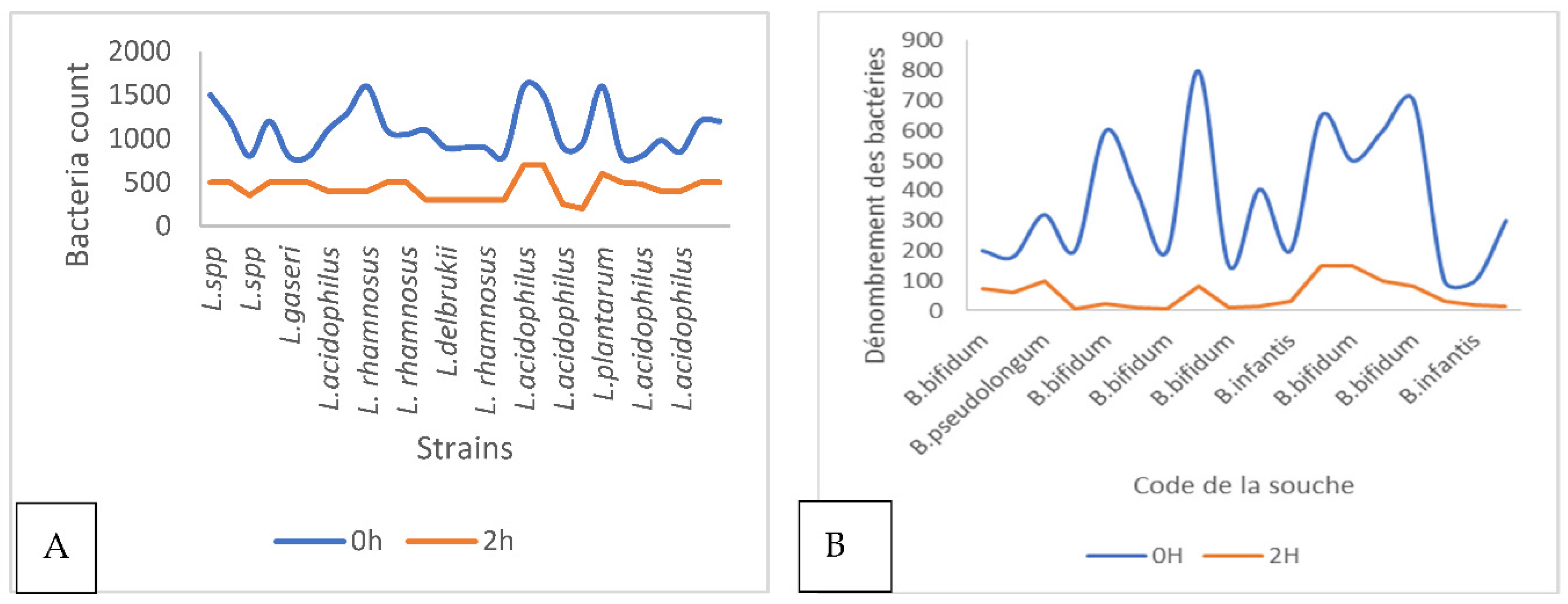
The bacteria were exposed to simulated stomach conditions (pH 1.5 with 3g/l pepsin) to assess their survival. After 1 and 2 hours of exposure, the survival of isolated lactic acid bacteria was evaluated under the combined effects of pH and pepsin. We specifically focused on 10 strains that were resistant at pH 1.5 to determine their ability to withstand these harsh gastric conditions compared to their survival at pH 1.5 alone. Under these conditions, all 10 strains showed resistance to both pepsin and low pH. However, there was a noticeable decline in their viability compared to when they were exposed to low pH alone. For instance, the survival rate of the L. gaseri strain 26C fell from 62.5% at low pH to 20% in the presence of bile salts, and for B. infantis strain 25M, it decreased from 62.5% to 5%. (Figure 7)

3. Discussion

Lactic acid bacteria, whose food habitat is milk and its derivatives, abound in a multitude of bacterial genera, including the Lactobacillus genus and the Bifidobacterium genus. Mother's milk is an essential food for the growth of infants, and its many properties have always been a solution to many infant health problems [5,27]. The present study set out to characterize Lactobacilli and Bifidobacteria with probiotic potential from breast milk in healthy breastfeeding women in Gabon. An interview was therefore conducted with these women to collect some socio-demographic data. Therefore, socio-demographic study was carried out and the level of knowledge about the importance of milk for both the infant and the mother was ascertained. From these data, it appears that the majority of breastfeeding women has no medical history and is in the age group between 26 and 30 years old and has a secondary level of education. They are aware that breastfeeding contributes to the well-being of the newborn and the mother. For example, these women know that breastfeeding protects against certain diseases and facilitates the loss of weight gained during pregnancy. They also believe that breastfeeding plays a role in reducing the risk of postpartum hemorrhage and delaying the return of menstruation.
The identification of Lactobacillus and Bifidobacterium strains isolated from human breast milk from different lactation periods was the first important point in the study. The isolation and identification of bacterial species from milk samples revealed the predominance of Lactobacilli and Bifidobacteria. Lactobacillus acidophilus and Bifidobacterium infantis were the most representative species across different types of breast milk. These findings align with previous studies indicating the prevalence of these bacterial species in breast milk and their potential role in maternal-infant health [5,27]. These results are also in line with studies carried out in several countries such as Slovenia, the United States, Brazil and Algeria using more advanced techniques, which reported that lactic acid bacteria belonging to the genera Lactobacillus and Bifidobacterium were isolated and characterized [28,31]. However, further research is warranted to explore the specific strains and their functional properties in breast milk. The isolation and purification of Lactobacilli on MRS agar medium and MRS supplemented with 0.05% cysteine chloride led to the collection of 68 strains.
In this study, bacterial species present in different types of breast milk, including colostrum, transitional milk, and mature breast milk, were isolated and identified. Among the Lactobacillus species, Lactobacillus acidophilus was found to be the most predominant species in colostrum, transitional milk, and mature breast milk. This suggests that Lactobacillus acidophilus is a dominant species in breast milk, regardless of the lactation stage. In contrast, among the Bifidobacteria species, only Bifidobacterium infantis was isolated from all three types of breast milk, while Bifidobacterium pseudolungum was only isolated from colostrum. The presence of Lactobacillus acidophilus and Bifidobacterium infantis in breast milk is consistent with previous research indicating the importance of these bacterial species in the colonization and development of the infant's intestinal microbiota [25,32]. Lactobacillus acidophilus is known for its probiotic properties and its ability to support digestive health [32]. Its prevalence in breast milk suggests that it could contribute to establishing a healthy intestinal microbiota in breastfed infants, which is crucial for immune function and overall well-being. Similarly, Bifidobacterium infantis is recognized for its beneficial effects on infant health, including its role in promoting digestion, preventing infections, and modulating the immune system [32]. The consistent presence of Bifidobacterium infantis at different stages of lactation underscores its importance in early intestinal colonization and suggests that breast milk serves as a natural source of this beneficial bacterium for infants. Furthermore, the isolation of Bifidobacterium pseudolungum exclusively from colostrum highlights the dynamic nature of breast milk composition during the early stages of lactation. Colostrum, also known as "first milk," is rich in bioactive components and is highly diversified. This milk provide essential nutrients and immune factors to newborns [4]. The presence of specific bacterial species such as Bifidobacterium pseudolungum in colostrum may contribute to the early establishment of a diverse and beneficial intestinal microbiota in infants, thereby laying the groundwork for health benefits throughout life [4,32].
The strains isolated were catalase and oxidase negative, then homofermentative and heterofermentative. The results are in agreement with those of [33], who stated that all the Lactobacilli and Bifidobacteria isolated were catalase and oxidase negative. The assortment of species identified included L. collinoides, constituting 50% of the isolates, followed by B. bifidum at 33.3%, then B. infantis, L. paracasei, L. fermentum, and B. pseudolongum in decreasing order of frequency. This investigation's findings showed the presence of various lactobacilli and bifidobacteria species in breast milk. L. collinoides predominated at each examined stage, closely followed by B. bifidum. Conversely, L. fermentum and L. paracasei were exclusively detected in transitional milk. However, molecular identification using PCR indicated Lactobacillus acidophilus as the most prevalent species (25%), trailed by Lactobacillus rhamnosus (21.23%) and Lactobacillus plantarum (14.28%).
Lactobacillus gasseri and Lactobacillus casei were the least frequently isolated species, 10.72% and 7.14%, respectively. Additionally, only 10% of bifidobacterial isolates were definitively identified through PCR. The study demonstrated that Lactobacilli and Bifidobacteria strains exhibit resilience to a wide range of environmental stressors, including temperature variations and high salt concentrations. Moreover, the strains displayed remarkable survival under simulated gastric and intestinal conditions, indicating their ability to withstand the hostile gastrointestinal environment. These findings underscore the potential of these bacterial strains as probiotics, capable of delivering health benefits to the host despite harsh physiological conditions in the digestive tract [34]. The identified probiotic bacteria in our study have a higher survival rate compare to that of previously reported strains such as Lactobacillus rhamnosus and Lactobacillus casei under such low pH values [35]. Also, the assessment of antibiotic sensitivity showed high resistance among Lactobacilli and Bifidobacteria strains, highlighting their intrinsic resistance to antibiotics. Moreover, despite antibiotic resistance, several strains exhibited significant antibacterial activity against pathogenic bacteria, indicating their potential as probiotics for therapeutic applications. Notably, Bifidobacterium bifidum strain exhibited the strongest antimicrobial activity, suggesting its promising role in combating pathogenic infections.
It would be beneficial to consider purifying these bacteria and exploring their therapeutic potential, particularly for preterm infants and those suffering from gastrointestinal diseases and diarrhea. However, our study has some limitations, including our inability to collect all three types of milk from each mother and the relatively small number of breast milk samples. Additionally, we need to utilize next-generation PCR techniques to better characterize our strains.

4. Materials and Methods

4.1. Biological Material

The biological material consisted of 34 samples of breast milk collected from lactating women. Escherichia coli ATCC 25299 was used as the reference strain for the different tests.

4.2. Inclusion Criteria

Any Gabonese woman who was breastfeeding, not undergoing any treatment, in good health and, above all, who had given informed consent was included in the study.

4.3. Methods

4.3.1. Samples Collection

A survey questionnaire was administered to each participant to collect sociodemographic data and other important parameters. The biological samples consisted of three types of milk: colostrum, transitional milk, and mature milk. Samples were collected from mothers meeting the inclusion criteria and take care at the maternal and child health centre in Port-Gentil. The nipple and areola of the breast were cleaned with 70% alcohol before each sampling. The first drops of breast milk were allowed to flow, then the subsequent drops were collected in a sterile glass tube. The samples were identified and transported to the laboratory within 24 hours in a cooler equipped with cold packs for various analyses.

4.3.2. Bacteria Isolation from Breast Milk and Biochemical Identification

The first stage consisted of pre-enrichment of breast milk. For each sample, two solutions were prepared according to the protocol described by [4]. The first (SMs1) consisted of 5ml of sample and 20ml of sterile peptone water and the second (SMs2) of 5ml of sample and 20ml of sterile peptone water with 0.05% cysteine-HCl added. They were then incubated anaerobically at 37°C for 24 hours in oven. Next day, for the isolation of Lactobacillus strains, a series of decimal dilutions from 10-1 to 10-4 was performed from the preculture of sMs1. Then, 200µl of the 10-3 and 10-4 dilutions were taken and inoculated by flooding onto previously prepared Man Rogosa Sharp (MRS) agar plates.
For the isolation of Bifidobacterium strains, the same dilutions were used from the SMs2 preculture and then inoculated onto MRS agar plates supplemented with 0.05% cysteine-HCl. The inoculated agar plates were left to dry for 30 to 45 min on the bench and then incubated at 37°C for 24 to 48 hours under anaerobic conditions. Species of the genera Lactobacillus and Bifidobacterium were identified as described by [36] by analyzing physiological characteristics (macroscopic and microscopic examination after Gram staining, growth at different temperatures, culture on hostile media) and biochemical characteristics (catalase, oxidase, mobility in the presence of mannitol, TSI (Triple Sugar Iron) agar and sugar fermentation). Specifically, for presumptive Bifidobacterium colonies, the fructose-6-phosphate phosphoketolase (F6PPK) production test was performed. After incubation, the bacterial colonies obtained for the Lactobacillus and Bifidobacterium strains were purified. To do this, three successive sub-cultures were carried out using suspensions of the isolated colonies that were homogeneous and developed on MRS and MRS+Cysteine-HCl agar respectively. The purified strains thus obtained were stored in MRS and MRS + Cysteine-HCl broths with 40% (v/v) sterile glycerol at -20°C [36].

4.3.3. Molecular Identification

The genomic DNA extraction from isolated and purified strains was the first step in the molecular identification of lactic acid bacteria strains. It was extracted in accordance with the instructions for the Zymo Quick-DNA Miniprep Plus extraction kit. The optical density was checked by spectrophotometry between 200 and 4000nm to ensure the integrity of the extracted DNA. Once the DNA had been extracted, the specific genes of each bacterial species were analyzed using polymerase chain reaction (PCR) [23]. Simplex PCR amplification was carried out using primers targeting genes coding for 16s and 23s rRNAs and their intergenic spacer region, namely R16 and LbLMA1-rev. The PCR conditions were: initial denaturation at 95°C for 5min, followed by 30 cycles consisting of denaturation at 95°C for 30s, hybridization at 55°C for 30s, extension at 72°C for 30s and a final extension step for 7 min at 72°C. Migration were performed on a 1% agarose gel with a 100 base pair molecular weight marker. The Table 5 present the primers [4,24].

4.3.4. Antibiotic Susceptibility Testing (AST)

The sensitivity profile of the isolated Lactobacillus and Bifidobacterium strains was assessed using the plate diffusion method. A total of thirteen antibiotic were tested: Penicillin (10U/l); Ampicillin (10µg); Oxacilin (1µg); Cefoxitin (30µg); Vancomycin (10µg); Erythromycin (15µg); Tetracyclin (30µg); Clindamycin (2µg); Chloramphenicol (30µg); Streptomycin (10µg); Gentamicin (10µg); Nalidixic acid (30µg) and Trimethropim (5µg). Bacterial suspensions were prepared from pure strains cultures and turbidity adjusted to that of 0.5 MacFarland. The discs were deposited, and the plates were incubated in anaerobioses at 37°C for 24 hours. After incubation, the inhibition diameters were measured and interpreted in accordance with CASFM (2022) recommendations. E. coli ATCC 29522 strain was used as quality control.

4.3.5. In Vitro Probiotic Capacity of Strains

  • Antimicrobial activity
Agar well diffusion assays were utilized to test the antimicrobial activity as described by [25]. The antibacterial activity of Lactobacillus and Bifidobacterium strains was assessed on multi-resistant Salmonella typhimurium, Staphylococcus aureus strains isolated in the bacteriology laboratory at hospital and a reference strain of Escherichia coli ATCC 25299.
  • Growth at different temperatures
The growth of bacterial strains at a wide range of temperatures was assessed by inoculating them in MRS or MRS + Cysteine-HCl broths, depending on the nature of the strain, followed by incubation at 15°C and 30°C for 48 hours. The results were observed after 48 hours for the test carried out at 30°C and for four days for the test carried out at 15°C [1,25].
  • Cultivation in a hostile environment
The tolerance of isolated strains to sodium chloride was determined using the method described by [37]. Isolates were grown anaerobically at 37°C for 24 h in MRS and MRS+0.05% cysteine chloride broth, supplemented with sodium chloride (6.5%). One ml of these broths obtained after 24 hours were inoculated into MRS and MRS+0.05% cysteine chloride agar. The hyper-saline medium was prepared by adding 6.5 g of NaCl to 100 ml of each of the MRS and MRS + Cysteine-HCl broths. The turbidity of the medium indicates the growth of the strains.
  • Ability to survive in gastric conditions.
The ability of bacterial strains to survive in conditions mimicking those of the human stomach was performed using the technique described by [38]. Simulated gastric juice was prepared by adding 3g of pepsin to 1L of 0.5% NaCl solution. The preparation was adjusted to pH=1.5. One milliliter of each bacterial culture (109 cells/ml; OD=620 nm between 0.5 and 0.7) was added to 9 ml of simulated gastric juice. Next day, 0.1 ml of the seeded gastric juice was taken after 0 and 2 h of reaction between the solutions respectively, and then plated onto MRS and MRS + Cysteine - HCl agar plates. The number of viable bacteria was determined after 24 to 48 h of incubation under anaerobic conditions. The experiment was repeated three times. The survival rate was calculated using the following equation:
Survival rate (%) = (log CFU at 2h/log CFU at 1h) x 100 [40].
  • Ability to survive in simulated intestinal conditions
The ability of the strains to survive in conditions similar to those in the human small intestine was assessed using the technique described by [38]. For this, an intestinal juice was prepared as follows: 1 g/l pancreatin, was added to 0.5% NaCl, with or without 0.3% oxgall bile salts. Both preparations were adjusted to pH 8. Next, one milliliter of each bacterial culture (109 cells/ml) was inoculated into 9 ml of the two preparations. Finally, 0.1 ml of each preparation was taken at 0 h and 4 h of exposure and streaked onto MRS and MRS + Cysteine-HCl agar plates. Viable bacteria were counted after 24 to 48 h of incubation under anaerobic conditions. The experiment was repeated three times and the survival rate was calculated using the equation: Survival rate (%) = (log CFU at 2h/log CFU at 1h) x 100 [38].
  • Hemolytic activity
The hemolytic activity of Lactobacillus and Bifidobacterium probiotic strains was determined using the method described by [38]. Overnight cultures of probiotic strains were streaked onto Columbia agar plates containing 5% human blood and then incubated for 48 hours at 37°C. After incubation, the plates were examined to identify traces of β-hemolysis (clear zones around the colonies) and α-hemolysis (areas with greenish reflections around the colonies) [38].

4.4. Data Analysis

The results were expressed as mean ± standard deviation. Differences were considered significant at p < 0.05. Graphical presentations were generated using Excel and Graph Pad.

5. Conclusions

Lactic acid bacteria could be, good probiotic candidates in view of the fact that they are normal and beneficial components of many microbiota and in view of their long history of use as sucking microorganisms in the food industry. Several species of Lactobacilli and Bifidobacteria were isolated from the breast milk samples collected, with a predominance of Latobacillus acidophilus. These strains showed good probiotic properties and antibacterial activity. These results need to be deeply performed with a view to better identifying these strains and exploiting their probiotic properties in therapy.

Author Contributions

Conceptualization, G.L. and S.T.A; Methodology, N.M. ; Validation, G.L., S.T.A. and V.D.; Investigation, N.M. and P.E.N; Data curation, G.L., S.T.A., V.D. and L.B-M.; Writing—original draft preparation, B.B.L; Writing-review and editing, G.L., S.T.A., V.D. and L.B-M.; Supervision, , G.L., S.T.A., V.D. and L.B-M. All authors have read and agreed to the published version of the manuscript.

Funding

This research received no external funding.

Informed Consent Statement

Informed consent was obtained from all subjects involved in the study.

Data Availability Statement

The original contributions presented in the study are included in the article/supplementary material, further inquiries can be directed to the corresponding author/s.

Acknowledgments

We thank all the staff at the Maternal and Child Health Center of Port-Gentil (CSMI), as well as all the women who agreed to participate in the study.

Conflicts of Interest

The authors declare no conflicts of interest.

References

  1. Arboleya, S.; Ruas-Madiedo, P.; Margolles, A.; Solís, G.; Salminen, S.; De los Reyes-Gavilan, C.G.; Gueimonde, M. Characterization and in vitro properties of potentially probiotic Bifidobacterium strains isolated from breast-milk. Int. J. Food Microbiol. 2011, 149, 28–36. [Google Scholar] [CrossRef] [PubMed]
  2. Zuo, F.; Yu, R.; Feng, X.; Chen, L.; Zeng, Z.; Khaskheli, G.B.; Ma, H.; Chen, S. Characterization and in vitro properties of potential probiotic Bifidobacterium strains isolated from breast-fed infant feces. Ann. Microbiol. 2016, 66, 1027–1037. [Google Scholar] [CrossRef]
  3. Fortmann, I.; Marißen, J.; Siller, B.; Spiegler, J.; Humberg, A.; Hanke, K.; Faust, K.; Pagel, J.; Eyvazzadeh, L.; Brenner, K. Lactobacillus acidophilus/Bifidobacterium infantis probiotics are beneficial to extremely low gestational age infants fed human milk. Nutrients 2020, 12, 850. [Google Scholar] [CrossRef]
  4. Liu, W.; Chen, M.; Duo, L.; Wang, J.; Guo, S.; Sun, H.; Menghe, B.; Zhang, H. Characterization of potentially probiotic lactic acid bacteria and bifidobacteria isolated from human colostrum. J. Dairy Sci. 2020, 103, 4013–4025. [Google Scholar] [CrossRef] [PubMed]
  5. Jamyuang, C.; Phoonlapdacha, P.; Chongviriyaphan, N.; Chanput, W.; Nitisinprasert, S.; Nakphaichit, M. Characterization and probiotic properties of Lactobacilli from human breast milk. 3 Biotech 2019, 9, 398. [Google Scholar] [CrossRef] [PubMed]
  6. Łubiech, K.; Twarużek, M. Lactobacillus bacteria in breast milk. Nutrients 2020, 12, 3783. [Google Scholar] [CrossRef] [PubMed]
  7. Oddi, S.; Huber, P.; Duque, A.R.F.; Vinderola, G.; Sivieri, K. Breast-milk derived potential probiotics as strategy for the management of childhood obesity. Food Res. Int. 2020, 137, 109673. [Google Scholar] [CrossRef] [PubMed]
  8. Park, J.; Yi, D.Y. Comprehensive analysis of the effect of probiotic intake by the mother on human breast milk and infant fecal microbiota. J. Korean Med. Sci. 2021, 36. [Google Scholar] [CrossRef]
  9. Arshad, F.A.; Mehmood, R.; Hussain, S.; Khan, M.A.; Khan, M.S. Lactobacilli as probiotics and their isolation from different sources. Br. J. Res. 2018, 5, 43. [Google Scholar] [CrossRef]
  10. Juharji, H.; Albalawi, K.; Aldwaighri, M.; Almalki, A.; Alshiti, H.; Kattan, W.; Alqarni, M.; Alsulaimani, S.; AlShaikh, T.; Alsulaimani, F. Impact of Breastfeeding on Low Birthweight Infants, Weight Disorders in Infants, and Child Development. Cureus 2022, 14, e32894. [Google Scholar] [CrossRef]
  11. Rodríguez Arreola, A.; Solís Pacheco, J.R.; Lacroix, M.; Balcazar López, E.; Navarro Hernández, R.E.; Sandoval Garcia, F.; Gutiérrez Padilla, J.A.; García Morales, E.G.; Aguilar-Uscanga, B.R. In vivo assessment and characterization of lactic acid bacteria with probiotic profile isolated from human milk powder. Nutr. Hosp. 2020, 38, 152–160. [Google Scholar] [CrossRef]
  12. Lokossou, G.A.G.; Kouakanou, L.; Schumacher, A.; Zenclussen, A.C. Human Breast Milk: From Food to Active Immune Response With Disease Protection in Infants and Mothers. Front. Immunol. 2022, 13. [Google Scholar] [CrossRef] [PubMed]
  13. Javed, G.A.; Arshad, N.; Munir, A.; Khan, S.Y.; Rasheed, S.; Hussain, I. Signature probiotic and pharmacological attributes of lactic acid bacteria isolated from human breast milk. Int. Dairy J. 2022, 127, 105297. [Google Scholar] [CrossRef]
  14. Lyons, K.E.; Ryan, C.A.; Dempsey, E.M.; Ross, R.P.; Stanton, C. Breast milk, a source of beneficial microbes and associated benefits for infant health. Nutrients 2020, 12, 1039. [Google Scholar] [CrossRef] [PubMed]
  15. Gunyakti, A.; Asan-Ozusaglam, M. Lactobacillus gasseri from human milk with probiotic potential and some technological properties. LWT 2019, 109, 261–269. [Google Scholar] [CrossRef]
  16. Sakwinska, O.; Moine, D.; Delley, M.; Combremont, S.; Rezzonico, E.; Descombes, P.; Vinyes-Pares, G.; Zhang, Y.; Wang, P.; Thakkar, S.K. Microbiota in breast milk of Chinese lactating mothers. PLoS One 2016, 11, e0160856. [Google Scholar] [CrossRef]
  17. Selvamani, S., Dailin, D. J., Gupta, V. K., Wahid, M., Keat, H. C., Natasya, K. H., ... & El Enshasy, H. A. An insight into probiotics bio-route: translocation from the mother’s gut to the mammary gland. Applied Sciences 2021, 11(16), 7247. [CrossRef]
  18. Chong, H.-Y.; Tan, L.T.-H.; Law, J.W.-F.; Hong, K.-W.; Ratnasingam, V.; Ab Mutalib, N.-S.; Lee, L.-H.; Letchumanan, V. Exploring the potential of human milk and formula milk on infants’ gut and health. Nutrients 2022, 14, 3554. [Google Scholar] [CrossRef]
  19. Moossavi, S.; Azad, M.B. Origins of human milk microbiota: new evidence and arising questions. GutMicrobes 2020, 12, 1667722. [Google Scholar] [CrossRef]
  20. Mohammadi, F.; Eshaghi, M.; Razavi, S.; Sarokhalil, D.D.; Talebi, M.; Pourshafie, M.R. Characterization of bacteriocin production in Lactobacillus spp. isolated from mother’s milk. Microb. Pathog. 2018, 118, 242–246. [Google Scholar] [CrossRef]
  21. Amadoro, C.; Rossi, F.; Pallotta, M.L.; Gasperi, M.; Colavita, G. Traditional dairy products can supply beneficial microorganisms able to survive in the gastrointestinal tract. LWT 2018, 93, 376–383. [Google Scholar] [CrossRef]
  22. Shokryazdan, P.; Faseleh Jahromi, M.; Liang, J.B.; Ho, Y.W. Probiotics: From Isolation to Application. J. Am. Coll. Nutr. 2017, 36, 666–676. [Google Scholar] [CrossRef] [PubMed]
  23. D’Alessandro, M.; Parolin, C.; Patrignani, S.; Sottile, G.; Antonazzo, P.; Vitali, B.; Lanciotti, R.; Patrignani, F. Human Breast Milk: A Source of Potential Probiotic Candidates. Microorganisms 2022, 10, 1279. [Google Scholar] [CrossRef] [PubMed]
  24. Nikolopoulou, G.; Tsironi, T.; Halvatsiotis, P.; Petropoulou, E.; Genaris, N.; Vougiouklaki, D.; Antonopoulos, D.; Thomas, A.; Tsilia, A.; Batrinou, A. Analysis of the Major Probiotics in Healthy Women’s Breast Milk by Realtime PCR. Factors Affecting the Presence of Those Bacteria. Appl. Sci. 2021, 11, 9400. [Google Scholar] [CrossRef]
  25. Rajoka, M.S.R.; Mehwish, H.M.; Siddiq, M.; Haobin, Z.; Zhu, J.; Yan, L.; Shao, D.; Xu, X.; Shi, J. Identification, characterization, and probiotic potential of Lactobacillus rhamnosus isolated from human milk. LWT 2017, 84, 271–290. [Google Scholar] [CrossRef]
  26. Zacarías, M.F.; Binetti, A.; Laco, M.; Reinheimer, J.; Vinderola, G. Preliminary technological and potential probiotic characterisation of bifidobacteria isolated from breast milk for use in dairy products. Int. Dairy J. 2011, 21, 548–555. [Google Scholar] [CrossRef]
  27. Mehanna, N.S.; Tawfik, N.F.; Salem, M.M.; Effat, B.A.; Gad El-Rab, D.A. Assessment of potential probiotic bacteria isolated from breast milk. Middle-East J. Sci. Res. 2013, 14, 354–360. [Google Scholar] [CrossRef]
  28. de Souza Rodrigues, J.Z.; Passos, M.R.; de Macêdo Neres, N.S.; Almeida, R.S.; Pita, L.S.; Santos, I.A.; Silveira, P.H.S.; Reis, M.M.; Santos, I.P.; Ricardo, L. de O.N. Antimicrobial activity of Lactobacillus fermentum TcUESC01 against Streptococcus mutans UA159. Microb. Pathog. 2020, 142, 104063. [CrossRef]
  29. Talhi-Mekhici, M.; Cornu, B.; Talhi-Mehaya, R.; Sahraoui, D.; Dib, W.; Yazi, L.A.; Zemmour, A.; Nadjia, S.-O.; Kacem, M.; Vander Wauven, C. Phenotypic and Genotypic Identification of Bacteria from Women Breast-Milk and the Feces of their Childs in the Western Region of Algeria. J. Pure Appl. Microbiol. 2017, 11, 1767–1776. [Google Scholar] [CrossRef]
  30. Thongaram, T.; Hoeflinger, J.L.; Chow, J.; Miller, M.J. Human milk oligosaccharide consumption by probiotic and human-associated bifidobacteria and lactobacilli. J. Dairy Sci. 2017, 100, 7825–7833. [Google Scholar] [CrossRef]
  31. Tušar, T.; Žerdoner, K.; Bogovič Matijašić, B.; Paveljšek, D.; Benedik, E.; Bratanič, B.; Fidler, N.; Rogelj, I. Cultivable bacteria from milk from Slovenian breastfeeding mothers. Food Technol. Biotechnol. 2014, 52. [Google Scholar] [CrossRef]
  32. Blessing, E.N.; Chukwuemeka, I.S.; Ukeachu, C.D.; Onuawuchi, U.G. Antibacterial properties of probiotics bacterial isolated from human breast milk. World News Nat. Sci. 2020, 29, 290–297. [Google Scholar] [CrossRef]
  33. Toscano, M.; De Grandi, R.; Grossi, E.; Drago, L. Role of the human breast milk-associated microbiota on the newborns’ immune system: a mini review. Front. Microbiol. 2017, 8, 296917. [Google Scholar] [CrossRef] [PubMed]
  34. Pisano, M.B.; Viale, S.; Conti, S.; Fadda, M.E.; Deplano, M.; Melis, M.P.; Deiana, M.; Cosentino, S. Preliminary evaluation of probiotic properties of Lactobacillus strains isolated from Sardinian dairy products. Biomed. Res. Int. 2014. [CrossRef]
  35. Manini, F.; Casiraghi, M.C.; Poutanen, K.; Brasca, M.; Erba, D.; Plumed-Ferrer, C. Characterization of lactic acid bacteria isolated from wheat bran sourdough. LWT 2016, 66, 275–283. [Google Scholar] [CrossRef]
  36. Hadadji, M. Caractérisation Microbiologique et Biochimique des Bifidobactéries isolées à partir des selles de nourrissons: Etude des critères Technologiques et Thérapeutiques. 2007. [CrossRef]
  37. Abhijit, C.; Nur, H.; Nure, J.; Mostazir, F.; Morsaline, B.; Monzur, M.A. Screening of Lactobacillus spp. From Buffalo Yoghurt for Probiotic and Antibacterial Activity. J. Bacteriol. Parasitol. 2012, 3. [Google Scholar] [CrossRef]
  38. Bahri, F.; Lejeune, A.; Dubois-Dauphin, R.; El Mejdoub, T.; Boulahrouf, A.; Thonart, P. Characterization of Lactobacillus strains isolated from Algerian children faeces for their probiotic properties. Afr. J. Microbiol. Res. 2014, 8. [Google Scholar] [CrossRef]
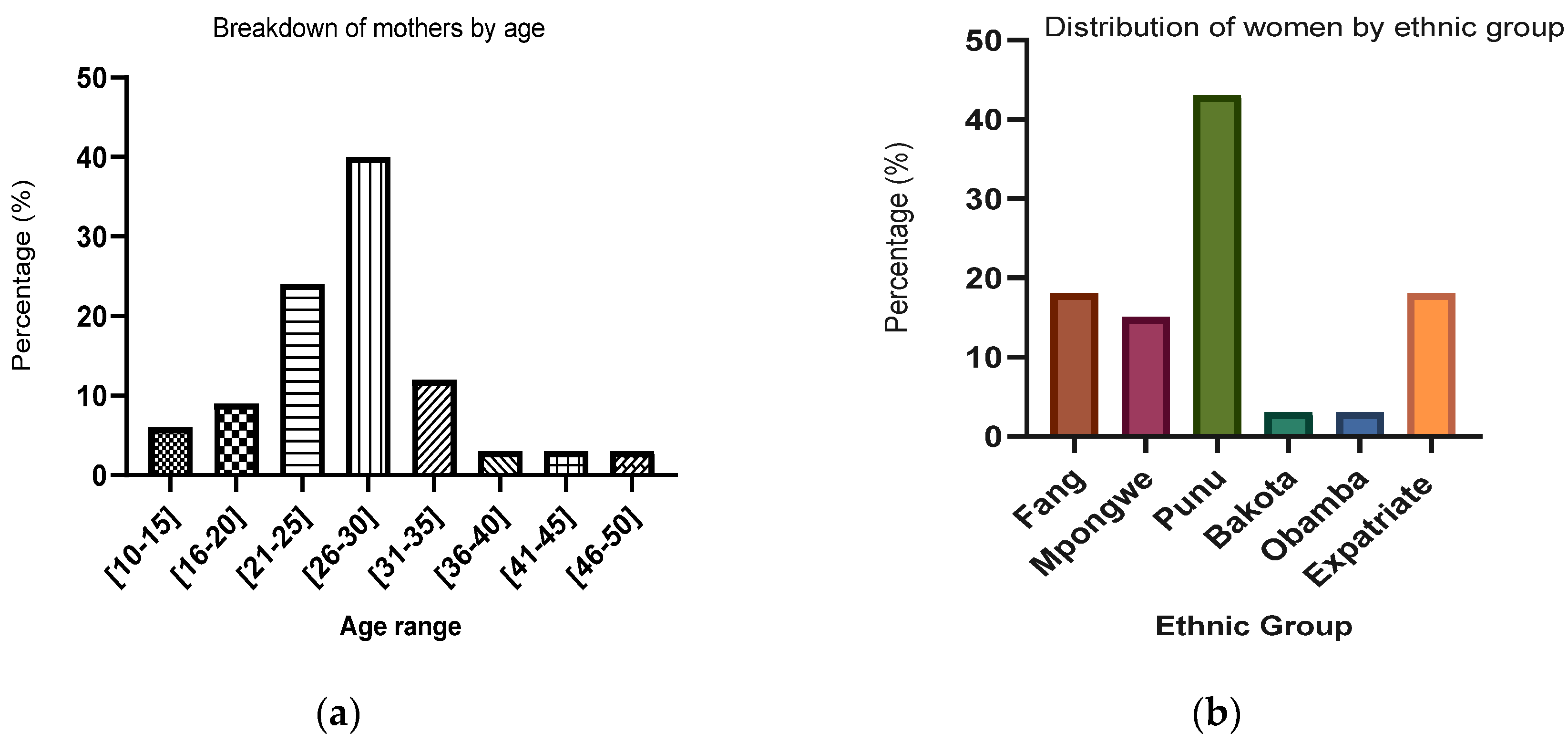
Figure 1. Socio-demographic data of population study.
Preprints 110941 g001aPreprints 110941 g001b
Figure 2. Distribution of isolated lactobacilli by type of milk.
Preprints 110941 g002
Figure 3. Proportion of Lactobacilli isolated by type of milk.
Preprints 110941 g003
Figure 4. Capacity of bacteria to grow in a range of temperatures.
Preprints 110941 g004
Figure 5. Bacteria ability to grow in a hostile environment.
Preprints 110941 g005
Figure 6. Survival of isolated lactic acid bacteria under simulated intestinal conditions (6A: pH 8, 6B: pH below 8).
Preprints 110941 g006
Figure 7. Survival of lactic acid bacteria isolated in gastric conditions with bile salts.
Preprints 110941 g007
Table 1. Women's knowledge of breastfeeding.
Parameters Good Knowledge % Bad Knowledge %
Composition of breast milk 50 50
Digestion of breast milk 80 20
Antibodies in breast milk 90 10
Allergies 40 60
Protective role of breast milk 65 35
Benefits of breast milk for babies 90 10
Improvement of the immune system 50 50
Duration of exclusive breastfeeding 66.66 33.33
Table 2. Frequency of bacterial species of Lactobacilli and Bifidobacteria isolated.
Lactobacillus Bifidobacteria
Colostrum
n=9
Transitional
breast milk
n=12
Mature
breast milk
n=7
Colostrum
n=5
Transitionall
breast milk
n=10
Mature breast
Milk
n=6
L. acidophilus 50% 30% 66,70% - - -
L. rhamnosus 25% 40% - - - -
L. gaseri 12,5% 10% - - - -
Lactobacillus spp 12,5% - 33,33% - - -
L. plantarum - 10% - - - -
L. delbrukii - 10% - - - -
B. infantis - - - 5,5% 44,5% 11,1%
B. pseudolungum - - - 5,5% - -
B. bifidum - - - 11,1% - 22,2%
Table 3. Resistance of potential probiotic bacteria isolated to antibiotics.
Antibiotics Disc Charge Lactobacillusspp L.
plantarum
L.
gaseri
B.
bifidum
B.
infantis
Penicilin 10U/L R R R R R
Ampicillin 10µg S S S R R
Oxacillin 1µg R R R R R
Cefoxitin 30µg R R R R R
Vancomycin 30µg R R R R R
Streptomycin 10µg S S S R R
Gentamicin 10µg S S S R R
Erythromycin 15µg R R R R R
Tetracyclin 30µg R R R R S
Clindamycin 2µg R R R R R
Chloramphenicol 30µg R R R R R
Nalidixic Acid 30µg R R R R R
Trimetroprim 5µg R R R R R
Legend: R = Resistant, S = Sensitive.
Table 4. Antibacterial activity profile of strains with high probiotic potential and their cell-free supernatant (CFS) against pathogenic bacteria.
Inhibition diameter (mm)
Bacteria Strains P1=Salmonella typhimurium P2=Staphylococcus aureus Escherichia coli
ATCC 25922
L. gaseri 8 8 8
CFS L. gaseri 0 6 0
Lactobacillus spp 8 8 8
CFS Lactobacillus. spp 0 6 0
L. plantarum 8 10 8
CFS L. plantarum 0 7 0
B. infantis 10 10 10
CFS B. infantis 0 6 0
B. bifidum 11 12 10
CFS B. bifidum 0 7 0
Table 5. List of primers used for Lactobacillus identification.
Bacterial species Primers Sequences (5’ to 3’) Size (pb)
All Lactobacillus IDL03R CCACCTTCCTCCGGTTTGTCA -
All Lactobacillus IDL04F AGGGTGAAGTCGTAACAAGTAGCC -
Lactobacillus casei IDL11F TGGTCGGCAGAGTAACTGTTGTCG 727
Lactobacillus acidophilus IDL22R AACTATCGCTTACGCTACCACTTTGC 606
Lactobacillus delbrueckii IDL31F CTGTGCTACACCTAGAGATAGGTGG 184
Lactobacillus gasseri IDL42R ATTTCAAGTTGAGTCTCTCTCTC 272
Lactobacillus reuteri IDL52F ACCTGATTGACGATGGATCACCAGT 1105
Lactobacillus plantarum IDL62R CTAGTGGTAACAGTTGATTAAAACTGC 428
Lactobacillus rhamnosus IDL73R GCCAACAAGCTATGTGTTCGCTTGC 448
Bifidobacterium lm26-f GATTCTGGCTCAGGATGAACG -
Bifidobacterium lm3-r CGGGTGCTCCCACTTTCATG -
Disclaimer/Publisher’s Note: The statements, opinions and data contained in all publications are solely those of the individual author(s) and contributor(s) and not of MDPI and/or the editor(s). MDPI and/or the editor(s) disclaim responsibility for any injury to people or property resulting from any ideas, methods, instructions or products referred to in the content.
Copyright: This open access article is published under a Creative Commons CC BY 4.0 license, which permit the free download, distribution, and reuse, provided that the author and preprint are cited in any reuse.
Prerpints.org logo

Preprints.org is a free preprint server supported by MDPI in Basel, Switzerland.

Subscribe

© 2024 MDPI (Basel, Switzerland) unless otherwise stated