Preprint
Article

Phytochemical Analysis on the Female Cone of Araucaria bidwillii Hook

Altmetrics

Downloads

89

Views

50

Comments

0

A peer-reviewed article of this preprint also exists.

Submitted:

24 July 2024

Posted:

24 July 2024

You are already at the latest version

Alerts
Abstract
In this work, the first phytochemical analysis ever performed on the whole cone of Araucaria bidwillii Hook is presented. This was carried out by means of column chromatography, NMR spectroscopy and MS spectrometry on the female cone, evidencing the presence of forty metabolites, thirty-three of which are primary metabolites whilst the remaining are secondary metabolites. A chemophenetic evaluation was also performed reporting seven new compounds for the species. These results are important since they provide more information on the phytochemistry of this species and also underline the need to study more of its populations.
Keywords: 
Subject: Biology and Life Sciences  -   Plant Sciences

1. Introduction

Araucaria bidwillii Hook. is a coniferous tree belonging to the Araucariaceae family, which is native to Australia, but it has now been introduced in several private and botanical gardens worldwide.
Its female cones (Figure 1) are characterized by an ovoidal shape and are sub-globulous, 30 cm high and 20 cm large, weighing up to 10 Kg, dark green colored, containing 50-100 small seeds which are elongated, elliptic, wingless, collected in a subtle, sharp tegument and light brown colored. They are produced by trees being at least 14 years old in April and ripen in June-September falling spontaneously [1].
The species is known to biosynthesize essential oil constituents, diterpenoids and biflavonoids [2]. Yet, these only come from phytochemical studies on the resin and leaves of different populations [2]. In fact, no work has ever been published on the phytochemical composition of the cones of this species and this represents the main reason why this work was begun. Actually, one study has focused on the seeds derived from these cones, purchased from Australia, and reporting the presence of primary metabolites like amino acids and fatty acids as well as a few secondary metabolites such as catechin, quercetin and epi-catechin [3].
The cones of this species have no specific ethnobotanical uses whilst their seeds are widely consumed by local Australian people in several forms and preparations for several purposes [4].
In this work, the female cone of a population of A. bidwillii collected in the experimental garden of the University of Rome “La Sapienza” located inside the University city, was phytochemically studied for its primary and secondary constituents in order to provide more general information about the species in this sense. In addition, a chemophenetic evaluation was performed, comparing its phytochemical results with those derived from the cones of other species of the genus and family in order to highlight similarities and differences. This part also represents a huge novelty of this work.

2. Materials and Methods

2.1. Plant Material

A single female cone of A. bidwillii (8.2 Kg) was collected in the experimental garden of the University of Rome “La Sapienza” located inside the University city (geographical coordinates 41°77'59'' N, 12°50'75'') in September 2022. The botanical identification as a female cone was performed by one of the authors (C.S.), an expert botanist working in the Botanical Garden of Rome and curator of also this experimental garden, by comparing its morphological features and history with those reported in the literature [1]. A part of this collection is stored in our laboratory for further reference under the voucher code AB18092022.

2.2. Chemicals and Materials

The following chemicals and materials were utilized for this phytochemical study: 96 % ethanol for the extraction procedure by maceration; n-butanol, distilled water and methanol as pure solvents or in mixture among them all to be used as eluting systems for the separation procedure of the total extract on open column chromatography using silica gel (40–63 μm) as stationary phase; 2N sulfuric acid for the developments of the TLCs; deuterated solvents (CDCl3, and D2O) for the identification of the metabolites by means of NMR spectroscopy; HPLC methanol for the identification of the metabolites by means of mass spectrometry.
All the solvents having RPE purity grade, if not differently specified, together with the deuterated solvents, the TLCs and HPLC-grade methanol were purchased from Merck (St. Louis, Missouri, USA) whereas silica gel was purchased from Fluka Analytical (Bergamo, Italy).

2.3. Instrumentations

The following instrumentations were used during this study: Rotavapor RII from Buchi for the evaporation procedure, UV lamp at the wavelength 254 nm for the visualization of TLCs; NMR spectra were recorded at 298 K on a Jeol JNM-ECZ 600R spectrometer with a magnet operating at 14.09 T corresponding to a proton resonance frequency of 600.19 MHz and equipped with a Jeol multinuclear z-gradient inverse probehead. 1H NMR spectra were acquired with 32 transients, a spectral width of 9013.7 Hz (corresponding to 15 ppm) and 64K data points for an acquisition time of 7.3 s. The recycle delay was set to 7.7 s to achieve complete resonance relaxation between successive scans. The chemical shifts were referenced to TMS (s, 0 ppm) for spectra in CDCl3, to the HDO signal (s, 4.79 ppm) for spectra in D2O; MS spectra were acquired on a triple quadrupole mass spectrometer PE-Sciex API-3000® (Perkin Elmer Sciex, Toronto, ON, Canada), equipped with an ESI source operating in the negative and/or positive ion mode, in a mass spectral range of 100–1000 m/z, with the capillary ion voltage set at 5000 V for the positive ionization and -4500 V for the negative one, with high-purity nitrogen used as a curtain gas (5 L/min) and air as nebulizer (2 L/min) and drying gas (30 psi), with the temperature to heat the drying gas set at 100 °C, with a flow rate of sample infusion of 20 μL/min, with 20 acquisitions per sample, with the full width at half maximum (FWHM) set at m/z 0.7 ± 0.1 in each mass-resolving quadrupole to operate with a unit resolution, with data acquired and elaborated by Analyst® 1.6 software (AB Sciex, Washington, USA).

2.4. Extraction and Separation Procedures

A part of the collected female cone (8.0 Kg) was opened, and its chips were inserted one by one, entire, in a flask which was filled with 5 L of 96 % ethanol till complete covering of the plant material. The whole was left to macerate for 21 days in order to have an exhaustive extraction of the plant material. At this point, the resulting solution was filtered, and the extracting solvent was departed from the solution at reduced pressure at the temperature of 50 °C. During this step, pH of the solution was checked in order to verify that it was not too acid (< 5.5) or too basic (> 8.5) since an extreme acidity or alkalinity might cause unwanted secondary reactions on the phytocomplex such as the hydrolysis of the ester and glycosidic bonds. In this case, pH was about 7.5. The extracting solvent was recovered and reutilized to extract the plant material for a second time and for the same time length to achieve a more complete extraction of the plant material. The resulting solution of this maceration was united with that derived from the previous step giving, in the end, a dried crude extract colored in light brown and weighing 36.6 g.
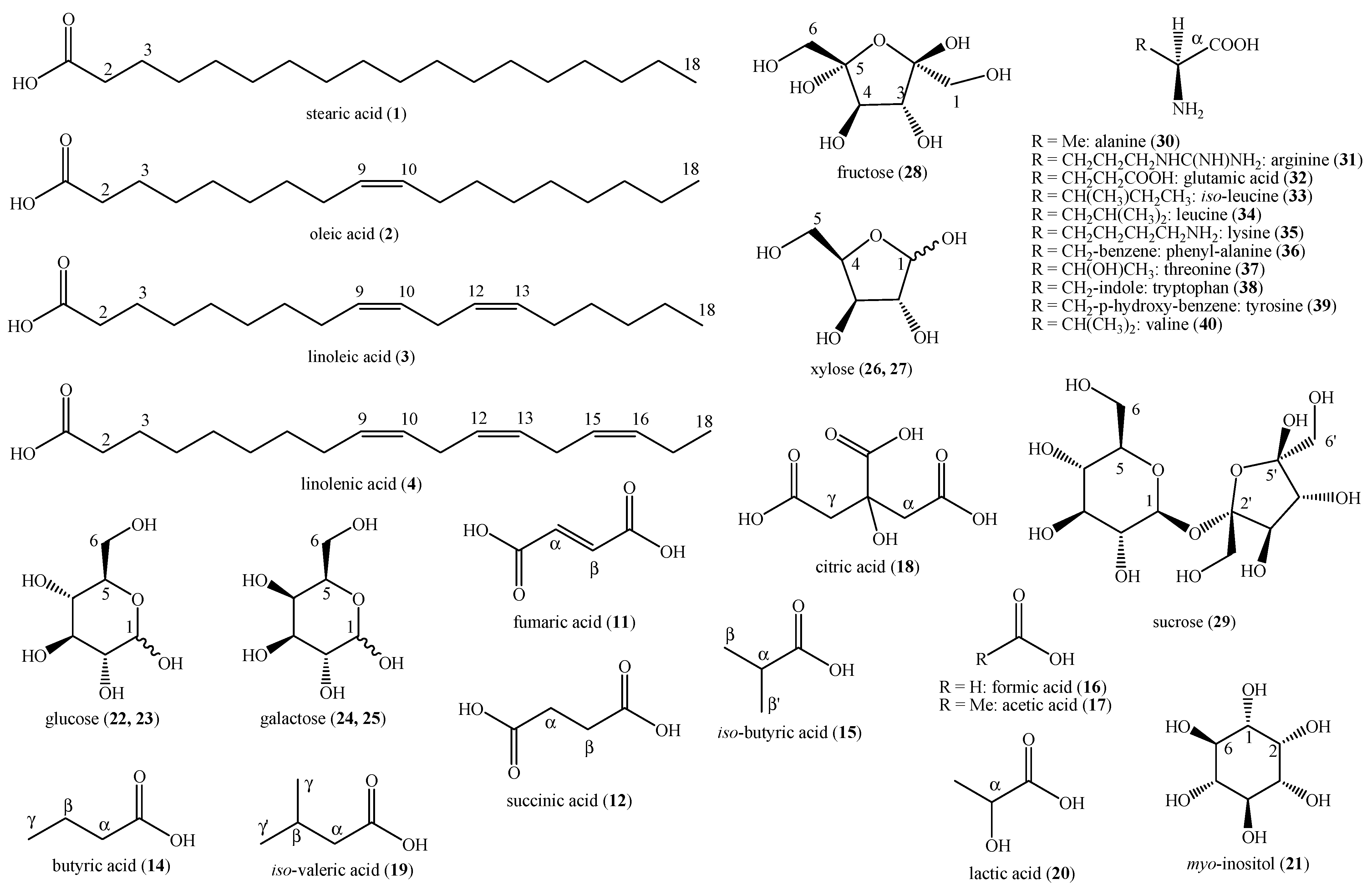
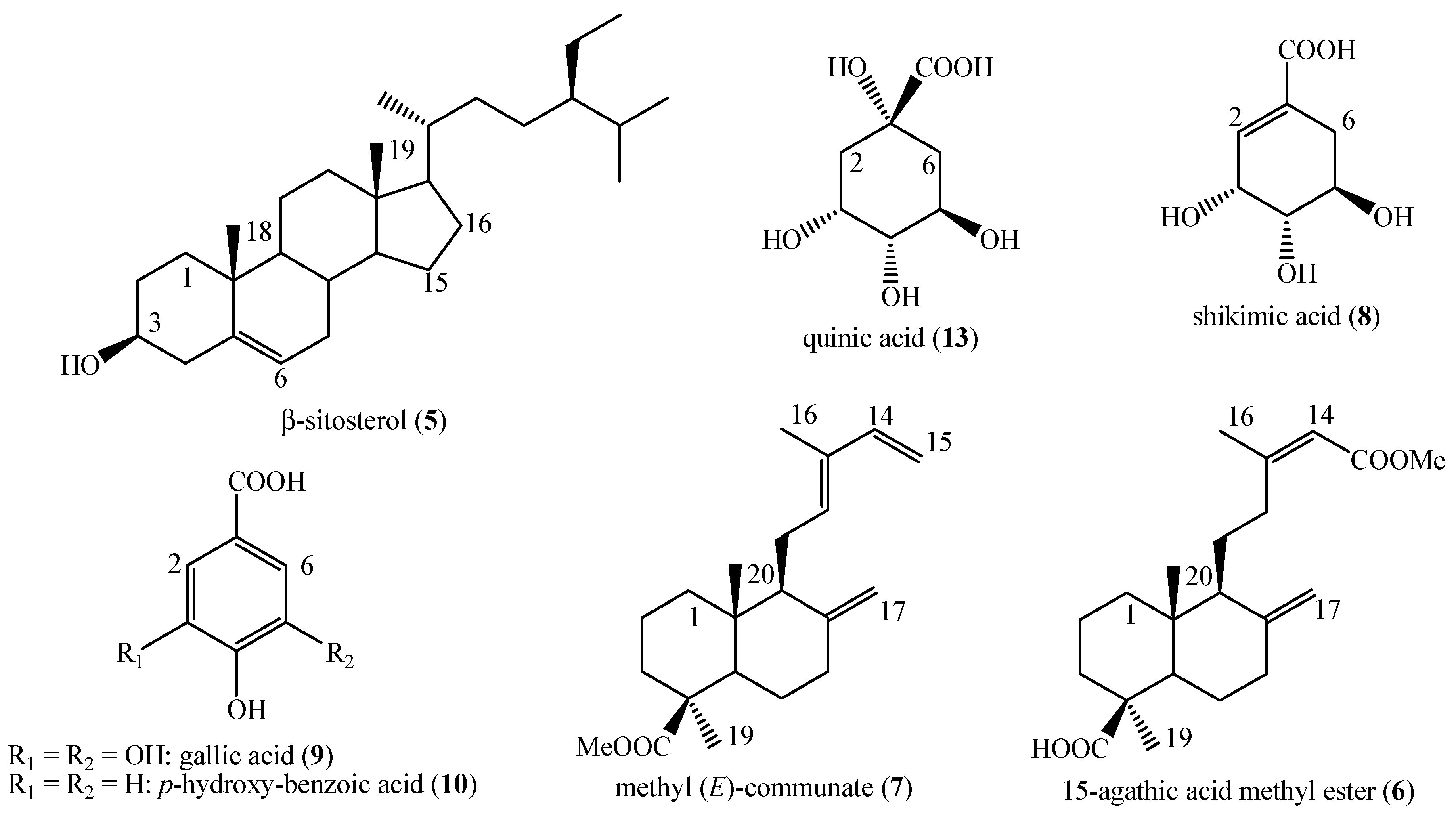
An aliquot of this extract (3.7 g) was subjected to a first separation procedure on column chromatography using 120.1 g of silica gel. The initial eluting system was a mixture of n-butanol and distilled water in concentration ratio of 82:18 v/v (400 mL) but during the chromatographic run, this mixture was changed in order to increase its polarity and let the elution of the most polar compounds passing to a mixture of n-butanol, methanol and distilled water in concentration ratio of 70:10:30 v/v/v (250 mL). During this procedure, all the compounds were identified by comparison of their spectroscopic and spectrometric data with those reported in the literature: stearic acid (1), oleic acid (2), linoleic acid (3), linolenic acid (4) [5,6], β-sitosterol (5) [5], 15-agathic acid methyl ester (6) [7] and methyl (E)-communate (7) [8] in mixture (ratio not calculable) from the assembly of fractions 3-18 for the total weight of 34.3 mg; shikimic acid (8) [7], gallic acid (9), p-hydroxy-benzoic acid (10), fumaric acid (11) and succinic acid (12) [5,6] from the assembly of fractions 94-100 for the total weight of 6.7 mg; shikimic acid (8) [7], p-hydroxy-benzoic acid (10), quinic acid (13), butyric acid (14), iso-butyric acid (15), formic acid (16), myo-inositol (21), α-glucose (22), β-glucose (23), α-galactose (24) and β-galactose (25) [5,6] in mixture (ratio not calculable) from the assembly of fractions 101-134 for the total weight of 4.5 mg; shikimic acid (8) [7], quinic acid (13), acetic acid (17), citric acid (18), iso-valeric acid (19), lactic acid (20), α-glucose (22), β-glucose (23), α-xylose (26), β-xylose (27), fructose (28), sucrose (29), alanine (30), arginine (31), glutamic acid (32), iso-leucine (33), leucine (34), lysine (35), phenyl-alanine (36), threonine (37), tryptophan (38), tyrosine (39) and valine (40) [5,6] in mixture (ratio not calculable) from the methanol column wash for the total weight of 28.2 mg.

2.5. NMR and MS Data of the Identified Compounds

Stearic acid (1): 1H NMR (600 MHz, CDCl3) δ: 2.31 (2H, t, J = 7.5 Hz, H-2), 1.62 (2H, m, H-3), 1.27 (28H, m, -(CH2)n-), 0.88 (3H, t, J = 6.9 Hz, Me-18). ESI-MS: m/z 307.5 [M+Na]+.
Oleic acid (2): 1H NMR (600 MHz, CDCl3) δ: 5.37 (2H, m, H-9 and H-10), 2.31 (2H, t, J = 7.5 Hz, H-2), 2.03 (4H, m, H-8 and H-11), 1.62 (2H, m, H-3), 1.27 (20H, m, -(CH2)n-), 0.88 (3H, t, J = 6.9 Hz, Me-18). ESI-MS: m/z 305.4 [M+Na]+.
Linoleic acid (3): 1H NMR (600 MHz, CDCl3) δ: 5.37 (4H, m, H-9 and H-10 and H-12 and H-13), 2.76 (2H, m, H-11), 2.31 (2H, t, J = 7.5 Hz, H-2), 2.03 (4H, m, H-8 and H-14), 1.62 (2H, m, H-3), 1.27 (14H, m, -(CH2)n-), 0.96 (3H, t, J = 6.9 Hz, Me-18). ESI-MS: m/z 303.5 [M+Na]+.
Linolenic acid (4): 1H NMR (600 MHz, CDCl3) δ: 5.37 (6H, m, H-9 and H-10 and H-12 and H-13 and H-15 and H-16), 2.78 (4H, m, H-11 and H-14), 2.31 (2H, t, J = 7.5 Hz, H-2), 2.03 (4H, m, H-8 and H-17), 1.62 (2H, m, H-3), 1.27 (8H, m, -(CH2)n-), 0.89 (3H, t, J = 6.9 Hz, Me-18). ESI-MS: m/z 301.3 [M+Na]+.
Β-sitosterol (5): 1H NMR (600 MHz, CDCl3) δ: 5.36-5.33 (1H, m, H-6), 3.56-3.53 (1H, m, H-3), 2.35-2.31 (2H, m, H-4), 2.01-1.96 (1H, m, Ha-7), 1.88-1.82 (3H, overlapped, Ha-1, Ha-2, Ha-16), 1.60-1.57 (2H, m, H-15), 1.54-1.45 (3H, overlapped, Hb-2, Hb-7, H-8), 1.26-1.23 (1H, m, Hb-16), 1.10-1.06 (1H, m, Hb-1), 1.02 (3H, s, Me-19), 1.00-0.98 (1H, m, H-14), 0.68 (3H, s, Me-18). ESI-MS: m/z 437.4 [M+Na]+.
15-agathic acid methyl ester (6): 1H NMR (600 MHz, CDCl3) δ: 5.63 (1H, d, J = 1.0 Hz, H-14), 4.86 (1H, br. s, H-17a), 4.51 (1H, br. s, H-17b), 3.67 (3H, s, COOMe), 2.14 (3H, s, Me-16), 1.12 (3H, s, Me-19), 0.73 (3H, s, Me-20). ESI-MS: m/z 371.5 [M+Na]+.
Methyl (E)-communate (7): 1H-NMR (600 MHz, CDCl3) δ: 6.31 (1H, dd, J = 17.8, 10.8, H-14), 5.37-5.34 (1H, overlapped, H-12), 5.31-5.27 (1H, overlapped, Ha-15), 5.08 (1H, br. s, Ha-17), 4.88 – 4.85 (1H, overlapped, Hb-15), 4.48 (1H, br. s, Hb-17), 3.46 (3H, s, 18-OMe), 1.67 (3H, s, Me-16), 1.25 (3H, s, Me-19), 0.67 (3H, s, Me-20). ESI-MS: m/z 339.4 [M+Na]+.
Shikimic acid (8): 1H-NMR (600 MHz, D2O) δ: 6.48-6.43 (1H, m, H-2), 4.31 (1H, t, J = 3.8 Hz, H-3), 4.31 (1H, t, J = 3.8 Hz, H-3), 3.91-3.87 (1H, m, H-5), 3.68-3.64 (1H, m, H-4), 2.60 (1H, dd, J = 18.3, 6.9 Hz, Ha-6), 2.10 (1H, dd, J = 18.3, 7.3 Hz, Hb-6). ESI-MS: m/z 197.3 [M+Na]+; m/z 173.2 [M-H]-.
Gallic acid (9): 1H-NMR (600 MHz, D2O) δ: 7.04 (2H, s, H-2 and H-6). ESI-MS: m/z 169.1 [M-H]-.
P-hydroxy-benzoic acid (10): 1H-NMR (600 MHz, D2O) δ: 7.75 (2H, d, J = 8.0 Hz, H-2 and H-6), 6.95 (2H, d, J = 8.0 Hz, H-3 and H-5). ESI-MS: m/z 161.2 [M+Na]+; m/z 137.0 [M-H]-.
Fumaric acid (11): 1H-NMR (600 MHz, D2O) δ: 6.51 (2H, s, H-α and H-β). ESI-MS: m/z 115.2 [M-H]-.
Succinic acid (12): 1H-NMR (600 MHz, D2O) δ: 2.38 (4H, s, H-α and H-β). ESI-MS: m/z 117.0 [M-H]-.
Quinic acid (13): 1H-NMR (600 MHz, D2O) δ: 4.14-4.10 (1H, m, H-4), 3.99-3.94 (1H, m, H-5), 3.57-3.53 (1H, m, H-3), 2.12-2.08 (4H, overlapped, Ha-2, Hb-2, Ha-6, Hb-6). ESI-MS: m/z 191.1 [M-H]-.
Butyric acid (14): 1H-NMR (600 MHz, D2O) δ: 2.27 (2H, t, J = 7.3 Hz, H-α), 1.38 (2H, m, H-β), 0.90 (3H, t, J = 7.4 Hz, H-γ). ESI-MS: m/z 87.2 [M-H]-.
Iso-butyric acid (15): 1H-NMR (600 MHz, D2O) δ: 2.40-2.37 (1H, m, H-α), 0.92 (6H, d, J = 7.0 Hz, H-β and H-β'). ESI-MS: m/z 87.2 [M-H]-.
Formic acid (16): 1H-NMR (600 MHz, D2O): 8.45 (1H, s, CH). ESI-MS: m/z 45.2 [M-H]-.
Acetic acid (17): 1H-NMR (600 MHz, D2O) δ: 1.92 (3H, s, Me). ESI-MS: m/z 59.1 [M-H]-.
Citric acid (18): 1H-NMR (600 MHz, D2O) δ: 2.71 (2H, d, J = 14.9 Hz, Ha-α and Ha-γ), 2.67 (2H, d, J = 14.9 Hz, Hb-α and Hb-γ). ESI-MS: m/z 191.1 [M-H]-.
Iso-valeric acid (19): 1H-NMR (600 MHz, D2O) δ: 2.15-2.05 (2H, m, H-α), 1.97-1.93 (1H, m, H-β), 0.91 (6H, d, J = 6.6 Hz, Me-γ and Me-γ'). ESI-MS: m/z 101.3 [M-H]-.
Lactic acid (20): 1H-NMR (600 MHz, D2O) δ: 4.11 (1H, q, J = 8.1 Hz, H-α), 1.31 (3H, d, J = 8.1 Hz, Me). ESI-MS: m/z 89.0 [M-H]-.
Myo-inositol (21): (600 MHz, D2O) δ: 4.07 (1H, t, J = 2.8 Hz, H-1), 3.64 (2H, m, H-4 and H-6), 3. 53 (2H, m, H-2 and H-3), 3.28 (1H, m, H-5). ESI-MS: m/z 202.7 [M+Na]+; m/z 179.1 [M-H]-.
A-glucose (22): 1H-NMR (600 MHz, D2O) δ: 5.22 (1H, d, J = 3.7 Hz, H-1), 3.87-3.84 (1H, m, H-5), 3.80-3.74 (2H, m, H-6), 3.75-3.72 (1H, m, H-3), 3.59-3.54 (1H, m, H-2), 3.47-3.41 (1H, m, H-4). ESI-MS: m/z 202.7 [M+Na]+; m/z 179.1 [M-H]-.
B-glucose (23): 1H-NMR (600 MHz, D2O) δ: 4.65 (1H, d, J = 8.0 Hz, H-1), 3.91-3.89 (1H, m, Ha-6), 3.79-3.75 (1H, m, Hb-6), 3.53-3.50 (1H, m, H-3), 3.49-3.46 (1H, m, H-5), 3.44-3.40 (1H, m, H-4), 3.29-3.25 (1H, m, H-2). ESI-MS: m/z 202.7 [M+Na]+; m/z 179.1 [M-H]-.
A-galactose (24): 1H-NMR (600 MHz, D2O) δ: 5.26 (1H, d, J = 3.7 Hz, H-1), 4.10-4.07 (1H, m, H-5), 4.00-3.97 (1H, m, H-3), 3.79-3.75 (2H, m, H-2 and H-4), 3.73-3.70 (2H, m, H-6). ESI-MS: m/z 202.7 [M+Na]+; m/z 179.1 [M-H]-.
B-galactose (25): 1H-NMR (600 MHz, D2O) δ: 4.59 (1H, d, J = 8.0 Hz, H-1), 3.99-3.96 (1H, m, H-4), 3.84-3.81 (2H, m, H-6), 3.74-3.71 (1H, m, H-5), 3.64-3.61 (1H, m, H-3), 3.51-3.48 (1H, m, H-2). ESI-MS: m/z 202.7 [M+Na]+; m/z 179.1 [M-H]-.
A-xylose (26): 1H-NMR (600 MHz, D2O) δ: 5.20 (1H, d, J = 3.7 Hz, H-1), 3.73-3.69 (1H, m, H-3), 3.65-3.62 (1H, m, H-4), 3.48-3.43 (2H, m, H-5), 3.37-3.34 (1H, m, H-2). ESI-MS: m/z 173.2 [M+Na]+; m/z 149.1 [M-H]-.
B-xylose (27): 1H-NMR (600 MHz, D2O) δ: 4.56 (1H, d, J = 7.9 Hz, H-1), 3.89-3.86 (2H, m, H-5), 3.71-3.68 (1H, m, H-3), 3.38-3.35 (1H, m, H-4), 3.19-3.16 (1H, m, H-2). ESI-MS: m/z 173.2 [M+Na]+; m/z 149.1 [M-H]-.
Fructose (28): 1H-NMR (600 MHz, D2O) δ: 4.15 (1H, d, J = 8.8 Hz, H-3), 4.04-4.01 (1H, m, H-4), 3.82 (2H, s, H-6), 3.71 (2H, s, H-1). ESI-MS: m/z 202.7 [M+Na]+; m/z 179.1 [M-H]-.
Sucrose (29): 1H-NMR (600 MHz, D2O) δ: 5.40 (1H, d, J = 3.9 Hz, H-1), 4.20 (1H, d, J = 8.8 Hz, H-3'), 4.07-4.03 (1H, m, H-4'), 3.90-3.84 (3H, m, H-5 and H-6), 3.80 (2H, s, H-6'), 3.77-3.74 (1H, m, H-3), 3.70 (2H, s, H-1'), 3.58-3.53 (1H, m, H-2), 3.49-3.45 (1H, m, H-4). ESI-MS: m/z 365.5 [M+Na]+; m/z 341.3 [M-H]-.
Alanine (30): 1H-NMR (600 MHz, D2O) δ: 3.80 (1H, q, J = 7.1 Hz, H-α), 1.49 (3H, d, J = 7.1 Hz, Me). ESI-MS: m/z 88.1 [M-H]-.
Arginine (31): 1H-NMR (600 MHz, D2O) δ: 3.77-3.74 (1H, m, H-α), 3.25 (2H, t, J = 8.9 Hz, H-δ), 1.94-1.91 (2H, m, H-β), 1.71-1.68 (2H, m, H-γ). ESI-MS: m/z 173.2 [M-H]-.
Glutamic acid (32): 1H-NMR (600 MHz, D2O) δ: 3.76-3.73 (1H, m, H-α), 2.37-2.32 (2H, m, H-β), 2.11-2.08 (2H, m, H-γ). ESI-MS: m/z 146.0 [M-H]-.
Iso-leucine (33): 1H-NMR (600 MHz, D2O) δ: 3.88-3.85 (1H, m, H-α), 2.00-1.97 (1H, m, H-β), 1.42-1.25 (2H, m, H-γ), 1.02 (3H, d, J = 7.0 Hz, Me-γ'), 0.95 (3H, t, J = 7.4 Hz, Me-δ). ESI-MS: m/z 130.2 [M-H]-.
Leucine (34): 1H-NMR (600 MHz, D2O) δ: 3.77-3.72 (1H, m, H-α), 1.74-1.70 (3H, m, H-β and H-γ), 0.99-0.95 (6H, m, H-δ and H-δ'). ESI-MS: m/z 130.2 [M-H]-.
Lysine (35): 1H-NMR (600 MHz, D2O) δ: 3.81-3.76 (1H, m, H-α), 3.03 (2H, t, J = 8.2 Hz, H-δ), 1.98-1.93 (2H, m, H-β), 1.70-1.67 (2H, m, H-γ), 1.49-1.45 (2H, m, H-δ). ESI-MS: m/z 145.1 [M-H]-.
Phenyl-alanine (36): 1H-NMR (600 MHz, D2O) δ: 7.43-7.40 (2H, m, H-3 and H-5), 7.39-7.36 (1H, m, H-4), 7.34-7.30 (2H, m, H-2 and H-6), 3.99-3.96 (1H, m, H-α), 3.28-3.25 (2H, m, H-β). ESI-MS: m/z 164.2 [M-H]-.
Threonine (37): 1H-NMR (600 MHz, D2O) δ: 4.26-4.23 (1H, m, H-β), 3.82-3.78 (1H, m, H-α), 1.32 (3H, d, J = 6.6 Hz, Me). ESI-MS: m/z 118.1 [M-H]-.
Tryptophan (38): 1H-NMR (600 MHz, D2O) δ: 7.74-7.71 (1H, m, H-4), 7.28-7.25 (1H, m, H-6), 7.21-7.18 (1H, m, H-5), 4.08-4.03 (1H, m, H-α), 3.31-3.29 (2H, m, H-β). ESI-MS: m/z 203.2 [M-H]-.
Tyrosine (39): 1H-NMR (600 MHz, D2O) δ: 7.20 (2H, d, J = 8.8 Hz, H-2 and H-6), 6.89 (2H, d, J = 8.8 Hz, H-3 and H-5), 3.95-3.91 (1H, m, H-α), 3.18-3.14 (2H, m, H-β). ESI-MS: m/z 180.3 [M-H]-.
Valine (40): 1H-NMR (600 MHz, D2O) δ: 3.64-3.60 (1H, m, H-α), 2.31-2.28 (1H, m, H-β), 1.05 (3H, d, J = 7.1 Hz, Me-γ'), 0.99 (3H, d, J = 7.0 Hz, Me-γ). ESI-MS: m/z 116.1 [M-H]-.

3. Results

The phytochemical analysis on the female cone of A. bidwillii collected in the experimental garden of the University of Rome “La Sapienza” led to the identification of forty compounds, thirty-three of which are primary metabolites whilst the remaining seven are secondary metabolites.
The identified primary metabolites are stearic acid (1), oleic acid (2), linoleic acid (3), linolenic acid (4), fumaric acid (10), succinic acid (11), butyric acid (13), iso-butyric acid (14), formic acid (15), acetic acid (17), citric acid (18), iso-valeric acid (19), lactic acid (20), myo-inositol (21), α-glucose (22), β-glucose (23), α-galactose (24), β-galactose (25), α-xylose (26), β-xylose (27), fructose (28), sucrose (29), alanine (30), arginine (31), glutamic acid (32), iso-leucine (33), leucine (34), lysine (35), phenyl-alanine (36), threonine (37), tryptophan (38), tyrosine (39) and valine (40) (Figure 2).
The identified secondary metabolites are β-sitosterol (4), 15-agathic acid methyl ester (5), methyl (E)-communate (6), shikimic acid (7), gallic acid (8), p-hydroxy-benzoic acid (9) and quinic acid (12), instead (Figure 3).

4. Discussion

The identified compounds belong to six different classes of natural compounds i.e., fatty acids (1-4), terpenoids (5-7), organic acids (8-20), polyols (21), saccharides (22-29), amino acids (30-40).
All these compounds have been identified for the first time in a whole cone of A. bidwillii. Yet, stearic acid (1), oleic acid (2), linoleic acid (3), linolenic acid (4), α-glucose (22), β-glucose (23), fructose (28), sucrose (29), alanine (30), arginine (31), glutamic acid (32), iso-leucine (33), leucine (34), lysine (35), phenyl-alanine (36), threonine (37), tyrosine (39) and valine (40) have, actually, been reported from the seeds derived from the female cones of this species [3]. Henceforth, their occurrence was also confirmed, during this study, from another population given that seeds are part of the female cones. On the other hand, it would be quite interesting to discover whether their presence here is due to these seeds or due to also other parts of the cone and for this, a further phytochemical analysis on the female cone deprived of the seeds is suggested for the future.
In general, the seeds and the female cone showed some important differences in their phytochemical composition, and this regarded primary metabolites but mainly secondary metabolites. As for the former ones, unlike the seeds, the female cone reported the presence of several primary organic acids such as fumaric acid (10), succinic acid (11), butyric acid (13), iso-butyric acid (14), formic acid (15), acetic acid (17), citric acid (18), iso-valeric acid (19) and lactic acid (20) but at the moment, it is impossible to establish their real absence in the seeds since they were not actually sought [3]. This aspect may indeed be an argument for future research on these cones, too. In the female cone, a minor number of fatty acids (four vs nine, including four in common) as well as a higher number of saccharides (eight vs four, including three in common) was evidenced with respect to the seeds [3] and this may be due to several intrinsic and extrinsic factors which need more in-depth phytochemical analyses considering more collection times. Indeed, for what concerns the secondary metabolite pattern, β-sitosterol (4), 15-agathic acid methyl ester (5), methyl (E)-communate (6), shikimic acid (7), gallic acid (8), p-hydroxy-benzoic acid (9) and quinic acid (12) were evidenced only in the female cone whereas no flavonoid was evidenced here unlike the seeds [3]. This also may be due to several intrinsic and extrinsic factors which need more in-depth phytochemical analyses considering more collection times as well as separating the nuts from the whole cone. This may also be an interesting argument for future research. Actually, the high amount of shikimic acid (7) evidenced in this study may even be responsible for this aspect given that this compound is well known to be one of the precursors for the biosynthesis of this type of compounds [9].
To the best of our knowledge, β-sitosterol (4), 15-agathic acid methyl ester (5), methyl (E)-communate (6), shikimic acid (7), gallic acid (8), p-hydroxy-benzoic acid (9) and quinic acid (12) were reported from this species for the first time, during this study. In fact, their occurrence has been already evidenced in other species of the genus as well as in other genera of the family [2]. Yet, their chemophenetic value is none for some compounds like β-sitosterol (4), shikimic acid (7), gallic acid (8), p-hydroxy-benzoic acid (9) and quinic acid (12) given that they are widespread metabolites in the plant kingdom evidenced in several families, even taxonomically distant [10,11,12,13,14]. On the other hand, the diterpenoids 15-agathic acid methyl ester (5) and methyl (E)-communate (6) have chemophenetic importance given that their occurrence is limited to only some species comprised in the Araucariaceae family. In fact, 15-agathic acid methyl ester (5) has been previously reported in Agathis and Araucaria species [7,15,16,17] whereas methyl (E)-communate (6) has been previously evidenced in Araucaria Juss species [18,19] and Wollemia nobilis W. G. Jones, K. D. Hill & J. M. Allen [8,20]. Thus, these compounds may be indeed employed as chemophenetic markers at the family level. As for the primary metabolites, they have no chemophenetic value as well since they can be found in all the plants. For this reason, the eventual presence of newly reported compounds of this type in the species was not actually verified also because of the lack of extremely specific data on this in the literature.
Comparing the phytochemical pattern of this whole female cone with the other few studies whole cones of the family, some similarities were evidenced even though not all these cones have been thoroughly studied for their primary and secondary metabolite contents. By consequence, these comparisons are extremely partial. In particular, with respect to the half-matured female cones of W. nobilis [20], nine compounds (6, 7, 11, 12, 20, 22, 23, 29, 30) were found in common. With respect to the male cones of W. nobilis, five compounds (6, 7, 22, 23, 29, 31) were found in common for the first collection [8] and four compounds (7, 22, 23, 29) for the second collection [21]. With respect to the unripe female cones of W. nobilis [22], only one compound (7) was found in common. These derived differences depend on several intrinsic and extrinsic factors again, and may surely be an argument for future research, too.

5. Conclusions

The first phytochemical analysis on the whole female cone of A. bidwillii evidenced the presence of forty primary and secondary metabolites, all newly identified compounds for the organ, and seven of which reported in the species for the first time as clearly pointed out by the chemophenetic evaluation. This evaluation also highlighted the need of future phytochemical researches on these cones and on cones of species belonging to this family, in general, in order to provide more general data on all of them given their quite scarcity in the literature.

Author Contributions

Conceptualization, C.F.; methodology, C.F., A.V., O.G., C.D.B., C.S.; investigation, C.F., A.V., O.G., C.D.B., M.F., C.S., F.S., M.S., D.D.V, F.A.; resources, C.S., D.D.V; writing—original draft preparation, C.F., A.V., O.G., C.D.B., M.F., C.S., F.S., writing—review and editing, C.F., A.V., C.D.B., M.F., C.S., F.S., M.S., D.D.V, F.A.; supervision, M.S., D.D.V., F.A. All authors have read and agreed to the published version of the manuscript. .

Funding

This research received no external funding.

Conflicts of Interest

The authors declare no conflicts of interest.

References

  1. Smith, I.A.; Butler, D. The Bunya in Queensland's Forests. Queensland Rev. 2002, 9(2), 31–38. [Google Scholar] [CrossRef]
  2. Frezza, C.; Venditti, A.; De Vita, D.; Toniolo, C.; Franceschin, M.; Ventrone, A.; Tomassini, L.; Foddai, S.; Guiso, M.; Nicoletti, M.; Bianco, A.; Serafini, M. Phytochemistry, chemotaxonomy, and biological activities of the Araucariaceae family - a review. Plants, 2020, 9, 888. [Google Scholar] [CrossRef] [PubMed]
  3. Moura Nadolny, J.; Best, O.; Netzel, G.; Shewan, H.M.; Thi Phan, A.D.; Smyth, H.E.; Stokes, J.R. Chemical composition of bunya nuts (Araucaria bidwillii) compared to Araucaria angustifolia and Araucaria araucana species. Food Res. Int. 2023, 163, 112269. [Google Scholar] [CrossRef] [PubMed]
  4. Huth, J. Introducing the Bunya Pine – A Noble Denizen of the Scrub. Queensland Rev. 2002, 9(2), 7–20. [Google Scholar] [CrossRef]
  5. Sciubba, F.; Di Cocco, M.E.; Gianferri, R.; Capuani, G.; De Salvador, F.R.; Fontanari, M.; Gorietti, D.; Delfini, M. Nuclear magnetic resonance-based metabolic comparative analysis of two apple varieties with different resistances to apple scab attacks. J. Agric. Food Chem. 2015, 63, 8339–8347. [Google Scholar] [CrossRef] [PubMed]
  6. Sciubba, F.; Capuani, G.; Di Cocco, M.E.; Avanzato, D.; Delfini, M. Nuclear magnetic resonance analysis of water soluble metabolites allows the geographic discrimination of pistachios (Pistacia vera). Food Res. Int. 2014, 62, 66–73. [Google Scholar] [CrossRef]
  7. Frezza, C.; Sciubba, F.; De Vita, D.; Toniolo, C.; Foddai, S.; Tomassini, L. , Petrucci, R.; Bianco, A.; Serafini, M. Non-volatile compounds from Araucaria columnaris (G.Forst.) Hook leaves. Biochem. Syst. Ecol. 2022, 103, 104430. [Google Scholar] [CrossRef]
  8. Venditti, A.; Frezza, C.; Sciubba, F.; Foddai, S.; Serafini, M.; Bianco, A. Terpenoids and more polar compounds from the male cones of Wollemia nobilis. Chem. Biodiversity 2017, 14, e1600332. [Google Scholar] [CrossRef] [PubMed]
  9. Rehan, M. Biosynthesis of diverse class flavonoids via shikimate and phenylpropanoid pathway. In Bioactive compounds - biosynthesis, characterization and applications; Queiroz Zepka, L., Casagrande do Nascimento, T., Jacob-Lopes, T., Eds. IntechOpen, London, UK, 2021; pp. 75392.
  10. Gupta, E. B-Sitosterol: predominant phytosterol of therapeutic potential. In Innovations in Food Technology; Mishra, P., Mishra, R.R., Adetunji, C.O. (eds). Springer, Singapore. pp. 465-477.
  11. Candeias, N.R.; Assoah, B.; Simeonov, S.P. Production and synthetic modifications of shikimic acid. Chem. Rev. 2018, 118, 10458–10550. [Google Scholar] [CrossRef] [PubMed]
  12. Badhani, B.; Sharma, N.; Kakkar, R. Gallic acid: a versatile antioxidant with promising therapeutic and industrial applications. RSC Adv. 2015, 5, 27540–27557. [Google Scholar] [CrossRef]
  13. Manuja, R.; Sachdeva, S.; Jain, A.; Chaudhary, J. A Comprehensive review on biological activities of p-hydroxy benzoic acid and its derivatives. Int. J. Pharm. Sci. Rev. Res. 2013, 22(2), 109–115. [Google Scholar]
  14. Benali, T.; Bakrim, S. , Ghchime, R., Benkhaira, N.; El Omari, N.; Balahbib, A.; Taha, D.; Zengin, G.; Hasan, M.M.; Bibi, S.; Bouyahya, A. Pharmacological insights into the multifaceted biological properties of quinic acid. Biotechnol. Gen. Engineer. Rev. 1–30. [CrossRef]
  15. Carman, R.M.; Marty, R.A. Diterpenoids* IX. Agathis microstachya oleoresin. Aust. J. Chem. 1966, 19, 2403–2406. [Google Scholar] [CrossRef]
  16. Poinar Jr., G. O. Resinites, with examples from New Zealand and Australia. Fuel Process. Technol. 1991, 28(2), 135–148. [Google Scholar] [CrossRef]
  17. Sahu, B.; Bhardwaj, N.; Chatterjee, E.; Dey, B.; Tripathi, N.; Goel, B.; Kushwaha, M. , Kumar, B. ; Singh, B.; Guru, S.K.; Jain, S.K. LCMS-DNP based dereplication of Araucaria cunninghamii Mudie gum-resin: identification of new cytotoxic labdane diterpene, Nat. Prod. Res. 2022, 36(24), 6207–6214. [Google Scholar] [PubMed]
  18. Frezza, C.; De Vita, D.; Fonti, L.; Giampaoli, O.; Dal Bosco, C.; Sciubba, F.; Venditti, A.; Scintu, C.; Attorre, F. Secondary metabolites of Araucaria cunninghamii Mudie from central Italy. Plant Biosyst. 2024, In press. [CrossRef]
  19. Patial, P.K.; Cannoo, D.S. Phytochemical profile, antioxidant potential and DFT study of Araucaria columnaris (G. Forst.) Hook. Branch extracts. Nat. Prod. Res. 2021, 35(22), 4611-4615.
  20. Frezza, C.; Venditti, A.; Scandurra, C.; Ciccòla, A.; Serafini, I.; Sciubba, F.; Foddai, S.; Franceschin, M.; Bianco, A.; Serafini, M. Phytochemical profile of Wollemia nobilis half-matured female cones and their potential ethnopharmacological and nutraceutical activities. J. Agric. Sci. Technol. A 2018, 8, 162–170. [Google Scholar]
  21. Venditti, A.; Frezza, C.; Vincenti, F.; Brodella, A.; Sciubba, F.; Montesano, C.; Franceschin, M.; Sergi, M.; Foddai, S.; Di Cocco, M.E.; Curini, R.; Delfini, M.; Bianco, A.; Serafini, M. A syn-ent-labdadiene derivative with a rare spiro-β-lactone function from the male cones of Wollemia nobilis. Phytochemistry 2019, 158, 91–95. [Google Scholar] [CrossRef] [PubMed]
  22. Venditti, A.; Frezza, C.; Rossi, G.; Sciubba, F.; Ornano, L.; De Vita, D.; Toniolo, C.; Tomassini, L.; Foddai, S.; Nicoletti, M.; Di Cocco, M.E.; Bianco, A.; Serafini, M. A new diterpene and other compounds from the unripe female cones of Wollemia nobilis. Nat. Prod. Res. 2021, 35(21), 3839–3849. [Google Scholar] [CrossRef] [PubMed]
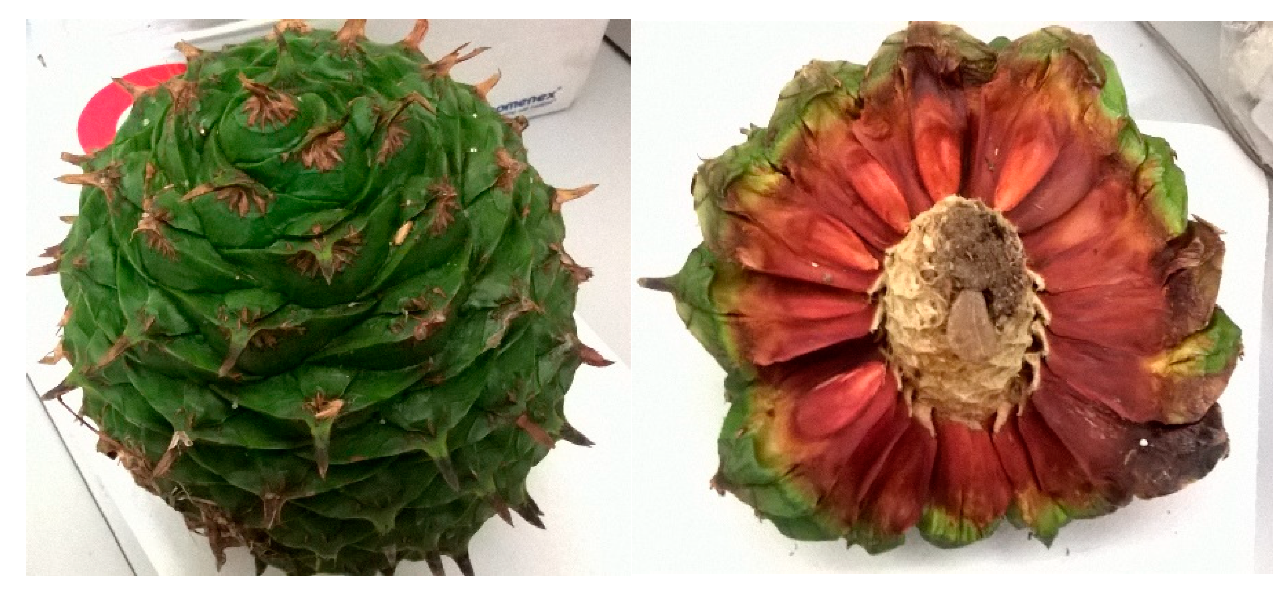
Figure 1. Images of the collected A. bidwillii cone.
Preprints 113122 g001
Figure 2. Structures of the identified primary metabolites in A. bidwillii cone.
Preprints 113122 g002
Figure 3. Structures of the identified secondary metabolites in A. bidwillii cone.
Preprints 113122 g003
Disclaimer/Publisher’s Note: The statements, opinions and data contained in all publications are solely those of the individual author(s) and contributor(s) and not of MDPI and/or the editor(s). MDPI and/or the editor(s) disclaim responsibility for any injury to people or property resulting from any ideas, methods, instructions or products referred to in the content.
Copyright: This open access article is published under a Creative Commons CC BY 4.0 license, which permit the free download, distribution, and reuse, provided that the author and preprint are cited in any reuse.
Prerpints.org logo

Preprints.org is a free preprint server supported by MDPI in Basel, Switzerland.

Subscribe

© 2024 MDPI (Basel, Switzerland) unless otherwise stated