Preprint
Review

Carbon Nanodots-Based Polymer Nanocomposite: A Potential Drug Delivery Armament of Phytopharmaceuticals

Altmetrics

Downloads

189

Views

97

Comments

0

Submitted:

22 July 2024

Posted:

24 July 2024

You are already at the latest version

Alerts
Abstract
Carbon nanodots (CNDs) have garnered significant attention as viable drug delivery vehicles in recent years, especially in the field of phytomedicine. Although there is much promise for therapeutic applications with phytomedicine, its effectiveness is frequently restricted by its low solubility, stability, and bioavailability. This paper offers a thorough synopsis of the developing field of phytomedicine drug delivery based on CND. It explores CND synthesis processes, surface functionalization strategies, and structural and optical characteristics. Additionally, the advantages and difficulties of phytomedicine are examined, with a focus on the contribution of drug delivery methods to the increased effectiveness of phytomedicine. The applications of CNDs in drug delivery are also included in the review, along with the mechanisms that underlie their improved drug delivery capabilities. Additionally, it looks at controlled release methods, stability augmentation, and phytomedicine loading tactics onto CNDs. The potential of polymeric carbon nanodots in drug delivery is also covered, along with difficulties and prospective directions going forward, such as resolving toxicity and biocompatibility issues. In summary, the present review highlights the encouraging contribution of CNDs to the field of drug delivery, specifically in enhancing the potential of phytomedicine for therapeutic purposes.
Keywords: 
Subject: Chemistry and Materials Science  -   Nanotechnology

1. Introduction

1.1. Background

A class of nanomaterials known as carbon nanodots (CNDs) has caught the attention of scientists because of its special qualities and broad range of uses. The amazing features of CNDs, including their programmable surface chemistry and biocompatibility, have led to increased interest in CND's application in phytopharmaceutical drug delivery. However, there are still flaws and shortcomings in the way that drugs are currently delivered for different dosage forms, such as problems with therapeutic efficacy, sustained release, and specificity. Carbon nanodots have attracted considerable attention owing to their distinctive advantages compared to other formulations. These nanodots possess unique properties, such as adjustable size, surface chemistry, and fluorescence characteristics, rendering them highly versatile for diverse applications. Additionally, their biocompatibility is noteworthy, with certain carbon nanodots demonstrating minimal toxicity, a vital feature for their potential biomedical use. Importantly, their synthesis is relatively straightforward, often achievable from readily available starting materials through simple methods. These attributes collectively contribute to the growing interest in carbon nanodots across various fields, promising novel solutions for a wide range of applications. These tiny carbon-based particles usually exhibit a spherical or quasi-spherical morphology and range in size from 1 to 10 nanometres [1,2]. Carbon nanodots, which are made up of carbon atoms arranged in a sp2-hybridized structure similar to graphene, have attracted interest from a variety of scientific and technological fields [2]. Carbon nanodots are synthesized using a variety of techniques, each of which helps to modify their characteristics for certain uses. Various methods have been utilized, including chemical vapor deposition, hydrothermal techniques, laser therapy, and pyrolysis of organic precursors. Typical precursors comprise organic compounds such as glucose and citric acid, which offer a carbon-rich base for the creation of nanodots [3]. The size, surface chemistry, and optical properties of the resultant carbon nanodots are all significantly influenced by the production process. The extraordinary optical qualities of carbon nanodots are one of their most notable characteristics. They are especially well-suited for use in bioimaging and medical diagnostics because of their high fluorescence [4]. The inherent fluorescence of CNDs enhances imaging technology, serving as a valuable contrast agent for improved visualization of biological structures and processes at the nanoscale. Because of this characteristic, carbon nanodots have been investigated extensively in the realm of medicine. Their biocompatibility and imaging abilities suggest that they may be useful for targeted medication delivery and non-invasive diagnostics. Carbon nanodots have a high surface area and a distinct electrical structure that makes them attractive candidates for a variety of uses [5]. Their surface is easily functionalizable with various chemical groups, increasing their adaptability to many applications. Carbon nanodots have proven effective in sensing applications for a variety of analytes, such as metal ions, pH variations, and biomolecules. Surface functionalization is a simple process that enables customized interactions with particular target molecules, which makes them useful instruments in the production of sophisticated sensors for healthcare, environmental monitoring, and other industries [6]. Another application where carbon nanodots show promise is catalysis. CNDs are effective catalysts for a variety of chemical processes due to their large surface area and distinctive electrical characteristics. Their catalytic potential is being investigated by researchers in a variety of processes, including environmental remediation and conventional chemical synthesis [7]. Carbon nanodots' versatility in catalysis highlights the contribution they provide to the advancement of effective and sustainable chemical transformations. Carbon nanodots are used in energy-related sectors in addition to sensing and catalysis. They are intriguing possibilities for usage in solar cells and supercapacitors due to their distinct electrical characteristics [8]. Researchers are examining strategies that exploit the capabilities of CNDs to optimize energy storage devices' efficiency and boost solar cells' performance. Carbon nanoparticles' biocompatibility and adjustable characteristics make them attractive for use in a variety of cutting-edge technologies [9]. A crucial feature of carbon nanodots that increases their potential for biological and medicinal applications is their biocompatibility. Because of their low toxicity, CNDs can be used in drug delivery systems to deliver therapeutic molecules to precise sites in a regulated way [10]. Their capacity to functionalize their surface with targeted ligands improves their precision in drug administration and presents a viable way to raise therapeutic treatment efficacy while lowering negative effects. The goals of carbon nanodot research continue to be improving synthesis techniques, finding new uses, and maximising the qualities of the material for particular purposes. Current studies seek to learn more about the basic properties of CNDs, which will open the door to advancements in a variety of industries, including energy storage, sensing technologies, medicine, and catalysis. Carbon nanodot research is interdisciplinary, which highlights how revolutionary they can be for a variety of industries and how they can help create new kinds of innovative materials with never-before-seen capabilities [11]. Given below are the different types of nanomaterials used for drug delivery in Figure 1.

1.2. Significance of CNDs in Drug Delivery Systems

In contemporary medicine, drug delivery systems are essential because they provide creative ways to improve the efficacy and security of therapeutic interventions. The importance of drug delivery systems resides in their capacity to resolve several issues related to traditional drug administration, which eventually enhances treatment outcomes and patient experiences [12]. These technologies seek to improve drug bioavailability, target distribution to particular body locations, and optimize drug pharmacokinetics. Achieving the best possible drug concentrations at the intended location with the least amount of systemic exposure is a major difficulty in traditional drug delivery. Drug delivery systems make the controlled release possible, guaranteeing a consistent and therapeutically effective drug concentration across the intended time [13]. By doing so, drug level variations are avoided, adverse effects are decreased, and patient compliance is increased. Drug delivery systems enhance treatment safety and efficacy by offering a more consistent and regulated release profile. One of the most important aspects of medication effectiveness is bioavailability, or the percentage of the medicine that is provided that enters the bloodstream and reaches the intended location. Due to variables including poor solubility, gastrointestinal tract breakdown, or fast metabolism, many medicines have low bioavailability. By addressing these obstacles, drug delivery systems can improve bioavailability [14]. Drugs that are poorly soluble, for instance, can be encapsulated by liposomes and nanoparticles to prevent degradation and enhance absorption. By ensuring that a higher percentage of the supplied medicine reaching the target, this increase in bioavailability maximises therapeutic effects [15]. One of the main characteristics of sophisticated medication delivery systems is targeted drug delivery. When drugs are administered conventionally, it frequently finds its way to non-target tissues, which can cause side effects and decreased efficacy. By delivering medications precisely to the intended location of action, targeted drug delivery systems reduce the likelihood of side effects [16]. This is especially important for treating diseases like cancer, when localised therapy is preferred. For example, passive or active targeting methods can be used to create nanoparticles and liposomes so that they preferentially aggregate in tumour tissues, enhancing drug delivery to cancer cells while sparing healthy tissues. For instance, the blood-brain barrier prevents many medications from entering the central nervous system. Bypassing or penetrating these barriers with drug delivery devices creates new opportunities for treating diseases affecting the brain and other difficult anatomical regions [17]. Drug delivery systems have not only improved the distribution of conventional small-molecule medications but have also broadened their application to encompass nucleic acid-based medicines, gene therapies, and biologics. Stability and delivery issues are common for these intricate and delicate compounds. Precision medicine has entered a new era with the safe and efficient delivery of biologics and gene treatments to their desired targets made possible by advanced drug delivery technologies like lipid nanoparticles and viral vectors [18]. The potential of drug delivery systems to improve patient comfort and adherence only serves to highlight how important they are. By lowering the frequency of medication delivery, controlled-release formulations can increase patient compliance. This is especially helpful for long-term medication adherence, which is necessary for managing chronic disorders and averting problems [19]. Drug delivery systems are important for getting beyond the body's physiological barriers. Drug delivery systems are essential to the medical revolution because they improve the accuracy, effectiveness, and safety of therapeutic interventions. Because of its special qualities and biocompatibility, carbon nanodots (CNDs) have become one of the many nanomaterials being investigated for drug delivery [20]. Drug delivery systems are important because they assist in issues such systemic toxicity, low bioavailability, and lack of selectivity that arise with traditional drug administration. Researchers hope to overcome these obstacles and usher in a new era of focused and controlled medication delivery by utilising the unique properties of carbon nanodots [21]. Carbon nanodots provide a nanoscale platform for drug delivery applications; their sizes typically fall between 1 and 10 nanometers. Because of their small size, which promotes interactions at the cellular and molecular levels, medicines can be delivered to particular tissues or cells with accuracy and efficiency. One important aspect of CNDs' importance for drug administration is their biocompatibility [22]. Because of their low toxicity, these nanodots are unlikely to hurt biological systems. Developing drug carriers that can successfully navigate the intricate biological environment and deliver therapeutic payloads without causing detrimental side effects requires biocompatibility [23]. The importance of carbon nanodots in drug delivery systems is largely due to their optical characteristics, especially their intense fluorescence. This fluorescence creates opportunities for theranostic applications, which combine therapy and diagnostics on a single platform, in addition to helping with bioimaging for real-time drug carrier tracking. Approaches to personalized medicine are made possible by the real-time visualization and monitoring of drug delivery mechanisms [24]. Carbon nanodots must be surface functionalized to be customized for a specific drug delivery application. Targeting ligands, medicinal compounds, or other biomolecules can be attached to CNDs by adding different functional groups on their surface [25]. Because of their adaptability, researchers can create drug carriers with a particular affinity for particular cells or tissues, increasing drug delivery's selectivity and reducing off-target effects. Moreover, functionalized carbon nanodots can be designed to react to external factors like pH variations or certain enzymes, causing regulated drug release at the intended location. Carbon nanodots are used in drug delivery systems because of their ability to overcome biological barriers [26]. The blood-brain barrier is one physiological barrier that CNDs can be made to cross, making it easier to deliver medications to anatomical sites that would otherwise be difficult. This capacity is especially important for treating neurological illnesses because the blood-brain barrier's limited permeability makes drug administration extremely difficult [27]. Additionally, there is plenty of room for loading and transporting therapeutic payloads on carbon nanodots due to their large surface area. CNDs offer a variety of drug loading techniques, including encapsulation, adsorbing, and covalent attachment. In order to ensure that a sufficient amount of the therapeutic substance reaches the target site to exercise its intended effects, this loading capacity is crucial for optimising drug delivery systems [28]. Beyond conventional small molecule medications, carbon nanodots are incredibly versatile. A vast array of medicinal substances, including proteins, peptides, and nucleic acids (such DNA and RNA), have been demonstrated to be delivered by them via their promise [29]. This creates novel possibilities for treating diseases where traditional drug delivery techniques could be ineffective, such as cancer and genetic abnormalities. A major development in the realm of nucleic acid therapies and biological products is the capacity of CNDs to precisely deliver and protect sensitive biomolecules. To sum up, the importance of carbon nanodot-based drug delivery systems originates from their capacity to overcome the obstacles related to traditional drug administration [30]. CNDs are potential vehicles for targeted and regulated drug administration because of their special mix of small size, biocompatibility, optical characteristics, surface functionalization, and barrier-crossing abilities. This field's continued study and development has the potential to completely transform medical care by providing more individualised and effective therapeutic interventions with fewer adverse effects and better patient outcomes. The use of carbon nanodots in drug delivery systems offers optimism for the advancement of medicine towards safer, more accurate, and effective treatment modalities as our understanding of them improves [31].

1.2. Significance of CNDs in Drug Delivery Systems

1.2. Emergence of Carbon Nanodots in Drug Delivery
The scientific and medical communities are particularly interested in the use of carbon nanodots (CNDs) in drug administration because they present a fresh and exciting opportunity to improve drug delivery methods. Carbon-rich precursors are the source of carbon nanodots, which are nanomaterials with typical diameters between 1 and 10 nanometres [32]. These nanodots are excellent prospects for a variety of biomedical applications, especially in the field of drug administration, due to their unique qualities, which include biocompatibility, biodegradability, eco-friendly, green-synthesis techniques fluorescence, and a high surface area. One of the main reasons carbon nanodots have become popular for use in drug delivery applications is their biocompatibility [33]. Because of their low toxicity and ability to be produced from organic precursors like glucose or citric acid, these nanodots rarely cause harm to biological systems. Their incorporation into medication delivery systems, where materials must effortlessly interact with the complex biological milieu without causing harm, depends on their biocompatibility [34]. Because carbon nanodots are naturally compatible with biological systems, they are a prospective vehicle for modern medicines. The rise of carbon nanodots in drug administration can be attributed in large part to their optical characteristics, particularly their intense fluorescence [35]. This fluorescence creates opportunities for theranostic applications in addition to allowing for real-time drug carrier monitoring and imaging. Theranostic is the process of combining therapy and diagnostics onto one platform to provide individualised and focused medical care. The fluorescence of carbon nanodots makes drug transport processes easier to see, improving the accuracy and effectiveness of therapeutic actions [36]. Charges and BBB based delivery" refers to drug delivery strategies that utilize either electrostatic charges or interactions with the blood-brain barrier (BBB) for targeted delivery of therapeutic agents to specific sites, particularly the brain. Utilizing charges and the blood-brain barrier (BBB) for drug delivery offers significant advantages in targeting neurological disorders. Drugs can be precisely delivered to the brain by taking advantage of charge-based interactions, which maximises therapeutic efficacy and minimises systemic side effects. Furthermore, BBB-based delivery techniques allow medicines to pass through the barrier, removing a significant obstacle in the treatment of disorders of the central nervous system [37]. Surface functionalization is an important factor in the adaptability of carbon nanodots in drug delivery. Different functional groups can be added to the surface of CNDs with ease, enabling the attachment of medicinal compounds, targeted ligands, or other biomolecules. Drug carriers with particular affinities to target cells or tissues can be designed with this customisation, which enhances drug delivery selectivity and reduces off-target effects [32]. Moreover, functionalized carbon nanodots can be designed to react to outside stimuli like pH variations or certain enzymes, causing regulated drug release at the intended location. The ability to interact at the nanoscale provided by the small size of carbon nanodots makes medication administration more accurate and effective by enabling the distribution of therapeutic substances to target tissues. Due to their small size and specific surface characteristics, nanoparticles can enter anatomical regions that would otherwise be inaccessible for targeted medication delivery. One example of a biological barrier they can easily cross is the blood-brain barrier. In difficult situations such as neurodegenerative disorders, nanoparticles provide a viable avenue for therapeutic treatments by utilising both active surface changes and passive mechanisms such as size and the Enhanced Permeability and Retention (EPR) effect. [38]. This capacity is especially important for treating neurological illnesses, as traditional drug delivery techniques might not be able to get pass these obstacles. Therapeutic payloads can be loaded and transported with plenty of room due to the high surface area of carbon nanodots. Different approaches for drug loading are available depending on whether the drug is covalently bonded, adsorbed, or encapsulated on the surface of CNDs. To maximise drug delivery systems and make sure that a sufficient amount of the therapeutic agent reaches the target site to have the desired effects, this loading capacity is crucial. Moreover, carbon nanodots' growing importance in drug delivery is attributed to their ability to transport a diverse array of therapeutic molecules [39]. CNDs have demonstrated potential in delivering nucleic acids (such DNA and RNA), proteins, and peptides in addition to conventional small molecule medicines. This potential creates novel therapy options for cancer, genetic abnormalities, and other illnesses where the effectiveness of traditional medication delivery techniques may be constrained [29]. A major development in the realm of nucleic acid therapies and biologics is the capacity of CNDs to precisely deliver and protect sensitive biomolecules. To sum up, the application of carbon nanodots in drug delivery is a revolutionary step forward for the field of nanomedicine. Their nanoscale size, surface functionalization abilities, optical characteristics, and biocompatibility make them useful and efficient carriers of medicinal drugs. Carbon nanodots have the potential to completely transform drug delivery systems as long as an investigation into this field is conducted. They can provide novel approaches for the precise, targeted, and controlled delivery of medicines of various medical applications [40] in life-threatening as well as chronic illnesses including cancer, viral diseases, and neurodegenerative disorders etc.

2. Phytomedicine in Drug Delivery

A timeless field with roots in ancient wisdom, phytomedicine is embracing the cutting edge of modern drug delivery technologies while navigating the prevailing modern currents [41]. The fusion of phytomedicine with state-of-the-art drug delivery technology, as demonstrated in "Carbon Nanodots-Based Drug Delivery of Phytomedicine," stands at the intersection of tradition and modernity and sets off on a path that surpasses traditional therapeutic approaches. The range of options available to us in phytomedicine becomes clear as we explore further. Beneath the abundance of bioactive substances is a dynamic interplay of holistic healing, an idea engrained in conventional medical practice. With a focus on overall health, phytomedicine aims to treat illnesses' underlying causes as well as their symptoms [42]. Investigation of this complex terrain reminds us that the effectiveness of plant-based medicines involves more than just single components; it involves the synergy of nature's pharmacopeia [43]. The symbiotic link between sophisticated medication delivery technologies and phytomedicine is a prominent feature of the modern period [44]. Optimizing the delivery of medicinal substances obtained from plants is the fundamental goal of this partnership [45]. Although the ancient healers had an innate understanding of the power of plant treatments, current technology allows us to fully realize their potential through targeted and deliberate delivery methods [46]. This harmonious dance portends well for a new chapter in healthcare history, one that combines the best aspects of conventional medicine with the cutting-edge techniques of contemporary medication delivery.

2.1. Carbon Nanodots (CNDs): Catalysts of Transformation

Carbon nanodots (CNDs), heralded as revolutionary agents in drug delivery, are fundamental to this metamorphosis. CNDs have their roots in nanotechnology and offer of several unique features. They are ideal phytomedicinal chemical carriers due to their large surface area, nanoscale particle size, and configurable surface functionalities. This increases the bar for phytomedicine since CNDs' biocompatibility and flexibility allow for more focused, accurate, and efficient drug administration [47].

2.1.1. Precision Delivery for Optimal Impact

The use of CNDs in medication delivery systems is going to revolutionise precision medicine. Targeting the delivery of phytomedicine to specific tissues or cells becomes possible. The utilisation of ancient plant knowledge in conjunction with nanoscale precision maximises therapeutic effects while mitigating adverse consequences [48]. The sophistication that may be gained when innovation and tradition come together is demonstrated by this exact delivery method.

2.1.2. Addressing Bioavailability Challenges

Though widely recognised for its comprehensive healing properties, phytomedicine encounters absorption challenges associated with pharmacokinetic failure that could potentially diminish its therapeutic impact [49]. In this situation, CNDs are crucial for overcoming these barriers. Due to its ability to conjugate or encapsulate phytomedicinal compounds, it ensures that these bioactive substances are more soluble and stable and reach their target in an easily absorbed state. By taking a deliberate approach to tackling bioavailability problems, phytomedicine can reach its full potential.

2.1.3. Guardians of Therapeutic Efficacy

Stability is essential for any drug delivery system to work, and in this regard, CNDs act as guardians. By stopping the degradation of phytomedicinal compounds, CNDs extend the duration of therapeutic activity [50]. This stabilisation ensures that phytomedicine's therapeutic potential persists throughout time, offering a dependable and durable solution for a variety of medical needs.
Therefore, it is clear from this combination of phytomedicine and enhanced drug delivery especially when seen through the lens of carbon nanodots-based drug delivery that this is both a scientific study and a reflection of how medicine is evolving. This symbiotic partnership, with its traditional foundation and new drive, has the power to fundamentally alter the way we view healing. As we traverse this complicated intersection of conventional treatments and cutting-edge technologies, we get a glimpse of a future in which the best features of both worlds come together to give the best possible therapeutic outcomes.

2.2. Overview of Phytomedicine

In order to properly contextualise the intricate tale of "Carbon Nanodots-Based Drug Delivery of Phytomedicine," it is imperative that we first discuss the historical background and progression of phytomedicine. This overview serves as a bridge between the state-of-the-art findings in the ground-breaking field of carbon nanodots (CNDs) and the conventional wisdom of plant-based therapy.

2.2.1. Historical Roots and Evolution

Throughout the ages of human civilization, phytomedicine has left enduring cultural imprints. Because they possessed an instinctive understanding of nature's pharmacy, healers from ancient times have exploited the medicinal properties of plants for millennia [51]. From Native American herbalists to Indian Ayurvedic sages, the lengthy history of phytomedicine attests to its ongoing efficacy. But as we learn more about "Carbon Nanodots-Based Drug Delivery of Phytomedicine," the historical continuum becomes more meaningful. It becomes proof of how flexible phytomedicine is, fitting in seamlessly with the evolving landscape of medical practices.

2.2.2. Bioactive Compounds in Phytomedicine

Examining the enormous pool of bioactive compounds found in phytomedicine reveals an unparalleled diversity in plant pharmacopoeia. Alkaloids, flavonoids, terpenoids, and polyphenols come together to provide a complex mosaic of therapeutic effects [52]. This symphony of bioactive molecules, with origins in conventional medicine, paves the way for the inclusion of carbon nanodots. Every class of chemicals engages in a cooperative dance within the plant matrix, in addition to possessing distinct therapeutic characteristics. The originality of CNDs is based on the synergy that is the hallmark of phytomedicine.
As we explore the complex environment, it becomes clear that the bioactive substances in phytomedicine are not merely stand-alone treatments but rather integral parts of a whole healing process. Rather from being a historical anecdote, the interdependence within the plant matrix becomes a driving force behind the integration of contemporary medication delivery systems.

2.2.3. Catalysts of Transformation: Carbon Nanodots (CNDs)

CNDs play a pivotal role in the modern drug delivery narrative, bringing about a revolutionary period. These nanoscale luminaries, who emerged from the field of nanotechnology, add a fresh perspective to the long history of phytomedicine. They become excellent carriers for phytomedicinal chemicals because of their enormous surface area, nanoscale size, and customisable functions [53]. This integration, which unites the accuracy of contemporary science with age-old therapeutic expertise, is not a break from tradition but rather a step forward.
The novelty of CNDs is not only in their technical proficiency but also in how well they fit into the entire philosophy of phytomedicine. CNDs serve as the channels via which the historical effectiveness of treatments derived from plants converges with the accuracy required by contemporary drug delivery [54]. Not only do they operate as transporters, but they also coordinate a symbiotic dance that enhances the medicinal effects of phytomedicine.

2.2.4. Precision Delivery for Optimal Impact

Heralding a new age in precision medicine, the use of CNDs into drug delivery systems aligns with the comprehensive goals of phytomedicine. Theoretical capabilities of traditional healers become tangible with the tailored delivery mechanisms made possible by CNDs [55]. It becomes an artistic endeavour to customise phytomedicine delivery to particular tissues or cells, and CNDs serve as the paintbrush strokes that create the ideal therapeutic effect. By serving as a link between the cutting-edge discoveries of modern medicine and the historical landscape of plant cures, the precise delivery mechanism becomes a monument to the sophistication possible when tradition and innovation intersect [56].

2.3. Addressing Bioavailability Challenges

CNDs provide novel approaches to address the problems associated with phytomedicine's bioavailability. The capacity of CNDs to conjugate or encapsulate phytomedicinal chemicals is a deliberate strategy to overcome bioavailability obstacles rather than just a technical workaround. By improving solubility and stability, these nanoscale carriers make sure that the bioactive ingredients in phytomedicine reach their desired targets in a form that the body can easily absorb [57]. The synergistic combination of phytomedicine and CNDs provides a modern answer to the historical problem of realising the full potential of plant-derived therapies. CNDs are sentinels that protect stability and prolong the duration of therapeutic efficacy. CNDs provide protection against degradation, which has the potential to diminish the effectiveness of phytomedicines over time. These nanoscale entities stabilise the plant and guarantee that its medicinal properties hold true over time [58]. This contribution goes beyond the technical side of medicine distribution; it is a dedication to sustainability that provides a strong answer for a range of healthcare requirements. As a result, the introduction of carbon nanodots into the field of phytomedicine is not a break from the past but rather a way to balance precision and historical efficacy. The resulting synergy bears witness to phytomedicine's adaptable character, which welcomes innovation while maintaining its core principles. The voyage transcends the traditional and modern dichotomies as we navigate this complex confluence, creating a story where the best practices from each era join together to provide the best possible therapeutic results. The investigation of "Carbon Nanodots-Based Drug Delivery of Phytomedicine" turns into a journey not just through the pages of history and the halls of contemporary science, but also towards a future in which the harmonious fusion of traditional knowledge and cutting-edge technology shapes the face of healthcare.

2.3.1. Advantages of Phytomedicine

Phytomedicine is a medicinal method with several benefits that is based on the use of chemicals produced from plants [59]. These therapeutic substances have a strong cultural and traditional legitimacy due to their natural origin and long history of use. Plants include a wide range of bioactive chemicals, such as alkaloids, flavonoids, terpenoids, and polyphenols, which together contribute to their diverse and abundant medicinal capabilities [60]. This diversity highlights the holistic character of phytomedicine by enabling the treatment of various illnesses with a single plant or extract. Furthermore, in keeping with the tenets of personalised medicine, the holistic healing approach goes beyond treating symptoms to address the root causes of illnesses. When it comes to side effects and safety, phytomedicine frequently has an advantage over synthetic medications. The complex interplay between the many chemicals found in plants frequently reduces the negative effects of their isolated synthetic counterparts. Moreover, phytomedicine is an affordable and easily accessible alternative to traditional medicine in areas where healthcare practices are strongly rooted in traditional medicine [61]. Phytoextracts are a rich source of natural substances that can be used as carbon sources for nanoparticle synthesis, which is why they are used as precursors for the development of Carbon NanoDots (CNDs). Many organic components found in phytoextracts, including proteins, phenolic compounds, and carbohydrates, can be carbonised to produce carbon-based nanoparticles like CNDs. This strategy provides an environmentally benign and sustainable way to synthesise CNDs, and it may also give the nanoparticles bioactive characteristics from the plant source, increasing their usefulness in biomedicine and other areas.

2.3.2. Challenges of Phytomedicine

Despite all of its benefits, phytomedicine faces a number of obstacles that require careful thought [62]. Among these difficulties, standardisation and quality control rank first. The natural fluctuation in plant growth environments, harvesting schedules, and extraction techniques makes it more difficult to create constant potency and effectiveness. Strict protocols are therefore required to guarantee consistency throughout various phytomedicine batches. One of the main obstacles to the widespread application of phytomedicines in conventional medicine is the lack of knowledge regarding their complex mechanisms of action [63]. To properly understand these systems and incorporate them into traditional healthcare methods, more research is necessary. Another obstacle to the efficient application of phytomedicines is their bioavailability. When taken orally, many bioactive chemicals have low bioavailability, which restricts their potential applications as therapeutic agents. Developing novel strategies to improve these chemicals' availability and absorption within the body is necessary to meet this problem. Furthermore, much research is required to clarify and appropriately manage the possible interactions between phytomedicines and conventional pharmaceuticals [64]. Cultural prejudices and the various legal systems around the world could make it difficult for phytomedicine to become widely accepted, necessitating coordinated efforts to close these gaps.

2.4. Advantages of Carbon Nanodots-Based Drug Delivery for Phytomedicine

The incorporation of carbon nanodots into phytomedicine drug delivery systems represents a paradigm change, opening up new avenues for overcoming long-standing obstacles. There are numerous benefits, the most notable of which is the notable increase in bioavailability. Carbon nanodots have the capacity to enhance the solubility and stability of phytomedicines, thereby guaranteeing the best possible absorption and utilisation within the body [65]. This discovery could greatly increase the medicinal benefits of substances obtained from plants. One of the most revolutionary benefits is that carbon nanodots can deliver drugs specifically to the right places. By means of careful engineering, these nanodots can be precisely engineered to overcome physiological barriers and target individual tissues or cells. By limiting off-target effects, this focused delivery maximises therapeutic benefit and minimises negative outcomes. By extending the release of phytomedicines over prolonged periods of time, carbon nanodots controlled release properties facilitate therapeutic optimisation even more. In addition to increasing efficacy, this regulated distribution encourages patient compliance, which is essential for positive therapeutic results. In addition, the combination of phytomedicines and carbon nanodots offers the exciting possibility of synergistic effects. The utilisation of these nanoscale carriers in conjunction with the complex blends of bioactive compounds present in plants has the potential to yield enhanced therapeutic effects, perhaps outperforming traditional drug delivery methods [66].

2.5. Challenges of Carbon Nanodots-Based Drug Delivery for Phytomedicine

Although carbon nanodots have several benefits in phytomedicine, integrating them is a difficult task. The most important of these is the absolute necessity of a thorough safety assessment. It is crucial to evaluate the possible toxicity and enduring impacts of carbon nanodots on human anatomy prior to their extensive clinical usage. Logistically difficult, the complicated synthesis of carbon nanodots demands careful attention to quality control and reproducibility when scaling up production for commercial application [67]. Clearance and biocompatibility concerns are important aspects that need careful examination. For safe and efficient use, it is essential to make sure that carbon nanodots blend in with the biological environment and to comprehend what happens to them inside the body. The complex regulatory environment around nanodot-based drug delivery devices calls for the creation of particular approval criteria [68]. Strict adherence to these recommendations is necessary to guarantee the moral and secure incorporation of this cutting-edge technology into standard medical procedures. Therefore, integrating carbon nanodots into phytomedicine drug delivery systems is a revolutionary step towards addressing long-standing obstacles. Although the benefits are great, resolving safety issues, producing nanodots on a large scale, and negotiating regulatory hurdles are essential stages in bringing this novel method to its full potential in clinical settings [69]. In order to guarantee the smooth integration of carbon nanodots with phytomedicine and launch a new era of precision medicine, a multidisciplinary and thorough approach is essential.

2.6. Role of Drug Delivery Systems in Enhancing Phytomedicine Efficacy

The medicinal potential of phytomedicine, which comes from plant sources, has been highly esteemed [70]. On the other hand, intrinsic difficulties such low bioavailability, restricted targeting, and regulated release may compromise its effectiveness. One revolutionary way to address these issues and greatly improve the overall effectiveness of phytomedicine is the use of cutting-edge drug delivery systems, with a focus on those based on carbon nanodots [71].

2.6.1. Enhanced Bioavailability

Low bioavailability is a common problem for phytomedicines, mostly because of things like poor solubility. Because of their distinct surface characteristics and nanoscale size, carbon nanodots are effective carriers to overcome this constraint. By increasing the solubility of hydrophobic phytomedicine components, they can optimise bioavailability by facilitating improved absorption and distribution within the body [72].

2.6.2. Targeted Drug Delivery

The remarkable ability of carbon nanodots for targeted medication delivery is an important factor in optimising treatment efficacy while reducing side effects. These nanodots' functionalization and surface alterations enable exact customisation to target particular tissues or cells. By ensuring that phytomedicines reach their intended site of action, this focused method maximises therapeutic benefits and reduces off-target effects [73].

2.6.3. Controlled Release

Attaining long-term therapeutic concentrations is essential for maximum effectiveness. One unique benefit of carbon nanodots is their ability to release phytomedicines under controlled conditions. Modulating the release kinetics guarantees a steady and progressive delivery, which not only improves efficacy but also encourages patient compliance by lowering the frequency of doses [65].

2.6.4. Synergistic Effects

There is a chance that the combination of phytomedicines with carbon nanodots will have synergistic effects. Increased stability and activity may result from interactions between nanodots and the various bioactive substances found in phytomedicines [74]. When combined, these components have the potential to produce more potent therapeutic effects than those of single or conventional drug delivery methods.

2.6.5. Overcoming Bioavailability Challenges

The low water solubility of hydrophobic chemicals found in many phytomedicines makes absorption difficult. By conjugating or encasing these hydrophobic chemicals, carbon nanodots can serve as carriers and enhance the solubility and consequent bioavailability of the molecules [75]. By maximising the amount of the supplied dose that reaches the systemic circulation, this novel technique maximises the therapeutic potential of phytomedicines.

2.6.6. Improving Cellular Uptake

Carbon nanodots' distinct surface characteristics and nanoscale size allow for improved cellular absorption of phytomedicines. This is especially important when it comes to intracellular targets. As effective carriers, the nanodots can help phytomedicines get through cell barriers and increase their total cellular bioavailability, which boosts their therapeutic effect. CNDs enhance cellular absorption of phytomedicines, potentially aiding uptake by macrophages and overcoming cellular barriers and could potentially aid in the uptake of phytomedicines by macrophages and other reticular endothelial system constituents [76].
Carbon nanodots (CNDs) have the potential to modulate and refine the immune system's responses. They can provide therapeutic benefits in diseases involving immunological dysregulation, such as infections or autoimmune diseases, by regulating immune activity and stimulating it when needed to avoid overreactions. Notwithstanding these positive results, more investigation is necessary to completely understand the immunomodulatory effects of CNDs and maximise their use in clinical contexts. Gaining insight into the ways in which CNDs engage with the immune system may open up new options for treatment approaches and illness control techniques [40].

2.6.7. Biocompatibility and Reduced Toxicity

Biocompatibility is essential for any medication delivery device to work. When thoughtfully developed, carbon nanodots exhibit low toxicity and great biocompatibility [77]. Their compatibility with biological systems reduces the possibility of negative consequences, and their biodegradability guarantees low long-term influence, all of which add to the overall safety of the phytomedicine delivery process.

2.6.8. Personalized Medicine Approach

The adaptability of carbon nanodots makes personalised medicine possible, enabling the customisation of drug delivery systems according to the unique characteristics of each patient [78]. By tailoring the phytomedicine to each patient's unique needs and response, it might potentially minimise side effects and maximise therapeutic success.
To summarise, the application of drug delivery systems especially those that make use of carbon nanodots goes beyond the traditional constraints connected to phytomedicine. Carbon nanodots present a novel platform for improving the effectiveness of phytomedicines by resolving issues with bioavailability, targeted distribution, controlled release, and promoting synergistic effects. In the field of natural therapies, this not only provides answers to current problems but also opens doors to a new era of precision and personalised therapy. It is highly promising that the integration of these cutting-edge drug delivery methods will transform the therapeutic landscape and enable phytomedicine to reach its full potential in clinical applications.

3. Carbon Nanodots: Properties and Synthesis

Carbon nanomaterials have been attracting a great deal of research interest in the past decades due to their unique properties such as biocompatibility, nontoxicity, high mechanical and thermal properties, easy functionalization, etc. Among all carbonaceous nanomaterials, carbon dots (CDs), with a size less than 10 nm, were first obtained during the purification of single-walled carbon nanotubes through preparative electrophoresis by Scrivens and co-workers in 2004 [79].
Over time, CDs have been proven to have many excellent properties. Referred to as fluorescent carbon due to their strong fluorescence properties, CDs exhibit superior fluorescence characteristics, including photostability, resistance to photobleaching, and non-blinking, when compared to traditional dyes. Additionally, increasing interest has been observed in the utilization of CDs in the fields of energy and catalysis [80,81], biological labelling, bioimaging, and gene/drug delivery [82,83]. This heightened interest is attributed to their unique properties, such as water-solubility, low toxicity, high chemical stability, and easy functionizability.

3.1. Sources, Structures and Properties of Carbon Nanodots

Generally, any carbon-based chemicals and materials can be used as a source for producing CDs. Various chemical precursors have been identified for the synthesis of CDs including, organic and inorganic precursor such as ammonium citrate [84], ethylene glycol [85], citric acid [86], Ethylene diamine tetraacetic acid (EDTA) [87], phytic acid [88], phenylenediamine [89], thiourea [90], carbon nanotube [91], graphite [92], and carbon nanotube [93] etc. Meanwhile, a large number of green carbon precursors have been used to produce CDs including fruits, fruit juices and fruit peels [94,95], animal and animal-derived such as chicken eggs [96] and silkworm [97], vegetables and spices [98], waste kitchen materials like frying oil [99] or waste paper [100], plant leaves and derivatives [101] etc.
It is worth noting that, while CDs typically showcase a dot-like structure in TEM, introducing unconventional precursors like rods, ribbons, and triangles can yield unexpected outcomes. Although this diversifies the appearance of CDs, it concurrently heightens the challenge of uncontrollable preparation. Successful management of this process necessitates accurate prediction of CD formation and strategic selection of precursors [102].
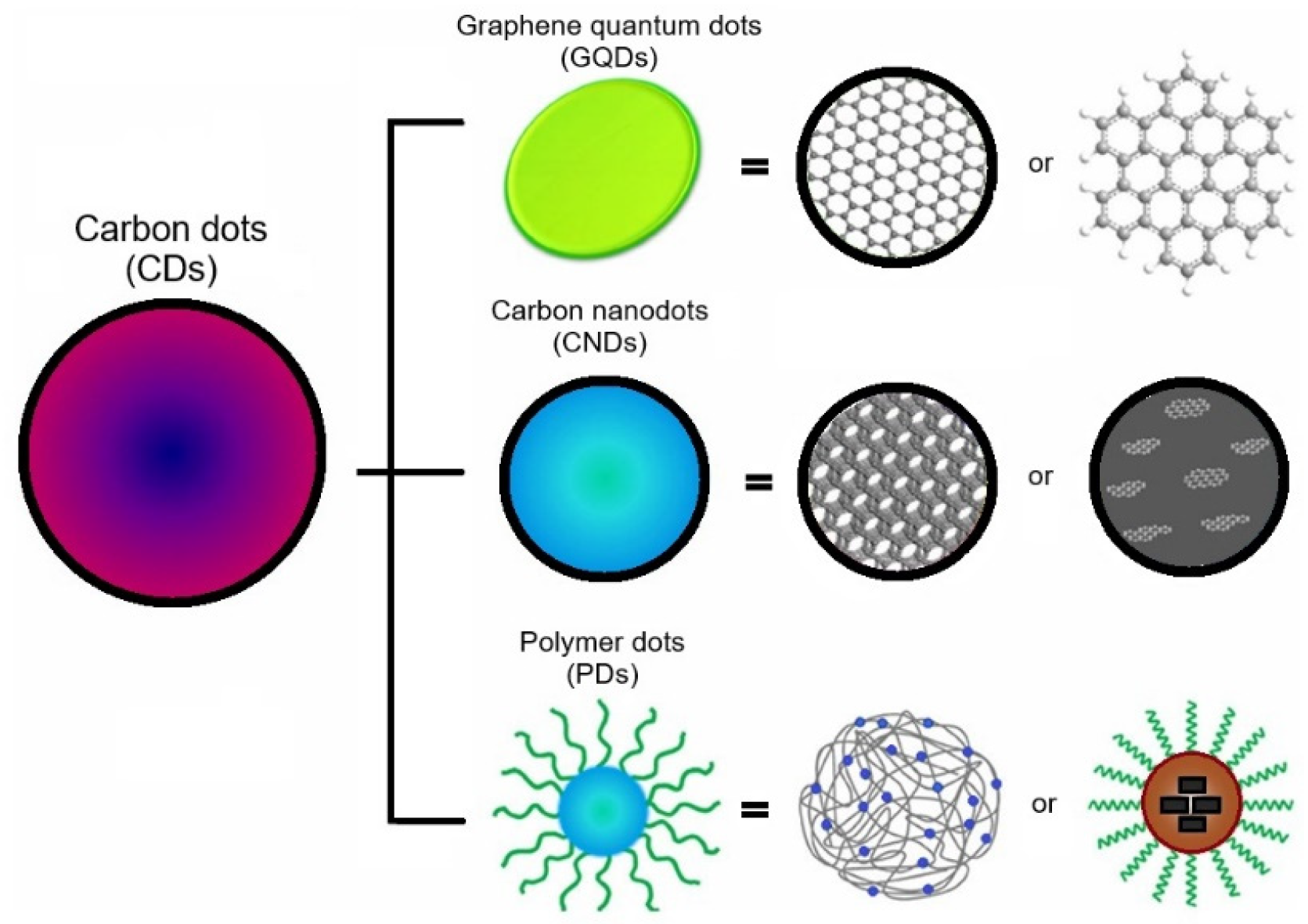
CDs are classified into three main groups, graphene quantum dots (GQDs), carbon nanodots (CNDs), and polymer dots (PDs) [103]. GQDs possess a crystalline structure and consist of a single or a few graphene layers with π conjugation. Chemically, it has more functional groups on the edges and shows an anisotropic shape, with lateral dimensions larger than the height. Mostly, these are synthesized in a circular or elliptical shape, whereas, CNDs are always spherical. CNDs are divided into two subgroups including carbon nanoparticles (CNPs) and carbon quantum dots (CQDs).
CNPs are amorphous without a clear crystal lattice and do not have any quantum confinement effect (QCE) while CQDs contain an obvious crystal lattice with sp2/sp3 carbons and have a quantum confinement effect [104,105]. CDs have various functional groups on the surface, which play a leading role in the photoluminescence (PL) behavior of these CDs. These functional groups could be oxygen-based, amino-based groups, polymer chains, etc. PDs are formed by the aggregation or crosslinking of monomers or linear polymers [106,107]. Figure 2 illustrates a typical structure of CNDs, GQDs, and PDs.
One of the most interesting properties that CDs have is their PL. It has been reported that CDs are effective in photon-harvesting in the short-wavelength region (260 to 390 nm) because of π−π* transition of C=C bonds and n-π transition of C=O bonds. It is also a known fact that CDs can emit wavelengths around the whole spectrum of visible light.
However, the PL of CDs, particularly in GQDs and CQDs, is generally influenced by the QCE and surface states. These states encompass functionalities, defects, heteroatom doping, edge configurations and synthesis methods. The photophysical characteristics of CDs, stemming from the QCE, are also significantly influenced by particle size. An increase in the size of CDs leads to a proportional growth in both absorption and emission wavelengths. This size-dependent PL phenomenon is attributed to a reduction in the bandgap, a consequence of π-electron delocalization. Thus, when a particular emission wavelength is desired for CDs, it necessitates specific shapes and sizes. Achieving this requires a meticulous selection of synthesis methods and precursors that offer control over the synthesis process for CDs [109]. Additionally, the quantum yield (QY) which is related to the emission wavelength is low for CNDs and they are more efficient in absorption of long wavelengths. Whereas, GQDs have higher QYs due to the layered structure and better crystallinity. The QYs of CDs depend on the synthesis method and carbon source. For instance, CDs prepared with aliphatic compounds as the carbon source exhibit blue-green luminescence with the QYs of 100% [110], while those prepared with aromatic compounds exhibit yellow emission [111]. CDs with red emission can be obtained by the addition of acid into the reaction mixture or using a different solvent such as DMF while the QYs are very low [112]. However, surface passivation is an efficient technique to improve the QYs and brightness of CDs [108,113]. The optical properties and PL mechanism of the CDs are extensively discussed in [102,107,113,114,115,116,117].

3.2. Synthesis and Production of Carbon Nanodots

CDs can be prepared by two main synthetic methods: “top-down” and “bottom-up” methods or in another type of classification, chemical, and physical methods. Each of these methods has some pros and cons which can be chosen regarding the application.
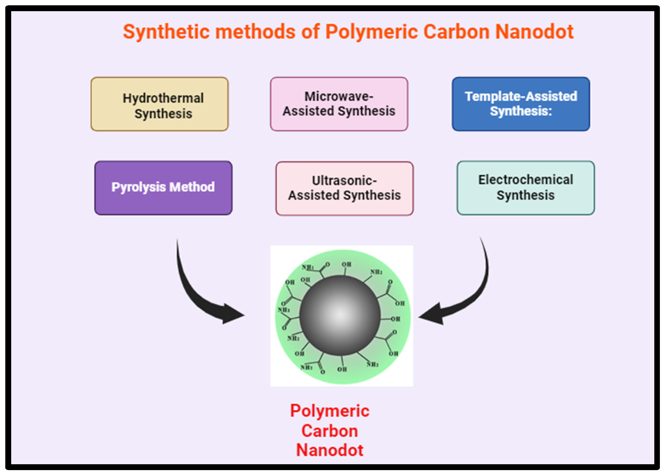
The bottom-up techniques lie in chemical synthesis and employ pyrolysis and some chemical reactions in order to carburize small organic molecules. hydrothermal treatment, microwave, thermal decomposition, template routes, plasma treatment are examples of bottom-up techniques to produce CDs in which the precursor shows lower requirements of carbon sources. Bottom-up methods have the advantages of simple surface modification in one-pot and synthesis of variety of structures and functionality of CDs. However, it is harder to modulate since side products can form that require further purification compared to top-down methods.
The top-down methods, by which CDs are generally formed through the electrochemical, chemical or physical cutting processes of relatively microscopic carbon structures, such as graphene oxide, carbon nanotubes, graphite powders etc., and obtained CDs resemble the structure of their precursors. The advantages of the top-down methods are the possibility of scaling up the production of CDs and well-defined CD structures. Top-down methods includ laser ablation, electrochemical oxidation, chemical oxidation, and ultrasonic synthesis. In the subsequent sections, a concise introduction to the selected top-down and bottom-up methods is provided.
Laser ablation is a straightforward method for producing CDs in which carbon precursor is irradiated by a laser. It is environmentally friendly and scalable production method however the energy consumption is high, QY is low, and cannot assure control over the size of the nanoparticle [118,119,120,121]. The nucleation and growth of CDs consequently its size and morphology depends on the laser pulse width. The CDs’ size and crystalline grains increased with increasing the laser pulse width [122].
In electrochemical synthesis is another top-down method which can be applied to different bulk carbon materials such as graphite rod, carbon fiber and carbon paste. In this method, two carbon-based electrodes are immersed in a water-based electrolyte. By applying redox potential, water electrolysis and H and OH radicals initiate at the edges and the defects of electrodes act s electrochemical scissors to form CDs [123,124]. Similar to laser ablation, this method is also low-cost, scalable and environmentally friendly and poor size control. However by tuning the parameters such as applied potential or starting materials CDs with narrow size distribution can be obtained [125]. The major drawback of this method is tedious purification process as the obtained CDs via electrochemical oxidation is highly hydrophilic [126].
In chemical oxidation a carbon precursor is oxidised by a strong oxidant to carbon dots. Various oxidants have been used to produce CDs such as acids (HNO3, H2SO4, NaNO3, etc), mixture of H2SO4/KMnO4/H2O2 [127], H2O2 [128] NaOH/H2O2 [129], and ozone [130]. The oxidation reaction usually requires purification in the case of acidic oxidation in order to remove the acid. Although chemical oxidation is an easy and low cost methods to produce CDs in a large scale with high QYs, lack of homogeneity in the size distribution of resulting CDs, risk of burning or explosion are its major disadvantages [131].
Using ultrasonic technology, carbon dots (CDs) are made by subjecting the reaction mixture to high-intensity ultrasonic waves. This causes cavitation and high-pressure vapor to develop, which facilitates the synthesis of CDs [132]. This approach presents several benefits, including its affordability, ease of use, and the ability to adjust emission wavelengths to suit different synthesis conditions [133]. However, the efficacy of this method relies on factors such as ultrasonic duration, intensity, and temperature. Variables such as ultrasonic duration, intensity, and temperature can influence various properties of CDs, including their size, emission wavelength, and surface characteristics. Longer ultrasonic exposure may result in larger CD sizes due to prolonged nucleation and growth, whereas higher intensities can augment cavitation intensity, impacting CD formation [134]. The method's affordability and simplicity make it a viable option for CD synthesis. Additionally, the ability to adjust emission wavelengths enhances its versatility for different applications [133]. Optimizing ultrasonic parameters can pose challenges, as it may require intricate adjustments and consume time. Furthermore, this technique may lack the precise control over reaction conditions offered by more sophisticated methods [134]. In comparison to alternative approaches, the ultrasonic method stands out for its cost-effectiveness, simplicity, and ability to customize CD properties. Nonetheless, achieving desired outcomes may necessitate meticulous parameter optimization due to the method's inherent limitations. The most common carbon sources for these techniques are carbon nanotubes, activated carbon and graphite.
Hydro/solvothermal methods are widely employed for the synthesis of carbon dots (CDs), In hydrothermal methods, an organic precursor's water solution is sealed in a hydrothermal reactor and heated to a high temperature for the reaction to occur. Whereas, in solvothermal method, organic solvents with high boiling points are used instead of water [135]. Parameters such as temperature, pressure, precursor concentration, and reaction time significantly influence CD characteristics. Higher temperatures and longer reaction times generally result in smaller CD sizes due to enhanced nucleation and growth kinetics. The advantages of hydrothermal synthesis include its low cost, eco-friendliness, and ability to produce CDs with tailored properties. However, disadvantages include the requirement for specialized equipment and potential challenges in parameter optimization [136]. Recently, spherical water-soluble CQDs (about 1-3 nm) have been prepared from lemon peel waste using a cost-effective hydrothermal approach. The produced CQDs were observed to have a quantum yield of about 14% and excellent photoluminescence properties with high aqueous stability and oxygen-rich surface functionalities. The prepared CQDs were used to design a highly sensitive fluorescent probe for the detection of Cr6+ ions with a detection limit of about 73 nM. Additionally, these CQDs were immobilized over electrospun TiO2 nanofibers. The photocatalytic activity for CQDs/TiO2 composite was seen to be about 2.5 times higher than pure TiO2 nanofibers when methylene blue dye was used as a model pollutant [137]. In another study, fluorescent CQDs (about 260-400 nm) were produced using Tamarindus indica leaves via a one-step hydrothermal treatment. These biocompatible CQDs were potentially applied in bio-imaging, disease diagnostics, sensing, and other analytical applications [138]. Citrus lemon juice was also used as a precursor of green synthesis of fluorescent CQDs (2-10 nm), through a one-pot hydrothermal approach. The prepared CQDs exhibited a 10.20% quantum yield. It was reported that the photoluminescence intensity depended on the pH of the solution and maximum intensity was obtained at pH of 6. Synthesized CQDs were also used for cell imaging and compared with the MTT assay to demonstrate its applicability as a fluorescent probe [139]. In another work, amorphous fluorescent CDs from orange waste peels were produced using the hydrothermal treatment at 180 °C. Consequently, a composite of CDs with zinc oxide was used as a photocatalyst for the degradation of naphthol blue-black azo dye under UV irradiation, and superior photocatalytic activity was reported [140].
Microwave (MW)-assisted method considered as a green bottom-up technique for CDs production, providing several benefits including simplicity, speed, and precise control. MW reactors operate on the principles of dipolar polarization and ionic conduction [141,142]. It encompasses a broad spectrum of electromagnetic waves ranging from 1 mm to 1 m, providing accelerated energies ideal for breaking down chemical bonds within precursor molecules. The manipulation of MW parameters significantly impacts the size and attributes of the synthesized CDs. Adjustments in MW power, synthesis duration, and precursor concentration influence the nucleation and growth kinetics of CDs, thereby influencing their size distribution [143]. Notably, higher MW power and shorter synthesis durations often yield smaller CD sizes owing to accelerated heating rates and rapid reaction kinetics. Moreover, variations in precursor composition and solvent selection can further adjust CD characteristics such as fluorescence emission, quantum yield, and surface properties [144]. For instance it has been observed that the QY of CDs increased with an increase in MW time while the QY decreased with decreasing of MW reaction temperature. MW is a rapid and effective and highly tunable technique for CDs production. However, the QY is low and it requires post-modification Moreover, it is high cost and energy consuming. Recently, highly stable and luminescent multicolour CQDs (4.85 ± 0.07 nm) were produced through microwave irradiation of Cydonia oblonga powder as a carbon source. Maximum emission intensity at 450 nm was observed for CQDs when it was excited at 350 nm and showed a quantum yield of 8.55% [145].
Pyrolysis/Carbonization considered as a bottom-up method for CDs production. It involves the decomposition of carbon precursors at high temperatures in the absence of oxygen. Various parameters such as precursor type, temperature, heating rate, and duration significantly influence CD size and properties. Higher pyrolysis temperatures typically lead to smaller CD sizes due to increased carbonization efficiency. The advantages of this method include its simplicity, scalability, and the ability to produce CDs with tailored properties. However, challenges may arise in achieving precise control over CD size and surface functionalities [146].
Other preparation techniques have been also proposed such as plasma treatment [147], supported synthesis [148,149,150], solution chemistry methods [151,152,153,154], cage-opening of fullerene [155], chemical vapor deposition [156], and template method [157]. The synthesized CDs, typically, gain photoluminescence properties by oxidation with nitric acid and surface-passivated by diamine-terminated organic molecules [94]. Interestingly, most of the resulting CDs exhibit blue or green emission. The synthesis route and source of carbon are important factors, which affect the size, shape, and properties of the final product. The size of CDs is very important for understanding quantum phenomena and also for biomedical applications and optoelectronics [158]. Generally, prepared CDs, regardless of their preparation technique, have a mixture of sizes, which requires complex separation methods to obtain monodisperse CDs. Some of the post-synthesis separation techniques include dialysis [159], chromatography [160,161] gel electrophoresis [94] and ultra-filtration [162]. Recently, CQDs were prepared by green ozone oxidation of lignite coal, which is abundant and cheap. In this case, also, synthesized CQDs (about 2-4 nm) were observed to be well dispersed in water, contain rich oxygen functional groups and excellent optical properties with a yield of 35%. It was observed that the fluorescence intensity of CQDs has a linear response to the Fe3+ concentration varying from 10 to 150 µmol/L. The detection limit was 0.26 µmol/L [163].

3.3. Surface Modification and Functionalization of Carbon Nanodots

Surface modification of CDs is a popular strategy to tailor their properties for specific applications and improving their performance. Surface functionalization/passivation and doping are two major techniques to chemically surface modify of CDs. The large number of CDs’ surface functional groups such as carboxylic groups, hydroxyl groups, amines, etc., make them hydrophilic and facilitate various surface functionalization and passivation. Surface functionalization/passivation of carbon dots refers to the process of treating the surface of CDs to enhance their stability, biocompatibility, and optical properties. The introduction of various functional groups imposes different defects on the CD surface affecting its excitation energy and leading to large variations in fluorescence emissions. Passivation typically involves the functionalization or coating of the surface of CDs with various organic, polymeric, inorganic or biological materials. It has several advantages: it prevents the surface defects from which the fluorescence originates; provides reactive sites for surface modification reactions and introduces new properties; enhances the potential application of CDs for specific sensing, bioimaging, drug delivery, optoelectronics and other specific tasks [164].
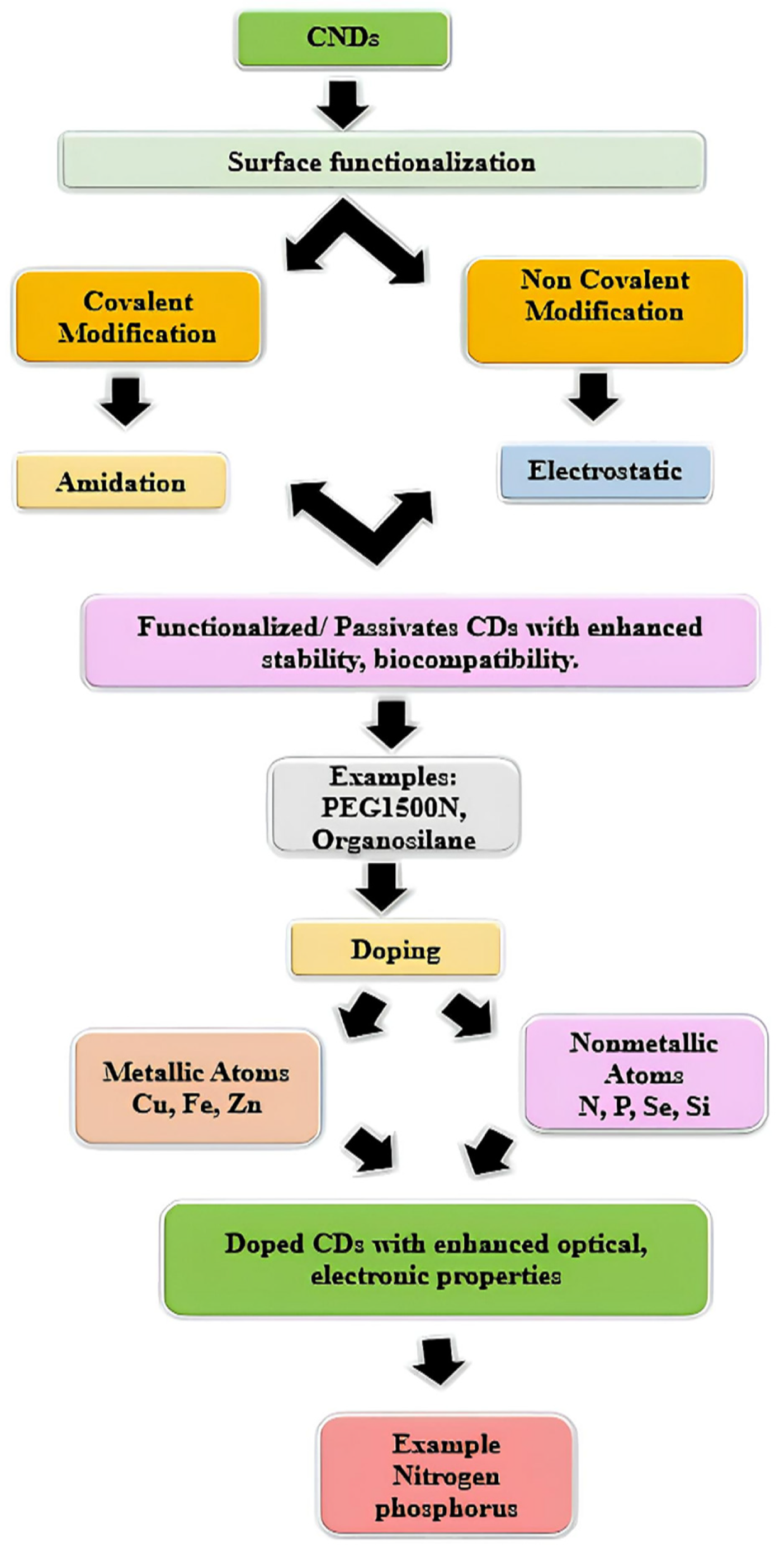
The functionalization/passivation strategies can be classified as covalent and non-covalent modification. The covalent modification includes amidation, sulylation, esterification, sulfonylation, and copolymerization, while the non-covalent modification includes electrostatic interactions, and π interaction [165]. An effective surface passivation can lead to high fluorescence intensities and high quantum yield of CDs [120]. Recently, Wang et al. have prepared CQDs with quantum yields of 60% through passivation with PEG1500N [166]. It has been reported that electrochemiluminescence activities depended on the surface passivation of CQDs. Surface passivation reduces the electrochemiluminescence activities and enhances the fluorescence properties [167]. Meanwhile, ultra-small CDs (0.9 nm) with QY of 47% have been produced through pyrolysis of anhydrous citric acid in organosilane at 240 ℃ for 1 min. The CDs functionalized with organosilane can be directly fabricated into a hybrid fluorescent film or monolith via a simple heating process, without using any additional components. It can be further converted into silica-encapsulated NPs by hydrolyzing and co-condensing the CDs with silica precursors for the biolabeling and imaging applications as well [168]. In another work, a methyl parathion sensor was established from CDs functionalized with tyrosine methyl ester through hydrothermal reaction using citric acid as a carbon precursor. The functionalized CDs show strong and stable photo-luminescence with a quantum yield of 3.8%. The obtained biosensors were used for the determination of different kinds of organophosphorus compounds [169]. Hydrothermal treatment of glucosamine produces amino-functionalized fluorescent CDs. The obtained CDs displayed stabilized green emission fluorescence at various excitation wavelengths and pH environments which are used to produce biosensors and selective detection of hyaluronidase [170]. In addition to the aforementioned modifying agents, polyethyleneimine [94], boronic acid [171], NH2-polyethylene-glycol (PEG200) and N-acetyl-L-cysteine (NAC) [172] were also used to functionalize CDs for the application in gene delivery and bioimaging, blood sugar sensing and Hg (II) sensing respectively.
Doping, is another modification technique that involves the introduction of impurities or foreign atoms into the carbon matrix. It offers a powerful means to tailor the properties of CDs and unlock new functionalities. Atomic doping enhances the florescence performance of CDs and classified into metallic atoms (such as Cu, Fe, Zn, etc.) or nonmetallic atoms ( nitrogen, phosphorus, selenium, silicon, etc.) [173]. Previous studies have demonstrated the effectiveness of doping in enhancing the optical, electronic, and catalytic properties of CDs. For instance, nitrogen doping has been shown to improve the photoluminescence quantum yield of CDs, making them more suitable for bioimaging and sensing applications. Similarly, phosphorus doping can enhance the photocatalytic activity of CDs for pollutant degradation [174]. The advantages of doping include improved stability, enhanced functionality, and tunable properties, which broaden the scope of CD applications in sensing, imaging, drug delivery, and energy conversion. However, doping may introduce defects or alter the surface chemistry of CDs, potentially affecting their biocompatibility and toxicity. Additionally, factors such as dopant type, concentration, synthesis method, and reaction conditions significantly influence the doping efficiency and resulting properties of CDs [175]. The schematic representation of the chemistry of CNDs is given in the figure below Figure 3.

4. Biomedical Applications of CNDs

CDs have been widely used for various biomedical applications due to their excellent PL properties as well as wavelength-tuneable emission properties, and unique electronic, mechanical, thermal, and chemical properties. In contrast to the traditional quantum dots (QDs) which essentially contain heavy metals are known to show toxicity and are environmentally hazardous. Whereas CDs are more safe and non-toxic towards both cell and animal levels, suggesting good environmental-friendly and biologically compatible for biomedical applications such as biosensing, bioimaging, gene and drug delivery [80,176]. In this context, in the recent past, various kinds of sensors have been designed using CDs to detect different targets such as DNA [177], heavy metals [178], glucose [179], proteins [180], H2O2 [181], nitrite [182], phosphate [183], etc. For instance, CDs prepared from flour and electrochemical carbonization of sodium citrate and urea show the PL emission which could be selectively quenched by Hg2+ with a detection limit of 0.5 nM and 3.3 nM respectively [184,185]. Other heavy metals such as Sn2+, Fe3+, Pb2+, Cr6+, Mn2+ and Cu2+ [186,187] were also detected using CDs. The CDs modified with boronic acid have been utilized for nonenzymatic blood glucose sensing. This novel sensor could detect the glucose level in the range of 9-900 µM with a detection limit of 1.5 µM. The plasma glucose concentration determined by this method was observed to be in good agreement with the values measured by a commercial blood glucose monitor which proves the high efficiency of the produced CDs-based sensor [171]. Recently, a CD-based fluorescence turn-on sensor was fabricated for hydrogen peroxide (H2O2) detection in aqueous solutions through a photo-induced electron transfer mechanism. The developed sensor exhibited good selectivity and sensitivity with a detection limit of 84 nM [188]. Table 1 presents examples of the application of CDs in biosensors.
The application of CDs as fluorescent labels for cellular imaging was first reported by Sun et al. CDs are a promising candidate for bioimaging purposes because of their low side effects and toxicity, excellent water solubility and visible-to-near infrared (NIR) emission properties [189,190,191]. Various types of cells have been imaged using CDs such as Ehrlich ascites carcinoma cells (EACs) [192], E.coli [152], HepG2 cells [192], NIH-3T3 fibroblast cells [193], HeLa cells [194], human lung cancer (A549) [195,196] etc. Jiang et al. [197] have reported the presence of photoluminescent (PL) CDs in commercial Nescafe instant coffee with a size of 4.4 nm and QY of about 5.5%. Coffee-derived CDs are directly applied in the imaging of carcinoma cells and small guppy fish without functionalization. CDs are also passivated with PPEI-EI for two-photon luminescence microscopy, used to image human breast cancer MCF-7 cells [82]. CDs exhibited bright PL both on the cytoplasm and in the cell membrane after 2h incubation at 37 ℃. The cellular uptake of CDs was temperature-dependent with no internationalization observed at 4℃. Similarly, Zhu et al. [198] have prepared a two-photon “turn-on” fluorescent probe, where CDs were used for imaging hydrogen sulfide in live cells and tissues.
Carbon dots are also proven to possess excellent gene/drug loading capability [199]. Besides their biocompatibility, nontoxicity and photoluminescence properties, small size and large surface area allow rapid cellular uptake. [200,201,202]. Recently, Lee et al. [203] have demonstrated DOX delivery in vitro and in vivo using CDs. The DOX was loaded on CDs via electrostatic interactions with 95% loading efficiency and induced death of HepG2 and MCF-7 cancer cells as well as tumours in mice. Interestingly, pure CDs preferably labeled the nucleus whereas CDs loaded with DOX were mainly distributed in the cytoplasm. Meanwhile, CD and alginate-based a smart stimuli-response drug delivery system was proposed by Majumdar et al. [204]. Here, carbon dots were coated on the surface of alginate beads and garlic extract (GE), which contains allicin, was taken as a model drug system. Interestingly, the amount of GE loaded on alginate beads coated with CDs was 60% higher than uncoated alginate beads. The loaded system shows pH-dependent controlled drug release, which results in increased therapeutic efficiency and controlled drug release upon the amount of pathogen (MRSA) present in the target. Jiao et al. [205] have developed a smart carrier for redox-responsive controlled drug delivery system by grafting carboxyl-abundant CDs to the surface of silica NPs where drug release was seen to be highly pH-dependent. Here, DOX loaded on the grafted silica have high drug loading up to 13.1% and exhibited a high cellular uptake and an excellent therapeutic effect on cancer cells by MTT (3-(4,5-Dimethylthiazol-2-yl)-2,5-diphenyl tetrazolium bromide) assay. Other than drug delivery, gene delivery has been also successfully demonstrated with positively charged CDs by Yang et al. [206]. The CDs with positive charge link to plasmid DNA easily and efficiently transfect the therapeutic plasmid into cells with low cytotoxicity. Recently, Zhang et al. [207] have demonstrated hyaluronate (HA) and PEI functionalized CDs were internalized readily into the cytoplasm of cancer cells via HA-receptor-mediated endocytosis. These functionalized CDs were found to have excellent gene condensation compatibility via electrostatic attraction and protective capacity by preventing nuclease degradation. Table 1 presents various examples of biomedical applications of CDs.
Table 1. Examples of sources, preparation techniques and biomedical applications of CD.
Sources of carbon Preparation techniques Size
(nm)
Quantum Yield (%) Color Excitation wavelength (nm) Application Ref
rose-heart radish Hydrothermal 1.2- 6.0 13.6 Blue 330 Sensing Fe+3 [208]
Prunus persica (peach) Hydrothermal 8 15 Blue 325 Cellular imaging and oxygen reduction reaction [209]
Trapa bispinosa peel Hydrothermal 5-10 0.1 Green 365 Cellular imaging [210]
Saccharum officinarum juice Hydrothermal 3 5.67 Blue 365 Cellular imaging of bacteria and yeast [211]
Unripe fruit extract of Prunus mume Hydrothermal 9 16 Blue 355 Cellular imaging [212]
Apple juice Hydrothermal 4.5 4.27 Blue 360 Imaging of mycobacterium and fungal cells [95]
Chionanthus retusus fruit extract Hydrothermal 5 9 Blue 365 Metal ion sensing and imaging of fungal cells [213]
Pseudo-stem of banana Hydrothermal 1-3 48 Green - Sensing Fe+3, Imaging of Hela and MCF-7 cells* [214]
Honey Solvothermal 2 19.18 Blue 365 Sensing Fe+3 and imaging of Hep-2 and Hela cells* [215]
Garlic Hydrothermal 11 17.5 Blue 365 Cellular imaging and free radical scavenging [216]
Sweet potato Hydrothermal 3.39 8.64 Blue 365 Fe+3 sensing and cellular imaging [217]
Walnut shell Hydrothermal 3.4 - Green 360-460 Cellular imaging [218]
Glycerine and PEG Microwave 3-4 Blue 365 Nitrite sensing [182]
Bloomed algae Microwave 8 13 Blue 365 In Vitro imaging [219]
Tissue paper Microwave 4.2 93 Blue - Determination of Glutathione [220]
Kidney beans Hydrothermal 20-30 8 green 340 Cellular imaging [221]
Water Chestnut and onion Hydrothermal 3.5 12 Green-Blue 400-600 Sensing of Cu (II) and Imaging of Coenzyme A [222]
Food waste-derived Ultrasonic 4.6 2.85 Blue-Red 400-470 In vitro bioimaging [223]
Beer Gel filtration chromatography 2.5 7.39 Blue 360 Breast cancer cell imaging and drug delivery [224]
Lignin biomass Ultrasonic and hydrothermal 2-6 21 Blue
Green
Red
310-420-540 Cellular imaging [225]
Onion waste Hydrothermal 15 28 Blue
Green
Red
408-488-561- Sensoring of Fe3+ and cellular imaging [226]
Bee pollens Hydrothermal 1-2 6-12.8 Blue-green 365 Cellular imaging and catalysis [227]
Coriander leaves Hydrothermal 2.4 6.48 green 320 Sensoring of Fe3+ and cellular imaging [228]
Grape seed Microwave 1-8 31.79 multicolor 250-550 Nucleus imaging and pH sensing [229]
Carrot Hydrothermal 2.3 7.60 Blue 365 Drug delivery [230]
Sugarcane molasses Hydrothermal 1.9 5.8 Blue 365 Sensoring of Fe3+ and cellular imaging [231]
Mango leaves Microwave 2-8 - Red 325 Cellular imaging and temperature sensors [232]
Papaya juice Hydrothermal 3 7 Blue
Green
Red
365
488
561
Cellular imaging [233]
Latex Microwave 2-8 - green 360-520 Metal sensing and cellular imaging [234]
Mangosteen pulp Hydrothermal 5 - Blue 330 Sensoring of Fe3+ and cellular imaging [235]
Lotus root Microwave 9.41 19 Blue 360 Heavy metal ion detection and cellular imaging [236]
Date kernel Hydrothermal 2.5 12.5 Blue 365 Sensing of drugs and cellular imaging [237]
Winter melon Hydrothermal 4.5–5.2 7.51 Blue 360 Cellular imaging [238]
* Hela: Human epithelial carcinoma, HepG2: Human hepatocarcinoma cells, MCF-7: Breast cancer cell.

5. Applications of Carbon Nanodots in Drug Delivery

5.1. Overview of Drug Delivery Systems

Drug delivery systems, or DDS, are essential to the progress of medicine because they aim to improve the convenience, safety, and effectiveness of therapeutic interventions. These systems entail the planning and execution of methods for releasing and transferring medications in a regulated way inside the body [239]. Their main objectives are to enhance medication pharmacokinetics, boost bioavailability, and guarantee targeted distribution to particular tissues or cells. Drug delivery systems comprise an array of methodologies, ranging from conventional oral tablets to cutting-edge nanotechnology-based carriers. This thorough analysis looks at the different kinds, intrinsic difficulties, and possible advancements in medication delivery systems in the future. In the past, oral pills, capsules, injections, and topical creams were the main traditional forms used for drug administration. These techniques had drawbacks despite their efficacy, such as unstable release profiles, low bioavailability, and poor drug solubility [240]. The development of sophisticated medication delivery systems has been prompted by the demand for more accurate and patient-friendly methods. There are several kinds of drug delivery systems. In order to increase patient compliance, oral drug administration options include both traditional tablets and capsules and controlled-release formulations. Rapid onset of action is ensured by injectable techniques like intramuscular and intravenous injections. Topical medication delivery offers localised therapies through the use of lotions, gels, ointments, and transdermal patches. Metered-dose inhalers and dry powder inhalers are two methods of inhalation medication administration that specifically target the respiratory system [241]. Liposomes, polymeric nanoparticles, and dendrimers are examples of nanoparticle-based drug delivery agents that provide targeted and controlled release. While implantable drug delivery systems, including drug-eluting implants, progressively release medications for localised treatment, microneedle-based systems enable transdermal drug delivery [242]. Approaches like antibody-drug conjugates (ADCs) and nanoparticle targeting, which improve specificity, are included in targeted medication delivery. Drug delivery systems have difficulties even with their importance. For drug carriers to be safely integrated into biological systems, biocompatibility and toxicity reduction are essential. For a treatment to be effective, it must maintain drug stability, achieve appropriate drug loading, and regulate release kinetics [243]. There are obstacles in overcoming biological barriers, like the blood-brain barrier, and immunogenicity is still an issue. Reproducibility and cost-effectiveness concerns must be addressed in order to scale up production for commercial use. Drug delivery has a bright future ahead of it with many developments and opportunities. Personalised medicine could lead to the creation of medication delivery methods specific to a person's genetic composition. Sensitive materials and sensor integration enable smart drug delivery systems to adjust to changes in the body's physiology. It is hoped that advanced nanocarriers would enable advances in gene and RNA therapy [244]. Emerging horizons include bioelectronics and digital medication delivery systems that may monitor patient responses and modify drug release accordingly. Furthermore, 3D printing technology has the potential to completely transform the way drugs are delivered by enabling the development of personalized systems with exact control over composition and structure. Drug delivery systems have experienced tremendous development and now provide a variety of methods to improve therapeutic interventions. Drug delivery technologies are changing the face of medicine, from traditional techniques to cutting-edge nanotechnology. The future will be characterized by overcoming obstacles and adopting cutting-edge technologies, with an emphasis on smart systems, personalised medicine, and cutting-edge treatments [245]. The figure given below demonstrates the various applications of Carbon Nanodot in Figure 4.

5.2. Role of Carbon Nanodots in Enhancing Drug Delivery

Utilizing the special qualities of these nanoscale carbon-based materials, carbon nanodots (CNDs) can be used to improve the delivery of drugs, opening up new possibilities in nanomedicine and addressing issues related to traditional drug administration. CNDs are interesting candidates for drug delivery applications because of their unique properties, which include biocompatibility, fluorescence, and a high surface area [246]. The capacity of CNDs to increase the solubility and bioavailability of medications is one of their main advantages. Drug research frequently encounters the challenge of poor solubility, which restricts the potency of medicinal medicines [247]. Because of its large surface area, CNDs can be used as carriers for hydrophobic medications, improving their solubility and allowing enhanced body absorption. Furthermore, one essential quality that guarantees low toxicity and compatibility with biological systems is the biocompatibility of CNDs. Drug carriers must possess this attribute since any negative effects might compromise the treatment's effectiveness and safety. Because of their biocompatibility, CNDs can be used in a variety of drug delivery methods, offering an effective way to transfer therapeutic substances with a low chance of side effects [248]. Drug delivery systems are being revolutionized by the optical features of CNDs, especially their intense fluorescence, which helps with real-time imaging and tracking. Because CNDs glow, drug carriers can be seen, making it possible for researchers to track their movements throughout the body and evaluate how well drugs are delivered. This has great potential for therapeutic applications as well as research and development, providing a way to optimize and customize drug administration based on patient reactions [249]. The function of CNDs in drug distribution is further enhanced by surface functionalization. Targeting ligands, medicinal compounds, or other biomolecules can be attached to CNDs by simply adding different functional groups on their surface. Because of their adaptability, medication carriers can be made to match the way they interact with particular cells or tissues [250]. Drug release can be regulated and targeted by engineering functionalized CNDs to react to external stimuli, such as pH changes or the presence of particular enzymes. This degree of accuracy in medication administration has a key role in reducing side effects and raising the therapeutic index of medications. Because CNDs are tiny, interactions at the cellular and molecular levels are facilitated, which is beneficial for drug delivery. They can cross physiological barriers, such as the blood-brain barrier, which is a difficult therapy issue for neurological illnesses, but due to their nanoscale size it is possible. The delivery of medications to the central nervous system by CNDs shows promise in treating diseases like brain tumours and neurodegenerative disorders [251]. Beyond conventional small molecule medications, CNDs show promise in providing a broad spectrum of therapeutic substances. They are appropriate transporters for proteins, peptides, and nucleic acids (such DNA and RNA) due to their flexibility. This skill is especially important in the emerging field of gene and RNA therapies, because successful interventions depend on the targeted delivery of genetic material. The potential of CNDs in developing nucleic acid therapies and biologics is highlighted by their capacity to distribute and protect sensitive biomolecules. Another domain in which CNDs excel is the recently developing subject of targeted medication delivery. The surface of CNDs can be functionalized with certain ligands to enable the construction of drug carriers that bind to target cells or tissues with preference [252]. By using a targeted strategy, medication distribution becomes more particular and therapeutic agents are delivered to their intended location more precisely. For example, CNDs can be designed to precisely target cancer cells during cancer treatment, reducing harm to healthy tissues and enhancing the overall therapeutic result. The special combination of characteristics that carbon nanodots possess biocompatibility, fluorescence, surface functionalization abilities, and nanoscale dimensions defines their role in improving drug delivery. Their ability to tackle issues related to traditional drug administration is facilitated by these characteristics. Carbon nanodots have the potential to completely transform drug delivery systems as this field of study develops, providing cutting-edge approaches for the precise, targeted, and controlled distribution of therapeutic substances in a variety of medical applications [253]. The following figure demonstrates the role of CNDs in Drug delivery enhancement in Figure 5.

5.3. Mechanisms of Drug Delivery Using Carbon Nanodots

With the advent of nanotechnology, the field of drug delivery has experienced tremendous developments, and among the different types of nanomaterials, carbon nanodots (CNDs) have attracted a lot of interest. These carbon-based materials at the nanoscale, which usually have special qualities that make them interesting options for improving drug delivery systems. We explore the complex mechanisms behind CND-assisted drug delivery in this extensive study, looking at how they can enhance solubility, biocompatibility, surface functionalization, real-time imaging, and targeted drug administration [254].

5.3.1. Increasing Bioavailability and Solubility

The main way that CNDs help with drug distribution is by solving the problem of poor solubility, which is a problem that many pharmaceutical compounds have. Because of their distinct surface chemistry and large surface area, CNDs are used as carriers for hydrophobic medications. Because the CND surface is hydrophilic, stable connections between hydrophobic medicines and it are made more soluble. This improved solubility therefore helps to raise the drug's bioavailability, which makes sure that a higher percentage of the medication is absorbed into the bloodstream and increases its therapeutic effect [255].

5.3.2. Decreased Toxicity and Biocompatibility

In order for any drug delivery system to be successful, biocompatibility is essential. One important factor in the success of CNDs in drug delivery applications is their innate biocompatibility. Because of their guarantee of low immunogenicity and cytotoxicity, CNDs are appropriate for use in biological system interactions. Because CNDs are compatible with biological settings, there is a lower chance of side effects, making them a safe and well-tolerated drug delivery platform. The effective integration of CND-based drug delivery systems into clinical applications depends in particular on this mechanism [256].

5.3.3. Imaging and Tracking in Real Time

The potent fluorescence that CNDs display is utilised as a potent tool for tracking and imaging in real time during drug delivery. This optical characteristic makes it possible to monitor drug carriers inside the body non-invasively, giving important information on how they are distributed, accumulate, and clear out of the system. One important factor in the optimisation of drug delivery systems is the real-time imaging capacity of CNDs. Researchers are able to see drug carriers' travel and make sure they arrive at their destinations precisely. Clinical applications where customised and site-specific treatments may be made based on individual patient responses appear to be promising applications of this mechanism [257].

5.3.4. Functionalization of Surfaces

One important method that increases CNDs' adaptability in drug delivery applications is surface functionalization. Different functional groups can be added to the surface of CNDs with ease, enabling the attachment of medicinal compounds, targeted ligands, or other biomolecules. Drug carriers with particular affinities to target cells or tissues can be designed because of their customisation, which enhances drug delivery selectivity and reduces off-target effects. Additionally, functionalized CNDs can be designed to react to outside stimuli like pH variations or the presence of particular enzymes, allowing for precise drug delivery at the intended location. Personalising the surface characteristics of CNDs is a potent way to achieve precise medication distribution [258].

5.3.5. Nanoscale Aspects for Improved Cellular Communication

One essential factor that promotes interactions at the nanoscale and allows for improved cellular connections is the small size of CNDs. Because of their nanoscale size, CNDs can pass across blood-brain barriers (BBB) and cell membranes, among other physiological barriers. Because the BBB prevents many therapeutic substances from entering the central nervous system, breaking through it is especially difficult when it comes to drug delivery. CNDs show promise in delivering medications to the brain because of their small size and ability to modify surfaces, creating novel possibilities for the treatment of neurological illnesses. CNDs can be useful instruments for cellular and molecular drug delivery that is both efficient and targeted [259].

5.3.6. Transport of Different Therapeutic Agents

Beyond conventional small molecule medications, CNDs have an adaptable mechanism that allows them to transport a variety of therapeutic substances, such as proteins, peptides, and nucleic acids (DNA and RNA). This feature is especially noteworthy in the quickly developing field of gene and RNA therapeutics, where precise genetic material delivery is essential for effective interventions. For the safe and effective distribution of delicate biomolecules to target cells while maintaining their structural integrity and functionality, CNDs offer a robust and biocompatible platform. CNDs are useful instruments for developing the fields of nucleic acid therapies and biologics because of their capacity to carry a variety of therapeutic payloads [260].

5.3.7. Mechanisms for Targeted Drug Delivery

A paradigm-shifting mechanism made possible by CNDs is the targeted drug delivery strategy, which advances precision medicine. Researchers may develop CND-based drug carriers with particular ligands that bind to target cells or tissues selectively by surface functionalization. Therapeutic substances are more precisely delivered to their intended location due to this focused mechanism, which also improves drug delivery specificity. To improve the overall therapeutic success and minimize harm to healthy organs, CNDs can be designed to specifically target cancer cells during cancer treatment. Targeted drug delivery helps improve overall patient outcomes by lowering systemic exposure and increasing the therapeutic index of medications. This could reduce negative effects [261].

6. Phytomedicine-Loaded Carbon Nanodots

Carbon nanodots loaded with phytomedicine are a state-of-the-art example of how nanotechnology and natural medicine can work together to overcome many obstacles in traditional phytomedicine delivery. By utilising the special qualities of carbon nanodots, this novel synergy improves the bioavailability, targeted administration, and general efficacy of chemicals originating from plants [262].

Synthesis and Characterization

The process of creating carbon nanodots loaded with phytomedicine is a laborious one that usually uses hydrothermal or other specialised techniques. These nanodots are perfect carriers for phytomedicines because of their unique surface functions and nanoscale size. Carbon nanodots' surface characteristics can be adjusted to best encapsulate and distribute different kinds of bioactive substances [263].

Improved Bioavailability

When it comes to overcoming the bioavailability constraints of phytomedicines, carbon nanodots are essential [264]. These nanodots improve the solubility of hydrophobic components and stop them from aggregating and degrading by conjugating or encasing plant-derived chemicals. The enhancement of solubility has a noteworthy impact on the bioavailability of phytomedicines loaded, guaranteeing effective absorption and dispersion throughout the biological system [265].

Targeted Drug Delivery

One of the main benefits of personalised medicine is the ability to precisely target particular tissues or cells through the functionalization of carbon nanodots [266]. By interacting with particular biomolecules or receptors, surface changes let the loaded phytomedicines reach their targeted sites of action. By reducing adverse effects and off-target consequences, this focused administration maximises therapeutic efficacy.

Controlled Release Kinetics

Carbon nanodots’-controlled release properties elevate the delivery of phytomedicine to a higher level. These nanodots allow for a controlled and prolonged release of the loaded phytomedicines by varying the surface characteristics or adding responsive components. By preserving ideal medication concentrations for prolonged periods of time, this controlled release profile improves therapeutic effects and may lessen the frequency of administration [267].

Biocompatibility and Safety

Because of their low toxicity and great biocompatibility, phytomedicine-loaded carbon nanodots are safe to use in biological systems. The effects of these nanodots on cellular architecture, metabolic pathways, and general physiological functions are the subject of much research [268]. Carbon nanodots' biodegradable nature adds to their safety profile and allays worries about long-term exposure.

Intracellular Delivery and Cellular Uptake

Enhancing cellular absorption is a critical component for intracellular targets, and carbon nanodots' nanoscale size makes this possible. It is possible to effectively transport phytomedicines over cellular barriers, guaranteeing their delivery to targeted cell compartments. The greater potency and overall bioavailability of the loaded phytomedicines are a result of this increased cellular absorption [269].

Encapsulation of Diverse Phytochemicals

Carbon nanodots loaded with phytomedicine are adaptable vehicles that can hold a wide variety of phytochemicals, such as polyphenols, terpenoids, alkaloids, and flavonoids. This adaptability broadens the application of this delivery technology across different herbal medicines by enabling the encapsulation of complex mixes present in medicinal plants [270].

Application in Multicomponent Phytomedicine Formulations

Phytomedicine-loaded carbon nanodots present an opportunity to formulate complicated, multicomponent delivery systems in the context of traditional herbal treatments that contain multiple active components. By delivering synergistic plant-derived components simultaneously, this method can enhance therapeutic effects and replicate the holistic aspect of conventional herbal preparations.

Future Perspectives and Challenges

Natural therapies could undergo a revolution with the introduction of carbon nanodots loaded with phytomedicine into the medication delivery system. Research is still ongoing in several areas, including scalability, standardised synthesis techniques, and long-term safety assessments. Subsequent efforts will concentrate on optimizing these delivery systems based on nanodots for broad clinical use, with a focus on regulatory compliance and reproducibility [271]. To sum up, the creation of carbon nanodots loaded with phytomedicines is a cutting-edge in the field of sophisticated medication administration. With its capacity to target delivery, ensure regulated release, and overcome bioavailability limitations, this novel technique has enormous promise to further the medicinal applications of chemicals produced from plants. The current research in this area marks the beginning of a new era in which the combination of natural medicines and nanotechnology creates opportunities for improved treatment results.

6.1. Loading Strategies for Phytomedicine

Recent studies have thrown light on novel strategies and developments in this emerging field, offering insightful information about phytomedicine loading tactics onto carbon nanodots. Modern technologies and a greater comprehension of the interactions between nanomaterials have led to the adaptation and improvement of these tactics, which are essential for maximizing the effectiveness of medicine administration.

Physical Adsorption: Recent Developments

Current research has looked into creative approaches to improve phytomedicine's physical adsorption onto carbon nanodots [272]. Through specialised chemical interactions, surface modification of nanodots with particular functional groups, including amino or hydroxyl groups, has been studied to increase adsorption affinity [273]. Furthermore, sophisticated characterisation methods have been used to accurately comprehend and control the non-covalent forces regulating adsorption, such as spectroscopic studies and computational modelling. The goal of these improvements is to get over issues with adsorption strength variability and environmental sensitivity.

Covalent Bonding: Progress in Precision

The goal of covalent bonding strategy advancements has been to increase loading process precision. Researchers have looked into site-specific functionalization of carbon nanodots to facilitate selective covalent interaction with certain areas of phytomedicine molecules [274]. This focused strategy reduces inadvertent changes and maintains the integrity of the loaded chemicals. Furthermore, current studies have focused on the creation of green chemistry strategies that maximise covalent bonding without sacrificing the phytomedicine's therapeutic qualities [275]. These strategies make use of environmentally friendly solvents and reaction conditions.

Encapsulation during Nanodot Synthesis: Tailoring Synthesis Conditions

Recent developments in the field of encapsulation during nanodot synthesis have focused on optimising synthesis conditions for enhanced control. Novel techniques like template-assisted procedures and microfluidic-assisted synthesis have been investigated to obtain a more accurate phytomedicine compound encapsulation. These methods address issues related to consistent distribution and possible changes in the therapeutic qualities of loaded phytomedicine during synthesis, providing scalability and reproducibility [276].

Emerging Trends and Future Directions

There is an increasing interest in using computer modelling and sophisticated nanomaterial engineering to forecast and optimise loading interactions, according to recent research trends. The complex dynamics of phytomedicine-nanodot interactions are being understood and optimal loading configurations are predicted through the use of computational simulations and machine learning techniques. Furthermore, new developments in surface modification methods, such molecular imprinting and plasma treatment, have the potential to improve loading strategy selectivity [277]. By enabling the development of customised binding sites on nanodot surfaces, these methods guarantee a more effective and regulated loading of phytomedicine components. It is expected that a better comprehension of nanomaterial behaviour and advanced loading procedures would aid in the creation of next-generation drug delivery systems as research at the nexus of nanotechnology and phytomedicine advances. In situ characterization techniques combined with real-time monitoring approaches have the potential to significantly transform our capacity to precisely regulate and optimise phytomedicine loading onto carbon nanodots, creating new opportunities for individualised and focused therapeutic interventions.

6.2. Stability and Bioavailability Enhancement

Recent work on phytomedicine-loaded carbon nanodots has yielded important advances to improve the stability of encapsulated bioactive chemicals as well as novel approaches to increase bioavailability. These developments, which are based on cutting-edge techniques and technologies, provide an early look at more reliable and effective medication delivery methods in the future.

Enhanced Stability through Nanoengineering

Utilising cutting-edge nanoengineering methods to increase the stability of carbon nanodots loaded with phytomedicine has been the focus of recent research. It has been investigated to produce protective shells around the nanodots by nanoencapsulation techniques, such as lipid-based formulations and polymeric coatings, to shelter the encapsulated phytomedicine from environmental stressors [278]. Furthermore, surface alterations like the application of silica or graphene oxide coatings have shown improved stability, delaying premature deterioration and maintaining the bioactivity of the loaded chemicals.

Smart Nanomaterials for Controlled Release

Recent studies have investigated the incorporation of smart nanomaterials into carbon nanodots as a means of achieving controlled and sustained release mechanisms. The creation of intelligent drug delivery systems has sparked research into stimuli-responsive nanocomposites, such as pH-sensitive polymers and temperature-responsive hydrogels. These technologies provide customised and on-demand delivery of phytomedicine components by precisely controlling release kinetics in response to particular physiological situations.

Advances in Nanotoxicology and Biocompatibility

Recent study has focused on ensuring the safety and biocompatibility of carbon nanodots loaded with phytomedicines. Utilising cutting-edge imaging methods including live-cell imaging and super-resolution microscopy, advanced nanotoxicology investigations have thoroughly evaluated the interactions between nanodots and biological systems at the cellular and subcellular levels [279]. This thorough knowledge has facilitated the creation of safer nanomaterials, reducing the possibility of cytotoxic effects and opening the door for their use in biomedicine.

Precision Bioavailability Enhancement

Advancements in bioavailability enhancement tactics in recent times have utilised precision approaches, which involve customised surface changes and carrier interactions. Improvements in lipid nanoparticle and micelle technologies have served as an inspiration for the development of nanoscale drug delivery vehicles, which have shown to better solubilize hydrophobic phytomedicine components and increase their bioavailability [280]. To further maximise bioavailability, studies have also looked at the use of ligand-targeted nanocarriers, which take use of ligand-receptor interactions to enable tailored distribution and uptake.

Emerging Trends and Future Directions

Trends in stability and improving bioavailability are moving in the direction of a multidisciplinary strategy. The amalgamation of computational modelling, artificial intelligence, and systems biology is increasingly common in order to forecast and enhance the behaviour of nanomaterials in intricate biological contexts. Biosensors and imaging modalities are among the real-time monitoring approaches that are being used to offer dynamic insights into the in vivo fate of carbon nanodots loaded with phytomedicine. Another well-known trend in nanotechnology is the hunt for "green" materials. To address sustainability issues, researchers are investigating biodegradable nanomaterials and ecologically friendly synthesis techniques [281]. The goal of surface modification innovations like bioinspired coatings and biomimetic methods is to produce nanocarriers that are less immunogenic and more biocompatible. As the field advances, it is anticipated that these recent research insights and technological innovations will propel phytomedicine-loaded carbon nanodots towards broader clinical applications, offering more effective, safe, and targeted therapeutic interventions.

6.3. Controlled Release Mechanisms

With a focus on accuracy, flexibility, and responsiveness, recent research has made significant advancements in the field of controlled release mechanisms for carbon nanodots loaded with phytomedicine. The field of controlled drug delivery is being shaped by cutting-edge technologies and creative approaches, which provide insights into how nanotechnology might be used to maximise therapeutic results.

Advanced Surface Engineering for Tuneable Release

In order to give carbon nanodots variable releasing capabilities, recent investigations have investigated sophisticated surface engineering techniques [282]. Dynamic changes in release kinetics are possible by functionalizing the nanodot surface with stimuli-responsive polymers or biomimetic coatings. The focus has been on pH-responsive systems in particular, which allow for controlled release in response to variations in pH that occur in various physiological compartments. The accuracy attained with these surface alterations guarantees a customised release profile in line with therapeutic requirements.

Microfluidic-Assisted Synthesis for Controlled Encapsulation

One notable development in the field of encapsulation techniques is microfluidic-assisted synthesis. By allowing for exact encapsulation of phytomedicine components within carbon nanodots, this approach offers tight control over the production process. The continuous and uniform mixing of reactants made possible by microfluidic devices produces nanodots with consistent characteristics [283]. This degree of control makes drug-loaded nanodots more reproducible and helps to ensure a more regulated and predictable release of phytomedicine.

Temperature-Responsive Systems for On-Demand Release

Temperature-responsive components incorporated into carbon nanodots have been the subject of recent studies, allowing for on-demand medication delivery [284]. These devices use the natural temperature fluctuations that occur in physiological settings to initiate regulated release. It is now possible to adjust the rate of drug release in nanodot formulations in response to particular thermal cues by using thermoresponsive polymers or materials that are susceptible to external heat sources. This strategy has potential for use in situations where exact control over release dynamics is essential.

Advancements in Biodegradable Nanocarriers

A crucial factor in the development of controlled release systems is biodegradability. The creation of biodegradable nanocarriers that can deliver phytomedicine in a regulated manner as they decompose over time has been the focus of recent developments. Researchers have looked into using biocompatible materials, including certain polymers and lipid-based nanoparticles, to make nanocarriers that break down gradually. This allows the release kinetics to be in line with therapeutic requirements and reduces the possibility of long-term accumulation issues [285].

Emerging Trends and Future Prospects

A convergence of disciplines characterises emerging developments in controlled release mechanisms for carbon nanodots loaded with phytomedicine. Predicting and optimising release patterns based on a variety of criteria, such as pharmacological qualities, patient-specific data, and nanomaterial features, is becoming more popular by integrating nanotechnology with AI and machine learning [286].
In addition, the latest developments in real-time monitoring methods, such biosensors and in vivo imaging, provide dynamic insights into the spatiotemporal behaviour of nanodots inside the body [287]. This real-time feedback loop makes adaptive release strategy modifications possible, improving the efficacy and adaptability of controlled drug delivery systems.
Emerging trends in the field suggest that regulated release mechanisms for carbon nanodots loaded with phytomedicines may change as a result of these recent discoveries and technological advancements. This will present prospects for personalised and precision medicine with improved therapeutic precision.

7. Polymeric Carbon Nanodots

7.1. Introduction to Polymer Nanocomposites

Over the past two decades, considerable scientific and technological interest has centred on polymer nanocomposites (PNCs). Initially, focus was primarily on understanding synthetic and physical chemistry, colloidal particle physics, and various properties of these materials. Recently, there has been a shift towards comprehending and harnessing the unique physics of polymers within PNCs. This shift is driven by the realization that achieving truly engineered and functional nanocomposites requires a deeper understanding of their structure-property-processing relationship. Modern developments in the realm of nanomaterials include polymeric carbon nanodots (CNDs) and polymer nanocomposites, each of which offers a distinct set of qualities and uses. Since they are so adaptable, polymeric CNDs which are essentially nanoscale carbon-based particles have attracted a lot of attention. These nanoparticles, which are useful for applications including bioimaging, sensors, and drug administration, usually have a size range of a few to roughly 10 nanometers [288]. They also have unique characteristics like high photoluminescence. By adding nanoscale fillers, such as nanoparticles or nanotubes, to polymer matrices, however, material characteristics can be improved, as in the case of polymer nanocomposites. Superior mechanical, thermal, and electrical properties are added to the resultant composite materials by the interaction of polymers and nanofillers [289]. Polymeric CNDs are distinguished by their distinct composition and structure. The majority of the carbon atoms in the core are grouped either crystalline or amorphously, resembling graphene. Functional groups including hydroxyl, carboxyl, and amino groups are present on the surface of these nanodots, which enhances their solubility, stability, and reactivity. The size and characteristics of the nanodots are affected by the many techniques used in the synthesis of polymeric CNDs, including hydrothermal synthesis, microwave-assisted synthesis, and template-assisted synthesis [290]. Conversely, to produce materials with improved performance, polymers are blended with nanoscale fillers to make polymer nanocomposites. When nanofillers are added, the material behaves differently from a pure polymer, providing increases in stiffness, strength, and other important characteristics. Polymeric CNDs are distinguished by their distinct composition and structure. The majority of the carbon atoms in the core are grouped either crystalline or amorphously, resembling graphene [291]. On the other hand, polymer nanocomposites comprise a wider range of materials in which nanofillers enhance overall performance and polymers function as matrices. The addition of nanofillers improves the composite's mechanical, thermal, and electrical qualities, making it appropriate for use in the automotive, electronics, and aerospace industries. Melt blending, solution mixing, and in-situ polymerization are the synthesis techniques used to create polymer nanocomposites; each has an impact on the properties of the finished product [292].

7.2. Structural Composition

Polymer nanocomposites' structural elements are distinguished from traditional composites by the accurate dispersion of nanofillers inside the polymer matrix. In this nanoscale configuration, the polymer matrix is finely interwoven with nanofillers, which can be nanoparticles, nanotubes, or other nanomaterials. Since it has a significant impact on the composite material's overall performance, this careful dispersion is essential. It's a laborious process with major consequences to distribute nanofillers uniformly throughout the polymer matrix. Given that they are nanoscale, the nanofillers are smaller than those that are usually employed in traditional composite materials. Its reduced size makes it possible for the polymer to be more thoroughly and uniformly integrated, which enhances its desirable qualities. A uniform dispersion of the nanofillers highlights the enhanced attributes, which include greater stiffness, strength, and thermal stability. This structural configuration at the nanoscale contrasts sharply with traditional composites, where the fillers are frequently bigger and less uniformly distributed. Uniform dispersion is more difficult to achieve in typical composites, and the unequal distribution of the filler elements can lead to variances in the properties of the final material. By optimising the advantages of the nanofillers and enhancing the material's overall performance, Polymer Nanocomposites' nanoscale accuracy guarantees that the improved properties are consistently manifested throughout [293].

7.3. Various Synthetic Methods of Polymeric Carbon Nanodots

The process for generating polymeric carbon nanodots (CNDs) involves a range of techniques designed to produce carbon particles at the nanoscale with particular characteristics. Many well-established methods are used to synthesise these polymeric CNDs, which are preferred for their improved stability, biocompatibility, and flexible functionalization potential. In order to control particle size and surface functions, hydrothermal synthesis requires exposing carbon precursors to high temperatures and pressures in an aqueous environment [279]. By using microwave irradiation to speed the reaction kinetics, however, microwave-assisted synthesis produces polymeric CNDs with regulated characteristics quickly and effectively. Using templates like polymers or proteins, template-assisted synthesis allows for fine control over the morphology of the CNDs during production by dictating their size and shape [294]. Pyrolysis is a versatile process that uses a variety of carbon-rich sources and offers sustainability by carbonising organic precursors which are often sourced from biomass under regulated circumstances. Using ultrasonic vibrations, bigger carbon structures can be gently and energy-efficiently broken down into nanoscale CNDs through the process of ultrasonic-assisted synthesis. With electrochemical synthesis, one can precisely regulate the synthesis conditions by inducing the creation of polymeric CNDs with the use of electrical energy [295]. Lastly, under benign and eco-friendly circumstances, photochemical synthesis uses light as an energy source to start reactions that result in the synthesis of polymeric CNDs. The resultant polymeric CNDs are adaptable for a variety of applications, including biological imaging, sensors, and optoelectronics, and can be further characterised and functionalized. The desired characteristics and planned uses of the polymeric CNDs inform the synthesis technique selection, which reflects the dynamic and varied nature of the nanomaterial sector. Various methods of synthesis of Polymeric Carbon Nanodots are shown in Figure 6.

7.3.1. Role of Polymer Nanocomposites in Drug Delivery

By addressing issues with traditional drug delivery systems, these nanocomposites which are made of polymers and nanoscale materials have opened the door for more accurate and regulated drug release. The potential of polymer nanocomposites to encapsulate and shield medications, preventing their untimely breakdown or excretion from the body, is one of its main benefits. This is especially important for medications that are difficult to degrade in an adverse biological environment or have low bioavailability. The medication is protected from the environment by nanocomposites, which also guarantee its stability until it reaches the intended location [296]. It is possible to modify the drug delivery system's characteristics by adding elements at the nanoscale, like nanoparticles, to polymer matrices. Some features, including targeting, can be built into these nanoparticles to allow the nanocomposites to selectively aggregate at the sick tissue while sparing healthy cells. By minimising systemic side effects, this focused drug administration improves the overall therapeutic result [297]. In addition, these composites' nanoscale size makes it easier for them to pass across biological barriers like blood-brain barriers and cell membranes. This makes it possible to administer medications to regions that were previously unreachable or difficult to treat, so broadening the range of illnesses that can be treated, including neurological conditions. In order to achieve controlled and prolonged medication release, polymer nanocomposites are also essential [298]. The release kinetics of the medication enclosed can be precisely tailored by researchers by carefully crafting the polymer matrix and modifying the composition of the nanoparticles. This helps maintain therapeutic concentrations over an extended length of time, lowering the frequency of dose and increasing patient compliance. It is especially helpful for chronic illnesses that call for continuous and controlled drug administration. The capacity of polymer nanocomposites to encapsulate a broad variety of medicinal substances, such as proteins, nucleic acids, and tiny molecules, is another example of their versatility [75]. The creation of multifunctional drug delivery devices that can administer combination medicines is made possible by this flexibility. For example, a nanocomposite may carry a gene therapy payload and a chemotherapeutic medication at the same time, resulting in a synergistic effect that improves treatment outcomes. Another crucial component of polymer nanocomposites in drug administration is their biodegradability [299]. The fact that a lot of these polymers break down gradually into inert byproducts helps to prevent the delivery system from building up within the body. This feature is especially helpful since it reduces the possibility of long-term side effects and permits the carrier to be cleared after its medication delivery purpose is completed [300]. Drug delivery has advanced even further with the creation of stimuli-responsive polymer nanocomposites. These intelligent materials are able to react to particular bodily stimuli, such variations in pH, temperature, or the presence of enzymes. This responsiveness improves accuracy and lessens off-target effects by enabling triggered medication release at the target spot [301]. For example, a medicine encapsulated in a nanocomposite that is made to withstand the acidic environment of tumours can release the drug only in the malignant tissue. Additionally, polymer nanocomposites have shown great promise in addressing drug resistance, a critical obstacle in the treatment of several illnesses, most notably cancer. These composites' multifunctionality enables the co-delivery of medications with various modes of action, addressing several drug resistance routes and raising the possibility of successful therapy. To sum up, polymer nanocomposites have a revolutionary function in drug delivery and provide a host of benefits over conventional drug delivery methods. These nanocomposites offer a platform for accurate, focused, and managed medication delivery. Future customised and extremely successful therapeutic interventions appear to be in the cards as this field of study progresses and more complex polymer nanocomposites are created [302].

7.3.2. Properties of Polymeric Carbon Nanodots

Polymeric carbon nanodots have outstanding photoluminescence, which is one of their prominent features. In the presence of visible or ultraviolet light, these nanodots can release visible light. Because CNDs emit light and can be adjusted in intensity, this feature is especially useful for bioimaging and fluorescence imaging applications, which require sensitive and accurate identification of biological processes and structures. Additionally, the capacity to regulate the emission wavelength. When these nanodots are subjected to ultraviolet or visible light, they can release visible light. The strong and adjustable emission of CNDs makes it possible to precisely and sensitively identify biological structures and processes, making this trait very beneficial for applications in fluorescence imaging and bioimaging [303]. Their further usefulness in multicolour imaging and diagnostics is increased by the controllable emission wavelength. Biocompatibility is another well-known characteristic of polymeric CNDs, which is essential for biomedical applications. Their composition guarantees low cytotoxicity and immunogenicity; they are frequently made from biocompatible polymers or carbon precursors [304]. Because of this, they can be used in a variety of biological settings, such as medication delivery, imaging in vivo and in vitro, and other biomedical applications. Polymeric CNDs have the potential to be safe and useful medical instruments because of their biocompatibility. The adaptable surface chemistry of polymeric carbon nanodots is another significant characteristic. It is simple to alter or functionalize the surface of CNDs with different groups, such as carboxyl, amino, or hydroxyl groups. The CNDs can be given extra functions by attaching particular ligands, biomolecules, or medications due to their adjustable surface chemistry. Functionalized polymeric CNDs exhibit great adaptability to individual requirements, as they can be customised for specialised applications like as sensing, targeted medication delivery, or other uses [305]. Excellent water solubility is another feature of polymeric CNDs that makes them useful for biological and environmental applications. Because of their hydrophilic character, these nanodots are easily incorporated into biological systems by dispersing in aqueous solutions. This solubility is especially useful in drug administration applications, where stable physiological conditions and effective dispersion are essential for effective therapeutic results. Moreover, polymeric CNDs show stability under a range of environmental circumstances. Because of their resilience, they can tolerate variations in pH, temperature, and light exposure, which guarantees their performance and functioning over time. The practical applications of CNDs in a variety of sectors, from electronics to biology, depend on this stability [306]. One of the notable characteristics that helps explain polymeric carbon nanodots' consistent and predictable performance is their homogeneity in size and shape. The manufacturing of nanodots with precise sizes and shapes is made possible by the controlled synthesis techniques used in their creation [307]. For uses like catalysis, sensing, or electrical devices where exact control over the characteristics of nanomaterials is critical this uniformity is vital [308]. To sum up, polymeric carbon nanodots have a number of amazing qualities that make them very appealing for a variety of uses. Their promise in several sectors like materials science, biomedical research, and sensing technology is enhanced by their photoluminescence, stability, water solubility, biocompatibility, and size uniformity. The special qualities of polymeric CNDs are anticipated to open up even more creative uses as research in this field develops, solidifying their status as useful nanomaterials across a wide range of scientific and technical fields [309].

7.4. Applications of Polymeric Carbon Nanodots in Drug Delivery

As a result of its distinct qualities and range of functions, polymeric carbon nanodots (CNDs) have become a viable nanomaterial for drug delivery applications [310]. The use of polymeric CNDs in drug delivery has been discussed, along with their applications and underlying mechanics.
Applications for Drug Delivery Using Polymeric Carbon Nanodots:
  • Delivery of Drugs with Specificity:
To distribute drugs in a targeted manner, polymeric CNDs can be functionalized with certain ligands, including peptides or antibodies. Target cells or tissues' surface receptors are the only receptors that the functionalized nanodots can bind to with selectivity. The tailored strategy improves overall treatment efficacy, minimises off-target effects, and concentrates the therapeutic payload at the intended site to increase drug delivery efficiency [311].
  • Imaging and Diagnosis:
Polymeric CNDs are perfect for imaging and diagnostic applications due to their exceptional photoluminescent characteristics. CNDs can allow for real-time medication delivery process monitoring when they are utilised as imaging agents. Furthermore, the vivid and adjustable fluorescence of CNDs can help see biological structures, which can help with illness monitoring and diagnostics.
  • Theranostics:
The potential for polymeric CNDs to integrate therapeutic and diagnostic functions onto a single platform is known as theranostics. Researchers can create systems that not only carry medications to the target region but also provide real-time feedback on the effectiveness of the treatment by combining therapeutic chemicals and imaging probes into the CNDs. Precision therapies and personalised treatment will be greatly impacted by this integrated approach [312].
  • pH-Responsive Drug Release:
It is possible to incorporate pH-responsive components into polymeric CNDs due to their changeable surface chemistry. CNDs can undergo controlled disintegration or structural changes that release medications that are contained in acidic environments, like those seen in tumour tissues. By maximising the delivery of therapeutic payloads to sick tissues and reducing exposure to healthy cells, this pH-responsive behaviour improves drug release precision [313].
  • Combination Therapy:
Combination therapy is made possible by the platform that polymeric CNDs provide, which enables the simultaneous loading of several therapeutic drugs with various modes of action. When treating complex diseases like cancer, where a combination of medicines may be more effective in reducing drug resistance and improving treatment results, this is very helpful.
  • Intracellular Delivery:
Drug distribution into cells is made easier by the nanoscale size of CNDs, which allows them to pass through cell membranes. This characteristic makes medications that target intracellular processes more effective in treating patients by enabling the delivery of medications to particular cellular compartments or organelles.
In order to transfer stem cells to diabetic wounds, a novel pH and thermosensitive hydrogel made of chitosan (CTS) and sADM modified with carbon nanodots (ND) from onion peels is presented in the work by Bankoti et. al. where enhanced diabetic wound healing can be achieved with the help of this hybrid hydrogel's several advantages, which include enhanced hydrophilicity, sustained release of ND, antibacterial and antioxidant qualities, promotion of stem cell delivery, and excellent biocompatibility and biodegradability. In addition to stimulating angiogenesis and scavenging reactive oxygen species (ROS), the hydrogel containing ND also makes it easier for stem cells to be encapsulated and delivered to the wound site, hastening wound closure and encouraging tissue regeneration without the formation of scars. In addition, the hydrogel has a moderate level of cytocompatibility and antimicrobial activity, which guarantees low cytotoxicity and supports cell viability. The study highlights the potential of capped or biopolymer-doped carbon nanodots in biomedical applications, especially advanced wound healing, where their multifunctional properties are crucial for tackling the intricate issues related to diabetic wounds, ultimately resulting in improved clinical outcomes [314].

7.5. Polymeric Carbon Nanodots' Mechanisms of Drug Delivery

Targeting Passively: The enhanced permeability and retention (EPR) effect, which is seen in tumours where leaky vasculature permits the preferential accumulation of nanoparticles, can be exploited by polymeric CNDs. CNDs can accumulate at the tumour site through passive targeting, which increases the amount of medication delivered to cancer cells.
Active Targeting: Targeting ligand-functionalized polymeric CNDs allow for active targeting. Ligands, which are recognised and bind to specific receptors on the surface of target cells, can be peptides or antibodies. This process facilitates the internalisation of CNDs and the subsequent release of therapeutic payloads. By improving drug delivery's specificity through active targeting, systemic adverse effects are decreased.
Drug Release That Is pH-Responsive: Polymeric CNDs have the ability to react to pH variations. It is possible for CNDs to experience structural alterations that result in the release of encapsulated medications in the acidic environment of tumours or certain cellular compartments. The accuracy and effectiveness of treatment are enhanced by this pH-responsive behaviour, which guarantees that drug release is initiated at the targeted spot [306].
Cellular Uptake and Endocytosis: Polymeric CNDs' interactions with cells are influenced by their surface characteristics and nanoscale size. Different endocytic mechanisms, including caveolae- or clathrin-mediated endocytosis, can be used to internalise CNDs. CNDs' efficiency in intracellular drug delivery is attributed to their capacity to manoeuvre these cellular processes.
Sustained Release: The medicine enclosed in CNDs can be released gradually due to the polymer matrix's architecture. Researchers can attain a regulated and extended-release profile by modifying the drug's interactions with the polymer or the polymer's rate of breakdown. When treating long-term illnesses that call for constant medication administration, this sustained release method is especially helpful [315].
Intracellular Trafficking: Polymeric CNDs go through intracellular trafficking after being absorbed by cells. In order to maximise the effects of drug delivery, it is essential to comprehend the routes and destiny of CNDs within cells. Drug delivery can provide a greater therapeutic effect by optimising drug release at certain subcellular sites through manipulation of CND intracellular trafficking.
To sum up, polymeric carbon nanodots have proven to have great promise for drug delivery applications. They provide a flexible platform for the precise and regulated release of medicinal molecules. Polymeric CNDs have distinct qualities like photoluminescence, biocompatibility, and programmable surface chemistry along with well-defined drug delivery systems that make them attractive options for furthering the science of nanomedicine. More advancements in the creation and use of polymeric CNDs are anticipated as this field of study develops, perhaps leading to the creation of more specialised and efficient drug delivery systems [316].

8. Challenges and Future Perspectives

8.1. Current Challenges in Carbon Nanodots-Based Drug Delivery

Carbon nanodots (CNDs) have emerged as promising candidates for drug delivery applications due to their unique physicochemical properties, biocompatibility, and potential for targeted therapy. However, several challenges persist in harnessing the full potential of CNDs for effective drug delivery systems. Understanding and addressing these challenges are critical for advancing this field towards practical biomedical applications.
One of the primary challenges in utilizing CNDs for drug delivery is the lack of standardized synthesis methods and characterization techniques. CNDs can be synthesized using various approaches such as laser ablation, chemical oxidation, or microwave-assisted methods, leading to variations in size, surface chemistry, and optical properties. This diversity makes it difficult to establish universal protocols for drug loading, stability assessment, and performance evaluation [317]. Stability of CNDs under physiological conditions is crucial for their successful application in drug delivery. Many CNDs are prone to aggregation, which can compromise their dispersibility and effectiveness. Moreover, ensuring long-term biocompatibility and minimizing potential toxicity are ongoing challenges. The effects of CNDs on cellular function, metabolism, and immunogenicity require thorough investigation to ensure their safe use in vivo. To improve CNDs' biocompatibility, researchers have concentrated on improving their surface functionalization and production techniques. Potential harmful effects are lessened by the selection of precursor materials and the use of biocompatible polymers. By adding hydrophilic functional groups to the surface, including hydroxyl or amino groups, CNDs' water solubility and biocompatibility are increased and negative interactions in biological systems are reduced [318].
Achieving high drug loading capacity while maintaining controlled release kinetics is another hurdle. As aforementioned, the surface chemistry of CNDs plays a pivotal role in drug loading efficiency and release behaviour. Engineering CNDs with specific functional groups can enhance drug binding affinity and regulate release profiles. However, optimizing these parameters to achieve therapeutic concentrations at target sites remains a complex task. Enhancing targeted delivery and tissue-specific accumulation of drug-loaded CNDs is essential for maximizing therapeutic efficacy and minimizing off-target effects.
Scaling up the synthesis of CNDs for industrial production without compromising quality and cost-effectiveness is a practical challenge. Large-scale production methods must be developed to meet the demand for clinical translation. Additionally, cost-effective strategies for functionalizing CNDs and loading drugs efficiently are essential for widespread adoption in healthcare settings. Lastly, navigating regulatory pathways and demonstrating the safety and efficacy of CND-based drug delivery systems are critical steps towards clinical translation. Establishing robust preclinical models that accurately mimic human physiology and disease states is imperative. Addressing these regulatory and translational hurdles requires interdisciplinary collaboration between researchers, clinicians, and regulatory authorities [319].

8.2. Future Directions and Emerging Trends

Thanks to new developments in nanotechnology and developing trends, there are exciting potential for the use of carbon nanodots (CNDs) in medication delivery in the future. Looking ahead, several future directions and emerging trends are shaping the trajectory of CND-based drug delivery. Future efforts will concentrate on designing multifunctional CND-based nanoplatforms capable of simultaneous drug delivery, imaging, and therapy. Integrating functionalities such as targeting ligands, stimuli-responsive elements, and imaging agents into CNDs will enable precise control over drug release kinetics and enhance therapeutic efficacy [320]. Theranostic CND platforms combining therapeutic and diagnostic capabilities will gain prominence. These systems can enable real-time monitoring of drug delivery, therapeutic response, and disease progression, thereby guiding personalized treatment strategies.
Smart CNDs provide focused and customised drug delivery, reducing adverse effects and maximising therapeutic benefits. They do this by reacting to particular cues within the body, such as pH changes or the presence of biomolecules. Further advancements in surface engineering techniques will facilitate the development of CNDs with tailored physicochemical properties. Fine-tuning surface chemistry and structure can optimize biocompatibility, cellular uptake, and biodistribution, leading to improved pharmacokinetics and reduced off-target effects [321]. It is anticipated that the investigation of new biocompatible precursors and environmentally friendly synthesis techniques would solve environmental issues and improve the sustainability of CND-based drugs. Furthermore, research endeavours can concentrate on refining large-scale production techniques to expedite the transition of CNDs from laboratory-based to real-world clinical applications. Personalised drug delivery tactics based on unique patient characteristics may become possible as our understanding of the interactions between CNDs and biological systems expands. Customising CNDs to a patient's unique traits could transform treatment modalities by increasing effectiveness and reducing side effects. Fundamentally, the convergence of innovation, sustainability, and personalised medicine holds the key to the future of CND-based medication delivery, offering revolutionary approaches to a variety of health issues. Collaborative endeavours between academia, industry, and regulatory agencies will be essential for bringing CND-based drug delivery technologies to the bedside [322].

8.3. Patents and Publications on CNDs

8.3.1. Publications of CNDs

Table 2. Publications of CNDs.
No Title Findings Results Discussions Ref
1 Effect of carbon nano-dots (CNDs) on structural and optical properties of PMMA polymer composite Strong intermolecular contacts, an improved amorphous phase, well-dispersed CNDs, increased photoluminescence, a shifted refractive index, and well-defined electron transitions were all displayed by the PMMA/CNDs nanocomposite films that were created using the solution cast process. Amorphous PMMA/CNDs nanocomposite films with enhanced complexation and optical characteristics were created in this work. Improved photoluminescence and UV-Vis absorption point to the material's applicability for photonic devices, LEDs, and other optoelectronic applications. The stability, optical qualities, and amorphous phase of PMMA were all enhanced by the addition of CNDs. Their promise in nanotechnology devices is highlighted by their enhanced photoluminescence and UV-Vis absorption, which imply suitable for optoelectronic applications including LEDs and photodetectors. [323]
2 One-step synthesis and characterization of N doped carbon nanodots for sensing in organic media The N-doped CNDs have useful applications in organic media without further functionalization due to their high quantum yield, excitation wavelength-dependent emission, and upconversion characteristics. N-doped CNDs with 78% QY were created from PMA and showed excellent selectivity when it came to detecting nitroaromatic explosives by the quenching of fluorescence in organic environments. In spite of their remarkable selectivity and possible commercial uses, such as self-cleaning surfaces, bioimaging of hydrophobic structures, and antiwetting, these N-doped CNDs hold great promise for the detection of nitroaromatic explosives. [324]
3 pH-dependent synthesis of novel structure-controllable polymer carbon NanoDots with high acidophilic luminescence and super carbon dots assembly for white-light-emitting diodes Different nanodot structures are produced by pH-dependent synthesis. White light from SCNDs is appropriate for LEDs. Investigation by PL reveals distinct emission paths. For carbon nanodots, theoretical computations reveal information on their electrical properties. Changing the pH led to different topologies of carbon nanodots. Super-small carbon nanodots (SCNDs) showed promise for LED technology at pH < 1. They radiated white light. Unique emission channels were found via PL research, which improved knowledge of carbon-based fluorescence. pH regulation provides customised nanodot morphology, which is essential for a variety of uses. The white emission of SCNDs offers opportunities for affordable LEDs. Understanding of carbon-based fluorescence is advanced by insights into PL processes, which direct future study. [325]
4 Freestanding luminescent films of nitrogen-rich carbon nanodots toward large-scale phosphor-based white-light-emitting devices Under UV light, CNDs produced vivid visible light that was appropriate for phosphor applications. Solid-state quenching was avoided via large-scale freestanding luminous films distributed across a polymer matrix, allowing for flexible, scalable, and thermally robust solid-state lighting systems. Nitrogen-rich carbon nanodots (CNDs) with a restricted size range and a well-developed graphitic structure were produced by carbonising polyacrylamide using an emulsion template. A high quantum yield of 40% was achieved in the fabrication of large-scale luminous films CNDs with desired features were produced via synthesis using an emulsion-templated carbonisation process. Because polymer matrix dispersion avoided quenching, flexible lighting systems on a vast scale could be made possible. Under realistic circumstances, white LEDs showed steady emission spectra, demonstrating the promise of CND-based solid-state lighting. [326]
5 Photoluminescence of argan-waste-derived carbon nanodots embedded in polymer matrices
Excitation-dependent emission was demonstrated by blue-emitting CND-polymer nanocomposites, with the blue spectrum exhibiting the highest emission. By placing CNDs within optically transparent matrices, PLQY was increased by two to three times, reaching a 29.6% improvement.
For use in photonic conversion layers on solar PV cells, luminizing carbon nanodots (CNDs) derived from argan waste were distributed in poly(styrene-co-acrylonitrile) and cyclo-olefin copolymer matrices to generate thin films with a 30% PL conversion efficiency.
After being distributed in transparent polymers, CNDs made from argan waste maintained their long-term luminescence characteristics and increased radiative efficiency by two to three times. Because thin films are easily processed, they may be used as photonic down-conversion layers to improve the efficiency of solar cells, especially when UV light is used. [327]
6 Oxidative synthesis of highly fluorescent boron/nitrogen co-doped carbon nanodots enabling detection of photosensitizer and carcinogenic dye P-CNDs with PEI passivation showed increased fluorescence after being synthesised in a simple manner. Protoporphyrin (PPD) introduction enabled fluorescence switch-off, allowing dye-doped nanoprobes with a limit of detection (LOD) of 9.9 pM−0.37 nM for Sudan red III (SRIII) and 15 pM for PPD.
The synthesis of boric acid and N-(4-hydroxyphenyl) glycine by hydrothermal oxidative method resulted in the straightforward production of carbon nanodots (CNDs) co-doped with silicon and nitrogen. polyethyleneimine (PEI) surface passivation improved fluorescence and monodispersity, resulting in polymerized CNDs (P-CNDs) with a 23.71% quantum yield. Highly fluorescent B/N co-doped CNDs are easily synthesised and have potential uses in a number of fields. The surface passivation of PEI enhances monodispersity and fluorescence. PPD and SRIII may be detected with high sensitivity using dye-doped nanoprobes, indicating the possibility of useful sensing and detection applications.
[328]
7 Fluorescent nitrogen-doped carbon nanodots synthesized through a hydrothermal method with different isomers N-oxide group production was aided by the o-PD precursor, whereas "lattice N" functionalities were inserted by hydrothermal synthesis using the m-PD precursor. While strongly quenched in propylene glycol methyl ether acetate (PGMEA), fluorescence was bright in polar solvents. Radiative emission from N-atom substitutions and N- and O-rich edge groups produced an ultrahigh quantum yield. Using o-, m-, and p-phenylenediamine (PD) isomers, N-functionalized carbon nanodots (CNDs) were created hydrothermally, allowing for exact control of the N/C atomic ratio (20.2-25.7 at.%). In polar solvents, CNDs showed up to 99% ultrahigh quantum yield of strong fluorescence.
N-functionalized CNDs can have their characteristics precisely tuned by the hydrothermal process, which has applications in optical, sensing, energy storage/conversion, and biological devices. Strong fluorescence in polar solvents suggests that high-performing nanomaterials may find use in a range of fields.
[329]
8 Synthesis of carbon nanodots from sugarcane syrup, and their incorporation into a hydrogel-based composite to fabricate innovative fluorescent microstructured polymer optical fibers The synthesis of CNDs is scalable, economical, and sustainable. Because of their N and O-rich edge groups, functionalized CNDs showed excellent quantum yields (85–99%), which increased their potential for use in optical and sensing applications. Using a home microwave oven, CNDs with a 3 nm diameter and low polydispersity were created from sugarcane syrup. They were added to an optical fibre and fluorescent hydrogel composite and displayed fluorescence.
This work offers a green synthesis approach for CNDs that might potentially replace costly and harmful compounds used in optical fibres. The novel hydrothermal method improves the fluorescence and application of CNDs by precisely controlling N-functionalization. [330]
9 Synthesis of highly stable red-emissive carbon polymer dots by modulated polymerization: from the mechanism to application in intracellular pH imaging†
This study offers a green technique for the synthesis of CNDs that might replace costly, hazardous compounds. R-CPDs' surface state and crosslink enhanced emission (CEE) effect are the sources of their red emission. In optical fibres, they exhibit excellent stability, biocompatibility, and appropriateness for intracellular pH monitoring in HeLa cells. The novel hydrothermal method improves the fluorescence and application of CNDs by precisely controlling N-functionalization. Red-emissive carbon polymer dots (R-CPDs) were synthesised at 80°C and shown resistance to photobleaching, stability in high salinity, high pH sensitivity (pH 4–6), and adjustable solvent-color effect (λem 528–600 nm).
This study presents a straightforward, controlled technique for producing highly stable, biocompatible long-wavelength emitting R-CPDs. On the processes of photoluminescence, cellular uptake, and multifunctional uses, more study is required.
[331]
10 Synthesis of surface molecularly imprinted poly-o-phenylene diamine/TiO2/ carbon nanodots with a highly enhanced selective photocatalytic degradation of pendimethalin herbicide under visible light Adsorption and selectivity were enhanced by the imprinted cavities and particular recognition sites on MIP. Photocatalytic activity was increased by the redshifted absorption to visible areas and the lowered band gap energy. The primary species responsible for PM photodegradation were O2% radicals. Using PM herbicide as a template, a TiO2/CNDs/MIP nanocomposite was created. Under visible light, it demonstrated a high adsorption capacity (86.1 mg/g), good selectivity, and increased photodegradation efficiency (95%).
The TiO2/CNDs/MIP nanocomposite, with high stability and reusability, effectively adsorbs and degrades PM due to its unique structure. It offers a promising photocatalyst for environmental pollutant removal, leveraging lower energy to produce reactive species. [332]
11 Direct solvent-derived polymer-coated nitrogen-doped carbon nanodots with high water solubility for targeted fluorescence imaging of glioma N-CNDs demonstrated superior dispersibility, shown minimal cytotoxicity, and improved passive targeting to facilitate glioma fluorescence imaging. NMP was used in the synthesis as a source of carbon and nitrogen as well as a solvent. A direct solvothermal process was used to create pN-CNDs, which produced 5–15 nm particles with a quantum yield of 8.4%, sustained fluorescence, and great water solubility. They facilitated in vivo fluorescence imaging by penetrating glioma cells. An effective method for producing functional carbon nanomaterials with promise for glioma-targeted imaging is the straightforward solvothermal synthesis of pN-CNDs. Their chemical makeup, growth, and targeting methods require more investigation. [333]
12 Evolution and synthesis of carbon dots: from carbon dots to carbonized polymer dots Different from conventional CDs, CPDs are characterised by partial carbonisation of polymer clusters. The lack of control over structure and performance in current synthesis methods limits their applicability in biolabeling, sensing, LEDs, and other areas. The unique polymer/carbon hybrid structure of CPDs, a novel type of carbon dots, is apparent. Different bottom-up synthesis techniques show how synthesis circumstances affect the structures and characteristics of CPDs.
Future studies should concentrate on comprehending the principles of CPD synthesis, reaction mechanisms, and formation processes in order to achieve regulated synthesis and maximise their potential for use in a variety of disciplines. [334]
13 Design, synthesis, and functionalization strategies of tailored carbon nanodots Tuning the emissive, electrochemical, and chiroptical characteristics of the CDs was made possible by regulating the reaction conditions. Their surface chemistry was further modified by post-functionalization, which increased their potential for use in a variety of applications, including energy conversion, sensing, and imaging. They synthesised nitrogen-doped carbon nanodots (CDs) that generate blue light by a bottom-up, microwave-assisted hydrothermal process. These CDs were effectively utilised in hybrid and composite systems and showed tunable optoelectronic characteristics.
CDs are appropriate for biomedical and energy applications due to their low cost, low toxicity, and strong photostability. Controlling synthesis for improved structural and performance regulation should be the main goal of future study, since this will increase their usefulness in many other domains.
[335]
14 Facile synthesis of multicolor photoluminescent polymer carbon dots with surface-state energy gap-controlled emission
The multicolor photoluminescence of PCDs was found to be mostly controlled by the surface state, namely the C=N functional groups. As the C=N concentration rose, the band gap shrunk and the emission peak moved.
Hydroquinone and ethylenediamine were used to develop multicolor emissive PCDs that emitted green, blue, and yellow fluorescence. These PCDs demonstrated outstanding solubility, high stability, and wavelength-independent photoluminescence upon stimulation. A brand-new, gentle, and simple process was created to create PCDs with outstanding water solubility and brilliant, steady emissions. A thorough characterization revealed how important surface states are in dictating the photoluminescence characteristics of PCDs. [336]
15 Synthesis separation, and characterization of small and highly fluorescent nitrogen-doped carbon nanodots NCNDs demonstrated strong luminescence, excellent fluorescence quantum yields (up to 0.46), and ease of functionalization. The surface states, which are impacted by various emission centres and traps, have a significant impact on the fluorescence.
Using a microwave-assisted process, nitrogen-doped CNDs (NCNDs) were created, producing particles with a narrow size distribution, adjustable fluorescence emission, and superior water solubility. Their size and surface characteristics were further improved using size-exclusion chromatography. NCNDs with exceptional optical characteristics were manufactured via a straightforward, programmable microwave-assisted approach that controlled both surface and size. These adaptable NCNDs may find use in biomedicine, bioimaging, and optoelectronics [337]
16 Ultrahigh-yield synthesis of N-doped carbon nanodots with down-regulating ROS in zebrafish With a larger C=C percentage, the synthesis yield of CNDs rose by 3.3 times. By adding nitrogen in the forms of pyridinic-like N (74%) and NH2 (26%), the antioxidative qualities against ROS were strengthened. A novel approach using carbon-carbon double bonds achieved a record-breaking 85.9% yield in synthesizing nitrogen-doped carbon nanodots (CNDs). These CNDs significantly reduced reactive oxygen species (ROS) by 68% in zebrafish. A viable method for creating antioxidative nitrogen-doped CNDs is the idea of increasing synthesis yield via carbon-carbon double bonds. These CNDs may be used as nanodrugs to treat illnesses associated with ageing.
[338]

8.3.2. Patents on CNDs

Table 3. Patents on CNDs.
No Title Patent number Findings Date Ref
1 Metal enhanced photoluminescence from carbon nanodots US 10,837,904B2 The invention enhances the detectable emissions of carbon nanodots through Metal-Enhanced Fluorescence (MEF). By positioning carbon nanodots at an optimal distance from plasmon-supporting materials like silver island films, this technique significantly improves brightness, photostability, and detectability, making it highly effective for biological imaging applications. Nov. 17, 2020 [339]
2 Nanomaterials with enhanced US 11,478,433B2 Based on medicinal natural products, supramolecular particles improve bioavailability, stabilise in acidic conditions, and distribute therapeutic agents efficiently, leading to better treatment results for diabetes or tumours. Oct. 25, 2022 [340]
3 Carbon nano-dot, and preparation method and application thereof
CN102849722B
The technique solves quenching problems in the production of highly fluorescent carbon nano-dots, which may be used for cryptography, photovoltaics, and biological imaging, among other things. It is straightforward and inexpensive. 2012-08-29 [341]
4 Nanocarbon composite structure having ruthenium oxide trapped therein
US7572542B2
The nanocarbon composite, incorporating ruthenium oxide within graphene via Ketjen black and ultracentrifugal reaction, exhibits enhanced electrochemical activity, making it suitable for high-capacity capacitor applications in electrical energy storage. 2005-06-10 [342]
5 Traditional Chinese medicine bio-based carbon nanodots, preparation method thereof, fluorescent probe, traditional Chinese medicine pharmaceutical preparation and application CN111778018A
The innovation proposes a carbon nanodot based on ginsenoside that has a large number of surface functional groups that allow for flexible alterations and good stability. It is biocompatible with other substances and functions as a potential biological fluorescence probe. It also shows selective inhibition on PC12 cells. 2020-06-08 [343]
6 Preparation and regulation method of high-color quality fluorescent carbon nanodots CN109504375B
Using this technique, standard white light emission and near spectrum matching with sunlight are achieved, yielding high-quality fluorescent carbon nanodots with good colour attributes. 2018-12-12 [344]
7 Carbon nano-dot compound and preparation method thereof, fluorescent powder and light source material CN106833631B
A stable carbon nanodot complex with a silicon dioxide covering is presented in this invention, providing improved fluorescence qualities and resilience to environmental influences. 2017-02-04 [345]
8 Fused carbon dot, preparation method and application thereof CN113403068B
This innovation includes fused carbon dots manufactured by a straightforward, economical technique that exhibits improved near-infrared emission and good light-heat conversion. 2021-06-16 [346]

9. Conclusions

The utilization of carbon nanodots (CNDs) as carriers for phytomedicine delivery holds significant promise in overcoming the limitations associated with traditional drug delivery systems. By leveraging the unique properties of CNDs, such as their tunable surface chemistry, high loading capacity, and biocompatibility, researchers can enhance the solubility, stability, and bioavailability of phytomedicinal compounds. The review has highlighted various strategies for loading phytomedicine onto CNDs, including encapsulation and surface modification, as well as mechanisms for controlled release. Furthermore, the exploration of polymeric carbon nanodots offers additional avenues for tailored drug delivery systems with improved efficacy and biocompatibility. However, challenges such as biocompatibility and toxicity concerns need to be addressed through rigorous research and development efforts. Looking ahead, continued advancements in CND-based drug delivery systems are expected to revolutionize the field of phytomedicine, paving the way for novel therapeutic interventions with enhanced efficacy and reduced side effects.

Acknowledgments

The authors are thankful to Mahatma Gandhi University, Kerala, India; Center of Molecular and Macromolecular Studies, Polish Academy of Sciences, Poland; and Assam University, Silchar, India for enormous support in designing this noteworthy review study.

References

  1. Khan, S.; Dunphy, A.; Anike, M.S.; Belperain, S.; Patel, K.; Chiu, N.H.; Jia, Z. Recent advances in carbon nanodots: a promising nanomaterial for biomedical applications. Int. J. Mol. Sci. 2021, 22(13):6786.
  2. Meng-Chih, S. ; Yung, Hui, Y. Introduction to Carbon Structures. Carbon Nanomaterials for Bioimaging, Bioanal., Ther. 2019, 9, 1-4.
  3. Ðorđević., L.; Arcudi, F.; Prato, M. Ðorđević. L.; Arcudi, F.; Prato, M. Preparation, functionalization and characterization of engineered carbon nanodots. Nat. Protoc. 2019,14(10), 2931-53.
  4. Sawalha, S.; Assali, M.; Nasasrah, A.; Salman, M.; Nasasrah, M.; Jitan, M.; Hilal, H.S.; Zyuod, A. Optical properties and photoactivity of carbon nanodots synthesized from olive solid wastes at different carbonization temperatures. RSC Adv. 2022, 12(8), 4490–500. [Google Scholar] [CrossRef] [PubMed]
  5. Manisha, H.; Swetha, P.P.; Shim, Y.B.; Prasad, K.S. Revisiting fluorescent carbon nanodots for environmental, biomedical applications and puzzle about fluorophore impurities. Nano-Struct. Nano-Objects. 2019, 20, 100391.
  6. Vibhute, A.; Patil, T.; Gambhir, R.; Tiwari, A.P. Fluorescent carbon quantum dots: Synthesis methods, functionalization and biomedical applications. Appl. Surf. Sci. 2022, 11, 100311. [Google Scholar] [CrossRef]
  7. Singh, A.; Kafle, S.R.; Sharma, M.; Kim, B.S. Comprehensive Review on Multifaceted Carbon Dot Nanocatalysts: Sources and Energy Applications. Catal. 2023, 13(11), 1446. [Google Scholar] [CrossRef]
  8. Daoudi, W.; Tiwari, A.; Tyagi, M.; Singh, P.; Saxena, A.; Verma, D.K.; Dagdag, O.; Sharma, H.K.; Fuertes, P.O.; El Aatiaoui, A. Carbon dots nanoparticles: A promising breakthrough in biosensing, catalysis, biomedical and authers applications. Nano-Struct. Nano-Objects. 2024, 37, 101074.
  9. Kumar, Y.R.; Deshmukh, K.; Sadasivuni, K.K.; Pasha, S.K. Graphene quantum dot based materials for sensing, bio-imaging and energy storage applications: a review. RSC Adv. 2020, 10(40), 23861–98. [Google Scholar] [CrossRef] [PubMed]
  10. Cadranel, A.; Margraf, J. T.; Strauss, V.; Clark, T.; Guldi, D. M. Carbon nanodots for charge-transfer processes. Acc. Chem. Res. 2019, 52(4), 955–63. [Google Scholar] [CrossRef] [PubMed]
  11. Patra JK, Das G, Fraceto LF, Campos EV, Rodriguez-Torres MD, Acosta-Torres LS, Diaz-Torres LA, Grillo R, Swamy MK, Sharma S, Habtemariam S. Nano based drug delivery systems: recent developments and future prospects. Journal of nanobiotechnology. 2018, 16, 1-33.
  12. Sahu, T.; Ratre, Y.K.; Chauhan, S.; Bhaskar, L.V.; Nair, M.P.; Verma, H.K. Nanotechnology based drug delivery system: Current strategies and emerging therapeutic potential for medical science. J. Drug Deliv. Sci. Technol. 2021, 63, 102487. [Google Scholar] [CrossRef]
  13. Stielow, M.; Witczyńska, A.; Kubryń, N.; Fijałkowski, Ł.; Nowaczyk, J.; Nowaczyk, A. The Bioavailability of Drugs—The Current State of Knowledge. Mol. 2023, 28(24), 8038. [Google Scholar] [CrossRef]
  14. Buya, A.B.; Beloqui, A.; Memvanga, P.B.; Préat, V. Self-nano-emulsifying drug-delivery systems: From the development to the current applications and challenges in oral drug delivery. Pharm. 2020, 12(12), 1194. [Google Scholar] [CrossRef] [PubMed]
  15. Mendoza-Munoz, N.; Urbán-Morlán, Z.; Leyva-Gómez, G.; de la Luz Zambrano-Zaragoza, M.; Quintanar-Guerrero, D. Solid lipid nanoparticles: an approach to improve oral drug delivery. J Pharm. Pharm. Sci. 2021, 24, 509–32. [Google Scholar] [CrossRef]
  16. Yetisgin, A.A.; Cetinel, S.; Zuvin, M.; Kosar, A.; Kutlu, O. Therapeutic nanoparticles and their targeted delivery applications. Mol. 2020, 25(9), 2193. [Google Scholar] [CrossRef]
  17. Manikkath, J.; Manikkath, A.; Lad, H.; Vora, L.K.; Mudgal, J.; Shenoy, R.R.; Ashili, S.; Radhakrishnan, R. Nanoparticle-mediated active and passive drug targeting in oral squamous cell carcinoma: current trends and advances. Nanomed. 2023, 18(27), 2061–80. [Google Scholar] [CrossRef] [PubMed]
  18. Vargason, A.M.; Anselmo, A.C.; Mitragotri, S. The evolution of commercial drug delivery technologies. Nat. Biomed. Eng. 2021, 5(9), 951–67. [Google Scholar] [CrossRef] [PubMed]
  19. Shirvan, A.R.; Bashari, A.; Hemmatinejad, N. New insight into the fabrication of smart mucoadhesive buccal patches as a novel controlled-drug delivery system. Eur. Polym. J. 2019, 119, 541–50. [Google Scholar] [CrossRef]
  20. Mohammed, J.; Desu, P.K.; Namratha, J.R.; Rao, G.K. Applications of carbon dots (CDs) in drug delivery. Adv. Pharmacol. Pharm. 2023, 11(1), 36–45. [Google Scholar] [CrossRef]
  21. Wang, B.; Lu, S. The light of carbon dots: From mechanism to applications. Matt. 2022, 5(1), 110–49. [Google Scholar] [CrossRef]
  22. Bayda, S.; Amadio, E.; Cailotto, S.; Frión-Herrera, Y.; Perosa, A.; Rizzolio, F. Carbon dots for cancer nanomedicine: a bright future. Nanoscale Adv. 2021, 3(18), 5183–221. [Google Scholar] [CrossRef]
  23. Huang, W.L.; Hsu, C.J.; Wang, J.H.; Hsu, Z.H.; Lu, K.H.; Wang, W.D.; Huang, C.C. Low-Toxicity Antibacterial Carbon Nanodots for Wound Dressings and Food Packaging. ACS Appl. Nano Mater. 2023, 6(20), 19200–9. [Google Scholar] [CrossRef]
  24. Nair, A.; Haponiuk, J.T.; Thomas, S.; Gopi, S. Natural carbon-based quantum dots and their applications in drug delivery: A review. Biomed. Pharmacotherapy. 2020, 132, 110834. [Google Scholar] [CrossRef]
  25. Ratre, P.; Nazeer, N.; Kumari, R.; Thareja, S.; Jain, B.; Tiwari, R.; Kamthan, A. ; Srivastava RK, Mishra PK. Carbon-Based Fluorescent Nano-Biosensors for the Detection of Cell-Free Circulating MicroRNAs. Biosensors. 2023, 13(2), 226.
  26. Hosnedlova, B.; Kepinska, M.; Fernandez, C.; Peng, Q.; Ruttkay-Nedecky, B.; Milnerowicz, H.; Kizek, R. Carbon nanomaterials for targeted cancer therapy drugs: A critical review. Chem. Rec. 2019, 19(2-3), 502-22.
  27. Guido, C.; Baldari, C.; Maiorano, G.; Mastronuzzi, A.; Carai, A. ; Quintarelli. ; C.; De Angelis, B.; Cortese, B.; Gigli, G.; Palamà, I.E. Nanoparticles for diagnosis and target therapy in pediatric brain cancers. Diagnostics. 2022, 12(1), 173. [Google Scholar]
  28. Hong, G.; Diao, S.; Antaris, A. L.; Dai, H. Carbon nanomaterials for biological imaging and nanomedicinal therapy. Chem. Rev. 2015, 115(19), 10816–906. [Google Scholar] [CrossRef]
  29. Panwar, N.; Soehartono, A.M.; Chan, K.K.; Zeng, S.; Xu, G.; Qu, J.; Coquet, P.; Yong, K.T.; Chen, X. Nanocarbons for biology and medicine: sensing, imaging, and drug delivery. Chem. Rev. 2019, 119(16), 9559–656. [Google Scholar] [CrossRef]
  30. Karami, M.H.; Abdouss, M.; Rahdar, A.; Pandey, S. Graphene quantum dots: Background, synthesis methods, and applications as nanocarrier in drug delivery and cancer treatment: An updated review. Inorg. Chem. Commun. 2024, 11, 112032. [Google Scholar] [CrossRef]
  31. Dhamodharan, D.; Byun, H.S.; Shree, M.V.; Veeman, D.; Natrayan, L.; Stalin, B. Carbon nanodots: synthesis, mechanisms for bio-electrical applications. J. Ind. Eng. Chem. 2022, 110, 68–83. [Google Scholar] [CrossRef]
  32. Sivasankarapillai, V.S.; Kirthi, A.V.; Akksadha, M.; Indu, S.; Dharshini, U.D.; Pushpamalar, J.; Karthik, L. Recent advancements in the applications of carbon nanodots: Exploring the rising star of nanotechnology. Nanoscale Adv. 2020, 2(5), 1760–73. [Google Scholar] [CrossRef]
  33. Ostadhossein, F.; Pan, D. Functional carbon nanodots for multiscale imaging and therapy. Wiley Interdisciplinary Reviews. Nanomed. Nanobiotechnol. 2017, 9(3), e1436. [Google Scholar] [CrossRef]
  34. Cohen, E.N.; Kondiah, P.P.; Choonara, Y.E.; du Toit, LC.; Pillay, V. Carbon dots as nanotherapeutics for biomedical application. Curr. Pharma. des. 2020, 26(19), 2207–21. [Google Scholar] [CrossRef] [PubMed]
  35. Paramanantham, P.; Anju, V.T.; Dyavaiah, M.; Siddhardha, B. Applications of carbon-based nanomaterials for antimicrobial photodynamic therapy. Microbial Nanobionics. Basic Res App. 2019, 237–59. [Google Scholar]
  36. Chiari-Andréo, B.G.; Abuçafy, M.P.; Manaia, E.B.; da Silva, B.L.; Rissi, N.C.; Oshiro-Junior, J.A.; Chiavacci, L.A. Drug delivery using theranostics: an Overview of its use, advantages and safety assessment. Curr. Nanosci. 2020, 16(1), 3–14. [Google Scholar] [CrossRef]
  37. Ding, S.; Khan, A. I.; Cai, X.; Song, Y.; Lyu, Z.; Du, D.; Dutta, P.; Lin, Y. Overcoming blood–brain barrier transport: Advances in nanoparticle-based drug delivery strategies. Mater. Today. 2020, 37, 112–25. [Google Scholar] [CrossRef] [PubMed]
  38. Cohen, E.N.; Kondiah, P.P.; Choonara, Y.E.; du Toit, L.C.; Pillay, V. Carbon dots as nanotherapeutics for biomedical application. Curr. Pharm. Des. 2020, 26(19), 2207–21. [Google Scholar] [CrossRef]
  39. Gaur, M.; Misra, C.; Yadav, A.B.; Swaroop, S.; Maolmhuaidh, F.Ó.; Bechelany, M.; Barhoum, A. Biomedical applications of carbon nanomaterials: fullerenes, quantum dots, nanotubes, nanofibers, and graphene. Mater. 2021, 14(20), 5978. [Google Scholar] [CrossRef] [PubMed]
  40. Yazdani, S.; Mozaffarian, M.; Pazuki, G.; Hadidi, N.; Villate-Beitia, I.; Zárate, J.; Puras, G.; Pedraz, J.L. Carbon-Based Nanostructures as Emerging Materials for Gene Delivery Applications. Pharm. 2024, 16(2), 288. [Google Scholar] [CrossRef]
  41. Wynn, S.G.; Fougère, B.J. The roots of veterinary botanical medicine. Veterinary. Herbal. Med. 2007, 33–49. [Google Scholar]
  42. Jamal, A. Embracing nature's therapeutic potential: Herbal medicine. IJMRA. 2023, 2(1), 117–26. [Google Scholar]
  43. Chawla, S. How to develop more effective policies against crime: some reflections on drugs and crime research in an international context. Eur. J. Crim. Pol Res. 2004, 10(1), 85–98. [Google Scholar] [CrossRef]
  44. Baishya, R.; Hati, Boruah, J.L; Bordoloi, M.J.; Kumar, D.; Kalita, P. Novel drug delivery system in phytochemicals: modern era of ancient science. Herbal Medicine in India: Indigenous Knowledge, Practice, Innovation and its Value. 2020, 175-89.
  45. Chen, L.H.; Hu, J.N. Development of nano-delivery systems for loaded bioactive compounds: using molecular dynamics simulations. Crit. Rev. Food Sci. Nutr. 2024, 3, 1–22. [Google Scholar] [CrossRef]
  46. Gaikwad, D.; Sutar, R.; Patil, D. Polysaccharide mediated nanodrug delivery: A review. In6t. J. Biol. Macromol. 2024, 129547. [Google Scholar] [CrossRef] [PubMed]
  47. Pandey, V.K.; Tripathi, A.; Taufeeq, A.; Dar, A.H.; Samrot, A.V.; Rustagi, S.; Malik, S.; Bhattacharya, T.; Kovacs, B.; Shaikh, A.M. Significance and applications of carbon dots in anti cancerous nanodrug conjugate development: A review. Appl. Surf. Sci. 2024, 19, 100550. [Google Scholar] [CrossRef]
  48. Zeng, M.; Guo, D.; Fernández-Varo, G.; Zhang, X.; Fu, S.; Ju, S.; Casals, E. The Integration of Nanomedicine with Traditional Chinese Medicine: Drug Delivery of Natural Products and Other Opportunities. Mol. Pharmaceutics. 2022, 20(2), 886–904. [Google Scholar] [CrossRef]
  49. Jiang, H.; Zhang, W.; Li, X.; Xu, Y.; Cao, J.; Jiang, W. The anti-obesogenic effects of dietary berry fruits: A review. Food. Res. Int. 2021, 147, 110539. [Google Scholar] [CrossRef]
  50. Naik, G. G.; Minocha, T.; Verma, A.; Yadav, S. K.; Saha, S.; Agrawal, A. K.; Sahu, A. N. Asparagus racemosus root-derived carbon nanodots as a nano-probe for biomedical applications. J. Mater. Sci. 2022, 57(43), 20380–20401. [Google Scholar] [CrossRef]
  51. Atran, S. The trouble with memes: Inference versus imitation in cultural creation. Hum. Nat. 2001, 12(4), 351–81. [Google Scholar] [CrossRef] [PubMed]
  52. Larit, F.; León, F. Therapeutics to Treat Psychiatric and Neurological Disorders: A Promising Perspective from Algerian Traditional Medicine. Plants. 2023, 12(22), 3860. [Google Scholar] [CrossRef] [PubMed]
  53. Chen, J.; Hu, L. Nanoscale delivery system for nutraceuticals: Preparation, application, characterization, safety, and future trends. Food Eng. Rev. 2020, 12(1), 14–31. [Google Scholar] [CrossRef]
  54. Hung, P. L.; Chang, W. N.; Huang, L.T. Cardiovascular medicine. Curr. Opin. Pediatr, 2004, 16, 585-613.
  55. Liu, M.; Anderson, R. C.; Lan, X.; Conti, P. S.; Chen, K. Recent advances in the development of nanoparticles for multimodality imaging and therapy of cancer. Med. Res. Rev. 2020, 40(3), 909–930. [Google Scholar] [CrossRef] [PubMed]
  56. Sekar, R.; Basavegowda, N.; Jena, S.; Jayakodi, S.; Elumalai, P.; Chaitanyakumar, A.; Somu, P.; Baek, K. H. Recent developments in heteroatom/metal-doped carbon dot-based image-guided photodynamic therapy for cancer. Pharm. 2022, 14(9), 1869. [Google Scholar] [CrossRef] [PubMed]
  57. Alexander, A.; Patel, R. J.; Saraf, S.; Saraf, S. Recent expansion of pharmaceutical nanotechnologies and targeting strategies in the field of phytopharmaceuticals for the delivery of herbal extracts and bioactives. J. Control. Release. 2016, 241, 110–124. [Google Scholar] [CrossRef] [PubMed]
  58. Singh, A.R.; Desu, P.K.; Nakkala, R.K.; Kondi, V.; Devi, S.; Alam, M.S.; Kesharwani, P. Nanotechnology-based approaches applied to nutraceuticals. Drug Deliv. Transl. Res. 2021, 1–15. [Google Scholar]
  59. Lin, J.; Yin, X.; Zeng, Y.; Hong, X.; Zhang, S.; Cui, B.; Yang, D. Progress and prospect: Biosynthesis of plant natural products based on plant chassis. Biotechnol. Adv. 2023, 108266. [Google Scholar] [CrossRef]
  60. Dar, R.A.; Shahnawaz, M.; Ahanger, M.A.; ul Majid, I. Exploring the Diverse Bioactive Compounds from Medicinal Plants: A Review. J. Phytopharmacol, 2023, 12, 189-195.
  61. Abdel-Aziz, S. M.; Aeron, A.; Kahil, T. A. Health benefits and possible risks of herbal medicine. Microbes in food and health. 2016, 97–116. [Google Scholar]
  62. Cai, R.; Shan, Y.; Du, F.; Miao, Z.; Zhu, L.; Hang, L.; Wang, Z. Injectable hydrogels as promising in situ therapeutic platform for cartilage tissue engineering. International J. Biol. Macromol. 2024, 129537. [Google Scholar] [CrossRef]
  63. Parveen, B.; Parveen, A.; Parveen, R.; Ahmad, S.; Ahmad, M.; Iqbal, M. Challenges and opportunities for traditional herbal medicine today, with special reference to its status in India. Ann Phytomed. 2020, 9(2), 97–112. [Google Scholar] [CrossRef]
  64. Pandey, M.; Debnath, M.; Gupta, S.; Chikara, S.K. Phytomedicine: An ancient approach turning into future potential source of therapeutics. J. Pharmacognosy. Phytother. 2011, 3(3), 27–37. [Google Scholar]
  65. Naik, G.G.; Pratap, R.; Mohapatra, D.; Shreya, S.; Sharma, D.K.; Parmar, A.S.; Sahu, A.N. From phytomedicine to photomedicine: quercetin-derived carbon nanodots—synthesis, characterization and healthcare applications. J. Mater. Sci. 2023, 58(34), 13744–13761. [Google Scholar] [CrossRef]
  66. Gaikwad, D.; Sutar, R.; Patil, D. Polysaccharide mediated nanodrug delivery: A review. Int. J. Biol. Macromol. 2024, 129547. [Google Scholar] [CrossRef]
  67. Qiang, R.; Huang, H.; Chen, J.; Shi, X.; Fan, Z.; Xu, G.; Qiu, H. Carbon quantum dots derived from herbal medicine as therapeutic nanoagents for rheumatoid arthritis with ultrahigh lubrication and anti-inflammation. ACS Appl. Mater. Interfaces. 2023, 15(32), 38653–64. [Google Scholar] [CrossRef]
  68. Viswanath, B.; Kim, S. Influence of nanotoxicity on human health and environment: The alternative strategies. Revi Environ Contam T. 2017, 242, 61–104. [Google Scholar]
  69. Osazee, F.O.; Mokobia, K.E. , Ifijen, I.H. The Urgent Need for Tungsten-Based Nanoparticles as Antibacterial Agents. Biomed. Mater. Dev. 2023, 1-16.
  70. Khan, J.; Ikbal, A.M.A.; Debnath, B.; Rajkhowa, A.; Choudhury, P.D.; Sen, S.; Folorunsho, A.A. Management of diabetes mellitus by nano-based drug delivery with special reference to phytosomes. Pharmaceutical and Biosciences Journal. 2021, 11–28. [Google Scholar] [CrossRef]
  71. Kaurav, H.; Verma, D.; Bansal, A.; Kapoor, D.N.; Sheth, S. Progress in drug delivery and diagnostic applications of carbon dots: a systematic review. Front. Chem, 2023, 11, 1227843.
  72. Gunasekaran, T.; Haile, T.; Nigusse, T.; Dhanaraju, M.D. Nanotechnology: an effective tool for enhancing bioavailability and bioactivity of phytomedicine. Asian Pac. J. Trop. Biomed. 2014, 4, 1–7. [Google Scholar] [CrossRef] [PubMed]
  73. Debnath, R.; Jamatia, K.; Choudhury, P.D.; Sen, S.; Saha, S.; Ikbal, A.M.A. Niosome: A Prominent Carrier in Advanced Drug Delivery System. Pharm Biosci J. 2024, 1–19. [Google Scholar]
  74. Naik, G.G.; Minocha, T.; Verma, A.; Yadav, S.K.; Saha, S.; Agrawal, A.K.; Sahu, A. N. Asparagus racemosus root-derived carbon nanodots as a nano-probe for biomedical applications. Journal of materials science, 2022, 57(43), 20380-20401.
  75. Truskewycz, A.; Yin, H.; Halberg, N.; Lai, D.T.; Ball, A.S. , Truong, V.K., Cole, I. Carbon dot therapeutic platforms: administration, distribution, metabolism, excretion, toxicity, and therapeutic potential. Small. 2022, 18(16), 2106342.
  76. Annisa, W.D.; Permatasari, F.A.; Iskandar, F.; Rachmawati, H. Functionalized Phytochemicals-Embedded Carbon Dots Derived from Medicinal Plant for Bioimaging Application. ACS Appl. Bio Mater. 2023, 7(1), 114–23. [Google Scholar] [CrossRef]
  77. Babu Busi, K.; Palanivel, M.; Kanta Ghosh, K.; Basu Ball, W.; Gulyás, B.; Padmanabhan, P.; Chakrabortty, S. The multifarious applications of copper nanoclusters in biosensing and bioimaging and their translational role in early disease detection. Nanomater. 2022, 12(3), 301. [Google Scholar] [CrossRef] [PubMed]
  78. Cutshaw, G.; Uthaman, S.; Hassan, N.; Kothadiya, S.; Wen, X.; Bardhan, R. The emerging role of Raman spectroscopy as an omics approach for metabolic profiling and biomarker detection toward precision medicine. Chem. Rev. 2023, 123(13), 8297–346. [Google Scholar] [CrossRef] [PubMed]
  79. Xu, X.; Ray, R.; Gu, Y.; Ploehn, H.J.; Gearheart, L. ; Raker K, Scrivens W.A. Electrophoretic analysis and purification of fluorescent single-walled carbon nanotube fragments. \J. Phy. Org. Com. 2004, 126(40), 12736-7.
  80. Baker, S.N.; Baker, G.A. Luminescent carbon nanodots: emergent nanolights. Angewandte Chemie International Edition. 2010, 49(38), 6726–44. [Google Scholar] [CrossRef] [PubMed]
  81. Shen, J.; Zhu, Y.; Yang, X.l.; Li, C. Graphene quantum dots: emergent nanolights for bioimaging, sensors, catalysis and photovoltaic devices. Chem Comm. 2012, 48(31), 3686–99. [Google Scholar] [CrossRef] [PubMed]
  82. Cao, L.; Wang, X.; Meziani, M. J.; Lu, F.; Wang, H.; Luo, P. G.; Lin, Y.; Harruff, B. A.; Veca, L.M.; Murray, D.; Xie, S.Y. Carbon dots for multiphoton bioimaging. J. Am. Chem. Soc. 2007, 129(37), 11318–9. [Google Scholar] [CrossRef]
  83. Yang, S.T.; Cao, L.; Luo, P.G.; Lu, F.; Wang, X.; Wang, H.; Meziani, M. J.; Liu, Y.; Qi, G.; Sun, Y. P. Carbon dots for optical imaging in vivo. J. Am. Chem. Soc. 2009, 131(32), 11308–9. [Google Scholar] [CrossRef]
  84. Wang, Q.; Huang, X.; Long, Y.; Wang, X.; Zhang, H.; Zhu, R.; Liang, L.; Teng, P.; Zheng, H. Hollow luminescent carbon dots for drug delivery. Carbon. 2013, 59, 192–9. [Google Scholar] [CrossRef]
  85. Fang, L.; Zhang, L.; Chen, Z.; Zhu, C.; Liu, J.; Zheng, J. Ammonium citrate derived carbon quantum dot as on-off-on fluorescent sensor for detection of chromium (VI) and sulfites. Mater. Lett. 2017, 191, 1–4. [Google Scholar] [CrossRef]
  86. Jaiswal, A.; Ghosh, S.S.; Chattopadhyay, A. One step synthesis of C-dots by microwave mediated caramelization of poly (ethylene glycol). Chem Comm. 2012, 48(3), 407–9. [Google Scholar] [CrossRef]
  87. Schneider, J.; Reckmeier, C.J.; Xiong, Y.; von Seckendorff, M.; Susha, A. S.; Kasák, P.; Rogach, A.L. Molecular fluorescence in citric acid-based carbon dots. J. Phy. Chem C. 2017, 121(3), 2014–22. [Google Scholar] [CrossRef]
  88. Liu, F.; Zhang, W.; Chen, W.; Wang, J.; Yang, Q.; Zhu, W.; Wang, J. One-pot synthesis of NiFe2O4 integrated with EDTA-derived carbon dots for enhanced removal of tetracycline. J. Chem. Eng. 2017, 310, 187–96. [Google Scholar] [CrossRef]
  89. Wang, W.; Li, Y.; Cheng, L.; Cao, Z.; Liu, W. Water-soluble and phosphorus-containing carbon dots with strong green fluorescence for cell labeling. J. Mater. Chem B. 2014, 2(1), 46–8. [Google Scholar] [CrossRef]
  90. Vedamalai, M.; Periasamy, A.P.; Wang, C.W.; Tseng, Y.T. ; Ho, L,C.; Shih, C.C.; Chang, H.T. Carbon nanodots prepared from o-phenylenediamine for sensing of Cu2+ ions in cells. Nanoscale 2014, 6(21), 13119-25.
  91. Wang, L.; Bi, Y.; Gao, J.; Li, Y.; Ding, H.; Ding, L. Carbon dots based turn-on fluorescent probes for the sensitive determination of glyphosate in environmental water samples. RSC adv. 2016, 6(89), 85820–8. [Google Scholar] [CrossRef]
  92. Shinde, D.B.; Pillai, V.K. Electrochemical preparation of luminescent graphene quantum dots from multiwalled carbon nanotubes. Chemi. Eur. J. 2012, 18(39), 12522–8. [Google Scholar] [CrossRef]
  93. Joseph, J.; Anappara, A.A. White-light-emitting carbon dots prepared by the electrochemical exfoliation of graphite. ChemPhysChem. 2017, 18(3), 292–8. [Google Scholar] [CrossRef]
  94. Tao, H.; Yang, K.; Ma, Z.; Wan, J.; Zhang, Y.; Kang, Z.; Liu, Z. In vivo NIR fluorescence imaging, biodistribution, and toxicology of photoluminescent carbon dots produced from carbon nanotubes and graphite. Small. 2012, 8(2), 281–90. [Google Scholar] [CrossRef] [PubMed]
  95. Mehta, V.N. , et al., One-step hydrothermal approach to fabricate carbon dots from apple juice for imaging of mycobacterium and fungal cells. Sensors and Actuators B: Chemical. 2015, 213, 434-443.
  96. Humaera, N.A.; Fahri, A.N.; Armynah, B.; Tahir, D. Natural source of carbon dots from part of a plant and its applications: A review. Luminescence. 2021, 36(6), 1354–64. [Google Scholar] [CrossRef] [PubMed]
  97. Zhang, Z.; Sun, W.; Wu, P. Highly photoluminescent carbon dots derived from egg white: facile and green synthesis, photoluminescence properties, and multiple applications. ACS Sustain Chem Eng. 2015, 3(7), 1412–8. [Google Scholar] [CrossRef]
  98. Feng, J.; Wang, W. J.; Hai, X.; Yu, Y.L.; Wang, J.H. Green preparation of nitrogen-doped carbon dots derived from silkworm chrysalis for cell imaging. J. Mater. Chem B. 2016, 4(3), 387–93. [Google Scholar] [CrossRef]
  99. Yin, B.; Deng, J.; Peng, X.; Long, Q.; Zhao, J.; Lu, Q.; Chen, Q.; Li, H.; Tang, H.; Zhang, Y.; Yao, S. Green synthesis of carbon dots with down-and up-conversion fluorescent properties for sensitive detection of hypochlorite with a dual-readout assay. Anlst. 2013, 138(21), 6551–7. [Google Scholar] [CrossRef]
  100. Xu, J.; Lai, T.; Feng, Z.; Weng, X.; Huang, C. Formation of fluorescent carbon nanodots from kitchen wastes and their application for detection of Fe3+. Luminescence. 2015, 30(4), 420–4. [Google Scholar] [CrossRef]
  101. Wei, J.; Zhang, X.; Sheng, Y.; Shen, J.; Huang, P.; Guo, S.; Pan, J.; Liu, B.; Feng, B. Simple one-step synthesis of water-soluble fluorescent carbon dots from waste paper. New J Chem. 2014, 38(3), 906–9. [Google Scholar] [CrossRef]
  102. Feng, Y.; Zhong, D.; Miao, H.; Yang, X. Carbon dots derived from rose flowers for tetracycline sensing. Talanta. 2015, 140, 128–33. [Google Scholar] [CrossRef]
  103. Wang, B.; Lu, S. The light of carbon dots: From mechanism to applications. Matter. 2022, 5(1), 110–49. [Google Scholar] [CrossRef]
  104. Ozyurt, D.; Al Kobaisi, M.; Hocking, R.K.; Fox, B. Properties, synthesis, and applications of carbon dots: A review. Carbon Trends. 2023, 100276. [Google Scholar] [CrossRef]
  105. Lin, L.; Zhang, S. Creating high yield water soluble luminescent graphene quantum dots via exfoliating and disintegrating carbon nanotubes and graphite flakes. Chem Comm. 2012, 48(82), 10177–9. [Google Scholar] [CrossRef]
  106. Nie, H.; Li, M.; Li, Q.; Liang, S.; Tan, Y.; Sheng, L.; Shi, W.; Zhang, S.X. Carbon dots with continuously tunable full-color emission and their application in ratiometric pH sensing. Chem Mater. 2014, 26(10), 3104–12. [Google Scholar] [CrossRef]
  107. Zhu, S.; Song, Y.; Zhao, X.; Shao, J.; Zhang, J.; Yang, B. The photoluminescence mechanism in carbon dots (graphene quantum dots, carbon nanodots, and polymer dots): current state and future perspective. Nano Res. 2015, 8, 355–81. [Google Scholar] [CrossRef]
  108. Bailey, R. E.; Nie, S. Alloyed semiconductor quantum dots: tuning the optical properties without changing the particle size. J. Am. Chem. Soc. 2003, 125(23), 7100–6. [Google Scholar] [CrossRef]
  109. Ma, P.; Sun, X.; Pan, W.; Yu, G.; Wang, J. Green and orange emissive carbon dots with high quantum yields dispersed in matrices for phosphor-based white LEDs. ACS sustain Chem Eng. 2020, 8(8), 3151–61. [Google Scholar] [CrossRef]
  110. Han, Z.; Ni, Y.; Ren, J.; Zhang, W.; Wang, Y.; Xie, Z.; Zhou, S.; Yu, S. F. Highly efficient and ultra-narrow bandwidth orange emissive carbon dots for microcavity lasers. Nanoscale. 2019, 11(24), 11577–83. [Google Scholar] [CrossRef]
  111. Yang, X.; Sui, L.; Wang, B.; Zhang, Y.; Tang, Z.; Yang, B.; Lu, S. Red-emitting, self-oxidizing carbon dots for the preparation of white LEDs with super-high color rendering index. Sci China Chem. 2021, 64(9), 1547–53. [Google Scholar] [CrossRef]
  112. Zheng, X.T.; Ananthanarayanan, A.; Luo, K.Q.; Chen, P. Glowing graphene quantum dots and carbon dots: properties, syntheses, and biological applications. Small. 2015, 11(14), 1620–36. [Google Scholar] [CrossRef]
  113. Han, M.; Zhu, S.; Lu, S.; Song, Y.; Feng, T.; Tao, S.; Liu, J.; Yang, B. Recent progress on the photocatalysis of carbon dots: Classification, mechanism and applications. Nano Today. 2018, 19, 201–18. [Google Scholar] [CrossRef]
  114. Ai, L.; Yang, Y.; Wang, B.; Chang, J.; Tang, Z.; Yang, B.; Lu, S. Insights into photoluminescence mechanisms of carbon dots: advances and perspectives. Sci. Bull. 2021, 66(8), 839–56. [Google Scholar] [CrossRef] [PubMed]
  115. Li, H.; Kang, Z.; Liu, Y.; Lee, S.T. Carbon nanodots: synthesis, properties and applications. J. Mater. Chem. 2012, 22(46), 24230–53. [Google Scholar] [CrossRef]
  116. Lim, S.Y.; Shen, W.; Gao, Z. Carbon quantum dots and their applications. Chem Soc Rev. 2015, 44(1), 362–81. [Google Scholar] [CrossRef]
  117. Mintz, K.J.; Zhou, Y.; Leblanc, R.M. Recent development of carbon quantum dots regarding their optical properties, photoluminescence mechanism, and core structure. Nanoscale. 2019, 11(11), 4634–52. [Google Scholar] [CrossRef]
  118. Sheng-Liang H, Pei-Kang B, Shi-Rui C, Jing S. Preparation of fluorescent carbon nanoparticles by pulsed laser. Chem J Chinese U. 2009, 30(8), 1497–500.
  119. Hu, S.L.; Niu, K.Y.; Sun, J.; Yang, J.; Zhao, N.Q.; Du, X.W. One-step synthesis of fluorescent carbon nanoparticles by laser irradiation. J Mater Chem. 2009, 19(4), 484–8. [Google Scholar] [CrossRef]
  120. Li, X.; Wang, H.; Shimizu, Y.; Pyatenko, A.; Kawaguchi, K.; Koshizaki, N. Preparation of carbon quantum dots with tunable photoluminescence by rapid laser passivation in ordinary organic solvents. Chem Comm. 2010, 47(3), 932–4. [Google Scholar] [CrossRef] [PubMed]
  121. Hu S, Liu J, Yang J, Wang Y, Cao S. Laser synthesis and size tailor of carbon quantum dots. J. Nanoparticle Res. 2011, 13, 7247–52.
  122. Ming, H.; Ma, Z.; Liu, Y.; Pan, K.; Yu, H.; Wang, F.; Kang, Z. Large scale electrochemical synthesis of high quality carbon nanodots and their photocatalytic property. Dalton Trans. 2012, 41(31), 9526–31. [Google Scholar] [CrossRef]
  123. Zhou, J.; Booker, C.; Li, R.; Zhou, X.; Sham, T.K.; Sun, X.; Ding, Z. An electrochemical avenue to blue luminescent nanocrystals from multiwalled carbon nanotubes (MWCNTs). J Am Chem Soc. 2007, 129(4), 744–5. [Google Scholar] [CrossRef] [PubMed]
  124. Bao, L.; Zhang, Z.L.; Tian, Z.Q.; Zhang, L.; Liu, C.; Lin, Y.; Qi, B.; Pang, D. W. Electrochemical tuning of luminescent carbon nanodots: from preparation to luminescence mechanism. Advanced materials (Deerfield Beach, Fla.). 2011, 23(48), 5801-6.
  125. Alaghmandfard, A.; Sedighi, O.; Rezaei, N.T.; Abedini, A.A.; Khachatourian, A.M.; Toprak, M.S.; Seifalian, A. Recent advances in the modification of carbon-based quantum dots for biomedical applications. Mater Sci Eng: C. 2021, 120, 111756.
  126. Wang, N.; Li, L.; Zhou, N.; Chen, S. Cage breaking of C60 into photoluminescent graphene oxide quantum dots: an efficient peroxidase mimic. physica status solidi (b). 2018, 255(4), 1700535. [Google Scholar] [CrossRef]
  127. Gu, S.; Hsieh, C.T.; Chiang, Y.M.; Tzou, D.Y.; Chen, Y.F.; Gandomi, Y.A. Optimization of graphene quantum dots by chemical exfoliation from graphite powders and carbon nanotubes. Mater Chem Phys. 2018, 215, 104–11. [Google Scholar] [CrossRef]
  128. Hu, S.; Wei, Z.; Chang, Q.; Trinchi, A.; Yang, J. A facile and green method towards coal-based fluorescent carbon dots with photocatalytic activity. Appl. Surf. Sci. 2016, 378, 402–7. [Google Scholar] [CrossRef]
  129. Liu, X.; Hao, J.; Liu, J.; Tao, H. Green synthesis of carbon quantum dots from lignite coal and the application in Fe3+ detection. InIOP Conference Series. Earth and Environmental Science 2018, 113, 012063. [Google Scholar] [CrossRef]
  130. Ventrella, A.; Camisasca, A.; Fontana, A.; Giordani, S. Synthesis of green fluorescent carbon dots from carbon nano-onions and graphene oxide. RSC adv. 2020, 10(60), 36404–12. [Google Scholar] [CrossRef]
  131. Sousa, H.B.; Martins, C.S.; Prior, J.A. You don’t learn that in school: An updated practical guide to carbon quantum dots. Nanomaterials. 2021, 11(3), 611. [Google Scholar] [CrossRef] [PubMed]
  132. Wang, F.; Wang, S.; Sun, Z.; Zhu, H. Study on ultrasonic single-step synthesis and optical properties of nitrogen-doped carbon fluorescent quantum dots. Fuller Nanotub Car Nanostr. 2015, 23(9), 769–76. [Google Scholar] [CrossRef]
  133. Kumar, R.; Kumar, V.B.; Gedanken, A. Sonochemical synthesis of carbon dots, mechanism, effect of parameters, and catalytic, energy, biomedical and tissue engineering applications. Ultrason. sonochem. 2020, 64, 105009. [Google Scholar] [CrossRef]
  134. Xu, M.; Li, Z.; Zhu, X.; Hu, N.; Wei, H.; Yang, Z.; Zhang, Y. Hydrothermal/Solvothermal Synthesis of Graphene Quantum Dots and Their Biological Applications. Nano Biomed. Eng. 2013, 5(2), 65–71. [Google Scholar] [CrossRef]
  135. Mohammed, S.J.; Omer, K.M.; Hawaiz, F.E. Deep insights to explain the mechanism of carbon dot formation at various reaction times using the hydrothermal technique: FT-IR, 13 C-NMR, 1 H-NMR, and UV-visible spectroscopic approaches. RSC adv. 2023, 13(21), 14340–9. [Google Scholar] [CrossRef]
  136. Tyagi, A.; Tripathi, K.M.; Singh, N.; Choudhary, S.; Gupta, R.K. Green synthesis of carbon quantum dots from lemon peel waste: applications in sensing and photocatalysis. RSC adv. 2016, 6(76), 72423–32. [Google Scholar] [CrossRef]
  137. Bano, D.; Kumar, V.; Singh, V.K.; Hasan, S.H. Green synthesis of fluorescent carbon quantum dots for the detection of mercury (II) and glutathione. New J Chem. 2018, 42(8), 5814–21. [Google Scholar] [CrossRef]
  138. Tadesse, A.; RamaDevi, D.; Hagos, M.; Battu, G.; Basavaiah, K. Facile green synthesis of fluorescent carbon quantum dots from citrus lemon juice for live cell imaging. Asian J. Nanosci. Mater. 2018, 1(1), 36–46. [Google Scholar]
  139. Prasannan, A.; Imae, T. One-pot synthesis of fluorescent carbon dots from orange waste peels. Ind. Eng. Chem. 2013, 52(44), 15673–8. [Google Scholar] [CrossRef]
  140. Polshettiwar, V.; Varma, R.S. Microwave-assisted organic synthesis and transformations using benign reaction media. Acc. Chem. Res. 2008, 41(5), 629–39. [Google Scholar] [CrossRef]
  141. De Medeiros, T.V.; Manioudakis, J.; Noun, F.; Macairan, J.R.; Victoria, F.; Naccache, R. Microwave-assisted synthesis of carbon dots and their applications. J Mater Chem C. 2019, 7(24), 7175–95. [Google Scholar] [CrossRef]
  142. Kung, J.C.; Tseng, I.T.; Chien, C.S.; Lin, S.H.; Wang, C.C.; Shih, C.J. Microwave assisted synthesis of negative-charge carbon dots with potential antibacterial activity against multi-drug resistant bacteria. RSC adv. 2020, 10(67), 41202–8. [Google Scholar] [CrossRef] [PubMed]
  143. Adeola, A.O.; Duarte, M.P.; Naccache, R. Microwave-assisted synthesis of carbon-based nanomaterials from biobased resources for water treatment applications: emerging trends and prospects. Front Carb. 2023, 2, 1220021. [Google Scholar] [CrossRef]
  144. He, G.; Shu, M.; Yang, Z.; Ma, Y.; Huang, D.; Xu, S.; Wang, Y.; Hu, N.; Zhang, Y.; Xu, L. Microwave formation and photoluminescence mechanisms of multi-states nitrogen doped carbon dots. Appl Surf Sci. 2017, 422, 257–65. [Google Scholar] [CrossRef]
  145. Ramezani, Z.; Qorbanpour, M.; Rahbar, N. Green synthesis of carbon quantum dots using quince fruit (Cydonia oblonga) powder as carbon precursor: application in cell imaging and As3+ determination. Colloids and Surfaces A: Physicochem Eng Asp. 2018, 549, 58-66.
  146. Qin, X.; Fu, C.; Zhang, J.; Shao, W.; Qin, X.; Gui, Y.; Wang, L.; Guo, H.; Chen, F. ; Jiang, L, Wu G. Direct preparation of solid carbon dots by pyrolysis of collagen waste and their applications in fluorescent sensing and imaging. Front Chem. 2022, 10, 1006389. [Google Scholar] [PubMed]
  147. Jiang, H.; Chen, F.; Lagally, M.G.; Denes, F.S. New strategy for synthesis and functionalization of carbon nanoparticles. Langmuir. 2010, 26(3), 1991–5. [Google Scholar] [CrossRef]
  148. Bourlinos, A.B.; Stassinopoulos, A.; Anglos, D.; Zboril, R.; Georgakilas, V.; Giannelis, E.P. Photoluminescent carbogenic dots. Chemistry of Materials. 2008, 20(14), 4539–41. [Google Scholar] [CrossRef]
  149. Liu, R.; Wu, D.; Liu, S.; Koynov, K.; Knoll, W.; Li, Q. An aqueous route to multicolor photoluminescent carbon dots using silica spheres as carriers. Angew. Chem. Int. Ed. Engl 2009, 48(25), 4598–601. [Google Scholar] [CrossRef]
  150. Zong, J.; Zhu, Y.; Yang, X.; Shen, J.; Li, C. Synthesis of photoluminescent carbogenic dots using mesoporous silica spheres as nanoreactors. Chem Comm. 2011, 47(2), 764–6. [Google Scholar] [CrossRef]
  151. Wang, J.; Xin, X.; Lin, Z. Cu 2 ZnSnS 4 nanocrystals and graphene quantum dots for photovoltaics. Nanoscale. 2011, 3(8), 3040–8. [Google Scholar] [CrossRef]
  152. Hamilton, I. P.; Li, B.; Yan, X.; Li, L.S. Alignment of colloidal graphene quantum dots on polar surfaces. Nano Lett. 2011, 11(4), 1524–9. [Google Scholar] [CrossRef] [PubMed]
  153. Mueller, M.L.; Yan, X.; Dragnea, B.; Li, L.S. Slow hot-carrier relaxation in colloidal graphene quantum dots. Nano Lett. 2011, 11(1), 56–60. [Google Scholar] [CrossRef] [PubMed]
  154. Liu, R. ; Wu, D; Feng, X. ; Müllen, K. Bottom-up fabrication of photoluminescent graphene quantum dots with uniform morphology. J. Am. Chem. Soc. 2011, 133(39), 15221–3. [Google Scholar]
  155. Lu, J.; Yeo, P.S.; Gan, C.K.; Wu, P.; Loh, K.P. Transforming C60 molecules into graphene quantum dots. Nat. Nanotechnol. 2011, 6(4), 247–52. [Google Scholar] [CrossRef] [PubMed]
  156. Fan, L.; Zhu, M.; Lee, X.; Zhang, R.; Wang, K.; Wei, J.; Zhong, M.; Wu, D.; Zhu, H. Direct synthesis of graphene quantum dots by chemical vapor deposition. Part. Syst. Charact. 2013, 30(9), 764–9. [Google Scholar] [CrossRef]
  157. Kurdyukov, D.A.; Eurov, D.A.; Stovpiaga, E.Y.; Kirilenko, D.A.; Konyakhin, S.V.; Shvidchenko, A.V.; Golubev, V.G. Template synthesis of monodisperse carbon nanodots. Solid State Phys. 2016, 58, 2545–9. [Google Scholar] [CrossRef]
  158. Li, H.; Kang, Z.; Liu, Y.; Lee, S.T. Carbon nanodots: synthesis, properties and applications. J. Mater. Chem. 2012, 22(46), 24230–53. [Google Scholar] [CrossRef]
  159. Liu, H.; Ye, T.; Mao, C. Fluorescent carbon nanoparticles derived from candle soot. Angew. Chem. 2007, 119(34), 6593–5. [Google Scholar] [CrossRef]
  160. Li, H.; He, X.; Kang, Z.; Huang, H.; Liu, Y.; Liu, J.; Lian, S.; Tsang, C.H.; Yang, X.; Lee, S.T. Water-soluble fluorescent carbon quantum dots and photocatalyst design. Angew Chem. 2010, 26(122), 4532–6. [Google Scholar] [CrossRef]
  161. Vinci, J.C.; Ferrer, I.M.; Seedhouse, S.J.; Bourdon, A.K.; Reynard, J.M.; Foster, B.A.; Bright, F.V.; Colón, L.A. Hidden properties of carbon dots revealed after HPLC fractionation. J. Phy. Chem. Lett. 2013, 4(2), 239–43. [Google Scholar] [CrossRef]
  162. Zheng, X.T.; Than, A.; Ananthanaraya, A.; Kim, D.H.; Chen, P. Graphene quantum dots as universal fluorophores and their use in revealing regulated trafficking of insulin receptors in adipocytes. ACS nano. 2013, 7(7), 6278–86. [Google Scholar] [CrossRef]
  163. Liu, X.; Hao, J.; Liu, J.; Tao, H. Green synthesis of carbon quantum dots from lignite coal and the application in Fe3+ detection. InIOP Conference Series. Earth Env Sci. 2018, 113, 012063. [Google Scholar] [CrossRef]
  164. Liu, W.; Li, C.; Ren, Y.; Sun, X.; Pan, W.; Li, Y.; Wang, J.; Wang, W. Carbon dots: surface engineering and applications. J. Mater. Chem B. 2016, 4(35), 5772–88. [Google Scholar] [CrossRef] [PubMed]
  165. Wang, H.; Ai, L.; Song, Z.; Nie, M.; Xiao, J.; Li, G.; Lu, S. Surface modification functionalized carbon dots. Chem. Euro. J. 2023, 29(65), e202302383. [Google Scholar] [CrossRef]
  166. Wang, X.; Cao, L.; Yang, S.T.; Lu, F.; Meziani, M.J.; Tian, L.; Sun, K.W.; Bloodgood, M.A.; Sun, Y.P. Bandgap-like strong fluorescence in functionalized carbon nanoparticles. Angewandte Chemie. 2010, 49(31), 5310. [Google Scholar] [CrossRef] [PubMed]
  167. Dong, Y.; Zhou, N.; Lin, X.; Lin, J.; Chi, Y.; Chen, G. Extraction of electrochemiluminescent oxidized carbon quantum dots from activated carbon. Chemistry of Materials. 2010, 22(21), 5895–9. [Google Scholar] [CrossRef]
  168. Zou, Y.; Yan, F.; Zheng, T.; Shi, D.; Sun, F.; Yang, N.; Chen, L. Highly luminescent organosilane-functionalized carbon dots as a nanosensor for sensitive and selective detection of quercetin in aqueous solution. Talanta 2015, 135, 145–8. [Google Scholar] [CrossRef]
  169. Hou, J.; Hou, J.; Dong, J.; Zhu, H.; Teng, X.; Ai, S.; Mang, M. A simple and sensitive fluorescent sensor for methyl parathion based on l-tyrosine methyl ester functionalized carbon dots. Biosens and Bioelectron. 2015,15, 68:20-6.
  170. Liu, S.; Zhao, N.; Cheng, Z.; Liu, H. Amino-functionalized green fluorescent carbon dots as surface energy transfer biosensors for hyaluronidase. Nanoscale. 2015, 7(15), 6836–42. [Google Scholar] [CrossRef]
  171. Shen, P.; Xia, Y. Synthesis-modification integration: one-step fabrication of boronic acid functionalized carbon dots for fluorescent blood sugar sensing. Anal Chem. 2014, 86(11), 5323–9. [Google Scholar] [CrossRef]
  172. Gonçalves, H.; Jorge, P.A.; Fernandes, J.R.; da Silva, J.C. Hg (II) sensing based on functionalized carbon dots obtained by direct laser ablation. Sensors and Actuators B: Chemical. 2010, 145(2), 702-7.
  173. Liang, S.; Wang, M.; Gao, W.; Zhao, X. Effects of elemental doping, acid treatment, and passivation on the fluorescence intensity and emission behavior of yellow fluorescence carbon dots. Opt. Mater. 2022, 128, 112471. [Google Scholar] [CrossRef]
  174. Barman, M.K.; Jana, B.; Bhattacharyya, S.; Patra, A. Photophysical properties of doped carbon dots (N, P, and B) and their influence on electron/hole transfer in carbon dots–nickel (II) phthalocyanine conjugates. J. Phys. Chem C. 2014, 118(34), 20034–41. [Google Scholar] [CrossRef]
  175. Redondo-Fernandez, G.; Canga, J.C.; Soldado, A.; Encinar, J.R.; Costa-Fernandez, J. M. Functionalized heteroatom-doped carbon dots for biomedical applications: A review. Anal. Chim Acta. 202334 1874. [CrossRef] [PubMed]
  176. Wang, K.; Gao, Z.; Gao, G.; Wo, Y.; Wang, Y.; Shen, G.; Cui, D. Systematic safety evaluation on photoluminescent carbon dots. Nanoscale Res Lett. 2013, 8, 1–9. [Google Scholar] [CrossRef] [PubMed]
  177. Wang, Y.; Wang, S.; Ge, S.; Wang, S.; Yan, M.; Zang, D.; Yu, J. Facile and sensitive paper-based chemiluminescence DNA biosensor using carbon dots dotted nanoporous gold signal amplification label. Anal Methods. 2013, 5(5), 1328–36. [Google Scholar] [CrossRef]
  178. Kumar, A.; Chowdhuri, A.R.; Laha, D.; Mahto, T.K.; Karmakar, P.; Sahu, S.K. Green synthesis of carbon dots from Ocimum sanctum for effective fluorescent sensing of Pb2+ ions and live cell imaging. Sens. Actuators B: Chem. 2017, 242, 679-86.
  179. Shi, W.; Wang, Q.; Long, Y.; Cheng, Z.; Chen, S.; Zheng, H.; Huang, Y. Carbon nanodots as peroxidase mimetics and their applications to glucose detection. Chem Comm. 2011, 47(23), 6695–7. [Google Scholar] [CrossRef] [PubMed]
  180. Dai, H.; Yang, C.; Tong, Y.; Xu, G.; Ma, X.; Lin, Y.; Chen, G. Label-free electrochemiluminescent immunosensor for α-fetoprotein: Performance of Nafion–carbon nanodots nanocomposite films as antibody carriers. Chem Comm. 2012, 48(25), 3055–7. [Google Scholar] [CrossRef] [PubMed]
  181. Zhao, J.; Yan, Y.; Zhu, L.; Li, X.; Li, G. An amperometric biosensor for the detection of hydrogen peroxide released from human breast cancer cells. Biosens Bioelectron. 2013, 41, 815–9. [Google Scholar] [CrossRef] [PubMed]
  182. Lin, Zhen.; L.Z, Xue.; Wei, X.W.; Chen, Hui, C.H.; Lin JinMing, L.J. Peroxynitrous-acid-induced chemiluminescence of fluorescent carbon dots for nitrite sensing. Anal. Chem. 2011, 83(21), 8245-8251.
  183. Zhao, H.X.; Liu, L.Q.; De Liu, Z.; Wang, Y.; Zhao, X.J.; Huang, C.Z. Highly selective detection of phosphate in very complicated matrixes with an off–on fluorescent probe of europium-adjusted carbon dots. Chem. Comm. 2011, 47(9), 2604–6. [Google Scholar] [CrossRef] [PubMed]
  184. Qin, X.; Lu, W.; Asiri, A.M.; Al-Youbi, A.O.; Sun, X. Microwave-assisted rapid green synthesis of photoluminescent carbon nanodots from flour and their applications for sensitive and selective detection of mercury (II) ions. Sens. Actuators B: Chem. 2013, 184, 156-62.
  185. Hou, Y.; Lu, Q.; Deng, J.; Li, H.; Zhang, Y. One-pot electrochemical synthesis of functionalized fluorescent carbon dots and their selective sensing for mercury ion. Anal. Chim. Acta. 2015, 866, 69–74. [Google Scholar] [CrossRef]
  186. Gogoi, N.; Barooah, M.; Majumdar, G.; Chowdhury, D. Carbon dots rooted agarose hydrogel hybrid platform for optical detection and separation of heavy metal ions. ACS applied materials & interfaces. 2015, 7(5), 3058-67.
  187. Mohd Yazid, S,N.; Chin, S.F.; Pang, S.C.; Ng, S.M. Detection of Sn (II) ions via quenching of the fluorescence of carbon nanodots. Microchim. Acta. 2013, 180, 137-43.
  188. Qian, Y.; Deng, Y.; Qiu, X.; Li, H.; Yang, D. Formation of uniform colloidal spheres from lignin, a renewable resource recovered from pulping spent liquor. Green Chem. 2014, 16(4), 2156–63. [Google Scholar] [CrossRef]
  189. Lan, M. ; Di Y, Zhu, X.; Ng, T. W.; Xia, J.; Liu, W..; Meng, X.; Wang, P.; Lee, C. S.; Zhang, W. A carbon dot-based fluorescence turn-on sensor for hydrogen peroxide with a photo-induced electron transfer mechanism. Chem. Comm. 2015, 51(85), 15574-7.
  190. Miao, P.; Han, K.; Tang, Y.; Wang, B.; Lin, T.; Cheng, W. Recent advances in carbon nanodots: synthesis, properties and biomedical applications. Nanoscale. 2015, 7(5), 1586–95. [Google Scholar] [CrossRef]
  191. Ray, S.C.; Saha, A.; Jana, N.R.; Sarkar, R. Fluorescent carbon nanoparticles: synthesis, characterization, and bioimaging application. J, Phys, Chem C. 2009, 113(43), 18546-51.
  192. Xu, Y.; Wu, M.; Liu, Y.; Feng, X.Z.; Yin, X.B.; He, X.W.; Zhang, Y.K. Nitrogen-doped carbon dots: a facile and general preparation method, photoluminescence investigation, and imaging applications. Chem. Eur. J. 2013, 19(7), 2276–83. [Google Scholar] [CrossRef]
  193. Zhang, X.; Wang, S.; Zhu, C.; Liu, M.; Ji, Y.; Feng, L.; Tao, L.; Wei, Y. Carbon-dots derived from nanodiamond: Photoluminescence tunable nanoparticles for cell imaging. J. Colloid. Interface Sci. 2013, 397, 39–44. [Google Scholar] [CrossRef] [PubMed]
  194. Dong, Y.; Pang, H.; Yang, H. B.; Guo, C.; Shao, J.; Chi, Y.; Li, C. M.; Yu, T. Carbon-based dots co-doped with nitrogen and sulfur for high quantum yield and excitation-independent emission. Angews Chem. Int. Ed. 2013, 52(30), 7800–4. [Google Scholar] [CrossRef]
  195. Wu, Z.L.; Zhang, P.; Gao, M.X.; Liu, C.F.; Wang, W.; Leng, F.; Huang, C.Z. One-pot hydrothermal synthesis of highly luminescent nitrogen-doped amphoteric carbon dots for bioimaging from Bombyx mori silk–natural proteins. J. Mater. Chem B. 2013, 1(22), 2868–73. [Google Scholar] [CrossRef]
  196. Jiang, C.; Wu, H.; Song, X.; Ma, X.; Wang, J.; Tan, M. Presence of photoluminescent carbon dots in Nescafe® original instant coffee: applications to bioimaging. Talanta. 2014, 127, 68–74. [Google Scholar] [CrossRef]
  197. Zhu, A.; Luo, Z.; Ding, C.; Li, B.; Zhou, S.; Wang, R.; Tian, Y. A two-photon “turn-on” fluorescent probe based on carbon nanodots for imaging and selective biosensing of hydrogen sulfide in live cells and tissues. Analyst. 2014, 139(8), 1945–52. [Google Scholar] [CrossRef] [PubMed]
  198. Ding, Z.; Li, F.; Wen, J.; Wang, X.; Sun, R. Gram-scale synthesis of single-crystalline graphene quantum dots derived from lignin biomass. Green Chem. 2018, 20(6), 1383–90. [Google Scholar] [CrossRef]
  199. Mohammadinejad, R.; Dadashzadeh, A.; Moghassemi, S.; Ashrafizadeh, M.; Dehshahri, A.; Pardakhty, A.; Sassan, H.; Sohrevardi, S.M.; Mandegary, A. Shedding light on gene therapy: Carbon dots for the minimally invasive image-guided delivery of plasmids and noncoding RNAs-A review. J. Adv. Res. 2019, 18, 81–93. [Google Scholar] [CrossRef]
  200. Gogoi, N.; Chowdhury, D. Novel carbon dot coated alginate beads with superior stability, swelling and pH responsive drug delivery. Journal of Materials Chemistry B. 2014, 2(26), 4089–99. [Google Scholar] [CrossRef]
  201. Karthik, S.; Saha, B.; Ghosh, S. K.; Singh, N.P. Photoresponsive quinoline tethered fluorescent carbon dots for regulated anticancer drug delivery. Chem. Comm. 2013, 49(89), 10471–3. [Google Scholar] [CrossRef] [PubMed]
  202. Lee, H.U.; Park, S.Y.; Park, E.S.; Son, B.; Lee, S. C.; Lee, J.W.; Lee, Y.C.; Kang, K.S.; Kim, M.I.; Park, H.G.; Choi, S. Photoluminescent carbon nanotags from harmful cyanobacteria for drug delivery and imaging in cancer cells. Sci. Rep. 2014, 4(1), 4665. [Google Scholar] [CrossRef] [PubMed]
  203. Majumdar, S.; Krishnatreya, G.; Gogoi, N.; Thakur, D.; Chowdhury, D. Carbon-dot-coated alginate beads as a smart stimuli-responsive drug delivery system. ACS Appl. Mater. Interfaces. 2016, 8(50), 34179–84. [Google Scholar] [CrossRef] [PubMed]
  204. Jiao, J.; Liu, C.; Li, X.; Liu, J.; Di, D.; Zhang, Y.; Zhao, Q.; Wang, S. Fluorescent carbon dot modified mesoporous silica nanocarriers for redox-responsive controlled drug delivery and bioimaging. J. Colloid Interface Sci. 2016, 483, 343–52. [Google Scholar] [CrossRef]
  205. Yang, X.; Wang, Y.; Shen, X.; Su, C.; Yang, J.; Piao, M.; Jia, F.; Gao, G.; Zhang, L.; Lin, Q. One-step synthesis of photoluminescent carbon dots with excitation-independent emission for selective bioimaging and gene delivery. J. Colloid Interface Sci. 2017, 492, 1–7. [Google Scholar] [CrossRef] [PubMed]
  206. Zhang, M.; Zhao, X.; Fang, Z.; Niu, Y.; Lou, J.; Wu, Y.; Zou, S.; Xia, S.; Sun, M.; Du, F. Fabrication of HA/PEI-functionalized carbon dots for tumor targeting, intracellular imaging and gene delivery. RSC Adv. 2017, 7(6), 3369–75. [Google Scholar] [CrossRef]
  207. Liu, W.; Diao, H.; Chang, H.; Wang, H.; Li, T.; Wei, W. Green synthesis of carbon dots from rose-heart radish and application for Fe3+ detection and cell imaging. Sens Actuators B Chem. 2017, 241, 190–8. [Google Scholar] [CrossRef]
  208. Atchudan, R.; Edison, T.N.; Lee, Y.R. Nitrogen-doped carbon dots originating from unripe peach for fluorescent bioimaging and electrocatalytic oxygen reduction reaction. J Colloid Interface Sci. 2016, 482, 8–18. [Google Scholar] [CrossRef] [PubMed]
  209. Mewada, A.; Pandey, S.; Shinde, S.; Mishra, N.; Oza, G.; Thakur, M.; Sharon, M.; Sharon, M. Green synthesis of biocompatible carbon dots using aqueous extract of Trapa bispinosa peel. Mater. Sci. Eng. C. 2013, 33(5), 2914–7. [Google Scholar] [CrossRef]
  210. Mehta, V.N.; Jha, S.; Kailasa, S.K. One-pot green synthesis of carbon dots by using Saccharum officinarum juice for fluorescent imaging of bacteria (Escherichia coli) and yeast (Saccharomyces cerevisiae) cells. Mater. Sci. Eng C. 2014, 38, 20–7. [Google Scholar] [CrossRef]
  211. Vanitha, M.; Aarthy, K.C.; Banupriya, D. GREEN SYNTHESIS OF NANOBIOCONJUGATES FOR BACTERIAL BIOFILM INHIBITION. GJMBT. 2021, 11(1), 1–6. [Google Scholar]
  212. Atchudan, R.; Edison, T. N.; Chakradhar, D.; Perumal, S.; Shim, J. J.; Lee, Y. R. Facile green synthesis of nitrogen-doped carbon dots using Chionanthus retusus fruit extract and investigation of their suitability for metal ion sensing and biological applications. Sens. Actuators B: CheM. 2017, 246, 497-509.
  213. Vandarkuzhali, S.A.; Jeyalakshmi, V.; Sivaraman, G.; Singaravadivel, S.; Krishnamurthy, K.R.; Viswanathan, B. Highly fluorescent carbon dots from pseudo-stem of banana plant: applications as nanosensor and bio-imaging agents. Sens. Actuators B: Chemical. 2017, 252, 894-900.
  214. Yang, X.; Zhuo, Y.; Zhu, S.; Luo, Y.; Feng, Y.; Dou, Y. Novel and green synthesis of high-fluorescent carbon dots originated from honey for sensing and imaging. Biosens Bioelectron. 2014, 60, 292–8. [Google Scholar] [CrossRef] [PubMed]
  215. Zhao, S.; Lan, M.; Zhu, X.; Xue, H.; Ng, T.W.; Meng, X.; Lee, C.S.; Wang, P.; Zhang, W. Green synthesis of bifunctional fluorescent carbon dots from garlic for cellular imaging and free radical scavenging. ACS appl. Mater. Interfaces. 2015, 7(31), 17054–60. [Google Scholar] [CrossRef] [PubMed]
  216. Shen, J.; Shang, S.; Chen, X.; Wang, D.; Cai, Y. Facile synthesis of fluorescence carbon dots from sweet potato for Fe3+ sensing and cell imaging. Mater. Sci. Eng C. 2017, 76, 856–64. [Google Scholar] [CrossRef]
  217. Cheng, C.; Shi, Y.; Li, M.; Xing, M.; Wu, Q. Carbon quantum dots from carbonized walnut shells: Structural evolution, fluorescence characteristics, and intracellular bioimaging. Mater. Sci. Eng C. 2017, 79, 473–80. [Google Scholar] [CrossRef]
  218. Ramanan, V.; Thiyagarajan, S.K.; Raji, K.; Suresh, R.; Sekar, R.; Ramamurthy, P. Outright green synthesis of fluorescent carbon dots from eutrophic algal blooms for in vitro imaging. ACS Sustain. Chem. Eng. 2016, 4(9), 4724–31. [Google Scholar] [CrossRef]
  219. Sivasankaran, U.; Jesny, S.; Jose, A.R.; Kumar, K.G. Fluorescence determination of glutathione using tissue paper-derived carbon dots as fluorophores. Anal. Sci. 2017, 33(3), 281–5. [Google Scholar] [CrossRef] [PubMed]
  220. Tripathi, K.M.; Tran, T.S.; Tung, T.T.; Losic, D.; Kim, T. Water soluble fluorescent carbon nanodots from biosource for cells imaging. J. Nanomater. 2017, 1–10. [Google Scholar] [CrossRef]
  221. Hu, Y.; Zhang, L.; Li, X.; Liu, R.; Lin, L.; Zhao, S. Green preparation of S and N Co-doped carbon dots from water chestnut and onion as well as their use as an off–on fluorescent probe for the quantification and imaging of coenzyme A. ACS Sustain. Chem. Eng. 2017, 5(6), 4992–5000. [Google Scholar] [CrossRef]
  222. Park, S.Y.; Lee, H.U.; Park, E.S.; Lee, S.C.; Lee, J.W.; Jeong, S.W.; Kim, C.H.; Lee, Y.C.; Huh, Y.S.; Lee, J. Photoluminescent green carbon nanodots from food-waste-derived sources: large-scale synthesis, properties, and biomedical applications. ACS. Appl. Mater. Interfaces. 2014, 6(5), 3365–70. [Google Scholar] [CrossRef]
  223. Wang, Z.; Liao, H.; Wu, H.; Wang, B.; Zhao, H.; Tan, M. Fluorescent carbon dots from beer for breast cancer cell imaging and drug delivery. Analytical Methods. 2015, 7(20), 8911–8917. [Google Scholar] [CrossRef]
  224. Ding, Z.; Li, F.; Wen, J.; Wang, X.; Sun, R. Gram-scale synthesis of single-crystalline graphene quantum dots derived from lignin biomass. Green Chem. 2018, 20(6), 1383–90. [Google Scholar] [CrossRef]
  225. Bandi, R.; Gangapuram, B.R.; Dadigala, R.; Eslavath, R.; Singh, S.S.; Guttena, V. Facile and green synthesis of fluorescent carbon dots from onion waste and their potential applications as sensor and multicolour imaging agents. RSC Adv. 2016, 6(34), 28633–9. [Google Scholar] [CrossRef]
  226. Zhang, J.; Yuan, Y.; Liang, G.; Yu, S.H. Scale-up synthesis of fragrant nitrogen-doped carbon dots from bee pollens for bioimaging and catalysis. Adv. Sci. 2015, 2(4). 1500002.
  227. Sachdev, A.; Gopinath, P. Green synthesis of multifunctional carbon dots from coriander leaves and their potential application as antioxidants, sensors and bioimaging agents. Analyst. 2015, 140(12), 4260–9. [Google Scholar] [CrossRef]
  228. Kumawat, M.K.; Thakur, M.; Gurung, R.B.; Srivastava, R. Graphene quantum dots for cell proliferation, nucleus imaging, and photoluminescent sensing applications. Sci. Rep. 2017, 7(1), 15858. [Google Scholar] [CrossRef] [PubMed]
  229. D’souza, S.L.; Chettiar, S.S.; Koduru, J.R.; Kailasa, S.K. Synthesis of fluorescent carbon dots using Daucus carota subsp. sativus roots for mitomycin drug delivery. Optik. 2018, 158, 893–900. [Google Scholar]
  230. Huang, G.; Chen, X.; Wang, C.; Zheng, H.; Huang, Z.; Chen, D.; Xie, H. Photoluminescent carbon dots derived from sugarcane molasses: synthesis, properties, and applications. RSC Adv. 2017, 7(75), 47840–7. [Google Scholar] [CrossRef]
  231. Kumawat, M.K.; Thakur, M.; Gurung, R.B.; Srivastava, R. Graphene quantum dots from mangifera indica: application in near-infrared bioimaging and intracellular nanothermometry. ACS Sustain. Chem. Eng. 2017, 5(2), 1382–91. [Google Scholar] [CrossRef]
  232. Kasibabu, B.S.; D’souza, S.L.; Jha, S.; Kailasa, S.K. Imaging of bacterial and fungal cells using fluorescent carbon dots prepared from carica papaya juice. J. Fluoresc. 2015, 25, 803–10. [Google Scholar] [CrossRef]
  233. Murugesan, B.; Sonamuthu, J.; Pandiyan, N.; Pandi, B.; Samayanan, S.; Mahalingam, S. Photoluminescent reduced graphene oxide quantum dots from latex of Calotropis gigantea for metal sensing, radical scavenging, cytotoxicity, and bioimaging in Artemia salina: A greener route. J. Photochem. Photobiol. B. 2018, 178, 371–9. [Google Scholar] [CrossRef]
  234. Yang, R.; Guo, X.; Jia, L.; Zhang, Y.; Zhao, Z.; Lonshakov, F. Green preparation of carbon dots with mangosteen pulp for the selective detection of Fe3+ ions and cell imaging. Appl. Surf. Sci. 2017, 423, 426–32. [Google Scholar] [CrossRef]
  235. Gu, D.; Shang, S.; Yu, Q.; Shen, J. Green synthesis of nitrogen-doped carbon dots from lotus root for Hg (II) ions detection and cell imaging. Appl. Surf. Sci. 2016, 390, 38–42. [Google Scholar] [CrossRef]
  236. Amin, N.; Afkhami, A.; Hosseinzadeh, L.; Madrakian, T. Green and cost-effective synthesis of carbon dots from date kernel and their application as a novel switchable fluorescence probe for sensitive assay of Zoledronic acid drug in human serum and cellular imaging. Anal. Chim. Acta. 2018, 1030, 183–93. [Google Scholar] [CrossRef] [PubMed]
  237. Feng, X.; Jiang, Y.; Zhao, J.; Miao, M.; Cao, S.; Fang, J.; Shi, L. Easy synthesis of photoluminescent N-doped carbon dots from winter melon for bio-imaging. Rsc Adv. 2015, 5(40), 31250–4. [Google Scholar] [CrossRef]
  238. Naik, G.G.; Pratap, R.; Mohapatra, D.; Shreya, S.; Sharma, D.K.; Parmar, A.S.; Sahu, A.N. From phytomedicine to photomedicine: quercetin-derived carbon nanodots—synthesis, characterization and healthcare applications. J. Mater. Sci. 2023, 58(34), 13744–13761. [Google Scholar] [CrossRef]
  239. Yadav, T.C.; Bachhuka, A. Tuning foreign body response with tailor-engineered nanoscale surface modifications: fundamentals to clinical applications. J. Mater. Chem. B, 2023, 11(33), 7834-7854.
  240. Leppert, W.; Malec–Milewska, M.; Zajaczkowska, R.; Wordliczek, J. Transdermal and topical drug administration in the treatment of pain. Mol. 2018, 23(3), 681. [Google Scholar] [CrossRef] [PubMed]
  241. Sahana, S.; Gautam, A.; Singh, R.; Chandel, S. A recent update on development, synthesis methods, properties and application of natural products derived carbon dots. Nat. Prod. Bioprospect. 2023, 13(1), 51. [Google Scholar] [CrossRef] [PubMed]
  242. Adepu, S.; Ramakrishna, S. Controlled drug delivery systems: current status and future directions. Mol. 2021, 26(19), 5905. [Google Scholar] [CrossRef] [PubMed]
  243. Zhang, H. Molecularly imprinted nanoparticles for biomedical applications. Adv Mater. 2020, 32(3), 1806328. [Google Scholar] [CrossRef]
  244. Das, A.; Adhikari, S.; Deka, D.; Baildya, N.; Sahare, P.; Banerjee, A.; Pathak, S. An updated review on the role of nanoformulated phytochemicals in colorectal cancer. Medicina. 2023, 59(4), 685. [Google Scholar] [CrossRef]
  245. Walia, S.; Sharma, C.; Acharya, A. Biocompatible Fluorescent nanomaterials for molecular imaging applications. nanomaterial-based biomedical applications in molecular imaging, diagnosis. Ther. 2023, 27-53.
  246. Javed, M.N.; Dahiya, E.S.; Ibrahim, A.M.; Alam, M.S.; Khan, F.A.; Pottoo, F.H. Recent advancement in clinical application of nanotechnological approached targeted delivery of herbal drugs. Nanophytomedicine: Concept to Clinic. 2020, 151-172.
  247. Soni, V.; Raizada, P.; Singh, P.; Cuong, H.N.; Rangabhashiyam, S.; Saini, A.; Nguyen, V.H. Sustainable and green trends in using plant extracts for the synthesis of biogenic metal nanoparticles toward environmental and pharmaceutical advances: A review. Environ. Res. 2021, 202, 111622. [Google Scholar] [CrossRef] [PubMed]
  248. John, V.L.; Nair, Y.; Vinod, T.P. Doping and surface modification of carbon quantum dots for enhanced functionalities and related applications. Part. Syst. Charact. 2021, 38(11), 2100170. [Google Scholar] [CrossRef]
  249. Arsene, M.L.; Răut, I.; Călin, M.; Jecu, M.L.; Doni, M.; Gurban, A.M. Versatility of reverse micelles: From biomimetic models to nano (bio) sensor design. Processes, 2021, 9(2), 345.
  250. Valizadeh, A.; Asghari, S.; Abbaspoor, S.; Jafari, A. , Raeisi, M., & Pilehvar, Y. Implantable smart hyperthermia nanofibers for cancer therapy: Challenges and opportunities. Nanomed. Nanobiot. 2023, 15(6), 1909.
  251. Vallejo, F.A.; Sigdel, G.; Veliz, E.A.; Leblanc, R.M.; Vanni, S.; Graham, R.M. Carbon Dots in Treatment of Pediatric Brain Tumors: Past, Present, and Future Directions. Int. J. Mol. Sci. 2023, 24(11), 9562. [Google Scholar] [CrossRef]
  252. Bhattacharya, T.; Shin, G. H.; Kim, J. T. Carbon dots: Opportunities and challenges in cancer therapy. Pharm. 2023, 15(3), 1019. [Google Scholar] [CrossRef] [PubMed]
  253. Yun, W.S.; Cho, H.; Jeon, S.I.; Lim, D.K.; Kim, K. Fluorescence-Based Mono-and Multimodal Imaging for In Vivo Tracking of Mesenchymal Stem Cells. Biomol. 2023, 13(12), 1787. [Google Scholar] [CrossRef] [PubMed]
  254. Peng, G.; Fadeel, B. Understanding the bidirectional interactions between two-dimensional materials, microorganisms, and the immune system. Adv. Drug Deliv. Rev. 2022, 114422. [Google Scholar] [CrossRef] [PubMed]
  255. Lee, H.P.; Gaharwar, A.K. Light-responsive inorganic biomaterials for biomedical applications. Adv. Sci. 2020, 7(17), 2000863. [Google Scholar] [CrossRef] [PubMed]
  256. Bakhle, S. S.; Avari, J.G. Development and characterization of solid self-emulsifying drug delivery system of cilnidipine. Chem. Pharm. Bull. 2015, 63(6), 408–17. [Google Scholar] [CrossRef] [PubMed]
  257. Yang, B.; Dong, Y.; Wang, F. ; Zhang. Nanoformulations to enhance the bioavailability and physiological functions of polyphenols. Mol. 2020, 25(20), 4613. [Google Scholar]
  258. Mauro, N.; Utzeri, M.A.; Varvarà, P.; Cavallaro, G. Functionalization of metal and carbon nanoparticles with potential in cancer theranostics. Mol, 2021, 26(11), 3085.
  259. Naik, G.G.; Pratap, R.; Mohapatra, D.; Shreya, S.; Sharma, D.K.; Parmar, A. S.; Sahu, A.N. From phytomedicine to photomedicine: quercetin-derived carbon nanodots—synthesis, characterization and healthcare applications. J. Mater. Sci. 2023, 58(34), 13744–13761. [Google Scholar] [CrossRef]
  260. Ji, Z.; Yin, Z.; Jia, Z.; Wei, J. Carbon nanodots derived from urea and citric acid in living cells: Cellular uptake and antioxidation effect. Langmuir. 2020, 36(29), 8632–8640. [Google Scholar] [CrossRef]
  261. Ji, D. K.; Dali, H.; Guo, S.; Malaganahally, S.; Vollaire, J.; Josserand, V.; Bianco, A. Multifunctional Carbon Nanodots: Enhanced Near-Infrared Photosensitizing, Photothermal Activity, and Body Clearance. Small Science. 2022, 2(2), 2100082. [Google Scholar] [CrossRef]
  262. Das, P.; Ganguly, S.; Saravanan, A.; Margel, S.; Gedanken, A.; Srinivasan, S.; Rajabzadeh, A. R. Naturally derived carbon dots in situ confined self-healing and breathable hydrogel monolith for anomalous diffusion-driven phytomedicine release. ACS Appl. Bio Mater. 2022, 5(12), 5617–5633. [Google Scholar] [CrossRef]
  263. Zahin, N.; Anwar, R.; Tewari, D.; Kabir, M.T.; Sajid, A.; Mathew, B.; Abdel-Daim, M.M. Nanoparticles and its biomedical applications in health and diseases: special focus on drug delivery. Environ. Sci. Pollut. Res. 2020, 27, 19151–19168. [Google Scholar] [CrossRef] [PubMed]
  264. Jain, K.K. Current status and future prospects of drug delivery systems. Drug Deliv. Sys. 2014, 1–56. [Google Scholar]
  265. Chatterjee, P.; Dhibar, S. Nanomaterial marvels: Pioneering applications and cutting-edge advancements in drug delivery. Nano. Med. Mater. 2023, 3(2):220.
  266. Ranganathan, R.; Madanmohan, S.; Kesavan, A.; Baskar, G.; Krishnamoorthy, Y.R.; Santosham, R.; Ponraju, D.; Rayala, S. K.; Venkatraman, G. Nanomedicine: towards development of patient-friendly drug-delivery systems for oncological applications. Int J Nanomedicine. 2012, 1043–60. [Google Scholar]
  267. Thakuria, A.; Kataria, B.; Gupta, D. Nanoparticle-based methodologies for targeted drug delivery—an insight. J. Nanoparticle Res. 2021, 1–30. [Google Scholar] [CrossRef]
  268. Ho, J.R.; Chien, J. Trends in translational medicine and drug targeting and delivery: new insights on an old concept—targeted drug delivery with antibody–drug conjugates for cancers. J. Pharm. Sci. 2014, 103(1), 71–7. [Google Scholar]
  269. Manzari, M.T.; Shamay, Y.; Kiguchi, H.; Rosen, N.; Scaltriti, M.; Heller, D.A. Targeted drug delivery strategies for precision medicines. Nature Reviews Materials. 2021, 6(4), 351–70. [Google Scholar] [CrossRef]
  270. Khan, F.A.; Narasimhan, K.; Swathi, C.S.; Mustak, S.; Mustafa, G.; Ahmad, M. Z.; Akhter, S. 3D printing technology in customized drug delivery system: current state of the art, prospective and the challenges. Curr. Pharm. Des. 2018, 24(42), 5049–61. [Google Scholar] [CrossRef]
  271. Zaidi, Z.; Maiti, N.; Ali, MI.; Sharma, G.; Moin, S.; Padhy, H.; Balaji, G.L.; Sundramurthy, V. P. Fabrication, characteristics, and therapeutic applications of carbon-based nanodots. J. Nanomater. 2022. [Google Scholar] [CrossRef]
  272. Luong, A.D.; Buzid, A.; Luong, J.H. Important roles and potential uses of natural and synthetic antimicrobial peptides (AMPs) in oral diseases: Cavity, periodontal disease, and thrush. J. Func. Biomater. 2022, 13(4), 175. [Google Scholar] [CrossRef]
  273. Patra, J.K.; Das, G.; Fraceto, L.F.; Campos, E.V.; Rodriguez-Torres, M.D.; Acosta-Torres, L.S.; Diaz-Torres, L.A.; Grillo, R.; Swamy, M.K.; Sharma, S.; Habtemariam, S. Nano based drug delivery systems: recent developments and future prospects. J. Nanobiotechnology. 2018, 16, 1–33. [Google Scholar] [CrossRef] [PubMed]
  274. Kashyap, B.K.; Singh, V.V.; Solanki, M.K.; Kumar, A.; Ruokolainen, J.; Kesari, K.K. Smart nanomaterials in cancer theranostics: challenges and opportunities. ACS omega. 2023, 8(16), 14290–320. [Google Scholar] [CrossRef] [PubMed]
  275. Sameer, M.; Arif, Y.; Aqil, A.; Nadaf, A.; Rafiya, K.; Hasan, N.; Kesharwani, P.; Ahmad, F. J. Carbon nanodots as a remedial nanovesicles for drug delivery. Eur. Polym. J. 2023, 14, 112515. [Google Scholar] [CrossRef]
  276. Merino, S.; Martín, C.; Kostarelos, K.; Prato, M.; Vázquez, E. Nanocomposite hydrogels: 3D polymer–nanoparticle synergies for on-demand drug delivery. ACS nano. 2015, 9(5), 4686–97. [Google Scholar] [CrossRef] [PubMed]
  277. Yazdani, S.; Mozaffarian, M.; Pazuki, G.; Hadidi, N.; Villate-Beitia, I.; Zárate, J.; Puras, G.; Pedraz, J.L. Carbon-Based Nanostructures as Emerging Materials for Gene Delivery Applications. Pharm. 2024, 16(2), 288. [Google Scholar] [CrossRef]
  278. Priyam, J.; Saxena, U. Therapeutic applications of carbon nanomaterials in renal cancer. Biotechnol. Lett. 2023, 45(11):1395-416.
  279. Fernandes, N.B.; Nayak, Y.; Garg, S.; Nayak, U.Y. Multifunctional engineered mesoporous silica/inorganic material hybrid nanoparticles: Theranostic perspectives. Coord. Chem. Rev. 2023, 478, 214977. [Google Scholar] [CrossRef]
  280. Iqbal, B.; Ali, J.; Baboota, S. Recent advances and development in epidermal and dermal drug deposition enhancement technology. Int. J. Dermatol. 2018, 57(6), 646–60. [Google Scholar] [CrossRef]
  281. Li, D.; Han, D.; Qu, S. N.; Liu, L.; Jing, P. T.; Zhou, D.; Ji, W.Y.; Wang, X.Y.; Zhang, T.F.; Shen, D.Z. Supra-(carbon nanodots) with a strong visible to near-infrared absorption band and efficient photothermal conversion. Light. Sci. Appl. 2016, 5(7), pp.e16120-e16120.
  282. Mao, Q.X.; Han, L.; Shu, Y.; Chen, X.W.; Wang, J.H. Improving the biocompatibility of carbon nanodots for cell imaging. Talanta. 2016, 161, 54–61. [Google Scholar] [CrossRef]
  283. Loukanov, A.; Mladenova, P.; Toshev, S.; Karailiev, A.; Ustinovich, E.; Nakabayashi, S. Real time monitoring and quantification of uptake carbon nanodots in eukaryotic cells. Microsc Res and Tech. 2018, 81(12), 1541–7. [Google Scholar] [CrossRef] [PubMed]
  284. Park, Y.; Yoo, J.; Lim, B.; Kwon, W.; Rhee, S.W. Improving the functionality of carbon nanodots: doping and surface functionalization. J. Mater. Chem A. 2016, 4(30), 11582–603. [Google Scholar] [CrossRef]
  285. Henna, T.K.; Raphey, V.R.; Sankar, R.; Shirin, V.A.; Gangadharappa, H.V.; Pramod, K. Carbon nanostructures: The drug and the delivery system for brain disorders. Int. J. Pharm. 2020, 587, 119701. [Google Scholar] [CrossRef]
  286. Saleemi, M.A.; Kong, Y.L.; Yong, P.V.; Wong, E.H. An overview of recent development in therapeutic drug carrier system using carbon nanotubes. J. Drug Deliv. Sci. Technol. 2020, 59, 101855. [Google Scholar] [CrossRef]
  287. Akharame, M.O.; Fatoki, O.S.; Opeolu, B.O.; Olorunfemi, D.I.; Oputu, O.U. Polymeric nanocomposites (PNCs) for wastewater remediation: an overview. Polym-Plast. Technol. Eng. 2018, 57(17), 1801-27.
  288. Fu, S.; Sun, Z.; Huang, P.; Li, Y.; Hu, N. Some basic aspects of polymer nanocomposites: A critical review. Nano Mater Sci. 2019, 1(1), 2–30. [Google Scholar] [CrossRef]
  289. Xia, C.; Zhu, S.; Feng, T.; Yang, M.; Yang, B. Evolution and synthesis of carbon dots: from carbon dots to carbonized polymer dots. Adv Sci. 2019, 6(23), 1901316. [Google Scholar] [CrossRef]
  290. De, Oliveira, A.D.; Beatrice, C.A. Polymer nanocomposites with different types of nanofiller. Nanocomposites-recent evolutions. 2018, 103-4.
  291. Keledi, G.; Hári, J.; Pukánszky, B. Polymer nanocomposites: structure, interaction, and functionality. Nanoscale. 2012, 4(6), 1919–38. [Google Scholar] [CrossRef]
  292. Singh, R.K.; Kumar, R.; Singh, D.P.; Savu, R.; Moshkalev, S.A. Progress in microwave-assisted synthesis of quantum dots (graphene/carbon/semiconducting) for bioapplications: a review. Mater. Today Chem. 2019, 12, 282–314. [Google Scholar] [CrossRef]
  293. Lan, G.; Yang, J.; Ye, R.P.; Boyjoo, Y.; Liang, J.; Liu, X.; Li, Y.; Liu, J.; Qian, K. Sustainable carbon materials toward emerging applications. Small Methods. 2021, 5(5), 2001250. [Google Scholar] [CrossRef]
  294. Rawat, P.; Nain, P.; Sharma, S.; Sharma, P.K.; Malik, V.; Majumder, S.; Verma, V. P.; Rawat, V.; Rhyee, J. S. An overview of synthetic methods and applications of photoluminescence properties of carbon quantum dots. Luminescence. 2023, 38(7), 845–66. [Google Scholar] [CrossRef]
  295. Purohit, A.; Panda, P.K. Thread of hope: Weaving a comprehensive review on electrospun nanofibers for cancer therapy. J. Drug Deliv. Sci. Technol. 2023, 27, 105100. [Google Scholar] [CrossRef]
  296. Wilczewska, A.Z.; Niemirowicz, K.; Markiewicz, K.H.; Car, H. Nanoparticles as drug delivery systems. Pharmacol. Rep. 2012, 64(5), 1020–37. [Google Scholar] [CrossRef] [PubMed]
  297. Zhou, Y.; Peng, Z.; Seven, E.S.; Leblanc, R.M. Crossing the blood-brain barrier with nanoparticles. J Control Release. 2018, 270, 290–303. [Google Scholar] [CrossRef] [PubMed]
  298. Shariati, L.; Esmaeili, Y.; Rahimmanesh, I.; Babolmorad, S.; Ziaei, G.; Hasan, A.; Boshtam, M.; Makvandi, P. Nanobased platform advances in cardiovascular diseases: Early diagnosis, imaging, treatment, and tissue engineering. Environ. Res. 2023, 116933. [Google Scholar] [CrossRef] [PubMed]
  299. Torchilin, V. P, Multifunctional, stimuli-sensitive nanoparticulate systems for drug delivery. Nat. Rev. Drug Discov. 2014, 13(11), 813–27. [Google Scholar] [CrossRef]
  300. Coelho, J.F.; Ferreira, P.C.; Alves, P.; Cordeiro, R.; Fonseca, A.C.; Góis, J.R.; Gil, M. H. Drug delivery systems: Advanced technologies potentially applicable in personalized treatments. EPMA J. 2010, 1, 164–209. [Google Scholar] [CrossRef] [PubMed]
  301. Das, S.S.; Bharadwaj, P.; Bilal, M.; Barani, M.; Rahdar, A.; Taboada, P.; Bungau, S.; Kyzas, G. Z. Stimuli-responsive polymeric nanocarriers for drug delivery, imaging, and theragnosis. Polym. 2020, 12(6), 1397. [Google Scholar] [CrossRef]
  302. Benko, A.; Medina-Cruz, D.; Vernet-Crua, A.; O’Connell, C. P.; Świętek, M.; Barabadi, H.; Saravanan, M.; Webster, T.J. Nanocarrier drug resistant tumor interactions: Novel approaches to fight drug resistance in cancer. Cancer Drug Resist. 2021, 4(2), 264. [Google Scholar] [CrossRef] [PubMed]
  303. Vithalani, R.; Patel, D.; Modi, C.K.; Suthar, D.H. Glowing photoluminescene in carbon-based nanodots: current state and future perspectives. J. Mater. Sci. 2020, 55, 8769–92. [Google Scholar] [CrossRef]
  304. Ding, H.; Zhou, X.X.; Wei, J. S.; Li, X.B.; Qin, B.T.; Chen, X.B.; Xiong, H.M. Carbon dots with red/near-infrared emissions and their intrinsic merits for biomedical applications. Carbon. 2020, 167, 322–44. [Google Scholar] [CrossRef]
  305. Bagyalakshmi, S.; Sivakami, A.; Pal, K.; Sarankumar, R.; Mahendran, C. Manufacturing of electrochemical sensors via carbon nanomaterials novel applications: a systematic review. J. Nanoparticle. Res. 2022, 24(10), 201. [Google Scholar] [CrossRef]
  306. MP, A.; Pardhiya, S.; Rajamani, P. Carbon dots: an excellent fluorescent probe for contaminant sensing and remediation. Small. 2022, 18(15), 2105579. [Google Scholar]
  307. Xiao. L.; Sun, H. Novel properties and applications of carbon nanodots. Nanoscale Horiz. 2018, 3(6), 565–97.
  308. Barhoum, A.; Meftahi, A. ; Kashef. Sabery, M.S., Momeni, Heravi, M.E., Eds.; A review on carbon dots as innovative materials for advancing biomedical applications: synthesis, opportunities, and challenges. J. Mater. Sci. 2023, 58(34), 13531-79.
  309. Aseri, V.; Kumari, P.; Nagar, V.; Godara, V. ; Kumar, Verma, R.; Awasthi, G.; Awasthi, K.K.; Sankhla, M.S. Synthetization and Functionalization of Natural, Polymer, and Quantum-Based Carbon Nanodots and Their Application in Biomedicine. Macromol. Symp. 2024, 413, 2300052.
  310. Gawrońska, M.; Kowalik, M.; Makowski, M. Recent advances in medicinal chemistry of ampicillin: Derivatives, metal complexes, and sensing approaches. Trac. 2022, 155, 116691. [Google Scholar]
  311. Chen, B.B.; Liu, M.L.; Huang, C.Z. Recent advances of carbon dots in imaging-guided theranostics. TrAC Trends. Anal. Chem. 2021, 134, 116116. [Google Scholar] [CrossRef]
  312. Deb, A.; Konwar, A.; Chowdhury, D. pH-responsive hybrid jute carbon dot-cotton patch. ACS Sustain. Chem. Eng. 2020, 8(19), 7394–402. [Google Scholar] [CrossRef]
  313. Bankoti, K.; Rameshbabu, A.P.; Datta, S.; Roy, M.; Goswami, P.; Roy, S.; Das, A.K.; Ghosh, S.K.; Dhara, S. Carbon nanodot decorated acellular dermal matrix hydrogel augments chronic wound closure. J. Mater Chem B. 2020; 8(40), 9277-94.
  314. Pardo, J.; Peng, Z.; Leblanc, R.M. Cancer targeting and drug delivery using carbon-based quantum dots and nanotubes. Mol. 2018, 23(2), 378. [Google Scholar] [CrossRef] [PubMed]
  315. Chen, Y.; Li, X. The utilization of carbon-based nanomaterials in bone tissue regeneration and engineering: respective featured applications and future prospects. Med. Nove Tech. Dev. 2022, 16, 100168. [Google Scholar] [CrossRef]
  316. Sajjadi, M.; Nasrollahzadeh, M.; Jaleh, B.; Soufi, G.J.; Iravani, S. Carbon-based nanomaterials for targeted cancer nanotherapy: Recent trends and future prospects. J. Drug Target. 2021, 9(7), 716–41. [Google Scholar] [CrossRef]
  317. Shi, Y.; Xu, H.; Yuan, T.; Meng, T.; Wu, H.; Chang, J.; Wang, H.; Song, X.; Li, Y.; Li, X.; Zhang, Y. Carbon dots: An innovative luminescent nanomaterial: Photovoltaics: Special Issue Dedicated to Professor Yongfang Li. Aggregate. 2022, 3(3), 108. [Google Scholar] [CrossRef]
  318. Shakeri, A.; Jarad, N.A.; Khan, S.; Didar, T.F. Bio-functionalization of microfluidic platforms made of thermoplastic materials: A review. Anal. Chim. Acta. 2022, 209, 339283. [Google Scholar] [CrossRef] [PubMed]
  319. Reina, G.; González-Domínguez, J.M.; Criado, A.; Vázquez, E.; Bianco, A.; Prato, M. Promises, facts and challenges for graphene in biomedical applications. Chem. Soc. Rev. 2017, 46(15), 4400–16. [Google Scholar] [CrossRef] [PubMed]
  320. Kumar, A.; Sharipov, M.; Turaev, A.; Azizov, S.; Azizov, I.; Makhado, E.; Rahdar, A.; Kumar, D.; Pandey, S. Polymer-based hybrid nanoarchitectures for cancer therapy applications. Polym. 2022, 14(15), 3027. [Google Scholar] [CrossRef] [PubMed]
  321. Kasouni, A.; Chatzimitakos, T.; Stalikas, C. Bioimaging applications of carbon nanodots: A review. C. 2019, 5(2), 19. [Google Scholar] [CrossRef]
  322. Chan, M.H.; Chen, B. G. ; Ngo, L, T.; Huang, W. T., Li, C.H., Liu, R.S., Eds.; Hsiao, M. Natural carbon nanodots: Toxicity assessment and theranostic biological application. Pharm. 2021, 13(11), 1874. [Google Scholar]
  323. Chan, M.H.; Chen, B. G.; Ngo, L, T.; Huang, W. T.; Li, C.H.; Liu, R.S.; Hsiao, M. Natural carbon nanodots: Toxicity assessment and theranostic biological application. Pharm. 2021, 13(11), 1874. [CrossRef]
  324. Cayuela, A.; Carrillo-Carrión, C.; Soriano, M.L.; Parak, W.J.; Valcárcel, M. One-step synthesis and characterization of N-doped carbon nanodots for sensing in organic media. Anal Chem. 2016, 88(6), 3178–85. [Google Scholar] [CrossRef] [PubMed]
  325. Lu, S.; Cong, R.; Zhu, S.; Zhao, X.; Liu, J.S.; Tse, J.; Meng, S.; Yang, B. pH-dependent synthesis of novel structure-controllable polymer-carbon nanodots with high acidophilic luminescence and super carbon dots assembly for white light-emitting diodes. ACS Appl. Mater Interfaces. 2016, 8(6), 4062–8. [Google Scholar] [CrossRef] [PubMed]
  326. Kwon, W.; Do, S.; Lee, J.; Hwang, S.; Kim, J.K.; Rhee, S.W. Freestanding luminescent films of nitrogen-rich carbon nanodots toward large-scale phosphor-based white-light-emitting devices. Chem Mater. 2013, 25(9), 1893–9. [Google Scholar] [CrossRef]
  327. Stan, C.S.; Elouakassi, N.; Albu, C.; Ania, C.O.; Coroaba, A.; Ursu, L.E.; Popa, M.; Kaddami, H.; Almaggoussi, A. Photoluminescence of Argan-Waste-Derived Carbon Nanodots Embedded in Polymer Matrices. Nanomater. 2023, 14(1), 83. [Google Scholar]
  328. Jahan, S.; Mansoor, F.; Naz, S.; Lei, J.; Kanwal, S. Oxidative synthesis of highly fluorescent boron/nitrogen co-doped carbon nanodots enabling detection of photosensitizer and carcinogenic dye. Anal. Chem. 2013, 85(21), 10232–9. [Google Scholar]
  329. Yang, P.C.; Ting, Y.X.; Gu, S.; Gandomi, Y.A.; Hsieh, C.T. Fluorescent nitrogen-doped carbon nanodots synthesized through a hydrothermal method with different isomers. J. Taiwan Inst. Chem. Eng. 2021, 123, 302–9. [Google Scholar] [CrossRef]
  330. Perli, G.; Soares, M.C.; Cabral, T.D.; Bertuzzi, D.L.; Bartoli, J.R.; Livi, S.; Duchet-Rumeau, J.; Cordeiro, C.M.; Fujiwara, E.; Ornelas, C. Synthesis of carbon nanodots from sugarcane syrup, and their incorporation into a hydrogel-based composite to fabricate innovative fluorescent microstructured polymer optical fibers. Gels. 2022, 8(9), 553. [Google Scholar] [CrossRef] [PubMed]
  331. Xia, J.; Chen, S.; Zou, G.Y.; Yu, Y.L.; Wang, J.H. Synthesis of highly stable red-emissive carbon polymer dots by modulated polymerization: from the mechanism to application in intracellular pH imaging. Nanoscale. 2018, 10(47), 22484–92. [Google Scholar] [CrossRef]
  332. Shahnazi, A.; Nabid, M.R.; Sedghi, R. Synthesis of surface molecularly imprinted poly-o-phenylenediamine/TiO2/carbon nanodots with a highly enhanced selective photocatalytic degradation of pendimethalin herbicide under visible light. React. Funct. Polym. 2020, 151, 104580. [Google Scholar] [CrossRef]
  333. Wang, Y.; Meng, Y.; Wang, S.; Li, C.; Shi, W.; Chen, J.; Wang, J.; Huang, R. Direct Solvent-Derived Polymer-Coated Nitrogen-Doped Carbon Nanodots with High Water Solubility for Targeted Fluorescence Imaging of Glioma. Small. 2015, 11(29), 3575–81. [Google Scholar] [CrossRef]
  334. Xia, C.; Zhu, S.; Feng, T.; Yang, M.; Yang, B. Evolution and synthesis of carbon dots: from carbon dots to carbonized polymer dots. Adv. Sci. 2019, 6(23), 1901316. [Google Scholar] [CrossRef]
  335. Arcudi, F.; Đorđević, L.; Prato, M. Design, synthesis, and functionalization strategies of tailored carbon nanodots. Acc. Chem. Res. 2019, 52(8), 2070–9. [Google Scholar] [CrossRef] [PubMed]
  336. Han, L.; Liu, S.G.; Dong, J.X.; Liang, J.Y.; Li, L.J.; Li, N.B.; Luo, H.Q. Facile synthesis of multicolor photoluminescent polymer carbon dots with surface-state energy gap-controlled emission. J. Mater. Chem C. 2017, 5(41), 10785–93. [Google Scholar] [CrossRef]
  337. Arcudi, F.; Đorđević, L.; Prato, M. Synthesis, separation, and characterization of small and highly fluorescent nitrogen-doped carbon nanodots. Angew. Chem. Int. Ed. 2016, 55(6), 2107–12. [Google Scholar] [CrossRef]
  338. Wang, L.; Li, B.; Li, L.; Xu, F.; Xu, Z.; Wei, D.; Feng, Y.; Wang, Y.; Jia, D.; Zhou, Y. Ultrahigh-yield synthesis of N-doped carbon nanodots that down-regulate ROS in zebrafish. J. Mater. Chem. B. 2017, 5(38), 7848–60. [Google Scholar] [CrossRef]
  339. https://patentimages.storage.googleapis.com/62/e4/c2/0addaa60e6dfef/US10837904.pdf.
  340. https://patentimages.storage.googleapis.com/03/96/a9/28ce82d2c74544/US11478433.pdf.
  341. https://patentimages.storage.googleapis.com/82/4d/1a/d6b3bc5dd09ffb/CN102849722B.pdf.
  342. https://patentimage zs.storage.googleapis.com/b6/28/0b/4149a74fffe4a8/US7572542.pdf.
  343. https://patentimages.storage.googleapis.com/a9/f9/8a/f57f618a5f3384/CN111778018A.pdf.
  344. https://patentimages.storage.googleapis.com/85/e0/b8/e07c9f2159c602/CN109504375B.pdf.
  345. https://patents.google.com/patent/CN106833631B/en.
  346. https://patents.google.com/patent/CN113403068B/en.
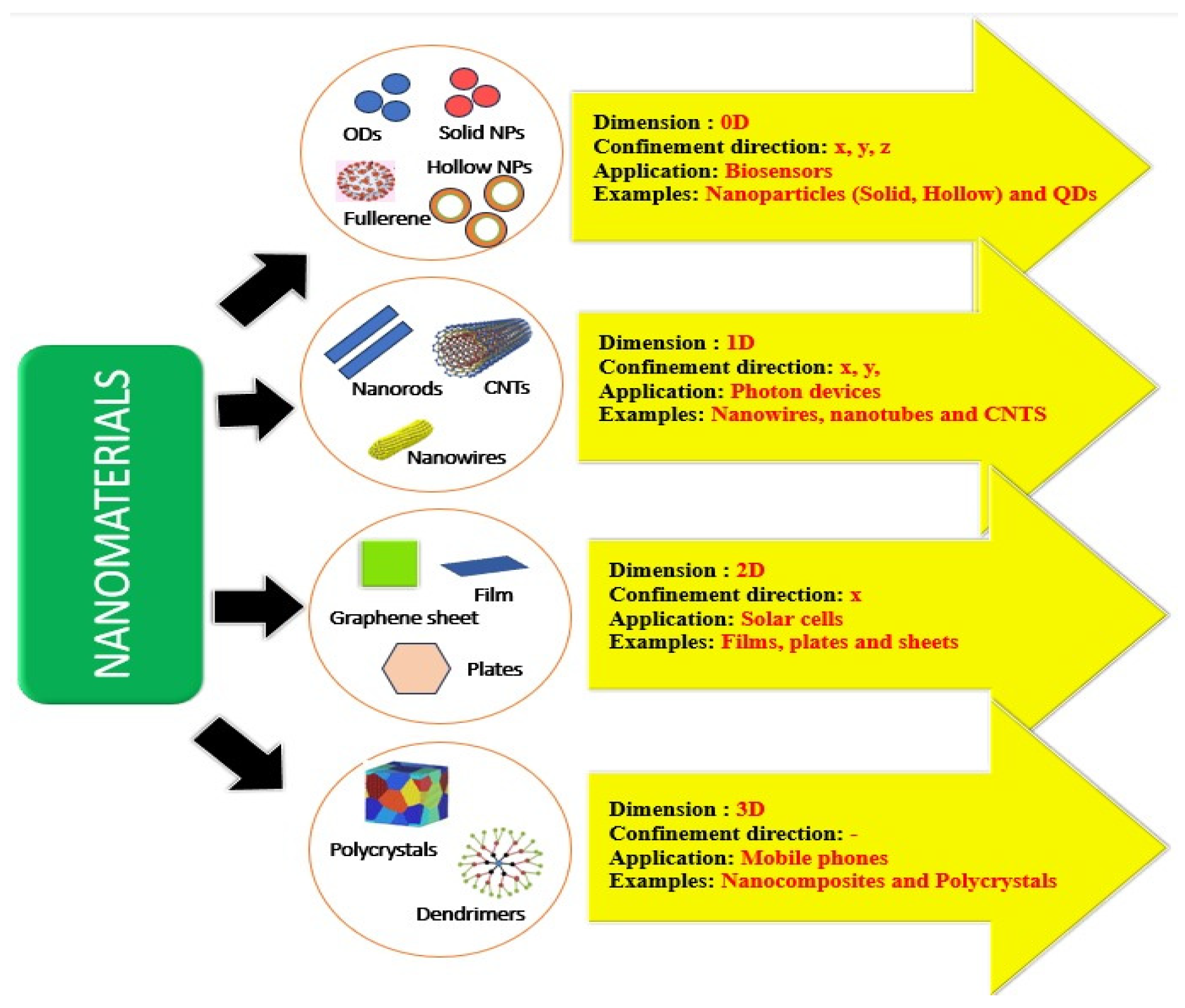
Figure 1. Different types of nanomaterials used for drug delivery.
Preprints 112913 g001
Figure 2. Schematic image of CNDs, GQDs, and PDs structure. Reproduced from ref. [108]. Copyright 2014. With permission from Springer.
Preprints 112913 g002
Figure 3. Schematic representation of the chemistry of CNDs.
Preprints 112913 g003
Figure 4. Different Applications of Carbon Nanodot.
Preprints 112913 g004
Figure 5. Role of CNDs in Drug delivery enhancement.
Preprints 112913 g005
Figure 6. Various methods of synthesis of Polymeric Carbon Nanodots.
Preprints 112913 g006
Disclaimer/Publisher’s Note: The statements, opinions and data contained in all publications are solely those of the individual author(s) and contributor(s) and not of MDPI and/or the editor(s). MDPI and/or the editor(s) disclaim responsibility for any injury to people or property resulting from any ideas, methods, instructions or products referred to in the content.
Copyright: This open access article is published under a Creative Commons CC BY 4.0 license, which permit the free download, distribution, and reuse, provided that the author and preprint are cited in any reuse.
Prerpints.org logo

Preprints.org is a free preprint server supported by MDPI in Basel, Switzerland.

Subscribe

© 2024 MDPI (Basel, Switzerland) unless otherwise stated