
Submitted:
01 August 2024
Posted:
01 August 2024
You are already at the latest version
Abstract
Dispersion of antibiotic in livestock farming represents a health concern worldwide, contributing to the spread of antimicrobial resistant bacteria through animals, the environment, and humans. Phenolic compounds could be alternatives to antibiotics, once drawbacks such as low water solubility, bioavailability, and reduced stability are overcome. Although nano- or microsized formulations could respond to these shortcomings they do not represent cost-effective options. Here, three phenolic compounds, obtained from wood processing manufacturers, were characterized, revealing suitable features such as antioxidant activity, size, and chemical and colloidal stability for in-field applications. The Minimum Inhibitory Concentration (MIC) of these colloidal suspensions was measured against six bacterial strains isolated from livestock. These particles showed different inhibition behaviors: Colloidal chestnut was effective against one of the most threatening antibiotic resistant pathogens such as S. aureus but was ineffective toward E. coli. On the other hand, colloidal pine showed a weak effect on S. aureus but specificity toward E. coli. The present proof-of-concept points at colloidal polyphenols as valuable alternatives for antimicrobial substitutes in the livestock context.
Keywords:
nanoparticles
; antimicrobials
; wood industry by-products
; AMR
1. Introduction
Livestock farming is an integral part of the food industry, providing for protein sources such as meat, milk, and eggs, that are further processed into products and by-products destined to human consumption [1]. Bacterial contaminations generate substantial detrimental effects, which not only compromise animal health, but also have significant implications for welfare and productivity along with the industrial chain. The use of antibiotics played a crucial role in preventing and treating bacterial infections, promoting animal health, and ensuring optimal productivity. However, their overuse and misuse contributed to the emergence and spread of antibiotic-resistant bacteria, such as Escherichia coli resistant to third- and fourth-generation cephalosporines, or methicillin-resistant Staphylococcus aureus (i.e., MRSA) [2]. Zoonotic diseases, which are transmitted between animals and humans, pose significant concerns in livestock productions. Consumption of contaminated food, such as meat or dairy products derived from animals hosting resistant bacteria, can contribute to their transmission to humans, posing a threat to public health [3]. Infections caused by resistant bacteria are more difficult to treat, leading to prolonged illness, increased healthcare costs, and higher mortality rates [4]. Addressing antimicrobial resistances requires promoting the responsible use of antimicrobial drugs, implementing surveillance programs, and exploring alternative approaches[5]. Polyphenols are widely recognized for their antioxidant and anti-inflammatory effects, which improve human health and contribute to disease prevention [6,7]. The antibacterial activity of polyphenols is believed to be related to the expression of specific genes in bacteria, leading to alterations in bacterial metabolism and virulence. On these bases, the potential for naturally occurring polyphenols to become alternatives to antimicrobials was explored [8]. These molecules downregulate the expression of genes involved in pathogenicity and upregulate the expression of genes related to stress response and antimicrobial resistance [9]. Furthermore, polyphenolic compounds showed potential as anti-biofilm agents [10] and antimicrobial effects against foodborne bacteria including Gram-positive and Gram-negative bacteria, and fungi [11].
Even though polyphenols demonstrated outstanding antibacterial potential, most of them presented poor water solubility, short pharmacokinetics half-lives, and low bioavailability[12]. Furthermore, environmental factors such as acidity, temperature variations, and enzymes compromise the stability of these substances, thereby further limiting their therapeutic activity. Nano-formulations reduced drastically the existing limitations to polyphenol use. They resulted in enhanced bioavailability and controlled drug-release, improving their suitability for human biomedical use as well as in livestock production applications [13]. Moreover, the denser particulate acted as a protective shield for polyphenols, preventing their degradation, enhancing their tissue absorption, and minimizing their potential toxicity in animals and humans[14]. Examples of existing nano-formulations, which were studied to improve polyphenolic agents include: Polymeric nanoparticles, liposomes, hydrogels, microspheres, transferosomes, solid lipid nanoparticles, metal-nanoparticles, drug-polymer conjugates, and nanostructured lipid carriers[14]. However, the translation of this kind of approach from human health care to large-scale productions of livestock food additives is far from being a trivial and easy-to-solve matter. When investigating their antimicrobial activities and the associated biological mechanisms, phenolic compounds were commonly studied as chemicals, and the attention was focused on their molecular structures and reactivity[15]. Their direct action on the microbial cell wall was attributed to epigallocatechin while inhibition of bacterial gene expression was ascribed to catechin, and anti-biofilm activity to rosmarinic acid. Some of these are non-specific (oxidative stress), while other mechanisms are specific [14,15].
This view does not consider that some natural phenolic compound sources such as wood, when subjected to chemical treatments and strong mechanical stress, typical of large-scale manufacturing plants, can generate micro and even nano-sized materials [16]. In other words, inexpensive polyphenolic micro- and nanoparticles are already available as by-products of industrial productions, processing natural material such as wood, therefore offering cost-effective antibiotic competitors for livestock infection management. It is worth mentioning that wood-derived materials were tested as antimicrobial agents, but chemical-physical features such as hydrodynamic size and zeta potentials were usually neglected during the interpretation of their action mechanisms[17]. These large-scale produced phenolic materials may display adequate chemical and colloidal stability, which are crucial pre-requisites for their in vivo application. Furthermore, their characterization could be helpful for understanding their antimicrobial activity. Based on this rationale, three compounds attained by wood industrial processing were first characterized, revealing optimal chemical-physical and colloidal characteristics. These colloidal materials were named depending on the respective source as follows: Colloidal chestnut A and B (CCA and CCB), deriving from chestnut trees from two distinct Italian regions, and colloidal pine (CP), from pine biomass. They were tested on six bacterial strains isolated from cattle, swine, and poultry (i.e., Pasteurella multocida, Staphylococcus aureus, Streptococcus suis, Mannhemia haemolitica, Escherichia coli, and Salmonella typhimurium).
Colloidal chestnut A (i.e., CCA) and colloidal pine (i.e., CP), displayed the most divergent behaviours, with opposite antioxidant power and highly different specificities reagarding their highest bacteriostatic performances. Although the observed selectivity is far from being easily explained, it points at the need of conceiving tailored phenolic combinations as possible alternatives to antimicrobials in livestock. Moreover, the colloidal perspective presented here paves the way to further studies overcoming the limited vision of a merely molecular-related activity of phenolic compounds.
2. Results
2.1. Morphologycal and Hydrodynamic Characterization
Hydrodynamic radii and the zeta potential (ζ) values of the carbonaceous materials were characterized by dynamic light scattering (DLS) as shown in Figure 1. According to DLS characterization both chestnut polyphenols comprised two distinct colloidal populations in the submicron and nano-size regions. The estimated hydrodynamic sizes of colloidal chestnut A (CCA) were 147 ± 2 nm and 793 ± 3 nm, displaying an average zeta potential (ζ) of −14 ± 1 mV (conductivity 0.098 mS cm−1 in water at 22 °C) whereas colloidal chestnut B (CCB) sizes were 120 ± 2 nm and 779 ± 1 nm, with and average ζ value was −14 ± 1 mV (conductivity 0.079 mS cm−1 in water at 22°C). Interestingly, colloidal pine (CP) was characterized by a minor contribution from sub-micron particles and an abundance of nano-sized material with dimensions in the region comprised from 10 to 100 nm, overall displaying an average hydrodynamic radius of 280 ± 60 nm and a quite remarkable average ζ value of −23.8 ± 0.4 mV (conductivity 0.091 mS cm−1 in water at 22°C). Indeed, DLS observation were corroborated by TEM micrographs showing that CCA and CCB were characterized by the relevant presence of submicron material, while CP was predominantly represented by poli-dispersed and very small spherical nanoparticles.
Although possessing a contained ζ in the 10–30 mV interval and, therefore, being classifiable (either positive or negative) from relatively stable to stable [18], these suspensions presented no sign of precipitation along with a six-months monitoring period and at concentrations in an aqueous milieu equal to 540, 340, and 50 g L−1 for CCA, CCB, and CP respectively. Thus, although phenolic compounds are commonly insoluble in water, these particulate materials produced rather stable colloidal suspensions, suitable for application purposes, and characterized by a significant fraction of nano-sized particles.
2.2. Chemical-Physical Characterization
In order to explain the colloidal nature of the investigated compounds, it can be assumed that they consist of supramolecular structures in the nano- and micro-size range [19], in which phenolic constituents are held together by a number of non-covalent bonds including, for example, aromatic stacking, hydrogen bonds, electrostatic interactions. In this view, Fourier Transform Infrared Spectroscopy (FTIR) was used to characterize these self-assembled molecular aggregates from a vibrational standpoint. All the wood derived powders share very similar FTIR profiles, comparable to the one of commercial tannic acid (TA), confirming an analogous chemical identity.
In order to provide a general description of this FTIR fingerprint, the overall vibrational contributions can be briefly summarized as follows: (1) a broad absorption comprised between 3300 and 3360 cm−1 ascribable to the stretching vibrations of the hydroxyl groups -OH [20,21]; (2) a peak at around 2930 cm−1 accompanied by a shoulder at around 2850 cm−1, clearly distinguishable only in Figure 1D and attributable to the symmetric stretching vibrations and antisymmetric -CH- of the CH2 and CH3 groups [21]; (3) A strong band associated to the carbonyl group C=O stretching in the region from 1730–1705 cm−1 [21,22,23,24]; (4) the signals in the 1605–1500 cm−1 range can be assigned to the deformation vibrations of the carbon–carbon bonds of the aromatic backbone [21]; (5) a peak in the 1440–1460 cm−1 interval, due to the stretching vibrations of C-C aromatic groups; (6) the characteristics bands of C-O stretching vibration falling at 1100–1300 cm−1 with very pronounced peaks at around 1100 cm−1 [21,24,25]. A more punctual spectral attribution is listed in Table 1; in general these results are similar to those reported in the literature for polyphenolic compounds as tannic acid [22,26].
Although the spectra were comparable in terms of vibrational contributions, the wood-derived materials reported an intensity decrease in many peaks located in the range between 1750 and 750 cm−1 when compared with tannic acid. In particular, these peaks were as sharp and strong as the ones observed in the control. This can be related to a higher degree of rigidity due the aggregated character of the molecules involved in the supramolecular assemblies, namely the documented nano- and micro-particles (vide supra).
Since the morphological and hydrodynamic characterization of the materials was interpreted as the result of the supramolecular clustering of polyphenols, kept together by non-covalent interactions, preliminary chemical stability tests were carried out by incubating the dried powders in different conditions and the effect of the chemical environment on the material was monitored thorough DLS analysis. The incubation in 96% ethanol led to slow dissolution of the powders, confirming the hydrophobic intermolecular interactions. Noteworthy, as the particulate disappeared, the liquid turned into a progressively darker suspension. Upon exposure to a strong alkaline milieu such as 3 M NaOH the powders instantaneously dissolved and turned to a black colored material as well. On the contrary, powders were not susceptible to acidic conditions up to 3 M HCl. This can be explained assuming that under acidic conditions molecular hydroxyl groups are non-ionized. Differently, when the pH was shifted to alkaline values the materials dissolved as a consequence of the electrostatic repulsion that took place at a molecular level between single polyphenols, as well as because of the increase of their hydrophilicity, being negatively charged due to the hydroxyl group deprotonation [27].
Overall, when the particulate form was degraded into its molecular constituents, as proved by the disappearance of the aforementioned particulate by DLS analysis, the former colloidal polyphenol was rapidly oxidized into a black material showing no antioxidant power according to Folin-Ciocalteâu. On these bases, aiming at marking possible differences among the three colloidal compounds, a more in-depth analysis of the disaggregation and oxidation behaviors was carried only in alkaline conditions. Hence the pH influence was explored in the 7–14 range and as function of time using DLS, UV-vis and measuring the antioxidant through the Folin-Ciocalteâu method. In Figure 3A,B, it is possible to appreciate the evolution of CCA and CCB UV-vis profile with the increase of pH, in particular the intensity of a band at around 280 nm, attributable to phenol aromatic rings, progressively diminished as the alkalinity enhanced, being almost zeroed at pH 14. In its place, the appearance of a new chromophore characterized by a broad band at around 450 nm, was registered, and ascribed to the occurrence of oxidation phenomena. Indeed, the emergence of a very similar spectral feature was reported for analogous chestnut materials upon the action of a strong oxidant [28]. DLS measurements confirmed that the samples, in these pH conditions, were below the limit of detection of the instrument, prompting that the colloidal materials were disaggregated into their molecular components and likely became susceptible to oxidation. Conversely, CP showed an increasingly more pronounced aromatic band at 280 nm in the examined range of pH, anyway accompanied by the appearance of a new chromophore signal already visible at pH 12, in the form of a broad shoulder centered at around 350 nm and ascribable to oxidation processes (Figure 3C) [29]. This can possibly be explained by considering the simultaneous occurrence of disaggregation and oxidation. However, differently from chestnut, a prevalence on the disaggregation phenomenon could be evinced observing the progress of pine UV-vis profile, along with the pH enhancement. Disaggregation was again witnessed by the disappearance of colloidal particles, as substantiated by DLS analysis.
Actually, from the comparison of kinetic behavior, monitored following the aromatic UV-vis fingerprint at 280 nm, it can be noted that the feature is influenced by pH in the case of CCA and CCB, but it is almost unaffected in the case of CP (Figure 3D–F). Anyway, the aforementioned emergence of new chromophores, witnessing the occurrence of oxidative processes, can be immediately appreciated by observing the drastic color change of chestnut and pine contents in the experimental tubes, turned to red and yellow respectively, fully in harmony with the band wavelengths documented by UV-vis characterization (Figure 3G). Finally, in good agreement with the optical analysis, the antioxidant power of chestnut compounds registered a decrease above pH 10. In particular CCB, the compound showing a more pronounced pH susceptibility according to the decrease of 280 nm UV-vis signal was also the one registering a decrease of the antioxidant power already at pH 10. CCA showed a more contained decrease of the phenol aromatic ring UV-vis feature and its antioxidant power was well-conserved until pH 12. Instead, pine showed an increase over approximatively the same pH, confirming that the oxidation phenomenon is apparently more relevant for chestnut than for pine. Nicely, in this range of pH, the antioxidant power against pH roughly follows a sigmoidal trend for all the examined compounds (Figure 3H). Thus, although it has been highlighted that it is the physical structure of these compounds that preserve the molecular cargos from degradation, it is unlikely that an actual antimicrobial effect could be exerted without the disaggregation of these supramolecular clusters. Hence, the different disaggregation-oxidation trends could be related to distinct antimicrobial activities (vide infra). Moreover, all these compounds gave rise to long-term stable water suspensions, combining the aforementioned chemical stability in normal environmental conditions with colloidal properties that are suitable for in vivo and in-field applications.
2.3. Minimum Inhibitory Concentration (MIC) of Phenolic Compounds on Selected Bacteria
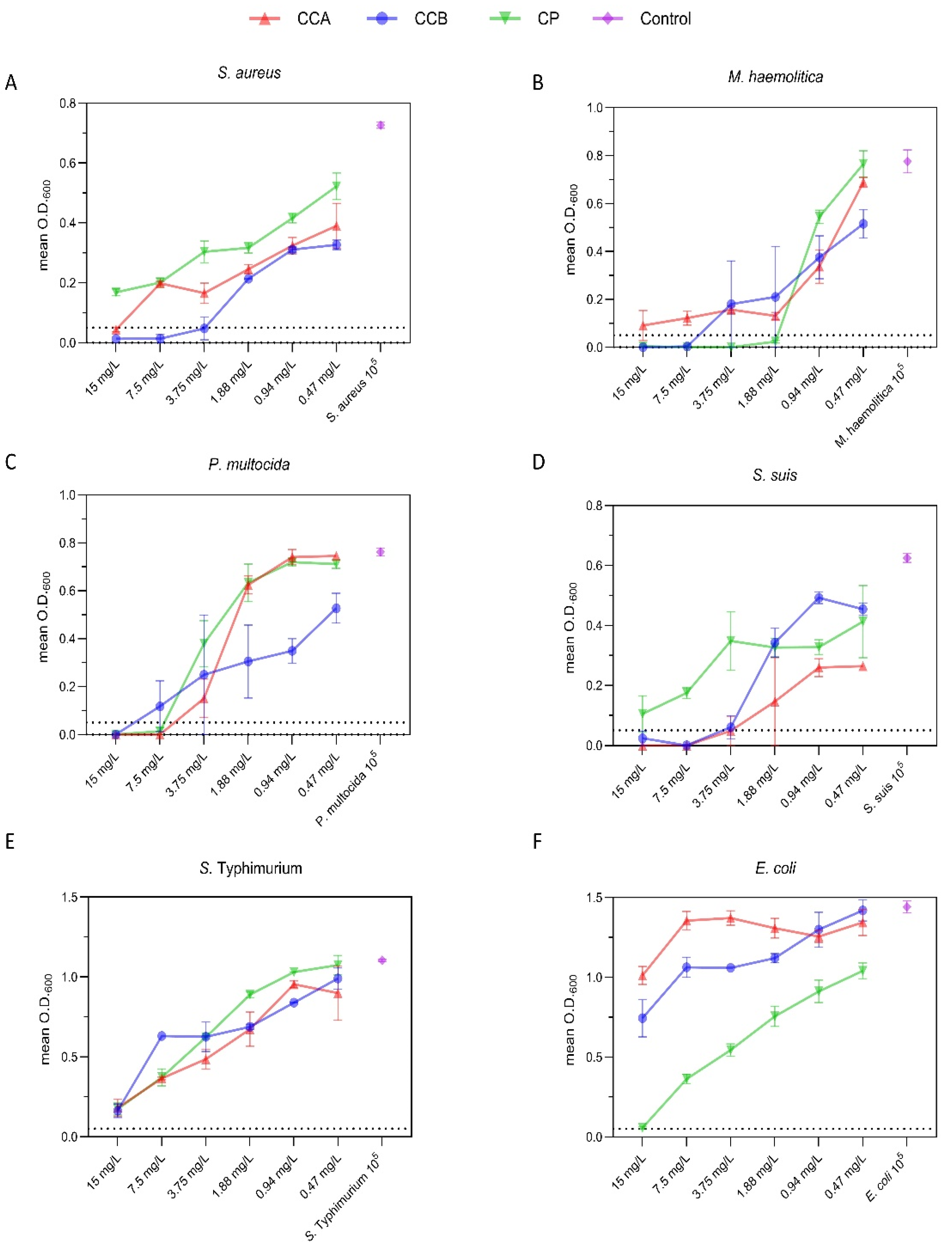
The minimum inhibitory concentration (MIC) is a widely accredited parameter for conveying the effectiveness of antimicrobial agents, consisting in the lowest concentration of a chemical, usually a drug, able to prevent the growth of bacteria or fungi in vitro [30,31]. The inhibitory effect of phenolic particulates was assessed against selected bacterial strains, isolated from livestock, i.e., Pasteurella multocida, Staphylococcus aureus, Streptococcus suis, Manneimia. haemolitica, Escherichia coli, and Salmonella Typhimurium (Table S1). The MIC of each compound was estimated using a microtiter assay by measuring the variation of the optical density at 600 nm (O.D.600) by UV-vis spectrometry, after 24h incubation at 37 °C. In Figure 4, for each bacterial strain the mean absorbance of six independent replicates was plotted against the polyphenol concentration in the range comprised between from 0.47 and 15.00 mg L−1.
Overall, the three materials displayed a concentration-dependent effect in the whole examined range and, most importantly, the growth inhibitory effect was significantly variable as a function of the specific pathogen. In order to better discriminate the variability of effect exerted by the colloidal compounds and the group of investigated bacteria, the differences in mean O.D. 600 were calculated (Table 2).
Figure 1.
MIC determination for CCA (blue), CCB (red), and CP (green) against S. aureus (A), M. haemolytica (B), P. multocida (C), S. suis (D), S. typhimurium (E), and E. coli (F). Each point in a logarithmic horizontal axis of colloid concentrations represents the mean O.D. of six independent replicates, with vertical whiskers indicating the standard deviation (SD). Horizontal dashed lines represent the MIC threshold set at a 0.05 increase in O.D. Bacterial strains inoculated without any compound were used as positive controls and are shown in purple.
3. Discussion
A number of reported examples substantiated that an essential condition to obtain antibacterial efficacy is to endow a particle with a positive surface charge, as the latter allows for more efficient interaction with the bacterial cell wall [32]. Indeed, as a consequence of the ionized substituents exposed to the extracellular environment, surfaces of bacterial cell possess net negative electrostatic charges [33]. This facet obviously contrasted with our experimental data as, although the three phenolic materials object of the current study showed suitable colloidal properties for in field applications, they have in common a negative zeta potential value. Thus, charge repulsion is likely to occur at the abiotic-biotic interface, hampering the contact between colloidal phenols and the target microorganisms. That said, notable exceptions to the aforementioned paradigm are available in literature and negative materials with antibacterial activity can be found [34]. Although the here observed phenomenon is far from being easily interpreted, it can be imagined that the colloids may disaggregate into their reactive molecular constituents in the proximity of the microorganisms. Although, it can be expected that disaggregation-oxidation processes would be driven by other factors than the pH in a real biological situation, alkalinity was a simple strategy for unveiling some distinctive traits in the investigated colloids. As emerged from the current chemical-physical characterization, these materials displayed completely different disaggregation-oxidation behaviour. Interestingly, CCB displayed the best overall antimicrobial performances and it showed the highest antioxidant power, according to both FRAP and Folin–Ciocalteâu assays, although being the most unstable among the investigated compounds. However, this reactivity-focused interpretation is not sufficient to explain all the examined cases, E. coli was not significantly susceptible to the CCB but it was strongly inhibited by the chemically stable CP. Indeed, the correlation of the antimicrobial activity of this kind of colloidal materials and their chemical-physical characteristics will surely merit several dedicated studies.
Overall, an inhibitory effect trend was observed in the examined range of colloid concentrations and it is worth to note that this trend significantly differed depending on the specific colloid-bacterium pair.
The growth of S. aureus was inhibited in the presence of 7.50 mg L−1 and 15.00 mg L−1 CCB and CCA, respectively, revealing a remarkable efficacy towards one of the most persistent and detrimental microorganisms worldwide. Indeed, this bacterium is known to be resistant to multiple antibiotics, making its eradication a challenging task [35,36]. On the other hand, the third compound, CP, was only able to exert a weak effect on this bacterium at the tested concentrations. In turn, M. haemolytica displayed susceptibility to CP at as little as 1.88 mg L−1, while only partial inhibition was observed for CCA in the whole examined range of concentrations. Meanwhile, MIC for CCB against M. haemolytica was 7.50 mg L−1, which is a significantly higher value when compared to that of CP, prompting a higher specificity of the pine-derived material for this bacterial species. M. haemolytica is a major livestock pathogen, which can harbour multiple resistant determinants and cause severe respiratory diseases in farm animals [37,38], resulting in weight gain reduction, decreased feed efficiency, and overall impaired productivity in livestock [39]. Inhibition against P. Multocida was obtained by all compounds, with concentrations ranging from 15.00 mg L−1 (CP) to 7.50 mg L−1 (CCB and CCA). Taken together, these results suggest that CP exhibited a selective antibacterial activity against the family Pasteurellaceae (P. multocida and M. haemolytica). Considering the high level of antimicrobial resistance commonly found in these two bacterial species [40], these findings are of great interest for both animal and human health. Indeed, P. multocida not only causes respiratory diseases in livestock animals, but it can be transmitted to humans [41,42] and strains beloning to this species commonly show mult-drug resistance pattern [43]. Futhermore, both P. multocida and M. haemolytica are known to acquire and transfer antimicrobial resistance determinants through horizontal gene transfer [44], which is the main driver of intra- and inter-species antimicrobial resistance spread [45].
Interestingly, CCA and CCB inhibited the proliferation of S. suis at very low concentrations, equal to 3.75 mg L−1 and 7.50 mg L−1, respectively, whereas CP struggled to approach the MIC threshold. Considering that S. suis is a major bacterial pathogen in pigs, linked to systemic infections and meningitis, leading to increased mortality, reduced growth rates, and significant economic losses for pig producers, any products able to treat or limit its infection is of great interest for the pig industry [46]. Indeed, S. suis strains show resistance to multiple antimicrobials [46] and pose a zoonotic risk, especially to workers exposed to infected animals [47]. Thus, these promising in vitro results suggest that the effectiveness of chestnut compounds against S. suis should be adequately exploited in real world scenarios.
Although all the compounds displayed comparable inhibition effect trends, hampering bacterial replication at the highest concentrations tested, none reached the MIC threshold for S. Typhimurium and E. coli. However, while the growth of E. coli was only slightly influenced by CCA and CCB, inhibition was observed when the E. coli strain was tested against CP. Indeed, exposure to increasing concentrations of CP was associated with a dose-dependent reduction in O.D.600, with 15 mg L−1 of this compound resulting in a mean O.D.600 increase equal to 0.057, which was just slightly above the adopted MIC threshold (i.e., an increase in turbidity of <0.05). Furthermore, significant differences of mean O.D.600 between CP and the other two compounds (vs CCA mean of differences = −0.662, 95% CI −0.343/−0.982, p = 0.028 and vs CCB mean of differences = −0.505, 95% CI −0.342/−0.668, p = 0.031) were observed. These findings indicate that CP could be a promising candidate as alternative to the use of antimicrobials for the control and treatment of E. coli infections. E. coli is widespread in livestock productions and known to carry multiple resistance genes [48], therefore any compound capable of inhibiting the growth of this bacterium could have a huge positive impact on livestock productions.
Some of the polyphenol-mediated mechanisms of antibacterial activity are non-specific (e.g., oxidative stress), while other mechanisms only act on certain bacteria (e.g., toxin inhibition) [14,15]. Specifically, Villanueva et al. [10] demonstrated an anti-biofilm activity of selected tannins inhibiting the growth of S. typhimurium, P. aeruginosa, E. coli, and S. aureus. Since biofilm generation is a common trait of many bacteria, polyphenolic particles could be explored for their possible use as biofilm disruptors.
Several research groups [49,50] explored different types of nanoparticles for microbial inhibition, with outcomes suggesting higher efficacy against Gram-negative bacteria compared to Gram-positive. The responsible factor is thought to be the difference between the membrane composition of these two groups, as Gram-positive bacteria have a thick and exposed peptidoglycan layer. Growth inhibition by phenolic modified nanomaterials was reported for tannic acid-modified nanoparticles effectively inhibiting Listeria monocytogenes [51]. Diverse mechanisms can be employed by nanoparticles, including adhesion to microbial cells, generation of reactive oxygen species (ROS), and interference of bacterial growth kinetics, however the specific action against bacteria requires a dedicated in depth analysis [52,53].
The current findings underline the potential of CCB, CCA, and CP as antimicrobial agents against a range of bacterial pathogens. Furthermore, the results suggest that the antimicrobial activity of the colloidal materials is specific to certain bacterial species. Indeed, the variations of the observed effects on the different bacteria envisage the influence of pathogen-specific factors and chemical-physical characteristics on antimicrobial efficacy, pointing at the existence of more sophisticated growth-inhibitory paths. Further research is required to understand the correlation between the chemical-physical properties and the antimicrobial activity and to study possible combinations of different colloids as actual alternatives to antibiotics.
4. Materials and Methods
4.1. Materials
All chemicals, which were purchased with the highest purity available, were used without any additional treatment. NaOH was purchased from J.T. Baker. Ethanol 96% was purchased from VWR. 2,4,6-tripyridyl-2-triazine, chloride acid (HCl), ferric chloride (FeCl3), sodium acetate, sodium carbonate, gallic acid, ammonium ferrous sulfate, ammonium ferrous sulfate were purchased from Merck. All solutions were prepared using ultrapure water from a Genie Direct-Pure water device (RephiLe Bioscience Ltd., Shanghai, China), with a resistivity of at least 18.0 MΩ·cm. Blood Agar (BA) medium, Muller Hinton Agar (MHA), Brain Heart Infusion (BHI) broth, and tannic acid were obtained from Oxoid (UK). All plasticware, including plates for Minimum Inhibitory Concentration (MIC) were single-used.
The three phenolic compounds object of the current study were gently provided by AINT s.r.l. (Advanced Iron Nano Technologies, Santa Croce, 510, Venice, Italy) as dried powders and in the form of water suspensions. AINT s.r.l. develops formulations for animal farming using selected materials from different large-scale worldwide manufacturers of wood by-products. Colloidal chestnut A and B (CCA and CCB) were extracted from two distinct Italian region chestnut trees, and colloidal pine (CP) was extracted from pine biomass.
4.2. Methods
Antioxidant activity determination was carried out using a UV-1800 spectrophotometer (Shimadzu, Columbia, MD, USA) according to Folin–Ciocalteâu (FC) and ferric reducing antioxidant power (FRAP) methods [54,55]. In the FC assay [55], gallic acid was used as calibration standard. The FC assay was carried out by pipetting 1 mL of sample into a 10-mL test tube, followed by the addition of 1 mL of diluted FC reagent (i.e., 1 part of FC reagent and 2 parts of water). The mixture was vortexed and incubated for 3 min at room temperature. Then, 2 mL of 10% sodium carbonate solution was added and the mixture was accurately vortexed for 20–30 s (time 0). After 15 min at room temperature, the absorbance of the colored reaction product was measured at 750 nm. The total phenols (TP) content in the extracts was calculated from a standard calibration curve obtained with different concentrations of gallic acid, ranging from 0 to 37.5 mg L−1 (Figure S1). The results were firstly calculated as mg of gallic acid equivalent (GAE) per kg of dry weight and finally expressed as normalized antioxidant effect.
Freshly prepared FRAP reagent contained 1 mmol L–1 of 2,4,6-tripyridyl-2-triazine and 2 mmol L–1 of ferric chloride (FeCl3) in sodium acetate 0.25 mol L–1 (pH= 3.6). A methanol-extracted volume of polyphenol sample (100 µL) was added to the FRAP reagent (1900 µL) and accurately mixed. After incubating the mixture at 20 °C for 4 min, the sample absorbance was determined at 593 nm. Calibration was performed with a standard curve (0–1200 µg mL–1 ferrous ion), obtained by the addition of freshly prepared ammonium ferrous sulfate (Mohr’s salt, (NH4)2SO4·Fe(SO4)·6H2O). FRAP values were calculated as µg mL–1 of ferrous ion (ferric reducing power) from three determinations, and finally expressed as normalized antioxidant effect.
Zeta-potential and size-distribution of colloidal materials were measured in water by dynamic light scattering (DLS) using a Zetasizer Nanoparticle analyzer ZEN3600 (Malvern Instrument, Malvern, UK). LogNormal-function was used to obtain statistical analysis on the size-distribution. High-resolution transmission electron microscopy (HR-TEM) micrographs of colloidal materials were attained with a HR-TEM TITAN 60–300 microscope equipped with an X-FEG type emission gun. One drop of the as-obtained suspension was placed on a carbon film coated copper grid and dried at room temperature. In order to perform stability tests, colloidal water suspensions were initially prepared at the final concentration of 1 g L−1 and then diluted to 50 mg L−1 in the different tested conditions including 96% ethanol, 3 M HCl, 3M NaOH and in the 7–14 pH interval.
After one hour of incubation under stirring at room temperature, the materials were characterized through DLS and Uv-vis spectroscopy. Their antioxidant powder was assessed by Folin-Ciocalteâu assay.
4.3. Minimum Inhibitory Concentration (MIC) Assay
Standardized inocula of six bacterial strains previously isolated from livestock (E. coli, M. haemolytica, P. multocida, S. aureus, S. typhimurium, and S. suis) were used for the experiments. The list of tested bacteria is reported in supplementary X. Strains stored at −80 °C were resuscitated in duplicates in Blood Agar and incubated at 37 °C. After 24 h a single colony from each plate was inoculated into 5 mL of Brain Heart Infusion (BHI) broth and incubated at 37 °C for 24 h. After this second incubation, broth cultures were diluted to a final concentration of 5.0 Log10 CFU (colony forming unit)/mL, which was used as the inoculum for the microtiter assays to assess the inhibitory effect of phenolic compounds. Microtiter assays were carried out in triplicates over two weeks. Briefly, serial dilutions (15.00, 7.50, 3.75, 1.88, 0.94 and 0.47mg L−1) of phenolic compounds were prepared and loaded (50 µL) onto 96-well flat-bottom microtiter plates (Sarstedt, Nümbrecht, Germany). Each dilution was then inoculated with either 150 µL of standardized inoculum or 150 µL of BHI. Wells inoculated with BHI and phenolic compounds were included to correct for potential absorbance interferences due to the phenolic compounds themselves. Negative control wells containing only the growth medium (without bacteria or nanoparticles) were included, as well as positive controls consisting of the inoculum without phenolic compounds. Before incubation (T0) at 37 °C for 24 (h) the optical density at 600 nm (O.D.600) was measured using a Spectrophotometer Multiskan GO Microplate Readers (Thermo Fisher Scientific, Waltham, Massachusetts, USA). Initial readings (T0) were taken to establish the baseline density. O.D.600 was then measured after 24 h of incubation. At both time-points, five measurements at five minutes interval were carried out. The optical density (mean O.D.600) of each phenolic compound dilution/inoculum combination was calculated as the average absorbance of the biological replicates over the two experiments. Standard deviation and coefficient of variation (CV) were also calculated to assess the data robustness (CV < 0.5). The minimum inhibitory concentration (MIC) was set as the lowest concentration of phenolic compounds that limited the increase of turbidity of all technical replicates to < 0.05, according to a published method [56]. Mean differences and 95% confidence intervals (95% CI) were calculated, and significant differences were set at a p value < 0.05.
5. Conclusions
Antimicrobials are used in livestock production to cure diseased animals and prevent the dissemination of bacteria with zoonotic potential along the food chain. However, the overuse of antimicrobials in the food chain might contribute to the emergence of resistant bacteria, posing a threat to both animal and human health. The ability of the tested colloidal polyphenol materials to inhibit the growth of specific bacterial pathogens suggests their potential application in controlling and preventing infections in livestock. In particular, they displayed different antimicrobial activities against selected bacterial pathogens. Chestnut derived colloids were effective against one of the most threatening antibiotic resistant pathogens such as S. aureus but was ineffective toward E. coli. On the other hand, the latter microorganism showed susceptibility to colloidal pine, a material that was, vice versa, ineffective against S. aureus. Indeed, if used in combinations, such phenol nanoparticles may represent realistic alternatives to traditional antibiotics in livestock productions.
The present interdisciplinary collaboration among biophysics, biotechnology, materials science, and microbiology paves the way to further research aimed at enhance our comprehension of the principles ruling the efficacy of these compounds, in respect to their natural source, their antioxidant properties, and their colloidal features. In addition, finding the correlation between their chemical-physical properties and the mechanism of action, their safety profile, their in vivo efficacy, and eventually the in-field application conditions are necessary to optimize the understanding and the broader implications in promoting sustainable and effective disease management in the livestock industry.
Supplementary Materials
The following supporting information can be downloaded at the website of this paper posted on Preprints.org.
Author Contributions
A.L., investigation, formal analysis, data curation, visualization, supervision; A.C., (Alessandro Cecconello) conceptualization, visualization, writing—review and editing, supervision; S.M., investigation, data curation, visualization; G.R., methodology, investigation; A.C., (Aura Cencini) data curation, formal analysis; C.N., methodology, investigation; F.T., visualization; A.K., investigation, H.N.M., investigation; R.T., methodology, supervision; E.S., methodology, supervision; C.N., methodology, investigation; A.P. writing—review and editing; F.V., resources, writing—review and editing, project administration; M.M., conceptualization, writing—original draft preparation, funding acquisition, supervision. All authors have read and agreed to the published version of the manuscript.
Funding
Aura Cencini was supported by the Italian Ministry of Education, University and research (MIUR) funds “Sentinel” and “Ecosistema dell’Innovazione”. Alessandro Cecconello was supported by REACT-EU PON “Ricerca e Innovazione 2014–2020” and “Supporting TAlent in ReSearch @ University of Padua—STARS Grants” 2023. Federica Tonolo was supported by “iNEST- Interconnected Nord-Est Innovation ECS00000043”.
Institutional Review Board Statement
Not applicable.
Informed Consent Statement
Not applicable.
Data Availability Statement
The original data are available upon reasonable request to the corre sponding author.
Acknowledgments
The authors would like to thank Jana Stráská from the Regional Centre of Advanced Technologies and Materials, Palacký University in Olomouc, Czech Republic for TEM images. M.M. and F.V. wish to thank Dr. Massimo Marchiori, Dr. Mauro Braga and AINT s.r.l. for the kind support, for providing the materials object of the current study, and for their precious frontline feedback from the market and field, in particular for what concerns livestock production and related infection management.
Conflicts of Interest
The authors declare no conflict of interest.
References
- Billen, G.; Aguilera, E.; Einarsson, R.; Garnier, J.; Gingrich, S.; Grizzetti, B.; Lassaletta, L.; Le Noë, J.; Sanz-Cobena, A. Reshaping the European Agro-Food System and Closing Its Nitrogen Cycle: The Potential of Combining Dietary Change, Agroecology, and Circularity. One Earth 2021, 4. [Google Scholar] [CrossRef]
- Lee, A.S.; De Lencastre, H.; Garau, J.; Kluytmans, J.; Malhotra-Kumar, S.; Peschel, A.; Harbarth, S. Methicillin-Resistant Staphylococcus Aureus. Nature Reviews Disease Primers 2018 4:1 2018, 4, 1–23. [Google Scholar] [CrossRef] [PubMed]
- Ahmad, N.; Joji, R.M.; Shahid, M. Evolution and Implementation of One Health to Control the Dissemination of Antibiotic-Resistant Bacteria and Resistance Genes: A Review. Front Cell Infect Microbiol 2023, 12. [Google Scholar] [CrossRef] [PubMed]
- Marshall, B.M.; Levy, S.B. Food Animals and Antimicrobials: Impacts on Human Health. Clin Microbiol Rev 2011, 24. [Google Scholar] [CrossRef] [PubMed]
- Van Boeckel, T.P.; Brower, C.; Gilbert, M.; Grenfell, B.T.; Levin, S.A.; Robinson, T.P.; Teillant, A.; Laxminarayan, R. Global Trends in Antimicrobial Use in Food Animals. Proc Natl Acad Sci U S A 2015, 112. [Google Scholar] [CrossRef]
- Scalbert, A.; Johnson, I.T.; Saltmarsh, M. Polyphenols: Antioxidants and Beyond. Am J Clin Nutr 2005, 81. [Google Scholar] [CrossRef]
- García-Lafuente, A.; Guillamón, E.; Villares, A.; Rostagno, M.A.; Martínez, J.A. Flavonoids as Anti-Inflammatory Agents: Implications in Cancer and Cardiovascular Disease. Inflammation Research 2009, 58. [Google Scholar] [CrossRef] [PubMed]
- Walsh, D.J.; Livinghouse, T.; Goeres, D.M.; Mettler, M.; Stewart, P.S. Antimicrobial Activity of Naturally Occurring Phenols and Derivatives Against Biofilm and Planktonic Bacteria. Front Chem 2019, 7. [Google Scholar] [CrossRef] [PubMed]
- Amalraj, A.; Pius, A.; Gopi, S.; Gopi, S. Biological Activities of Curcuminoids, Other Biomolecules from Turmeric and Their Derivatives – A Review. J Tradit Complement Med 2017, 7. [Google Scholar] [CrossRef]
- Villanueva, X.; Zhen, L.; Ares, J.N.; Vackier, T.; Lange, H.; Crestini, C.; Steenackers, H.P. Effect of Chemical Modifications of Tannins on Their Antimicrobial and Antibiofilm Effect against Gram-Negative and Gram-Positive Bacteria. Front Microbiol 2023, 13. [Google Scholar] [CrossRef]
- Manso, T.; Lores, M.; de Miguel, T. Antimicrobial Activity of Polyphenols and Natural Polyphenolic Extracts on Clinical Isolates. Antibiotics 2022, 11. [Google Scholar] [CrossRef] [PubMed]
- Li, Z.; Jiang, H.; Xu, C.; Gu, L. A Review: Using Nanoparticles to Enhance Absorption and Bioavailability of Phenolic Phytochemicals. Food Hydrocoll 2015, 43. [Google Scholar] [CrossRef]
- Liu, C.; Dong, S.; Wang, X.; Xu, H.; Yang, X.; Wu, S.; Jiang, X.; Kan, M.; Xu, C. Research Progress of Polyphenols in Nanoformulations for Antibacterial Application. Mater Today Bio 2023, 21. [Google Scholar] [CrossRef] [PubMed]
- Turuvekere Vittala Murthy, N.; Agrahari, V.; Chauhan, H. Polyphenols against Infectious Diseases: Controlled Release Nano-Formulations. European Journal of Pharmaceutics and Biopharmaceutics 2021, 161. [Google Scholar] [CrossRef] [PubMed]
- Renzetti, A.; Betts, J.W.; Fukumoto, K.; Rutherford, R.N. Antibacterial Green Tea Catechins from a Molecular Perspective: Mechanisms of Action and Structure-Activity Relationships. Food Funct 2020, 11. [Google Scholar] [CrossRef] [PubMed]
- Gao, W.; Fatehi, P. Lignin for Polymer and Nanoparticle Production: Current Status and Challenges. Canadian Journal of Chemical Engineering 2019, 97. [Google Scholar] [CrossRef]
- Munir, M.T.; Pailhories, H.; Eveillard, M.; Irle, M.; Aviat, F.; Dubreil, L.; Federighi, M.; Belloncle, C. Testing the Antimicrobial Characteristics of Wood Materials: A Review of Methods. Antibiotics 2020, 9. [Google Scholar] [CrossRef]
- Patel, V.; Agrawal, Y. Nanosuspension: An Approach to Enhance Solubility of Drugs. J Adv Pharm Technol Res 2011, 2. [Google Scholar] [CrossRef]
- Ariga, K.; Hill, J.P.; Lee, M. V.; Vinu, A.; Charvet, R.; Acharya, S. Challenges and Breakthroughs in Recent Research on Self-Assembly. In Proceedings of the Science and Technology of Advanced Materials; 2008; Vol. 9.
- Thummajitsakul, S.; Samaikam, S.; Tacha, S.; Silprasit, K. Study on FTIR Spectroscopy, Total Phenolic Content, Antioxidant Activity and Anti-Amylase Activity of Extracts and Different Tea Forms of Garcinia Schomburgkiana Leaves. LWT 2020, 134. [Google Scholar] [CrossRef]
- Stuart, B.H. Infrared Spectroscopy: Fundamentals and Applications. Infrared Spectroscopy: Fundamentals and Applications. [CrossRef]
- Pantoja-Castroa, M. a.; González-rodríGuez, H. Study by Infrared Spectroscopy and Thermogravimetric Analysis of Tannins and Tannic Acid. Rev Latinoam Quim 2011, 39. [Google Scholar]
- Ricci, A.; Olejar, K.J.; Parpinello, G.P.; Kilmartin, P.A.; Versari, A. Application of Fourier Transform Infrared (FTIR) Spectroscopy in the Characterization of Tannins. Appl Spectrosc Rev 2015, 50. [Google Scholar] [CrossRef]
- Silverstein, R.M.; Webster, F.X.; Kiemle, D.J. Spectrometric Identification of Organic Compounds 7ed 2005 - Silverstein, Webster & Kiemle.Pdf. Microchemical Journal 2005, 21. [Google Scholar]
- Tondi, G.; Petutschnigg, A. Middle Infrared (ATR FT-MIR) Characterization of Industrial Tannin Extracts. Ind Crops Prod 2015, 65. [Google Scholar] [CrossRef]
- Herdlevær, K.M.; Løhre, C.; Nodland, E.; Barth, T. Comparison of Calibration Models for Rapid Prediction of Lignin Content in Lignocellulosic Biomass Based on Infrared and Near-Infrared Spectroscopy. Results Chem 2022, 4. [Google Scholar] [CrossRef]
- Chen, M.; Li, R.; Gao, Y.; Zheng, Y.; Liao, L.; Cao, Y.; Li, J.; Zhou, W. Encapsulation of Hydrophobic and Low-Soluble Polyphenols into Nanoliposomes by Ph-Driven Method: Naringenin and Naringin as Model Compounds. Foods 2021, 10. [Google Scholar] [CrossRef] [PubMed]
- Moccia, F.; Piscitelli, A.; Giovando, S.; Giardina, P.; Panzella, L.; D’ischia, M.; Napolitano, A. Hydrolyzable vs. Condensed Wood Tannins for Bio-Based Antioxidant Coatings: Superior Properties of Quebracho Tannins. Antioxidants 2020, 9. [Google Scholar] [CrossRef] [PubMed]
- Seong, G.; Yoko, A.; Inoue, R.; Takami, S.; Adschiri, T. Selective Chemical Recovery from Biomass under Hydrothermal Conditions Using Metal Oxide Nanocatalyst. Journal of Supercritical Fluids 2018, 133. [Google Scholar] [CrossRef]
- Kowalska-Krochmal, B.; Dudek-Wicher, R. The Minimum Inhibitory Concentration of Antibiotics: Methods, Interpretation, Clinical Relevance. Pathogens 2021, 10. [Google Scholar] [CrossRef]
- Balouiri, M.; Sadiki, M.; Ibnsouda, S.K. Methods for in Vitro Evaluating Antimicrobial Activity: A Review. J Pharm Anal 2016, 6. [Google Scholar] [CrossRef]
- Li, Z.; Ma, J.; Ruan, J.; Zhuang, X. Using Positively Charged Magnetic Nanoparticles to Capture Bacteria at Ultralow Concentration. Nanoscale Res Lett 2019, 14, 1–8. [Google Scholar] [CrossRef]
- Wilson, W.W.; Wade, M.M.; Holman, S.C.; Champlin, F.R. Status of Methods for Assessing Bacterial Cell Surface Charge Properties Based on Zeta Potential Measurements. J Microbiol Methods 2001, 43, 153–164. [Google Scholar] [CrossRef] [PubMed]
- Salvioni, L.; Galbiati, E.; Collico, V.; Alessio, G.; Avvakumova, S.; Corsi, F.; Tortora, P.; Prosperi, D.; Colombo, M. Negatively Charged Silver Nanoparticles with Potent Antibacterial Activity and Reduced Toxicity for Pharmaceutical Preparations. Int J Nanomedicine 2017, 12. [Google Scholar] [CrossRef] [PubMed]
- Zadoks, R.N.; Allore, H.G.; Barkema, H.W.; Sampimon, O.C.; Wellenberg, G.J.; Gröhn, Y.T.; Schukken, Y.H. Cow- and Quarter-Level Risk Factors for Streptococcus Uberis and Staphylococcus Aureus Mastitis. J Dairy Sci 2001, 84. [Google Scholar] [CrossRef] [PubMed]
- Grundmann, H.; Aires-de-Sousa, M.; Boyce, J.; Tiemersma, E. Emergence and Resurgence of Meticillin-Resistant Staphylococcus Aureus as a Public-Health Threat. Lancet 2006, 368. [Google Scholar] [CrossRef]
- Rice, J.A.; Carrasco-Medina, L.; Hodgins, D.C.; Shewen, P.E. Mannheimia Haemolytica and Bovine Respiratory Disease. Anim Health Res Rev 2008, 8. [Google Scholar]
- Griffin, D. Bovine Pasteurellosis and Other Bacterial Infections of the Respiratory Tract. Veterinary Clinics of North America - Food Animal Practice 2010, 26. [Google Scholar] [CrossRef] [PubMed]
- Confer, A.W. Update on Bacterial Pathogenesis in BRD. Animal health research reviews / Conference of Research Workers in Animal Diseases 2009, 10. [Google Scholar] [CrossRef] [PubMed]
- Ziagham, A.; Gharibi, D.; Mosallanejad, B.; Avizeh, R. Molecular Characterization of Pasteurella Multocida from Cats and Antibiotic Sensitivity of the Isolates. Vet Med Sci 2024, 10. [Google Scholar] [CrossRef] [PubMed]
- Dabo, S.M.; Taylor, J.D.; Confer, A.W. Pasteurella Multocida and Bovine Respiratory Disease. Anim Health Res Rev 2008, 8. [Google Scholar]
- Punpanich, W.; Srijuntongsiri, S. Pasteurella (Mannheimia) Haemolytica Septicemia in an Infant: A Case Report. J Infect Dev Ctries 2012, 6. [Google Scholar] [CrossRef]
- Melchner, A.; van de Berg, S.; Scuda, N.; Feuerstein, A.; Hanczaruk, M.; Schumacher, M.; Straubinger, R.K.; Marosevic, D.; Riehm, J.M. Antimicrobial Resistance in Isolates from Cattle with Bovine Respiratory Disease in Bavaria, Germany. Antibiotics 2021, 10. [Google Scholar] [CrossRef] [PubMed]
- Michael, G.B.; Bossé, J.T.; Schwarz, S. Antimicrobial Resistance in Pasteurellaceae of Veterinary Origin. Microbiol Spectr 2018, 6. [Google Scholar] [CrossRef] [PubMed]
- Džidić, S.; Šušković, J.; Kos, B. Antibiotic Resistance Mechanisms in Bacteria: Biochemical and Genetic Aspects. Food Technol Biotechnol 2008, 46. [Google Scholar]
- Segura, M.; Aragon, V.; Brockmeier, S.L.; Gebhart, C.; de Greeff, A.; Kerdsin, A.; O’Dea, M.A.; Okura, M.; Saléry, M.; Schultsz, C.; et al. Update on Streptococcus Suis Research and Prevention in the Era of Antimicrobial Restriction: 4thinternationalworkshop on s. Suis. Pathogens 2020, 9. [Google Scholar] [CrossRef] [PubMed]
- Gottschalk, M.; Xu, J.; Calzas, C.; Segura, M. Streptococcus Suis: A New Emerging or an Old Neglected Zoonotic Pathogen? Future Microbiol 2010, 5. [Google Scholar] [CrossRef] [PubMed]
- Apostolakos, I.; Mughini-Gras, L.; Fasolato, L.; Piccirillo, A. Assessing the Occurrence and Transfer Dynamics of ESBL/PAmpC-Producing Escherichia Coli across the Broiler Production Pyramid. PLoS One 2019, 14. [Google Scholar] [CrossRef] [PubMed]
- Chapa González, C.; González García, L.I.; Burciaga Jurado, L.G.; Carrillo Castillo, A. Bactericidal Activity of Silver Nanoparticles in Drug-Resistant Bacteria. Brazilian Journal of Microbiology 2023, 54. [Google Scholar] [CrossRef] [PubMed]
- Sathiyaraj, S.; Suriyakala, G.; Dhanesh Gandhi, A.; Babujanarthanam, R.; Almaary, K.S.; Chen, T.W.; Kaviyarasu, K. Biosynthesis, Characterization, and Antibacterial Activity of Gold Nanoparticles. J Infect Public Health 2021, 14. [Google Scholar] [CrossRef] [PubMed]
- de Almeida Roger, J.; Magro, M.; Spagnolo, S.; Bonaiuto, E.; Baratella, D.; Fasolato, L.; Vianello, F. Antimicrobial and Magnetically Removable Tannic Acid Nanocarrier: A Processing Aid for Listeria Monocytogenes Treatment for Food Industry Applications. Food Chem 2018, 267, 430–436. [Google Scholar] [CrossRef]
- Theophel, K.; Schacht, V.J.; Schlüter, M.; Schnell, S.; Stingu, C.S.; Schaumann, R.; Bunge, M. The Importance of Growth Kinetic Analysis in Determining Bacterial Susceptibility against Antibiotics and Silver Nanoparticles. Front Microbiol 2014, 5. [Google Scholar] [CrossRef]
- Youssef, F.S.; El-Banna, H.A.; Elzorba, H.Y.; Galal, A.M. Application of Some Nanoparticles in the Field of Veterinary Medicine. Int J Vet Sci Med 2019, 7. [Google Scholar] [CrossRef] [PubMed]
- Benzie, I.F.F.; Strain, J.J. The Ferric Reducing Ability of Plasma (FRAP) as a Measure of “Antioxidant Power”: The FRAP Assay. Anal Biochem 1996, 239. [Google Scholar] [CrossRef] [PubMed]
- Singleton, V.L.; Orthofer, R.; Lamuela-Raventós, R.M. Analysis of Total Phenols and Other Oxidation Substrates and Antioxidants by Means of Folin-Ciocalteu Reagent. Methods Enzymol 1999, 299. [Google Scholar] [CrossRef]
- Tsai, T.H.; Tsai, T.H.; Chien, Y.C.; Lee, C.W.; Tsai, P.J. In Vitro Antimicrobial Activities against Cariogenic Streptococci and Their Antioxidant Capacities: A Comparative Study of Green Tea versus Different Herbs. Food Chem 2008, 110. [Google Scholar] [CrossRef] [PubMed]
Figure 1.
Hydrodynamic and morphological characterization of the colloidal phenols. DLS hydrodynamic radii of (A) CCA, (B) CCB, (C) CP. TEM micrographs of (D) CCA, (E) CCB, (F) CP.
Figure 1.
Hydrodynamic and morphological characterization of the colloidal phenols. DLS hydrodynamic radii of (A) CCA, (B) CCB, (C) CP. TEM micrographs of (D) CCA, (E) CCB, (F) CP.

Figure 2.
Chemical characterization of the phenol dried powders. FTIR profile of (A) CCA, (B) CCB, (C) CP, (D) commercial tannic acid, used as control. Comparison of the antioxidant powers through (E) FRAP and (F) Folin-Ciocalteâu assay.
Figure 2.
Chemical characterization of the phenol dried powders. FTIR profile of (A) CCA, (B) CCB, (C) CP, (D) commercial tannic acid, used as control. Comparison of the antioxidant powers through (E) FRAP and (F) Folin-Ciocalteâu assay.
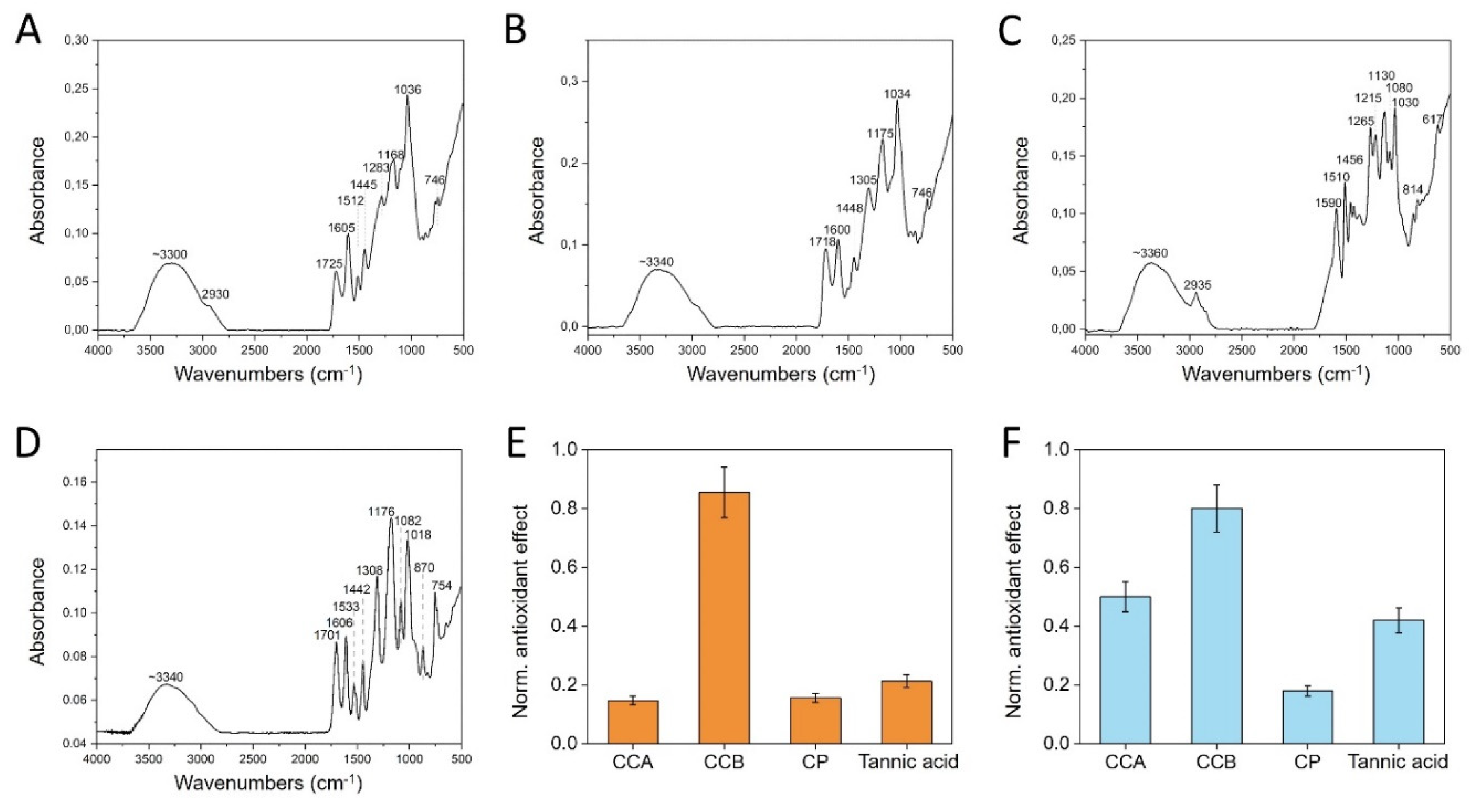
Figure 3.
Study of disaggregation and oxidation behaviors of CCA, CCB, CP. The effect of increasing pH documented by Uv-vis spectroscopy for (A) CCA, (B) CCB, (C) CP. Comparison of the evolution of the aromatic phenol ring Uv-vis peak at 280 nm as a function of time at (D) pH 7, (E) pH 8, (F) pH 12. (G) Comparison of color variation as a function of pH. (H) Comparison of antioxidant powder as a function of pH.
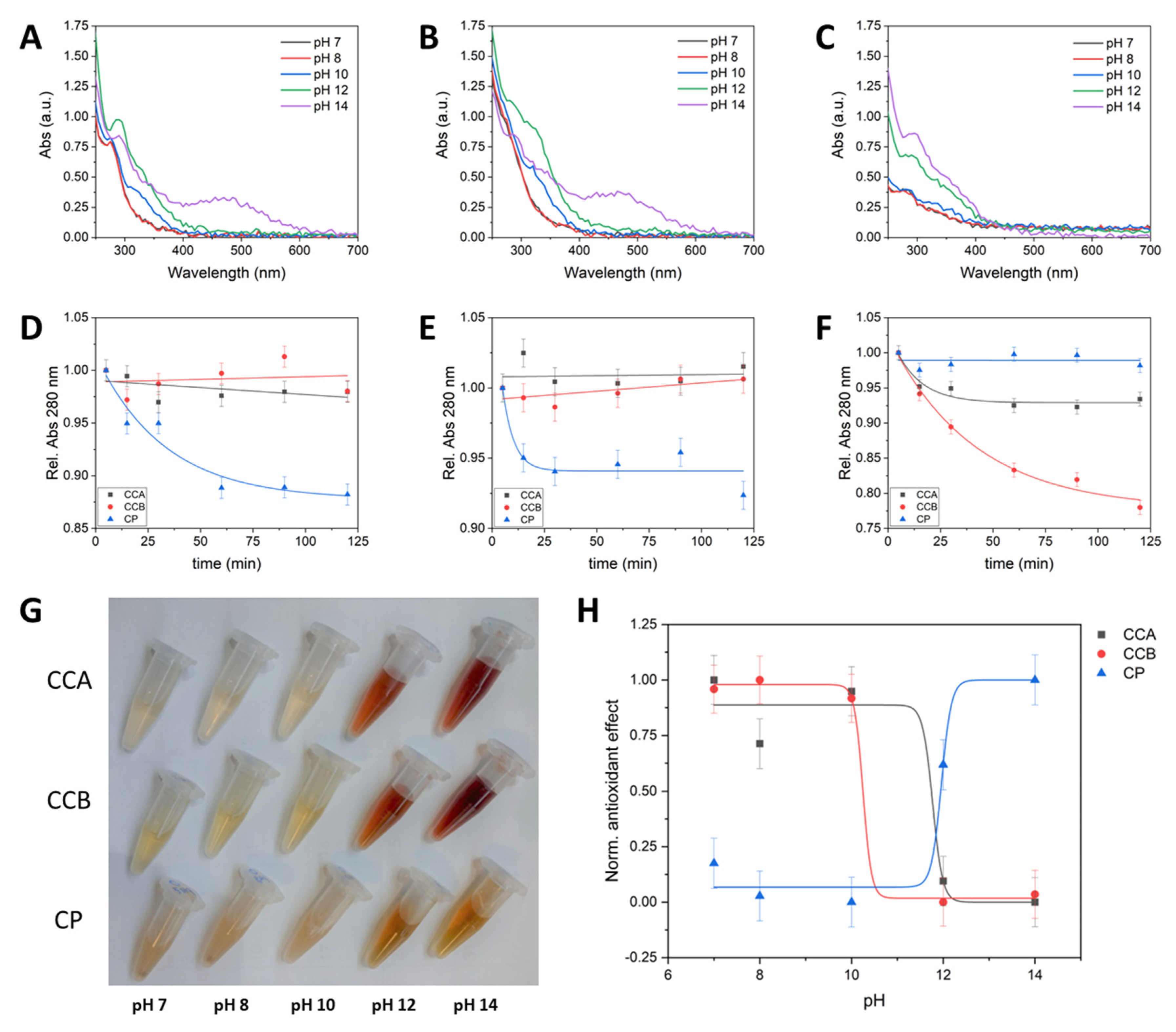

Table 1.
FTIR vibration attribution of the identified spectral feature wavelengths (cm−1) of CCA, CCB, CP and TA, as control.
Table 1.
FTIR vibration attribution of the identified spectral feature wavelengths (cm−1) of CCA, CCB, CP and TA, as control.
| Spectral identification | CCA | CCB | CP | TA |
|---|---|---|---|---|
| Stretching vibrations of the hydroxyl groups -OH | 3300 | 3340 | 3360 | 3350 |
| Symmetric stretching vibrations and antisymmetric -CH- |
2930 | 2935 2850 |
||
| Carbonyl group stretching C=O | 1725 | 1718 | 1701 | |
| Deformation vibrations of the aromatic backbone C-C |
1605 | 1600 1512 |
1590 1510 |
1605 1534 |
| Stretching vibrations of the aromatic backbone C-C | 1445 | 1448 | 1456 | 1443 |
| Stretching vibration of C-O | 1283 1168 |
1305 1175 |
1265 1215 1130 1080 |
1308 1175 1080 |
Table 2.
Differences in mean O.D.600 among the tested compounds.
| Bacteria | Compound | Mean O.D. | Comparator | Mean O.D. | Mean difference | 95% CI | p value | |
|---|---|---|---|---|---|---|---|---|
| lower | upper | |||||||
| S. aureus | CCA | 0.228 | CCB | 0.155 | 0.074 | 0.005 | 0.143 | 0.031 |
| CP | 0.322 | CCA | 0.228 | 0.094 | 0.040 | 0.147 | 0.041 | |
| CP | 0.322 | CCB | 0.155 | 0.167 | 0.106 | 0.229 | 0.014 | |
| M. haemolitica | CCA | 0.255 | CCB | 0.214 | 0.040 | −0.066 | 0.146 | >0.05 |
| CP | 0.223 | CCA | 0.255 | −0.031 | −0.181 | 0.118 | >0.05 | |
| CP | 0.223 | CCB | 0.214 | 0.009 | −0.178 | 0.195 | >0.05 | |
| P. multocida | CCA | 0.410 | CCB | 0.377 | 0.032 | −0.070 | 0.135 | >0.05 |
| CP | 0.258 | CCA | 0.410 | −0.151 | −0.344 | 0.042 | >0.05 | |
| CP | 0.258 | CCB | 0.377 | −0.119 | −0.350 | 0.112 | >0.05 | |
| S. suis | CCA | 0.120 | CCB | 0.229 | −0.109 | −0.222 | 0.004 | >0.05 |
| CP | 0.283 | CCA | 0.120 | 0.163 | 0.079 | 0.247 | 0.036 | |
| CP | 0.283 | CCB | 0.229 | 0.054 | −0.117 | 0.224 | >0.05 | |
| S. typhimurium | CCA | 0.593 | CCB | 0.656 | −0.063 | −0.203 | 0.076 | >0.05 |
| CP | 0.694 | CCA | 0.593 | 0.101 | 0.005 | 0.196 | >0.05 | |
| CP | 0.694 | CCB | 0.656 | 0.037 | −0.140 | 0.215 | >0.05 | |
| E. coli | CCA | 1.275 | CCB | 1.118 | 0.157 | −0.024 | 0.339 | >0.05 |
| CP | 0.612 | CCA | 1.275 | −0.662 | −0.343 | −0.982 | 0.028 | |
| CP | 0.612 | CCB | 1.118 | −0.505 | −0.342 | −0.668 | 0.031 | |
Disclaimer/Publisher’s Note: The statements, opinions and data contained in all publications are solely those of the individual author(s) and contributor(s) and not of MDPI and/or the editor(s). MDPI and/or the editor(s) disclaim responsibility for any injury to people or property resulting from any ideas, methods, instructions or products referred to in the content. |
© 2024 by the authors. Licensee MDPI, Basel, Switzerland. This article is an open access article distributed under the terms and conditions of the Creative Commons Attribution (CC BY) license (http://creativecommons.org/licenses/by/4.0/).
Copyright: This open access article is published under a Creative Commons CC BY 4.0 license, which permit the free download, distribution, and reuse, provided that the author and preprint are cited in any reuse.


