Preprint
Article

Salmonella Inactivation Model by UV-C Light Treatment in Chicken Breast

Altmetrics

Downloads

110

Views

50

Comments

0

A peer-reviewed article of this preprint also exists.

Submitted:

01 August 2024

Posted:

02 August 2024

You are already at the latest version

Alerts
Abstract
This study aims to evaluate the effectiveness of inactivating Salmonella enteritidis in fresh chicken breast by irradiation using a combination of short-wave UV (0, 3, 6, 9, 12 and 15 J / cm2) and a natural antimicrobial such as caffeine (0, 5, 10, 15 and 20 nM/g) as alternative proposals to conventional techniques to reduce pathogens in food. The temperatures used were from 2 to 22°C. The most suitable models were double Weibull in 60% of cases with an adjustment of R2 0.9903-0.9553 and Weibull + cola, in 46.67%, with an adjustment of R2 of 0.9998-0.9981. Noting that the most effective combination in the reduction of Salmonella was 12 J/cm2 of UV light and 15 nM/g of caffeine, with a reduction of 6 CFU/g and an inactivation rate of 0.72. The synergistic effect was observed by increasing caffeine and UV light. Furthermore, the physic-chemical characteristics of the food matrix were not affected by the combination of both technologies. Therefore, these results suggest that this combination can be used in the food industry to effectively inactivate Salmonella enteritidis, without deteriorating product quality.
Keywords: 
Subject: Public Health and Healthcare  -   Other

1. Introduction

The analysis of meat and meat products is a significant activity in the area of food safety and nutrition, since they represent an important and relatively broad component of the diet. Meat and meat products are covered by legislative requirements around the world [1]. In Europe, Regulation No. 853/2004 [2], is responsible for defining the conditions of legislation on meat and edible parts of animals, including blood. On the other hand, a meat product is referred to as the transformed product resulting from the transformation of the meat or from the new transformation of said transformed product so that the cut surface shows that the product has ceased to possess the characteristics of the fresh meat. Chicken breast is a very popular product because it is relatively inexpensive, has high nutritional value, and has many health benefits [3]. For this reason, a multitude of studies on the microbiological safety of this type of product are carried out [4], during the manufacturing and distribution phase that involves cutting, peeling, joining and branching [5].
Salmonellosis is a gastrointestinal disease caused by ingestion of food contaminated with Salmonella or by handling animals or animal products contaminated with Salmonella. Currently, it remains the second most commonly reported gastrointestinal infection in humans after campylobacteriosis and a major cause of foodborne outbreaks in the EU [6], therefore, it is considered a public health problem.
Salmonella can enter the food supply through various routes, such as fecal contamination from food handlers. Food-producing animals such as chickens, pigs, and cows harbor Salmonella serotypes that are human pathogens and can be reached by people through fresh foods such as eggs, meat, and dairy products. Salmonella foodborne infections often start from products such as custards, cream pies, meringues, pies, and eggnog made with raw eggs. Other foods that tend to intervene in salmonellosis outbreaks are meats and meat products, especially poultry, raw cured sausages, and other meats, milk, and dairy products [6].
Consequently, a variety of interventions based on chemical, physical, or biological agents have been developed to prevent bacterial contamination of foods [7,8,9]. Among the physical agents, there are traditional treatments such as thermal ones, and the current trend in the food industry is the implementation of non-thermal treatments, driven by the strong preference of consumers for fresh and minimally processed foods. These technologies include pulsed electric fields, UV light processing, minimal thermal processes and high-pressure batch or continuous processing, use of natural antimicrobials, among many others [10].
The technology used in this work was short-wave UV radiation, which has numerous advantages, including the ability to inactivate a wide range of pathogenic microorganisms [11,12], thus minimizing the loss of nutritional quality and sensory [13]. Among the main advantages is the absence of chemical residues or toxic compounds during treatment [10]. The microbicidal properties of short-wave UV light depend on the absorption of DNA from UV light, which induces distortions in the DNA molecule, inhibiting transcription and replication and eventually leading to cell death [14]. The application of this technology has been successfully applied as a non-thermal method for food decontamination [15,16,17].
Caffeine is a plant alkaloid present in plants such as coffee, tea, and cocoa [18], and it is a biologically active molecule used in the food and pharmaceutical industry. Also known by the IUPAC, as 1,3,7-trimethyl-1H-purine-2,6(3H,7H)-dione, it is a purine alkaloid that contains pharmacological properties as a therapeutic agent with analeptic activity [19]. Some studies have shown that caffeine can significantly decrease the survival of E. coli strains grown in laboratory media [20] chicken breast [21] and gram-negative bacteria [22].
Malettab& Were (2012) performed a study using caffeine in chicken breasts as a dietary matrix. They concluded that coffee filtering did not contribute significantly to the antimicrobial effect on chicken breasts, although there was a correlation between the presence of coffee filtrate and a slight decrease in Salmonella growth [23].
Quantitative Microbial Risk Assessment (QMRA) is a widely used formal process that seeks to integrate scientific information [24] into a computational framework that describes microbial behaviors and interactions and, therefore, persistence or inactivation within the medium. environment. As with all modeling approaches, it is important to understand uncertainty in the context of (1) data generation (or experimental error) and variability; (2) selection and evaluation of model structures; and (3) estimation of parameters [25].
Predictive microbiology can be defined as a specialized branch of food microbiology dedicated to studying and predicting microbial behavior against environmental factors and intrinsic to the microorganism, making use of mathematical functions for this purpose. To obtain these functions, the modeling process is carried out which is the use of mathematical equations that use physical and chemical laws to describe behavior [26].
The application of mathematical models to describe the inhibitory effect of UV-C treatment on Salmonella Enteritidis in food such as soy milk was published by Possas et al. (2018), where predictive models of inactivation are used to predict the shelf life of the product [27]. In the study, the primary and secondary models were made using computer programs such as GInaFIT v1,6 add-in for Excel®. Another study, reported by Keklik et al. (2012), described the effect that this type of technology had on different microorganisms (Salmonella Typhimurium, Listeria monocytogenes, and Salmonella Enteritidis), concluding that the survival curves of pathogens in poultry products exposed to pulsed UV light are not linear and that the Weibull model can generally be a valuable tool to describe the inactivation patterns of pathogenic microorganisms affiliated with poultry products [28].
Therefore, to contribute to quantitative risk assessment [29], it was proposed to carry out a combination of UV light and caffeine due to the existence of previously published studies that show that the combination of various treatments is more effective than if they are applied individually. This is the case of Pagal and Gabriel (2020), where the juice was chosen as the food matrix; it was concluded that, although the heating was more effective than short-wave UV treatment, the simultaneous combined treatment of mild heat and UV-C was more effective than the combined sequential treatments tested [30].
Having obtained such positive results in previous studies with the application of UV light and caffeine jointly and individually, and taking into account the current trend of consumers towards a more natural consumption of products, this study was proposed. This study aims to know the effect of the inactivation of Salmonella in the chicken breast by applying a short UV light treatment and to see if, by applying a natural antimicrobial component such as caffeine, the inactivation of the pathogen is also achieved. Finally, it is based on whether the two components individually manage to inhibit Salmonella; it is assumed that the combination of both treatments results in their inactivation capacity being enhanced.

2. Materials and Methods

2.1. Experimental Setup

The influence of temperature (from 2 to 22 ° C) was studied to optimize the application of UV-C technology for chicken breast decontamination. Five caffeine concentrations (0, 5, 10, 15 and 20 nM/g) were used during chicken breast UV-C treatments at different UV-C doses (0-15 J/cm2). These conditions were set to evaluate if the combination of UV-C irradiation with caffeine would act synergistically, resulting in an increase of Salmonella inactivation levels in comparison to using caffeine. This synergistic effect would result in the reduction of the exposure time of chicken breast to UV-C radiation needed to comply with microbiological criteria.

2.2. Chicken Breast Preparation and Characterization

Raw chicken breast meat was acquired in a local supermarket at Diamantina/MG (Brazil), the samples were transported in Styrofoam boxes to the Laboratory of Food Microbiology at the Federal University of Vales do Jequitinhonha e Mucuri for subsequent filleting and cold storage. Caffeine was purchased from Fluka Biochemika (Barcelona, Spain), and stock solutions were prepared to the concentration of 20g/L, and later the needed concentration was spread on the chicken breast fillet. Processing was performed at about 7°C in the Laboratory of Food Technology, with utensils previously sanitized with a solution of organic chlorine (dichlorocyanurate) at a concentration of 2 g/L. The operators were adequately protected with gloves, aprons, hats and masks to protect the product from contamination as much as possible. The raw chicken breasts were filleted manually using sterile knife.
A series of physicochemical analyses were performed for sample characterization before (control samples) and after UV-C treatments at different temperatures. Fat, protein, ash, moisture and total acidity were determined by following AOAC methods [31]. pH was measured using a digital pH meter (PHB-500, Prolab, Brazil). About 10 g of sample (raw chicken breast) was cut into small pieces to which 50 mL of distilled water was added, and a slurry was made using a blender; the pH was recorded. Moisture was determined by the oven drying method at 110C for 24 h. Total protein content was determined by the Kjeldhal method. Total lipids were evaluated by the Soxhlet method.
Carbohydrates and fiber analyses were not performed on these samples since they are not relevant compounds on chicken breasts. Before inoculation, samples were kept in incubators until the temperatures set in the experimental design were achieved and stabilized. Samples were also evaluated for the presence of non-inoculated Salmonella. Serial decimal dilutions of UV-C treated chicken breast samples were made in peptone water (0.9 % w/v). Subsequently, 1 mL aliquots of diluted samples were pour plated on dishes according to ISO 6579 (2007) using XLD (Xilose Lisine Desoxicolate Agar, Oxoid, UK) selective agar medium, a second trial was also performed with Colorex Salmonella, and CHROMagar Salmonella (Oxoid, UK) to ensure that the sample was free of Salmonella.

2.3. Inoculum Preparation and Samples Inoculation

The Salmonella enterica subsp. enterica serovar Enteritidis ATCC 13076 was selected for the experiments, as the contamination of chicken meat derivatives and processing environments with Salmonella spp. The strain was obtained lyophilized from the Spanish Type Culture Collection (CECT, Valencia, Spain). After strain reconstitution, stock cultures were maintained by regular subculture on Plate Count Agar (PCA, Oxoid, UK) and stored at 4 °C. Before each experiment, a loopful of the stock culture was transferred to Nutrient broth (Oxoid, UK) and incubated at 37 ºC, for 20 h. Then, 0.5 mL aliquots of Salmonella culture, previously diluted in peptone water (0.9 % w/v) (Merck, USA), were inoculated in chicken breast samples in order to reach a concentration of approximately 107 CFU/mL. Afterwards chicken breast samples were submitted to UV-C treatments.

2.4. UV-C Radiation of Chicken Breast Samples

The samples were submitted to UV-C irradiation inside a lab-scale chamber made of stainless steel, which was cleaned and disinfected with alcohol 70 % (v/v) before each experiment. The chamber was provided with one low-pressure mercury lamp with 9 W output, with dominant emission wavelength of 253.7 nm. The inner surface was black painted to avoid light reflection in the walls. Chicken breast samples of 20g and 2mm were submitted to UV-C treatments in inox rotator apparatus turning around an UV-C lamp. The sample was homogenized.
The sample was homogenized by tipping. The apparatus was spinning and upon reaching the opposite point in height the sample plummeted to the bottom, resuming the process. The apparatus, being stainless steel and exposed to UV-C light, was sanitized in the section to complete a rotation before receiving the sample dropping. The distance between the lamp and the samples was set at 10 cm. Different doses of irradiation were applied by changing the exposure time of chicken breast samples to UV-C radiation (0-72 min), at the room temperature set to 14°C. The UV-C doses were calculated according to Equation (1):
D = I × t
where D represents the dose of UV radiation (J/cm2); I Is the radiation intensity (mW/cm2); and t corresponds to the exposure time (s) of the product to UV light. The intensity obtained in this equipment with the configurations adopted was 3.45 mW/cm2. The ambient temperature was controlled with air conditioner, and monitored with an infrared thermometer (TR-300, Prolab, Brazil).

2.5. Microbial Analysis

Serial decimal dilutions of UV-C-treated chicken breast samples were made in peptone water (0.9 % w/v). Subsequently, 1 mL aliquots of diluted samples were pour-plated on XLD (Xilose Lisine Desoxicolate Agar) dishes according to ISO 6579 (2007) as a selective medium for Salmonella. In order to decrease the detection limit, 10 mL aliquots were pour-plated in macro XLD dishes (140 x 20 mm). The agar plates were incubated at 37 ˚C/24 h, and the number of survivors (CFU/mL) was determined by plate count methodology. All microbial analyses were conducted in triplicate.

2.6. Statistical Analysis

All the experiments were carried out in three different days to capture biological variability. Results were compared by Analysis of variance (ANOVA) followed by Tukey’s test (P ≤ 0.05), with the software Statistica® (Statsoft, Portugal).
Data Modelling
The survival curves of the test microorganism in UV-C-treated chicken breast were constructed by plotting the logarithm of the number of colony-forming units per mL of samples (log CFU/mL) against the UV-C dose (J/cm²).
The GInaFiT add-in for Excel® [32] was applied to look for the best model.

3. Results and Discussion

3.1. Physicochemical Characterization of the Chicken Breast

The results of the physicochemical characterization of the chicken breast (fat, protein, ash, humidity, pH, and total acidity), are presented in Table 1, in the results obtained, no statistically significant differences were detected between the chemical composition and other physicochemical attributes determined before and after caffeine treatments at different doses (P> 0.05). The chemical composition of chicken breast is consistent with other studies conducted on the effect of UV light treatment on the physicochemical characteristics of chicken breast where minimal changes or losses of nutritional and sensory quality have been observed [13,33]. Other studies on orange juice reported minimal changes, only in pH, titratable acidity, and soluble solids [30].

3.2. Behavior of Salmonella enteritidis at Different Doses of UV Light in Chicken Breast at Storage Temperatures from 2 to 22 ° C

To facilitate the understanding of the data, they were represented in Figure 1, showing a clear relationship between the amount of UV light applied and the decrease in microbiological development. The data shown indicates that temperature is not an influencing factor for microbiological inhibition. It can be observed that an entire temperature range at a dose of UV light of 15 J / cm2, that is, the highest dose with which this experiment has been carried out; the microbiological count does not vary. Slight changes are simply observed due to the variability of the sample (Table 2). It also was observed that, when applying the non-thermal treatment, the microbiological development decreases to a minimum of 3.5± 0.2 CFU / g in comparison with samples not treated (7.5± 0.2 CFU / g). We can conclude, therefore, that as the dose of the non-thermal treatment is increased, the lethality of the microorganism increases, without the temperature exerting a statistically significant effect on the result.
This behavior corresponds to that observed in other studies published in different foods, treated with the same technology, such as freshly cut lettuce inoculated with Escherichia coli O157: H7, Salmonella Typhimurium, and Listeria monocytogenes [34]; This inactivation has also been exposed in white grape and apple juices inoculated with Alicyclobacillus acidoterrestris spores, showing itself as an alternative to heat treatment [35]. This non-heat treatment has also been shown to be effective on chicken breast during storage in Campylobacter jejuni, Listeria monocytogenes, and Salmonella enterica serovar Typhimurium, without damaging the quality of the product [15]. Positive results were also shown in Escherichia coli O157: H7, Listeria monocytogenes, Pseudomonas aeruginosa, and Salmonella enterica in liquid egg white [36].
A primary model, the adjusted equation of the relation of cell counts on time, was implemented to evaluate the inactivation behavior of Salmonella enteritidis at different doses of UV light in chicken breasts stored at temperatures from 2 to 22 ° C. The survival curves of the test microorganism in UV-treated chicken breast were constructed by plotting the logarithm of the number of colony-forming units per ml of sample (log CFU / ml) versus the short-wave UV dose (J / cm²) at each temperature considered (Appendix A, Table A1).
For the treatment of the data obtained experimentally related to microbiological growth, a complement for Microsoft © Excel, GInaFIT, was used. It was useful to test ten different types of microbial survival models in user-specific experimental data that relate to the evolution of the microbial population over time. The values of the mean sum (root) of the squared errors (RMSE) and R2 were taken as a reference, as shown in Appendix Table A1, and are the models that best fit those applied in this experiment.
The Weibull model [37] (Equation 2) was fitted to survival curves using
log N N 0 = D δ p
where N is the number of survival cells (log CFU/mL) after UV-C treatments; N0 is the number of cells before UV-C treatments (log CFU/mL); D is the UV-C dose (J/cm²); p is the model shape parameter (dimensionless); and δ is the model scale parameter, and represents the dose required to have a first tenfold reduction of the population (J/cm²).
The inactivation rate for the Weibull model was (Kmax = 0.70-0.76), for the range of all temperatures studied in the highest UV dose (15 J/cm2).
This inactivation model has also been obtained in other studies related to Salmonella Typhimurium in dry fermented sausages, to which short-wave UV light has been applied [38]. On the other hand, this model has served to evaluate the inactivation effect, using the same non-thermal treatment, in Salmonella Enteritidis in a soymilk matrix [27]. It has also been shown that Escherichia coli O157: H7 and Listeria monocytogenes inoculated in apple juice, show this inactivation pattern when applying an isothermal treatment by microwave heating [39].
It can also reach a good fit with the biphasic model [40], and represented in the equation 3, where these values were adjusted to the survival curves using the GInaFIT add-in for Excel® [32], the best fit being the biphasic model.
〖Log〗_10 N=〖Log〗_10 N_0±〖Log〗_10 (f·e^((-〖Kmax〗_1·t))±(1-f)· e^((-〖Kmax〗_2·t)))
where N is the number of surviving cells (log CFU / ml) after UV-C treatments; N0 is the number of cells before UV-C treatments (log CFU / mL); f is fraction of the initial main subpopulation; t corresponds to the time of exposure of the product to UV light and Kmax is the constant of the rate of inactivation of the first order (1 / time unit) where Kmax1 (1.38-1.99) and Kmax2 (0.33-0.40) for the rate of decrease for initially larger and smaller populations (constant, after the shoulder and / or before the tail).
This inactivation model has also made a good fit in other food matrices such as dry persimmon where fungi such as Cladosporuim spp., Aspergillus spp., Penicillium spp., Alternaria spp., among others, showing that this technology is promising for surface food decontamination [41].
Other studies performed on chicken breast with UV light treatment at doses ranging from 0 to 0.5 J / cm2 resulted in a reduction of no more than 1.3 log CFU / g of Salmonella enterica serovar Typhimurium [15]. Treatment of 0.192 J / cm2 caused a reduction of up to 1.3 and 4.2 log10 CFU / cm2 on serovar Enteritidis [42].

3.3. Primary Model of the Inactivation of Salmonella enteritidis at Different Doses of Caffeine in Chicken Breast at a Constant Temperature of 14ºC

This study was decided to be carried out at a temperature close to refrigeration (14 ° C), which had previously resulted in the highest inactivation rates and could represent an abusive storage condition at low temperatures, where the risk of Salmonella on fresh chicken breasts is high. An analysis of variance (ANOVA) of the effect of different concentrations of caffeine was executed, adding Tuckey’s post hoc test (p ≤ 0.05), obtaining again non-significant values.
As previously described by Maletta & Were (2012)[23] coffee filtering by itself did not present a significant antimicrobial effect in chicken breast, although in our case there was a correlation between the presence of coffee and a slight decrease in Salmonella growth, as can be seen in Table 3 where the best result of pathogen reduction (1.5 log CFU / g) was in the highest caffeine concentration of 20 nM / g. In addition, caffeine has shown to continue to be effective in its inhibition against Salmonella depending on the dose used, for the two Salmonella serotypes both one day after storage, and during storage, presenting growth delays or total inhibition [23]. In the same line, the antimicrobial effect against E. coli O157: H7 has been demonstrated in liquid medium and skim milk [43,44]. Other studies demonstrated this antibacterial effect of Arabica coffee extracts against Streptococcus mutans [44].
When modeling the data through the GInaFIT program, based on R2 and RMSE values (Appendix Table A3), the model that showed the best fit is the linear logarithmic regression model [45], whose equation 4 shows the identified inactivation model:
N = N 0 · e K m a x · t
where N is the number of survival cells (log CFU / ml) after caffeine treatment; N0 is the number of cells before the treatments (log CFU / mL); t is corresponding to the product’s exposure time to UV light and Kmax (0.17 ± 0.01 in this model) is the first-order inactivation rate constant (1 / time unit).
Caffeine is one of the active pharmaceutical ingredients (API) most widely studied for co-crystallization. It forms caffeine co-crystals with organic substrates [46] and with different acids, such as maleic acid [47], oxalic acid [48], and malic and mesaconic acid [49], among others. The antibacterial activity showed that the co-crystal ([(1,10-PhenH±) (caf) ( P F 6 )]) has a better activity against bacteria: Escherichia coli, Staphylococcus aureus, Klebsiella pneumonia, Klebsiella oxytoca and Pseudomonas putida [50].
The experimental data exposed show that a dose of caffeine greater than 15.0 nM / g decreases the rate of inactivation of the microorganism (Table 3), so the substance would be wasted; the optimum point of application is 15.0 nM / g.

3.5. Inactivation Effect of UV and Caffein on Salmonella enteritidis

Regarding the statistical analysis, a battery of different tests was carried out to better describe the influence of caffeine and/or UV-C radiation on bacterial growth. So, we started from the premise that, knowing the “influence” of these factors separately, later we proceeded to analyze both treatments together on the mentioned growth.
A linear regression of these factors against bacterial growth was carried out without finding a clear significance, for all the tests the SPSS software, S. (2019) was used. SPSS for Windows. Release, 25. Preventing making a secondary model that will integrate the set of caffeine doses, UV light and temperature range.
The results obtained after combined treatment of UV-C and caffein (Table 4), showed that although apparently, they are influential factors for microbiological development, although statistical analysis indicated no significant differences.
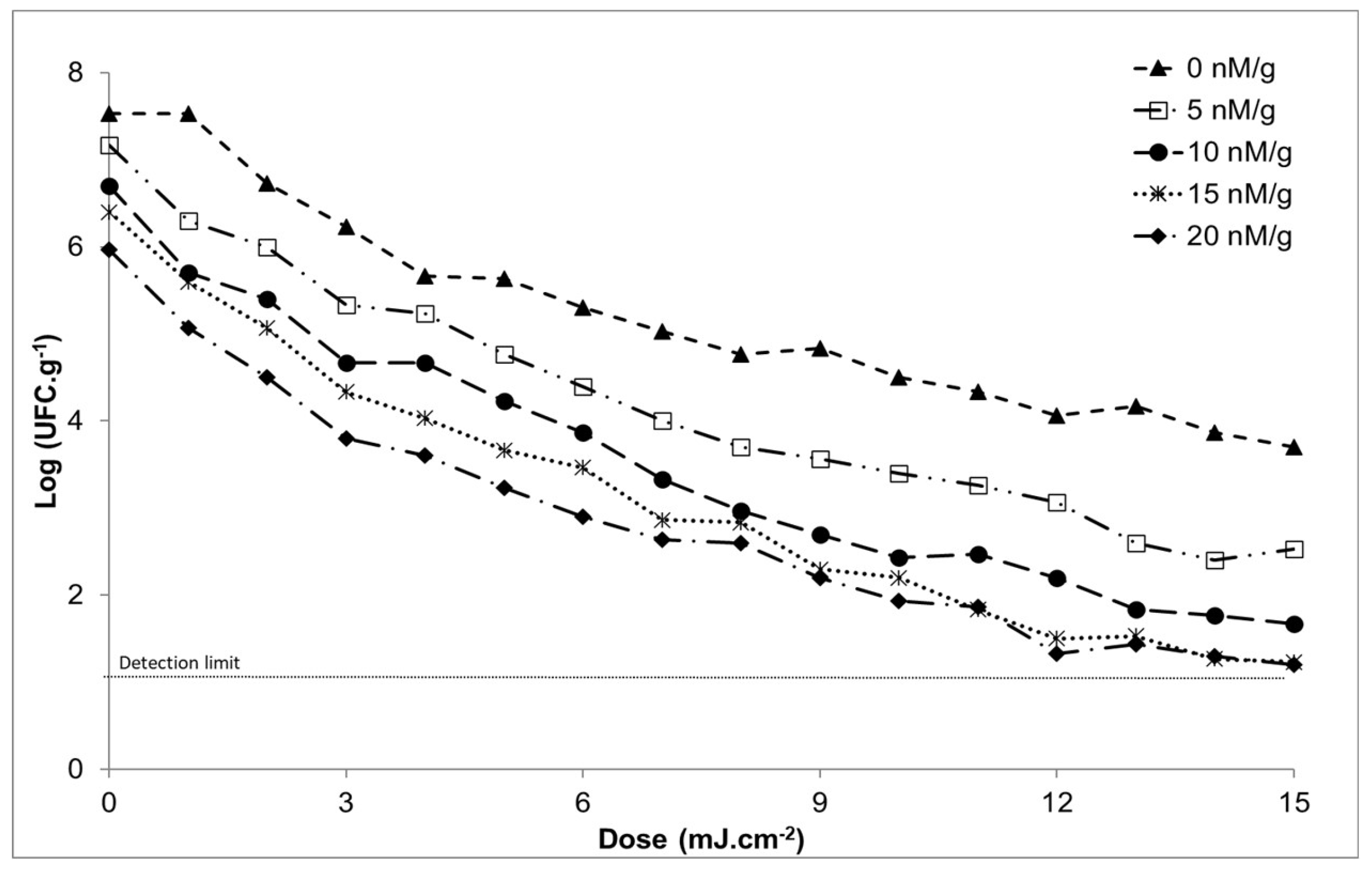
On the other hand, it is observed that when caffeine is applied (Figure 2), microbiological development decreases. If the microbiological development obtained in a sample that has not been subjected to UV light is observed, this is 7.5± 0.2 CFU / g. When the natural inhibiting substance is applied, this value decreases to a minimum of 6.0± 0.2 CFU / g. We can conclude, therefore, that as the dose of this substance increases, the lethality of the microorganism increases.
When modeling the data (Appendix Table A3) the double Weibull model (Equation 5) was the best adjusted to the survival curves using the GInaFiT add-in for Excel®:
N = N 0 ( 1 ± 10 α ) · 10 t 1 δ 1 p 1 ± α ± 10 1 δ 2 p 2
where N is the number of surviving cells (log CFU / ml) after UV-C treatments; N0 is the number of cells before UV-C treatments (log CFU / mL); t corresponds to the time of exposure of the product to UV light; α is the fraction of the first remaining subpopulation in the total population and is defined as the logarithm of f and is equivalent to α = log10 (N01 / N02); p is the shape parameter of the model (dimensionless); and δ is the scale parameter of the model, and represents the dose required to have a first ten-fold reduction in the population (J / cm²).
The Weibull model + tail [37] (Equation 6) was fitted to the survival curves using the GInaFiT add-in for Excel® [32]:
N = ( N 0 N r e s ) ·   10 t δ p   + N r e s
where N is the number of surviving cells (log CFU / ml) after UV-C treatments; N0 is the number of cells before UV-C treatments (log CFU / mL); and Nres is the starting point of the tail (log CFU / mL); t corresponds to the time of exposure of the product to UV light; p is the shape parameter of the model (dimensionless); and δ is the scale parameter of the model, and represents the dose required to have a first ten-fold reduction in the population (J / cm²).
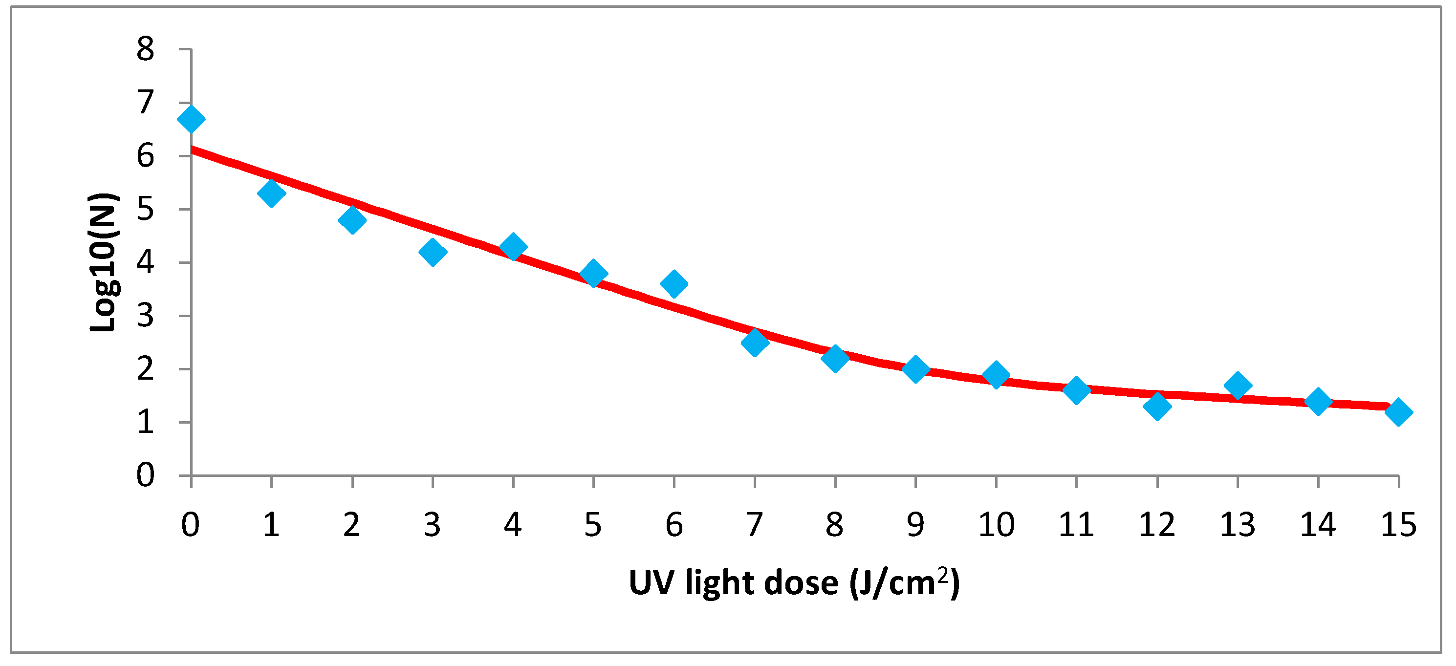
It can also reach a good fit with the biphasic model [40], represented in Figure 3 where the red curve is the fitted model and the blue diamond markers are the data observed in the study and represented in the equation 3, where these values were adjusted to the survival curves using the GInaFIT add-in for Excel® [32], the best fit being the biphasic model, equation 3.
Observing the results obtained, it can be affirmed that there is a greater reduction in microbiological development when applying both non-thermal treatments simultaneously than individually but sequentially, with the optimal application points being 15.0 nM / g and 7.0 J / cm2.
In summary, all the conditions previously described have a slight influence on Salmonella, but after performing the statistical analysis, it has been found that this influence is not significant, so it is not possible to fit our data into a secondary model.
In addition, the models are often compared with data from previously published studies to be validated using specific statistical methodologies. However, due to the lack of data in the consulted bibliography, the model could not be validated with the described precision and bias factors [51].

5. Conclusions

Finally, it can be concluded that the influence of caffeine and UV radiation on the inactivation of Salmonella enteritidis in fresh chicken breast is very promising when applied in combination, leading to reductions in the pathogen load of up to 6.3 log CFU/g. On the other hand, it is observed that in samples submitted only to caffeine the greatest reduction is 1.5 log CFU/g in the highest dose, and in the case of UV light, this is 4.0 CFU/g. By applying the predictive models, a good fit has been achieved in each of the cases studied, reaching R2 values from 0.9998 to 0.9553. Where the most effective combination in the reduction of Salmonella was 12 J/cm2 of UV light and 15 nM/g of caffeine.

Author Contributions

For research articles with several authors, a short paragraph specifying their individual contributions must be provided. The following statements should be used “Conceptualization and methodology, Mendes de Sousa and Posada Izquierdo; software and formal analysis, Palomo Manzano; writing—original draft preparation, García-Gimeno and Posada Izquierdo.; writing—review and editing, García-Gimeno and Posada Izquierdo; supervision and funding acquisition, Mendes de Souza.

Funding

National Council for Scientific and Technological Development (CNPQ-Brazil), the Research Support Foundation of the State of Minas Gerais (FAPEMIG, Brazil), the Federal University of Jequitinhonha and Mucuri Valley (Brazil), the AT17-5686 Project supported by FEDER funds and the Council of Knowledge, Research and University of the Junta of Andalucía. Poliana Mendes de Souza acknowledges the Fundación Carolina and Erasmus Plus program for the financial support to collaborate with the University of Cordoba.

Conflicts of Interest

“The authors declare no conflicts of interest.”

Appendix A

Table A1. Adjustments obtained with the GinaFiT program in chicken breast samples subjected to a temperature of 2 to 22 °C at ultraviolet light ranges of 0.3,6,9,12,15 J / cm2.
Temperature (°C) Model Log-linear regression Log-linear + shoulder Log-linear + tail Log-linear + shoulder + tail* Weibull Weibull
Fixed p-parameter
Weibull + tail* Double Weibull* Biphasic model* Biphasic + shoulder*
2 RMSE 0.5378 0.3117 0.2488 0.2270 0.2314 0.6375 0.1992 0.3061 0.1115 0.1577
R2 adj 0.8895 0.9629 0.9764 0.9803 0.9796 0.8448 0.9848 0.9642 0.9952 0.9905
RMSE 0.4814 0.2045 0.5039 0.2504 0.2490 0.5656 0.2990 0.2990 0.1897 0.2683
R2 adj 0.8356 0.9703 0.8198 0.9555 0.9560 0.7731 0.9366 0.9366 0.9745 0.9489
RMSE 0.4436 0.0544 0.4123 0.0666 0.0816 0.5277 0.1000 0.1000 0.0409 0.0548
R2 adj 0.9163 0.9987 0.9277 0.9981 0.9972 0.8815 0.9957 0.9957 0.9993 0.9987
4 RMSE 0.5337 0.4395 0.2810 0.3433 0.3971 0.6283 0.3441 0.3398 0.3403 0.4816
R2 adj 0.8555 0.9020 0.9599 0.9402 0.9200 0.7997 0.9399 0.9414 0.9412 0.8823
RMSE 0.7604 0.2047 0.6000 0.2508 0.6890 0.8932 0.2752 0.2757 0.2530 0.3577
R2 adj 0.7886 0.9847 0.8684 0.9770 0.8264 0.7083 0.9723 0.9723 0.9766 0.9532
RMSE 0.4052 0.2816 0.4647 0.3449 0.3130 0.4787 0.3834 0.3834 0.3388 0.4764
R2 adj 0.9226 0.9626 0.8983 0.9440 0.9538 0.8921 0.9307 0.9307 0.9459 0.8931
6 RMSE 0.4762 0.3261 0.3978 0.3994 0.3049 0.5619 0.3734 0.3714 0.2976 0.3834
R2 adj 0.8777 0.9427 0.9147 0.9140 0.9499 0.8298 0.9248 0.9256 0.9523 0.9207
RMSE 0.4121 0.2776 0.4638 0.3401 0.3362 0.4859 0.4117 0.4117 0.3033 0.4290
R2 adj 0.9224 0.9648 0.9017 0.9472 0.9484 0.8921 0.9225 0.9225 0.9580 0.9159
RMSE 0.7805 0.2751 0.3999 0.3369 0.5415 0.9133 0.3376 0.3376 0.2973 0.4183
R2 adj 0.6871 0.9611 0.9178 0.9417 0.8494 0.5715 0.9414 0.9414 0.9546 0.9101
8 RMSE 0.3096 0.1820 0.3039 0.1649 0.1374 0.3692 0.1683 0.1683 0.1407 0.1871
R2 adj 0.9338 0.9771 0.9362 0.9812 0.9870 0.9058 0.9804 0.9804 0.9863 0.9758
RMSE 0.5666 0.2787 0.3524 0.2952 0.2145 0.6709 0.3991 0.1879 0.1934 0.2735
R2 adj 0.8815 0.9713 0.9542 0.9678 0.9830 0.8339 0.9412 0.9870 0.9862 0.9724
RMSE 0.5213 0.3358 0.5927 0.4103 0.5159 0.6134 0.4429 0.4429 0.3960 0.5541
R2 adj 0.8753 0.9482 0.8388 0.9227 0.8778 0.8273 0.9100 0.9100 0.9280 0.8591
10 RMSE 0.6261 0.2772 0.6084 0.3395 0.3128 0.7352 0.3831 0.3831 0.3282 0.4637
R2 adj 0.8158 0.9639 0.8260 0.9459 0.9540 0.7460 0.9310 0.9310 0.9494 0.8990
RMSE 0.3984 0.0447 0.3334 0.0498 0.0534 0.4736 0.0654 0.0654 0.0576 0.0815
R2 adj 0.9098 0.9989 0.9368 0.9986 0.9984 0.8725 0.9976 0.9976 0.9981 0.9962
RMSE 0.2008 0.1225 0.1989 0.1496 0.1312 0.2440 0.1607 0.1607 0.1485 0.2074
R2 adj 0.9782 0.9919 0.9786 0.9879 0.9907 0.9678 0.9860 0.9860 0.9881 0.9767
12 RMSE 0.4075 0.1954 0.4325 0.2393 0.2121 0.4851 0.2598 0.2598 0.2396 0.3388
R2 adj 0.9337 0.9847 0.9253 0.9771 0.9820 0.9060 0.9730 0.9730 0.9771 0.9541
RMSE 0.5596 0.4233 0.4924 0.5145 0.4208 0.6560 0.5153 0.5153 0.5215 0.7376
R2 adj 0.8112 0.8920 0.8538 0.8404 0.8933 0.7406 0.8399 0.8399 0.8360 0.6720
RMSE 0.6649 0.2282 0.5477 0.2795 0.2549 0.7804 0.3122 0.3439 0.2651 0.3742
R2 adj 0.7877 0.9750 0.8560 0.9625 0.9688 0.7076 0.9532 0.9432 0.9663 0.9328
14 RMSE 0.4405 0.1211 0.4406 0.1483 0.1574 0.5228 0.1927 0.1927 0.1466 0.2073
R2 adj 0.9102 0.9932 0.9102 0.9898 0.9885 0.8736 0.9828 0.9828 0.9901 0.9801
RMSE 0.6994 0.5778 0.8634 0.7076 0.6137 0.8168 0.7842 0.7516 0.7011 0.9915
R2 adj 0.8035 0.8659 0.7006 0.7989 0.8487 0.7321 0.7530 0.7731 0.8026 0.6052
RMSE 0.6937 0.4784 0.4931 0.4847 0.4561 0.8119 0.5016 0.5111 0.5832 0.8248
R2 adj 0.7179 0.8658 0.8575 0.8623 0.8780 0.6136 0.8525 0.8469 0.8006 0.6011
16 RMSE 0.6937 0.4784 0.4931 0.4847 0.4561 0.8090 0.5016 0.2572 0.5832 0.8248
R2 adj 0.7179 0.8658 0.8575 0.8623 0.8780 0.6154 0.8525 0.9611 0.8006 0.6011
RMSE 0.5794 0.3891 0.5219 0.4765 0.3857 0.6813 0.4724 0.4724 0.4730 0.6690
R2 adj 0.8474 0.9312 0.8762 0.8968 0.9324 0.7890 0.8985 0.8985 0.8983 0.7966
RMSE 0.3476 0.2496 0.4014 0.3057 0.2895 0.4112 0.3546 0.3546 0.2924 0.4135
R2 adj 0.9408 0.9695 0.9211 0.9543 0.9590 0.9172 0.9385 0.9385 0.9581 0.9163
18 RMSE 0.5472 0.1900 0.5384 0.2327 0.2417 0.6454 0.2960 0.2960 0.2037 0.2881
R2 adj 0.8702 0.9843 0.8743 0.9765 0.9747 0.8194 0.9620 0.9620 0.9820 0.9640
RMSE 0.5348 0.1097 0.4345 0.0836 0.1070 0.6341 0.1311 0.1311 0.1411 0.1996
R2 adj 0.8933 0.9955 0.9296 0.9974 0.9957 0.8501 0.9936 0.9936 0.9926 0.9851
RMSE 0.4688 0.2374 0.3425 0.2908 0.2221 0.5531 0.2721 0.1114 0.2048 0.2683
R2 adj 0.8590 0.9638 0.9247 0.9457 0.9683 0.8037 0.9525 0.9920 0.9731 0.9538
20 RMSE 0.4526 0.4866 0.3186 0.3840 0.4636 0.5310 0.3634 0.3634 0.3901 0.5431
R2 adj 0.9136 0.9002 0.9572 0.9378 0.9094 0.8811 0.9443 0.9443 0.9358 0.8757
RMSE 0.8864 0.7444 0.9001 0.9117 0.7578 1.0330 1.0600 0.9281 0.9086 1.2849
R2 adj 0.6931 0.7836 0.6836 0.6753 0.7757 0.5831 0.5611 0.6636 0.6775 0.3551
RMSE 0.8106 0.4411 0.2494 0.3054 0.3199 0.9479 0.3062 0.1801 0.2368 0.3318
R2 adj 0.6465 0.8953 0.9665 0.9498 0.9450 0.5165 0.9496 0.9826 0.9698 0.9408
22 RMSE 0.7309 0.4878 0.8440 0.5974 0.6194 0.8531 0.6927 0.6750 0.5527 0.7817
R2 adj 0.7647 0.8952 0.6863 0.8428 0.8311 0.6795 0.7887 0.7994 0.8655 0.7309
RMSE 0.4208 0.2650 0.4531 0.3246 0.4211 0.4996 0.3983 0.3605 0.2931 0.4099
R2 adj 0.9262 0.9707 0.9144 0.9561 0.9261 0.8959 0.9338 0.9458 0.9642 0.9299
RMSE 0.4889 0.1759 0.2487 0.0997 0.1263 0.5780 0.0948 0.1169 0.1552 0.2194
R2 adj 0.8596 0.9818 0.9637 0.9942 0.9906 0.8038 0.9947 0.9920 0.9859 0.9717
Table A2. adjustments obtained with the GinaFiT program in samples of chicken breast subjected to ultraviolet light at a constant temperature of 14 ° C.
UVC
(J / cm2)
Model Log-linear regression Log-linear ±
shoulder
log-linear ± tail Log-linear ±
shoulder ± tail*
Weibull* Weibull
Fixed p-parameter*
Weibull ± tail* Biphasic model*
o RMSE 0.0399 0.0485 0.0488 0.0686 0.0486 0.0502 0.0688 0.0684
R2 adj 0.9958 0.9938 0.9937 0.9877 0.9938 0.9934 0.9876 0.9877
1 RMSE 0.1292 0.1458 0.1543 0.2051 0.1461 0.1615 0.2066 0.2034
R2 adj 0.9602 0.9493 0.9432 0.8997 0.9491 0.9378 0.8982 0.9013
2 RMSE 0.1026 0.0942 0.1256 0.1333 0.0938 0.1199 0.1325 0.1776
R2 adj 0.9786 0.9819 0.9679 0.9639 0.9821 0.9708 0.9643 0.9358
3 RMSE 0.0869 0.1032 0.1065 0.1428 0.1036 0.1047 0.1434 0.1506
R2 adj 0.9866 0.9812 0.9800 0.9639 0.9810 0.9806 0.9637 0.9599
4 RMSE 0.0710 0.0719 0.0868 0.0194 0.0763 0.0824 0.0121 0.1229
R2 adj 0.9928 0.9926 0.9892 0.9995 0.9917 0.9903 0.9998 0.9783
5 RMSE 0.0380 0.0417 0.0235 0.0219 0.0399 0.0526 0.0210 0.0332
R2 adj 0.9979 0.9975 0.9992 0.9993 0.9977 0.9960 0.9994 0.9984
6 RM5E 0.0678 0.0509 0.0785 0.0720 0.0546 0.0907 0.0772 0.0686
R2 adj 0.9932 0.9962 0.9909 0.9924 0.9956 0.9879 0.9912 0.9931
7 RMSE 0.1970 0.1234 0.0225 0.0121 0.1100 0.2511 0.0100 0.0060
R2 adj 0.9488 0.9799 0.9993 0.9998 0.9841 0.9168 0.9999 1.0000
8 RMSE 0.3864 0.1616 0.1104 0.1343 0.2113 0.4834 0.1289 0.0767
R2 adj 0.8186 0.9683 0.9852 0.9781 0.9457 0.7161 0.9798 0.9929
9 RMSE 0.3342 0.2330 0.0178 0.0184 0.2127 0.4198 0.0197 0.0250
R2 adj 0.8817 0.9425 0.9997 0.9996 0.9521 0.8134 0.9996 0.9993
10 RMSE 0.3360 0.2380 0.1128 0.1567 0.2191 0.4220 0.1587 0.1324
R2 adj J 0.8854 0.9425 0.9871 0.9751 0.9513 0.8191 0.9744 0.9822
11 RMSE 0.3088 0.2764 0.0988 0.1138 0.2582 0.3873 0.1055 0.1397
R2 adj J 0.8966 0.9172 0.9894 0.9860 0.9277 0.8373 0.9879 0.9788
12 RM5E 0.3199 0.2281 0.0528 0.0444 0.1942 0.4047 0.0507 0.0742
R2 adj 0.9261 0.9624 0.9980 0.9986 0.9728 0.8817 0.9981 0.9960
13 RMSE 0.4358 0.1895 0.0823 0.0271 0.2460 0.5451 0.0204 0.0401
R2 adj 0.8143 0.9649 0.9934 0.9993 0.9408 0.7094 0.9996 0.9984
14 RMSE 0.4375 0.2009 0.1448 0.1113 0.1834 0.5471 0.1206 0.1861
R2 adj 0.8130 0.9606 0.9795 0.9879 0.9671 0.7076 0.9858 0.9662
15 RM5E 0.3691 0.2540 0.0187 0.0255 0.2321 0.4628 0.0249 0.1502
R2 adj 0.8627 0.9350 0.9996 0.9993 0.9457 0.7841 0.9994 0.9773
Table A3. Adjustments obtained with the GinaFiT program in samples of chicken breast subjected to caffeine at a constant temperature of 14 ° C.
Caffeine
(nM/g)
Model Log-linear regression Log-linear + shoulder Log-linear + tail Log-linear + shoulder + tail Weibull Weibull
Fixed p-parameter
Weibull + tail Double Weibull Biphasic model Biphasic + shoulder
0 RMSE 0.3197 0.1481 0.2739 0.1518 0.1273 0.3398 0.1304 0.1127 0.1133 0.1159
R2 adj 0.9224 0.9833 0.9430 0.9825 0.9877 0.9123 0.9871 0.9903 0.9902 0.9898
RMSE 0.4962 0.4084 0.5130 0.4251 0.4063 0.5204 0.4531 0.4229 0.4126 0.4310
R2 adj 0.8446 0.8947 0.8339 0.8859 0.8958 0.8290 0.8704 0.8871 0.8925 0.8827
RMSE 0.4871 0.4191 0.4027 0.3896 0.4022 0.5107 0.3974 0.4271 0.4261 0.4451
R2 adj 0.8103 0.8595 0.8703 0.8786 0.8706 0.7915 0.8737 0.8542 0.8548 0.8416
5 RMSE 0.2630 0.1764 0.2711 0.1836 0.1915 0.2808 0.2038 0.1993 0.1842 0.1924
R2 adj 0.9683 0.9857 0.9663 0.9846 0.9832 0.9639 0.9810 0.9818 0.9845 0.9830
RMSE 0.4241 0.3509 0.3412 0.3107 0.3107 0.4489 0.4035 0.3060 0.3273 0.3418
R2 adj 0.9239 0.9479 0.9507 0.9592 0.9591 0.9147 0.9311 0.9604 0.9547 0.9505
RMSE 0.4881 0.4057 0.2958 0.2934 0.3486 0.5149 0.2935 0.2909 0.2943 0.3073
R2 adj 0.8743 0.9131 0.9538 0.9546 0.9359 0.8601 0.9545 0.9553 0.9543 0.9501
10 RMSE 0.3585 0.2517 0.2320 0.1685 0.2055 0.3821 0.3124 0.1814 0.2332 0.2435
R2 adj 0.9425 0.9717 0.9758 0.9873 0.9811 0.9347 0.9564 0.9853 0.9757 0.9735
RMSE 0.3575 0.2671 0.2505 0.2042 0.2267 0.3810 0.2119 0.2120 0.2442 0.2551
R2 adj 0.9479 0.9709 0.9744 0.9830 0.9791 0.9408 0.9817 0.9817 0.9757 0.9735
RMSE 0.5428 0.4842 0.4793 0.4753 0.4482 0.5713 0.4721 0.4831 0.4536 0.4738
R2 adj 0.8914 0.9136 0.9153 0.9167 0.9259 0.8797 0.9178 0.9140 0.9242 0.9173
15 RMSE 0.3702 0.3166 0.3647 0.3244 0.3147 0.3917 0.3276 0.3276 0.3029 0.3072
R2 adj 0.9524 0.9652 0.9538 0.9635 0.9656 0.9467 0.9627 0.9627 0.9681 0.9672
RMSE 0.4405 0.3679 0.2684 0.3225 0.2853 0.4675 0.3674 0.2129 0.3125 0.2181
R2 adj 0.9169 0.9420 0.9691 0.9554 0.9651 0.9064 0.9422 0.9806 0.9649 0.9796
RMSE 0.5291 0.3820 0.3091 0.2341 0.3112 0.5600 0.5696 0.2630 0.3125 0.3264
R2 adj 0.8995 0.9476 0.9657 0.9803 0.9652 0.8874 0.8835 0.9752 0.9649 0.9617
20 RMSE 0.4977 0.3750 0.3231 0.3128 0.3017 0.5258 0.2914 0.2395 0.2415 0.2523
R2 adj 0.8747 0.9288 0.9472 0.9505 0.9539 0.8601 0.9570 0.9710 0.9705 0.9678
RMSE 0.5530 0.3819 0.4105 0.3561 0.3043 0.5837 0.2890 0.2587 0.2598 0.2713
R2 adj 0.8589 0.9327 0.9222 0.9415 0.9573 0.8427 0.9614 0.9691 0.9688 0.9660
RMSE 0.3976 0.3359 0.3571 0.3244 0.3519 0.4188 0.3524 0.3533 0.3717 0.4890
R2 adj 0.9296 0.9498 0.9432 0.9531 0.9449 0.9219 0.9447 0.9444 0.9385 0.8935

References

  1. Townshend, A. Food and Nutritional Analysis - Meat and Meat Products. In Encyclopedia of Analytical Science; Elsevier Applied Science, 2019; pp 436–450.
  2. EU Parliament and Council. Regulation (EC) No. 853/2004 of the European Parliament and of the Council of April 29, 2004, which establishes specific hygiene rules for food of animal origin. DO L 2004, 139, 55. [Google Scholar]
  3. Chouliara, E.; Karatapanis, A.; Savvaidis, I. N.; Kontominas, M. G. Combined Effect of Oregano Essential Oil and Modified Atmosphere Packaging on Shelf-Life Extension of Fresh Chicken Breast Meat, Stored at 4 °C. Food Microbiol 2007, 24, 607–617. [Google Scholar] [CrossRef] [PubMed]
  4. Barbut, S. Poultry Products Processing. CRC Press eBooks, 2016.
  5. Yang, S.; Park, S. Y.; Ha, S. Do. A Predictive Growth Model of Aeromonas hydrophila on Chicken Breasts under Various Storage Temperatures. LWT - Food Science and Technology 2016, 69, 98–103. [Google Scholar] [CrossRef]
  6. Boelaert, F.; Tegegne, H. A.; Stoicescu, A.; Messens, W.; Hempen, M.; Rossi, M.; Sarno, E.; Rizzi, V.; Aznar, I.; Antoniou, S. E.; Baldinelli, F.; Van der Stede, Y.; Niskanen, T.; Young, J.; Merk, H.; Quinten, C.; Barco, L.; Mancin, M.; Garbo, A.; Patuzzi, I.; Lettini, A. A.; Losasso, C.; Mantovani, C.; Morabito, S.; Scavia, G.; Knijn, A.; Tozzoli, R.; Moro, O.; Gianfranceschi, M.; Suffredini, E.; Di Bartolo, I.; Delibato, E.; Anniballi, F.; Ianiro, G.; Altieri, I.; Maugliani, A.; Morales, M. A. G.; Casulli, A.; Cacciò, S.; Albert, D. The European Union One Health 2018 Zoonoses Report. EFSA Journal 2019, 17. [Google Scholar] [CrossRef]
  7. Aymerich, T.; Picouet, P. A.; Monfort, J. M. Decontamination Technologies for Meat Products. Meat Sci 2008, 78, 114–129. [Google Scholar] [CrossRef] [PubMed]
  8. Chen, J. H.; Ren, Y.; Seow, J.; Liu, T.; Bang, W. S.; Yuk, H. G. Intervention Technologies for Ensuring Microbiological Safety of Meat: Current and Future Trends. Comprehensive Reviews in Food Science and Food Safety. March 2012, pp 119–132. [CrossRef]
  9. Troy, D. J.; Ojha, K. S.; Kerry, J. P.; Tiwari, B. K. Sustainable and Consumer-Friendly Emerging Technologies for Application within the Meat Industry: An Overview. Meat Sci 2016, 120, 2–9. [Google Scholar] [CrossRef] [PubMed]
  10. Guerrero-Beltrán, J. A.; Barbosa-Cánovas, G. V. Review: Advantages and Limitations on Processing Foods by UV Light. Food Science and Technology International. June 2004, pp 137–147. [CrossRef]
  11. Gabriel, A. A. Inactivation of Escherichia coli O157:H7 and Spoilage Yeasts in Germicidal UV-C-Irradiated and Heat-Treated Clear Apple Juice. Food Control 2012, 25, 425–432. [Google Scholar] [CrossRef]
  12. Amanina, A. K. Z.; Rosnah, S.; Noranizan, M. A.; Alifdalino, S. UV-C Effect on Microbial Disinfection of Pineapple-Mango Juice Blend Using Dean-Vortex Technology. Food Res 2019, 3, 285–294. [Google Scholar] [CrossRef]
  13. Caminiti, I. M.; Palgan, I.; Muñoz, A.; Noci, F.; Whyte, P.; Morgan, D. J.; Cronin, D. A.; Lyng, J. G. The Effect of Ultraviolet Light on Microbial Inactivation and Quality Attributes of Apple Juice. Food Bioproc Tech 2012, 5, 680–686. [Google Scholar] [CrossRef]
  14. Koutchma, T.; Parisi, B. Biodosimetry of Escherichia Coli UV Inactivation in Model Juices with Regard to Dose Distribution in Annular UV Reactors. J Food Sci 2004, 69. [Google Scholar] [CrossRef]
  15. Chun, H. H.; Kim, J. Y.; Lee, B. D.; Yu, D. J.; Song, K. B. Effect of UV-C Irradiation on the Inactivation of Inoculated Pathogens and Quality of Chicken Breasts during Storage. Food Control 2010, 21, 276–280. [Google Scholar] [CrossRef]
  16. Barba, F. J.; Koubaa, M.; do Prado-Silva, L.; Orlien, V.; Sant’Ana, A. Mild Processing Applied to the Inactivation of the Main Foodborne Bacterial Pathogens: A Review. Trends in Food Science and Technology. Elsevier Ltd. August 1, 2017, pp 20–35. [CrossRef]
  17. Reichel, J.; Kehrenberg, C.; Krischek, C. UV-C Irradiation of Rolled Fillets of Ham Inoculated with Yersinia enterocolitica and Brochothrix thermosphacta. Foods 2020, 9. [Google Scholar] [CrossRef] [PubMed]
  18. Porciúncula, L. O.; Sallaberry, C.; Mioranzza, S.; Botton, P. H. S.; Rosemberg, D. B. The Janus Face of Caffeine. Neurochem Int 2013, 63, 594–609. [Google Scholar] [CrossRef] [PubMed]
  19. Madusanka, N.; Eddleston, M. D.; Arhangelskis, M.; Jones, W. Polymorphs, Hydrates and Solvates of a Co-Crystal of Caffeine with Anthranilic Acid. Acta Crystallogr B Struct Sci Cryst Eng Mater 2014, 70, 72–80. [Google Scholar] [CrossRef] [PubMed]
  20. Ibrahim, S. A.; Salameh, M. M.; Phetsomphou, S.; Yang, H.; Seo, C. W. Application of Caffeine, 1,3,7-Trimethylxanthine, to Control Escherichia coli O157:H7. Food Chem 2006, 99, 645–650. [Google Scholar] [CrossRef]
  21. Possas, A.; Valero, A.; García-Gimeno, R. M.; Pérez-Rodríguez, F.; Mendes de Souza, P. Combining UV-C Technology and Caffeine Application to Inactivate Escherichia Coli on Chicken Breast Fillets. Food Control 2021, 129. [Google Scholar] [CrossRef]
  22. Abosede, O. O.; Gordon, A. T.; Dembaremba, T. O.; Lorentino, C. M. A.; Frota, H. F.; Santos, A. L. S.; Hosten, E. C.; Ogunlaja, A. S. Trimesic Acid-Theophylline and Isopthalic Acid-Caffeine Cocrystals: Synthesis, Characterization, Solubility, Molecular Docking, and Antimicrobial Activity. Cryst Growth Des 2020, 20, 3510–3522. [Google Scholar] [CrossRef]
  23. Maletta, A. B.; Were, L. M. Effect of Coffee Filtrate, Methylglyoxal, Glyoxal, and Caffeine on Salmonella Typhimurium and S. Enteritidis Survival in Ground Chicken Breasts. J Food Sci 2012, 77. [Google Scholar] [CrossRef] [PubMed]
  24. Haas, C.; Rose, J.; Gerba, C. Quantitative Microbial Risk Assessment, 2nd Edition.; John Wiley & Sons Inc: Hoboken, New Jersey, 2014. [Google Scholar]
  25. Dean, K.; Wissler, A.; Hernandez-Suarez, J. S.; Nejadhashemi, A. P.; Mitchell, J. Modeling the Persistence of Viruses in Untreated Groundwater. Science of the Total Environment 2020, 717. [Google Scholar] [CrossRef] [PubMed]
  26. McMeekin, T.; Olley, J.; Ratkowsky, D.; Corkrey, R.; Ross, T. Predictive Microbiology Theory and Application: Is It All about Rates? Food Control 2013, 29, 290–299. [Google Scholar] [CrossRef]
  27. Possas, A.; Valero, A.; García-Gimeno, R. M.; Pérez-Rodríguez, F.; de Souza, P. M. Influence of Temperature on the Inactivation Kinetics of Salmonella Enteritidis by the Application of UV-C Technology in Soymilk. Food Control 2018, 94, 132–139. [Google Scholar] [CrossRef]
  28. Keklik, N. M.; Demirci, A.; Puri, V. M.; Heinemann, P. H. Modeling the Inactivation of Salmonella Typhimurium, Listeria monocytogenes, and Salmonella Enteritidis on Poultry Products Exposed to Pulsed UV Light. J Food Prot 2012, 75, 281–288. [Google Scholar] [CrossRef] [PubMed]
  29. Hessel, C. T.; de Freitas Costa, E.; Boff, R. T.; Pessoa, J. P.; Tondo, E. C. A Systematic Review and Bayesian Meta-Analysis about Salmonella spp. Prevalence on Raw Chicken Meat. Microb Risk Anal 2022, 21, 100205. [Google Scholar] [CrossRef]
  30. Pagal, G. A.; Gabriel, A. A. Individual and Combined Mild Heat and UV-C Processes for Orange Juice against Escherichia coli O157:H7. LWT 2020, 126. [Google Scholar] [CrossRef]
  31. AOAC International; Latimer, G. W. AOAC International. In Official methods of analysis of AOAC International; 2012.
  32. Geeraerd, A. H.; Valdramidis, V. P.; Van Impe, J. F. GInaFiT, a Freeware Tool to Assess Non-Log-Linear Microbial Survivor Curves. Int J Food Microbiol 2005, 102, 95–105. [Google Scholar] [CrossRef] [PubMed]
  33. Soro, A. B.; Harrison, S. M.; Whyte, P.; Bolton, D. J.; Tiwari, B. K. Impact of Ultraviolet Light and Cold Plasma on Fatty Acid Profile of Raw Chicken and Pork Meat. Journal of Food Composition and Analysis 2022, 114. [Google Scholar] [CrossRef]
  34. Kim, Y. H.; Jeong, S. G.; Back, K. H.; Park, K. H.; Chung, M. S.; Kang, D. H. Effect of Various Conditions on Inactivation of Escherichia coli O157:H7, Salmonella Typhimurium, and Listeria monocytogenes in Fresh-Cut Lettuce Using Ultraviolet Radiation. Int J Food Microbiol 2013, 166, 349–355. [Google Scholar] [CrossRef]
  35. Baysal, A. H.; Molva, C.; Unluturk, S. UV-C Light Inactivation and Modeling Kinetics of Alicyclobacillus acidoterrestris Spores in White Grape and Apple Juices. Int J Food Microbiol 2013, 166, 494–498. [Google Scholar] [CrossRef] [PubMed]
  36. Gabriel, A. A.; Vera, D. D.; Lazo, O. M. Y.; Azarcon, V. B.; De Ocampo, C. G.; Marasigan, J. C.; Sandel, G. T. Ultraviolet-C Inactivation of Escherichia coli O157:H7, Listeria monocytogenes, Pseudomonas aeruginosa, and Salmonella enterica in Liquid Egg White. Food Control 2017, 73, 1303–1309. [Google Scholar] [CrossRef]
  37. Albert, I.; Mafart, P. A Modified Weibull Model for Bacterial Inactivation. In International Journal of Food Microbiology; Elsevier, 2005; Vol. 100, pp 197–211. [CrossRef]
  38. Mutz, Y. S.; Rosario, D. K. A.; Bernardes, P. C.; Paschoalin, V. M. F.; Conte-Junior, C. A. Modeling Salmonella Typhimurium Inactivation in Dry-Fermented Sausages: Previous Habituation in the Food Matrix Undermines UV-C Decontamination Efficacy. Front Microbiol 2020, 11. [Google Scholar] [CrossRef] [PubMed]
  39. Siguemoto, É. S.; Gut, J. A. W.; Martinez, A.; Rodrigo, D. Inactivation Kinetics of Escherichia coli O157:H7 and Listeria monocytogenes in Apple Juice by Microwave and Conventional Thermal Processing. Innovative Food Science & Emerging Technologies 2018, 45, 84–91. [Google Scholar] [CrossRef]
  40. CERF, O. A Review Tailing of Survival Curves of Bacterial Spores. Journal of Applied Bacteriology 1977, 42, 1–19. [Google Scholar] [CrossRef] [PubMed]
  41. Gündüz, G. T.; Korkmaz, A. UV-C Treatment for the Inhibition of Molds Isolated from Dried Persimmons (Diospyros Kaki L.) and Modelling of UV-C Inactivation Kinetics. LWT 2019, 115. [Google Scholar] [CrossRef]
  42. Haughton, P. N.; Lyng, J. G.; Cronin, D. A.; Morgan, D. J.; Fanning, S.; Whyte, P. Efficacy of UV Light Treatment for the Microbiological Decontamination of Chicken, Associated Packaging, and Contact Surfaces. J Food Prot 2011, 74, 565–572. [Google Scholar] [CrossRef] [PubMed]
  43. Gyawali, R.; Adkins, A.; C. Minor, R.; Ibrahim, S. A. Behavior and Changes in Cell Morphology of Escherichia coli O157:H7 in Liquid Medium and Skim Milk in the Presence of Caffeine. CYTA - Journal of Food 2014, 12, 235–241. [CrossRef]
  44. Almeida, A. A. P.; Naghetini, C. C.; Santos, V. R.; Antonio, A. G.; Farah, A.; Glória, M. B. A. Influence of Natural Coffee Compounds, Coffee Extracts and Increased Levels of Caffeine on the Inhibition of Streptococcus mutans. Food Research International 2012, 49, 459–461. [Google Scholar] [CrossRef]
  45. Bigelow, W. D.; Esty, J. R. The Thermal Death Point in Relation to Time of Typical Thermophilic Organisms. J Infect Dis 1920, 27, 602–617. [Google Scholar] [CrossRef]
  46. Melnik, M.; Sprusansky, O.; Musil, P. Bio-Medical Aspects of Purine Alkaloids. Adv Biol Chem 2014, 04, 274–280. [Google Scholar] [CrossRef]
  47. Apshingekar, P. P.; Aher, S.; Kelly, A. L.; Brown, E. C.; Paradkar, A. Synthesis of Caffeine/Maleic Acid Co-Crystal by Ultrasound-Assisted Slurry Co-Crystallization. J Pharm Sci 2017, 106, 66–70. [Google Scholar] [CrossRef] [PubMed]
  48. Daurio, D.; Medina, C.; Saw, R.; Nagapudi, K.; Alvarez-Núñez, F. Application of Twin Screw Extrusion in the Manufacture of Cocrystals, Part I: Four Case Studies. Pharmaceutics 2011, 3, 582–600. [Google Scholar] [CrossRef] [PubMed]
  49. Leyssens, T.; Tumanova, N.; Robeyns, K.; Candoni, N.; Veesler, S. Solution Cocrystallization, an Effective Tool to Explore the Variety of Cocrystal Systems: Caffeine/Dicarboxylic Acid Cocrystals. CrystEngComm 2014, 16, 9603–9611. [Google Scholar] [CrossRef]
  50. El Hamdani, H.; El Amane, M.; Duhayon, C. Studies on the Syntheses, Structural Characterization, Antimicrobial of the CO-CRYSTAL 1,10-Phenanthrolin-1-IUM(1,10-PhenH+)-Caffeine(Caf)-Hexafluorophosphate. J Mol Struct 2018, 1155, 789–796. [Google Scholar] [CrossRef]
  51. Ross, T. Indices for Performance Evaluation of Predictive Models in Food Microbiology. Journal of Applied Bacteriology 1996, 81, 501–508. [Google Scholar] [CrossRef] [PubMed]
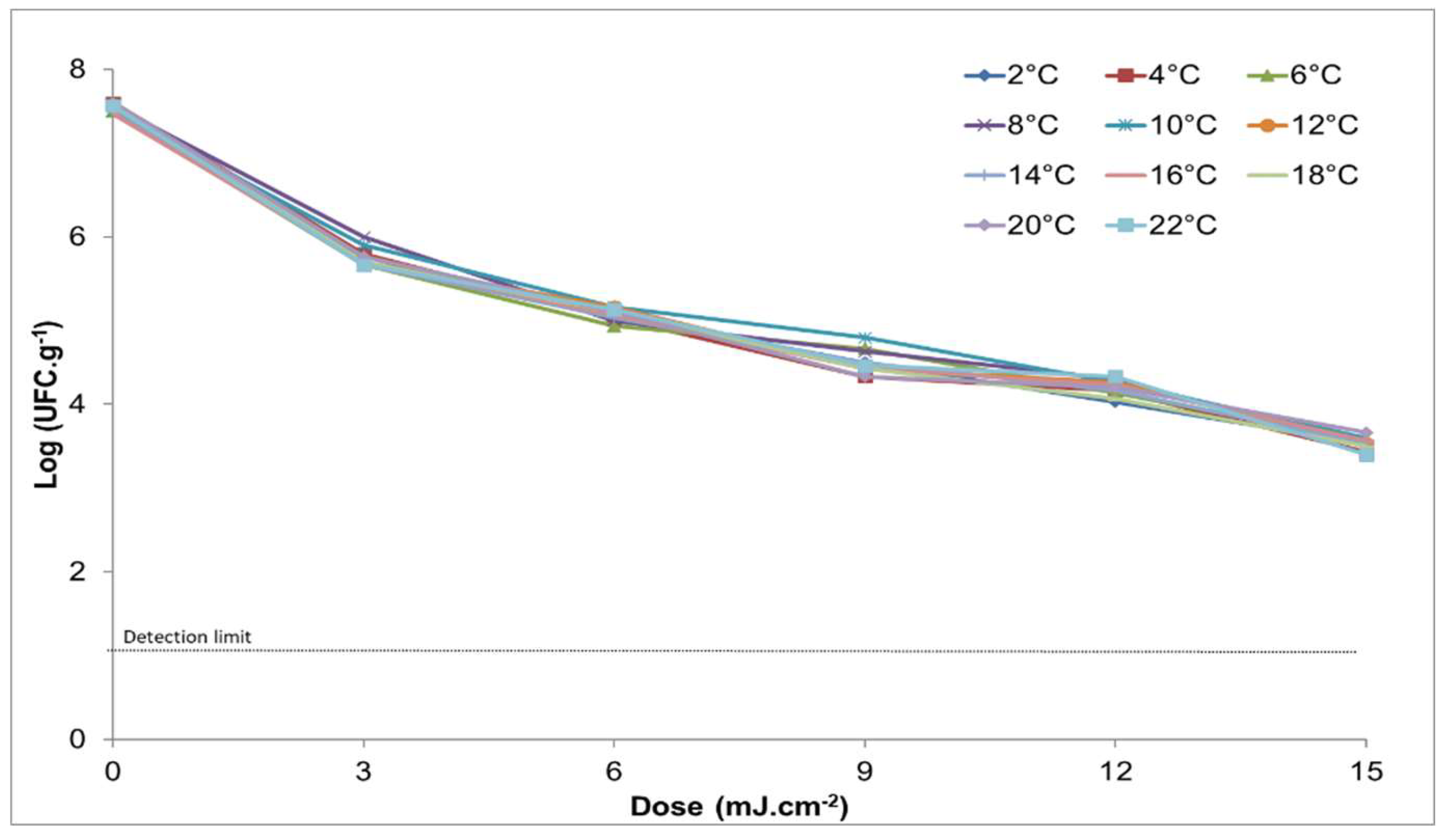
Figure 1. Graphic representation of the microbiological development (log CFU / g) when applying different doses of UV light (0,3,6,9,12,15) J / cm2 on the reduction of Salmonella at different temperatures.
Preprints 114063 g001
Figure 2. Graph that represents doses of UV light vs Log, at different doses of caffeine at a temperature of 14ºC.
Preprints 114063 g002
Figure 3. Fit of observed data (dotted points) for the biphasic model of the treatment of UV light with 15nM/g of caffeine.
Preprints 114063 g003
Table 1. Physical-chemical characterization (average ± SD) of the chicken breast after the application of caffeine at doses 0, 5, 10, 15 and 20 nM / g.
Caffeine
(nM / g)
Protein
(%)
Grease
(%)
Humidity
(%)
Ash
(%)
Total acidity
(%)
pH Absorption
coefficient (cm-1)
0 20.82 ± 0.18 2.85 ± 0.08 74.85 ± 2.28 1.69 ± 0.05 0.21 ± 0.05 5.87 ± 0.08 959.2 ± 46.7
5 20.97 ± 1.48 2.84 ± 0.07 76.56 ± 2.40 1.66 ± 0.10 0.24 ± 0.05 5.84 ± 0.06 961.5 ± 31.2
10 21.13 ± 2.00 2.86 ± 0.12 75.40 ± 2.18 1.67 ± 0.06 0.22 ± 0.03 5.82 ± 0.08 965.8 ± 42.9
15 20.77 ± 1.07 2.80 ± 0.09 76.69 ± 1.70 1.65 ± 0.08 0.23 ± 0.03 5.84 ± 0.05 961.3 ± 79.7
20 21.18 ± 1.70 2.86 ± 0.05 74.94 ± 2.71 1.67 ± 0.04 0.24 ± 0.02 5.82 ± 0.12 962.6 ± 49.1
Table 2. Microbiological counts (CFU / g) of the experimental data in chicken breast subjected to a dose of UV light between 0 and 15 J / cm2 at a temperature of 2 to 22ºC.
Dose (J / cm2)
T (° C) 0.0 3.0 6.0 9.0 12.0 15.0
2 7.5 ± 0.2 5.8 ± 0.2 5.1 ± 0.2 4.5 ± 0.3 4.0 ± 0.2 3.5 ± 0.2
4 7.6 ± 0.2 5.8 ± 0.3 5.0 ± 0.2 4.3 ± 0.4 4.2 ± 0.2 3.4 ± 0.3
6 7.5 ± 0.1 5.7 ± 0.3 4.9 ± 0.4 4.7 ± 0.1 4.1 ± 0.2 3.6 ± 0.2
8 7.6 ± 0.2 6.0 ± 0.1 5.0 ± 0.2 4.6 ± 0.3 4.3 ± 0.2 3.5 ± 0.3
10 7.5 ± 0.2 5.9 ± 0.1 5.2 ± 0.2 4.8 ± 0.1 4.3 ± 0.2 3.6 ± 0.1
12 7.6 ± 0.2 5.7 ± 0.1 5.2 ± 0.2 4.4 ± 0.3 4.3 ± 0.2 3.5 ± 0.3
14 7.5 ± 0.2 5.7 ± 0.1 5.0 ± 0.0 4.5 ± 0.1 4.2 ± 0.5 3.5 ± 0.5
16 7.5 ± 0.2 5.7 ± 0.2 5.1 ± 0.2 4.4 ± 0.2 4.2 ± 0.3 3.6 ± 0.4
18 7.6 ± 0.1 5.7 ± 0.2 5.1 ± 0.2 4.4 ± 0.2 4.1 ± 0.4 3.5 ± 0.3
20 7.6 ± 0.2 5.8 ± 0.3 5.1 ± 0.5 4.3 ± 0.4 4.2 ± 0.6 3.7 ± 0.3
22 7.6 ± 0.2 5.7 ± 0.2 5.1 ± 0.2 4.5 ± 0.2 4.3 ± 0.2 3.4 ± 0.3
Mean log (CFU / g) ± SD log (CFU / g).
Table 3. Effect of caffeine Microbiological counts (log CFU / g) of Salmonella in chicken breast at a constant temperature of 14ºC and the inactivation rate.
Caffeine dose (nM / g) Without caffeine* With caffeine* Inactivation rate
0 7.5 ± 0.2 7.5 ± 0.2 0.23
5 7.6 ± 0.2 7.2 ± 0.0 0.30
10 7.6 ± 0.2 6.7 ± 0.1 0.32
15 7.6 ± 0.2 6.4 ± 0.2 0.33
20 7.5 ± 0.2 6.0 ± 0.2 0.29
* Average log (CFU / g) ± SD log (CFU / g).
Table 4. Microbiological counts (CFU / g) of Salmonella in chicken breast subjected to different doses of UV light in combination of different doses of caffeine at 14ºC.
Caffeine (nM / g)
Dose (J / cm2) 0 5 10 15 20
0 7.5 ± 0.2 7.2 ± 0.0 6.7 ± 0.1 6.4 ± 0.2 6.0 ± 0.2
1 6.7 ± 0.2 6.3 ± 0.1 5.7 ± 0.1 5.6 ± 0.2 5.1 ± 0.2
2 6.2 ± 0.2 6.0 ± 0.1 5.4 ± 0.1 5.1 ± 0.2 4.5 ± 0.3
3 5.7 ± 0.1 5.3 ± 0.2 4.7 ± 0.2 4.3 ± 0.1 3.8 ± 0.2
4 5.6 ± 0.2 5.2 ± 0.2 4.7 ± 0.2 4.0 ± 0.2 3.6 ± 0.2
5 5.3 ± 0.1 4.8 ± 0.1 4.2 ± 0.2 3.7 ± 0.1 3.2 ± 0.2
6 5.0 ± 0.0 4.4 ± 0.1 3.9 ± 0.0 3.5 ± 0.1 2.9 ± 0.3
7 4.8 ± 0.3 4.0 ± 0.1 3.3 ± 0.2 2.9 ± 0.2 2.6 ± 0.3
8 4.8 ± 0.2 3.7 ± 0.3 3.0 ± 0.2 2.8 ± 0.6 2.6 ± 0.4
9 4.5 ± 0.1 3.6 ± 0.4 2.7 ± 0.5 2.3 ± 0.2 2.2 ± 0.1
10 4.3 ± 0.3 3.4 ± 0.2 2.4 ± 0.4 2.2 ± 0.2 1.9 ± 0.2
11 4.1 ± 0.3 3.3 ± 0.3 2.5 ± 0.5 1.8 ± 0.2 1.9 ± 0.4
12 4.2 ± 0.5 3.1 ± 0.3 2.2 ± 0.2 1.5 ± 0.1 1.3 ± 0.2
13 3.9 ± 0.4 2.6 ± 0.4 1.8 ± 0.2 1.5 ± 0.4 1.4 ± 0.3
14 3.7 ± 0.2 2.4 ± 0.2 1.8 ± 0.1 1.3 ± 0.2 1.3 ± 0.3
15 3.5 ± 0.5 2.5 ± 0.4 1.7 ± 0.3 1.2 ± 0.2 1.2 ± 0.2
Mean log (CFU / g) ± SD log (CFU / g).
Disclaimer/Publisher’s Note: The statements, opinions and data contained in all publications are solely those of the individual author(s) and contributor(s) and not of MDPI and/or the editor(s). MDPI and/or the editor(s) disclaim responsibility for any injury to people or property resulting from any ideas, methods, instructions or products referred to in the content.
Copyright: This open access article is published under a Creative Commons CC BY 4.0 license, which permit the free download, distribution, and reuse, provided that the author and preprint are cited in any reuse.
Prerpints.org logo

Preprints.org is a free preprint server supported by MDPI in Basel, Switzerland.

Subscribe

© 2024 MDPI (Basel, Switzerland) unless otherwise stated