1. Introduction
Coxofemoral dysplasia (CFD) (
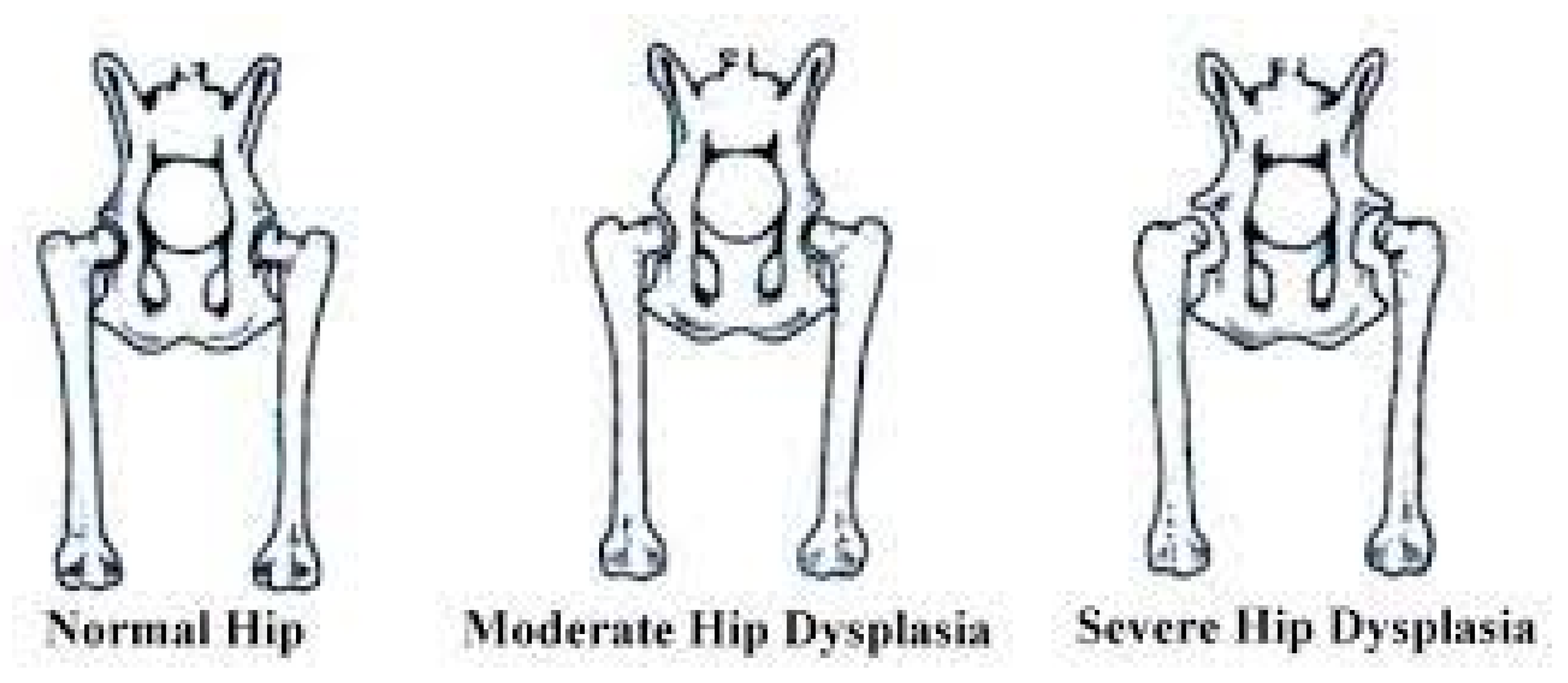
Figure 1) is a prevalent orthopedic disease characterized by joint incongruity and subsequent osteoarthritis and pain, influenced by complex interactions of multiple genes and environmental factors impacting disease susceptibility [1,3,8,37]. Although the coxofemoral joint is normal at birth, the imbalance between skeletal and muscular system growth rates during development imposes excessive loads on the femoral head, altering acetabular conformation and leading to irregular joint surface remodeling [6,36,38].
CFD can progress to painful secondary osteoarthritis and affect the dog’s behavior, manifesting as chronic lameness, muscle atrophy, and exercise reluctance [2,23,50]. While medical and surgical management can alleviate pain, they do not address underlying skeletal muscle conditions, which necessitates physiotherapy for effective treatment [57].
Clinical signs of CFD include pain, lameness, stiff gait, and muscle atrophy, with diagnosis relying on comprehensive medical history, clinical evaluation, and imaging [6,10,23]. Manifestations may remain latent for extended periods, becoming apparent with the onset of degenerative joint disease.
The pathophysiology involves muscle contractures, joint inflammation, and increased intra-articular pressure, contributing to pain and joint dysfunction [3,6,26,28]. Contractures of the adductor muscles, notably the pectineus, can cause microfractures during growth remodeling, exacerbating the condition.
In patients with osteoarthritis, increased intra-articular pressure and subchondral bone ischemia are prominent contributors to pain [3,6,26]. Understanding the multifaceted etiology and clinical manifestations of CFD is essential for targeted therapeutic interventions.
The selection of an appropriate treatment strategy for CFD depends on various factors, including the patient’s age, the severity of dysplasia, and the presence of any concurrent pathologies. The primary objectives are to alleviate pain, improve limb function, and optimize the patient’s overall quality of life.
Conventional surgical interventions are often inadequate for addressing the complex anatomical alterations associated with Canine Femoral Head Necrosis (CFD). Instead, comprehensive symptom management can be effectively achieved through physiotherapy (rehabilitation) combined with the integration of nutraceuticals. Unlike surgical approaches, physiotherapy specifically targets the underlying muscular, tendinous, and ligamentous changes associated with CFD [19,29,32,41].
For example:
Resection of the Head and Femoral Strain: No significant influence
Rehabilitation: Controls pain and resolves issues with extension and adduction
Resection of the Head and Femoral Strain: Improvement is observed, though often
Rehabilitation: Always leads to improvement due to muscle mass gain
Resection of the Head and Femoral Strain: Resolves the issue
Rehabilitation: Controlled through a sequence of passive mobilization exercises
Resection of the Head and Femoral Strain: No significant influence
Rehabilitation: Resolves contracture
Resection of the Head and Femoral Strain: No significant influence
Rehabilitation: Promotes hypertrophy
Resection of the Head and Femoral Strain: No significant influence
Rehabilitation: Controls pain and improves biomechanics
Resection of the Head and Femoral Strain: No significant influence
Rehabilitation: Resolves pain through exercises and laser therapy
Resection of the Head and Femoral Strain: Resolves the issue
Rehabilitation: Resolves through exercises and laser therapy
The physiotherapy regimen employed encompassed a multifaceted approach incorporating several specialized techniques tailored to the needs of the patient. These techniques included:
Manual Treatment: Application of massage and stretching protocols to promote joint mobility and tissue flexibility.
Balance and Proprioceptive Exercises: Activities designed to enhance proprioception and stability, crucial for improving limb function and gait.
Therapeutic Exercises: Prescribed exercises targeting specific muscle groups to promote strength and endurance.
Aquatic Treadmill Therapy: Utilization of an aquatic treadmill to facilitate low-impact exercise, fostering muscle conditioning and joint mobilization.
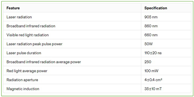
Super-Pulsed Class I Laser Therapy: Utilization of the ACTIVet PRO
® device from Multi Radiance Medical (Solon, OH, USA) for laser therapy sessions. This therapy is known for its analgesic and anti-inflammatory effects, aiding in pain management and tissue healing (
Table 1).
Recent studies indicate that various physical therapeutic modalities can contribute to the conservative or post-surgical treatment of Canine Hip Dysplasia (CHD) in dogs. Physiotherapy induces biological effects through physical action mechanisms, triggering biochemical responses in the body. The primary therapies recommended for individuals with osteoarthritis include laser therapy, due to its analgesic effects and low-impact exercises on an underwater treadmill,
2. Materials and Methods
2.1. Patient Details a 7-Year-Old Male Canine Setter, 23 kg Was the Subject of This Study
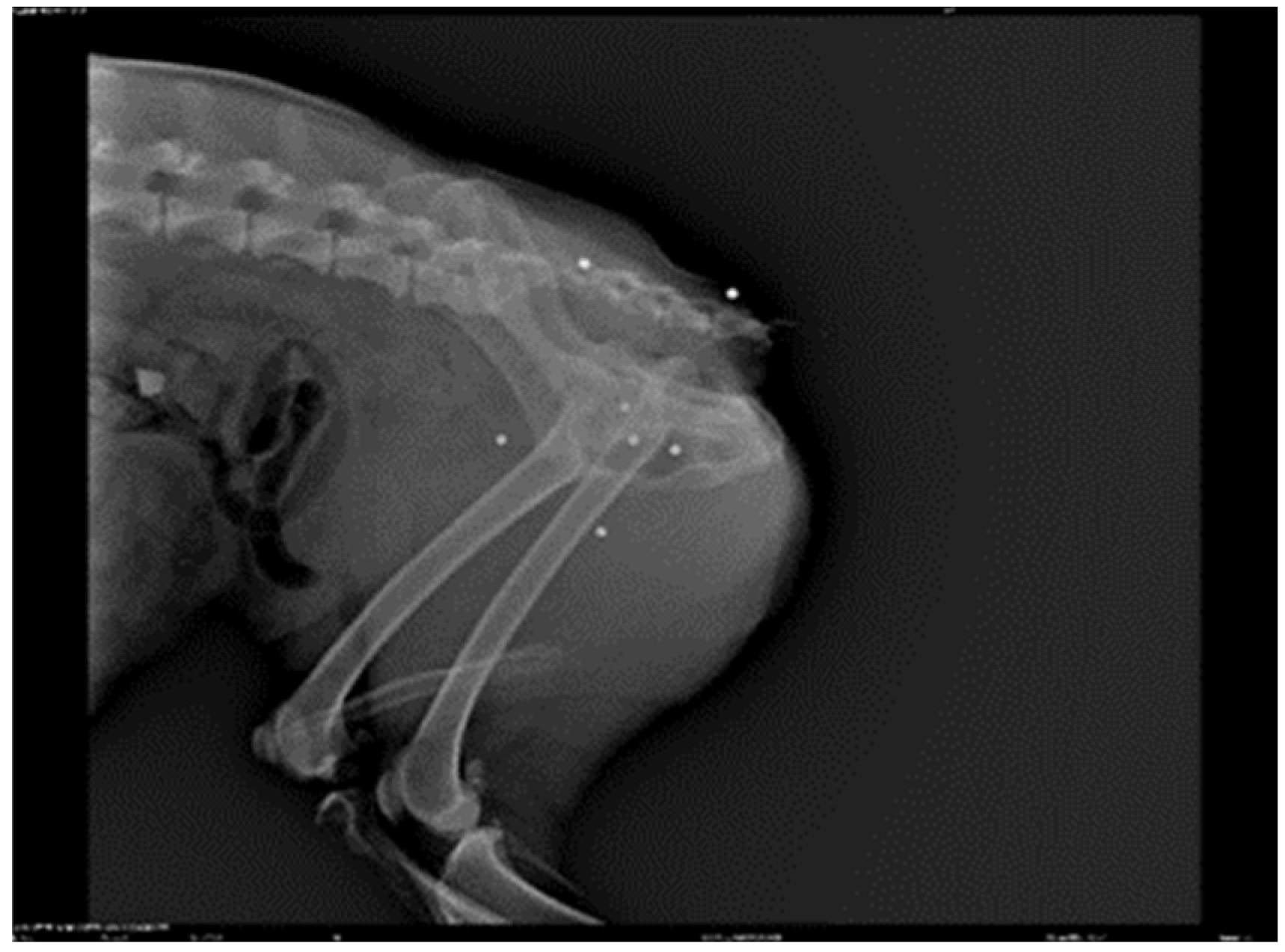
Figure 2.
Pre-surgery X-ray (lateral position) showing bilateral coxofemoral dysplasia and the presence of metallic artifacts (lead pellets from a firearm with which the animal was unfortunately shot).
Figure 2.
Pre-surgery X-ray (lateral position) showing bilateral coxofemoral dysplasia and the presence of metallic artifacts (lead pellets from a firearm with which the animal was unfortunately shot).
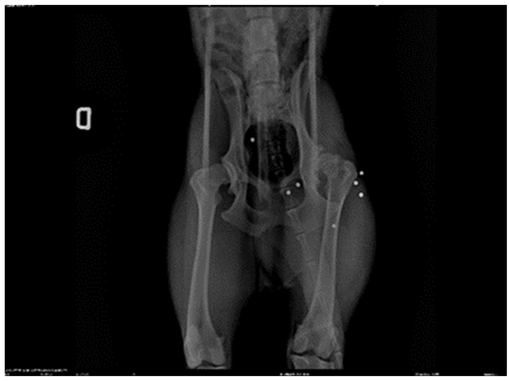
Figure 3.
Pre-surgery X ray -(ventro dorsal position) showing bilateral coxofemoral dysplasia and the presence of metallic artifacts (lead pellets from a firearm with which the animal was unfortunately shot.
Figure 3.
Pre-surgery X ray -(ventro dorsal position) showing bilateral coxofemoral dysplasia and the presence of metallic artifacts (lead pellets from a firearm with which the animal was unfortunately shot.
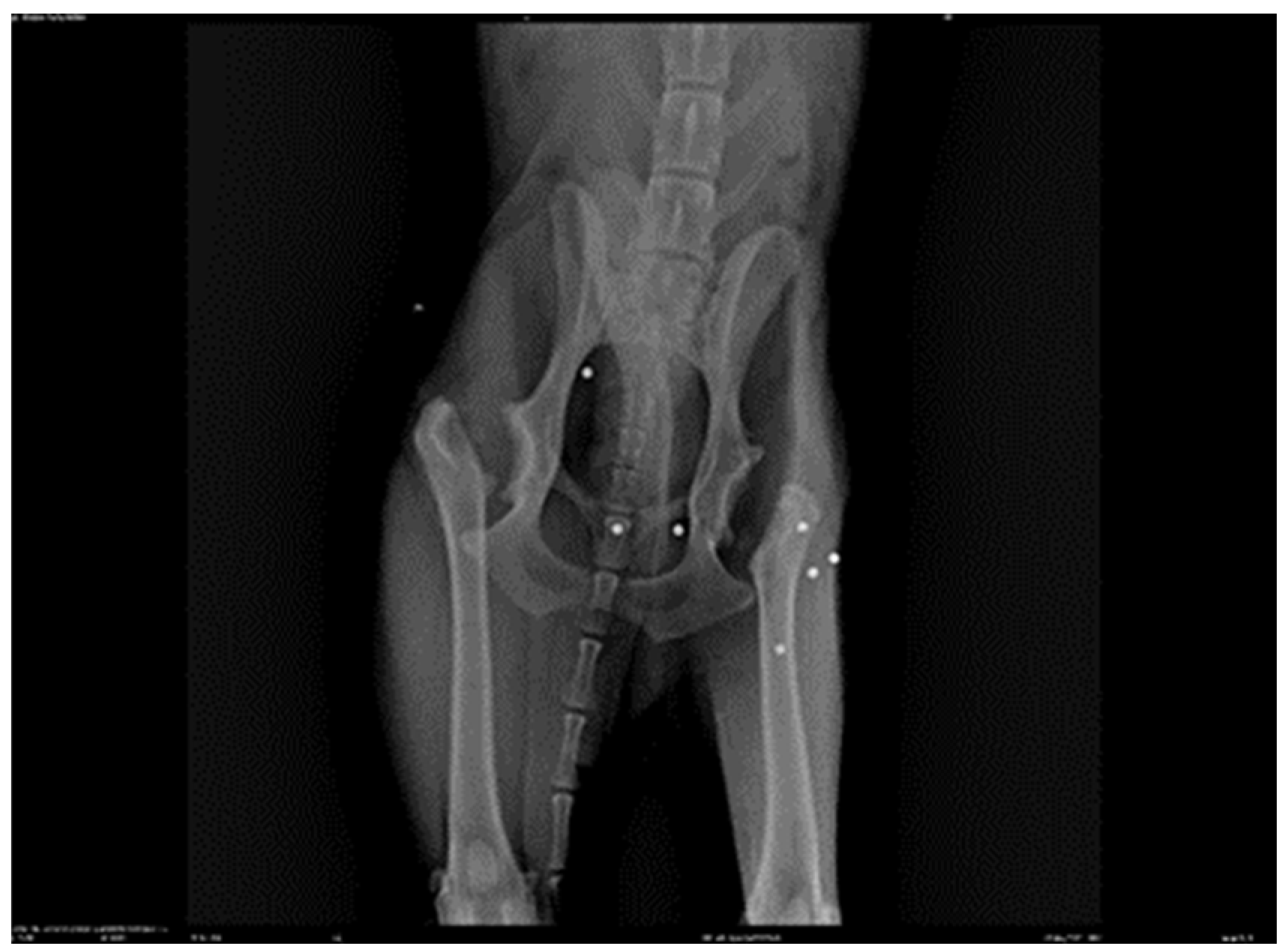
Figure 4.
X-ray after the second surgery. -(ventro dorsal position) with bilateral excision of the femoral head and neck.
Figure 4.
X-ray after the second surgery. -(ventro dorsal position) with bilateral excision of the femoral head and neck.
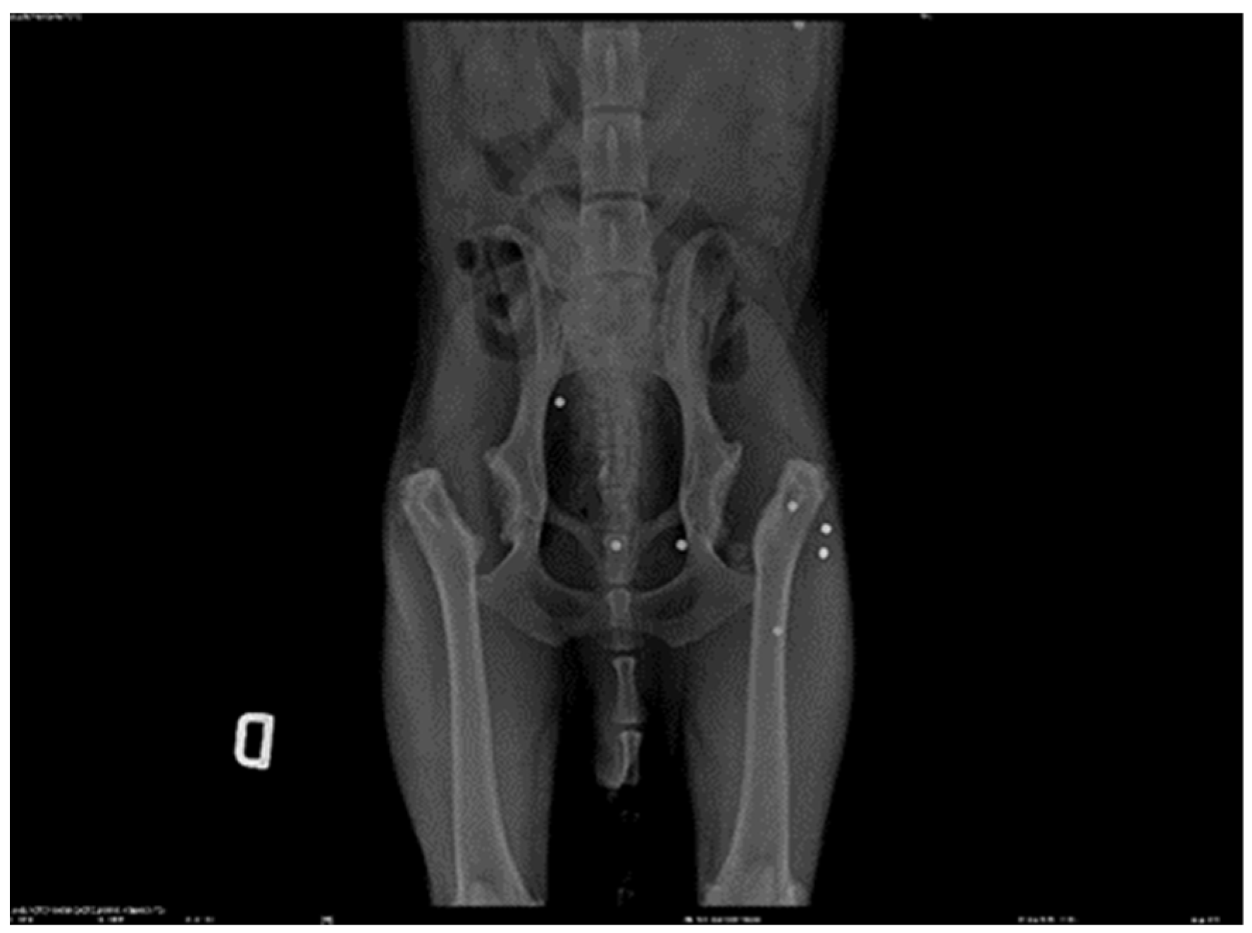
Figure 5.
X-ray after the second surgery. -(ventro dorsal position) with bilateral excision of the femoral head and neck Post 10 session, where you can see greater muscle mass in relation to photo 04.
Figure 5.
X-ray after the second surgery. -(ventro dorsal position) with bilateral excision of the femoral head and neck Post 10 session, where you can see greater muscle mass in relation to photo 04.
2.2. History
The canine was discovered abandoned near the municipal kennel and subsequently rescued. Following a medical examination and X-rays, the dog was diagnosed with severe coxofemoral dysplasia, necessitating femoral head ostectomy due to joint dislocation (Figure 2 and Figure 3). The initial surgery took place in March 2022, followed by a subsequent procedure in August 2022.
Between the two surgeries, no rehabilitation procedures were performed; only anti-inflammatory (non-steroidal anti-inflammatory drugs) and antibiotics were administered. The patient was referred for physiotherapy evaluation late (two months after the second surgery) because it was not walking well, exhibiting a grade four lameness, difficulty in rising and sitting, and in positioning to urinate. As the medical therapy was ineffective, the patient was referred for rehabilitation.
2.3. Assessment
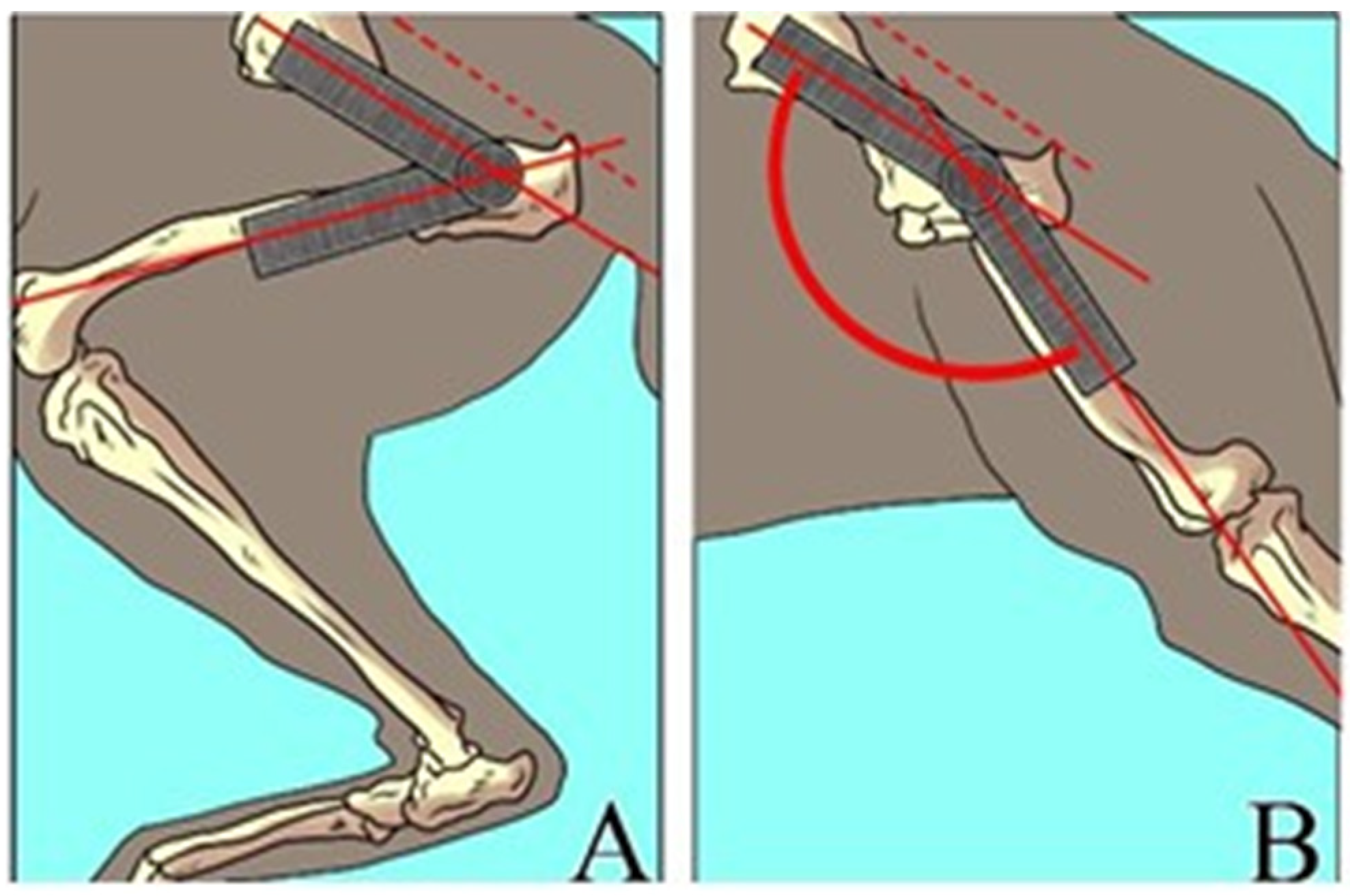
In October 2022, the patient exhibited pelvic extension pain, gluteal muscle atrophy, quadriceps and biceps femoris contractures, iliopsoas muscle contracture, lumbar-thoracic spine pain, and joint capsule retraction. The dog demonstrated difficulty in sitting and standing. Goniometric measurements indicated left hip flexion of 48º and extension of 158º, and right hip flexion of 47º and extension of 157º (normal flexion being 50º and extension 162º) [52]. Goniometry was consistently performed with the same goniometer, with the patient in lateral recumbency, depending on the limb being assessed. For instance, to measure the angles of the right pelvic limb, the patient was positioned in left lateral recumbency (with the left limb resting on the floor).
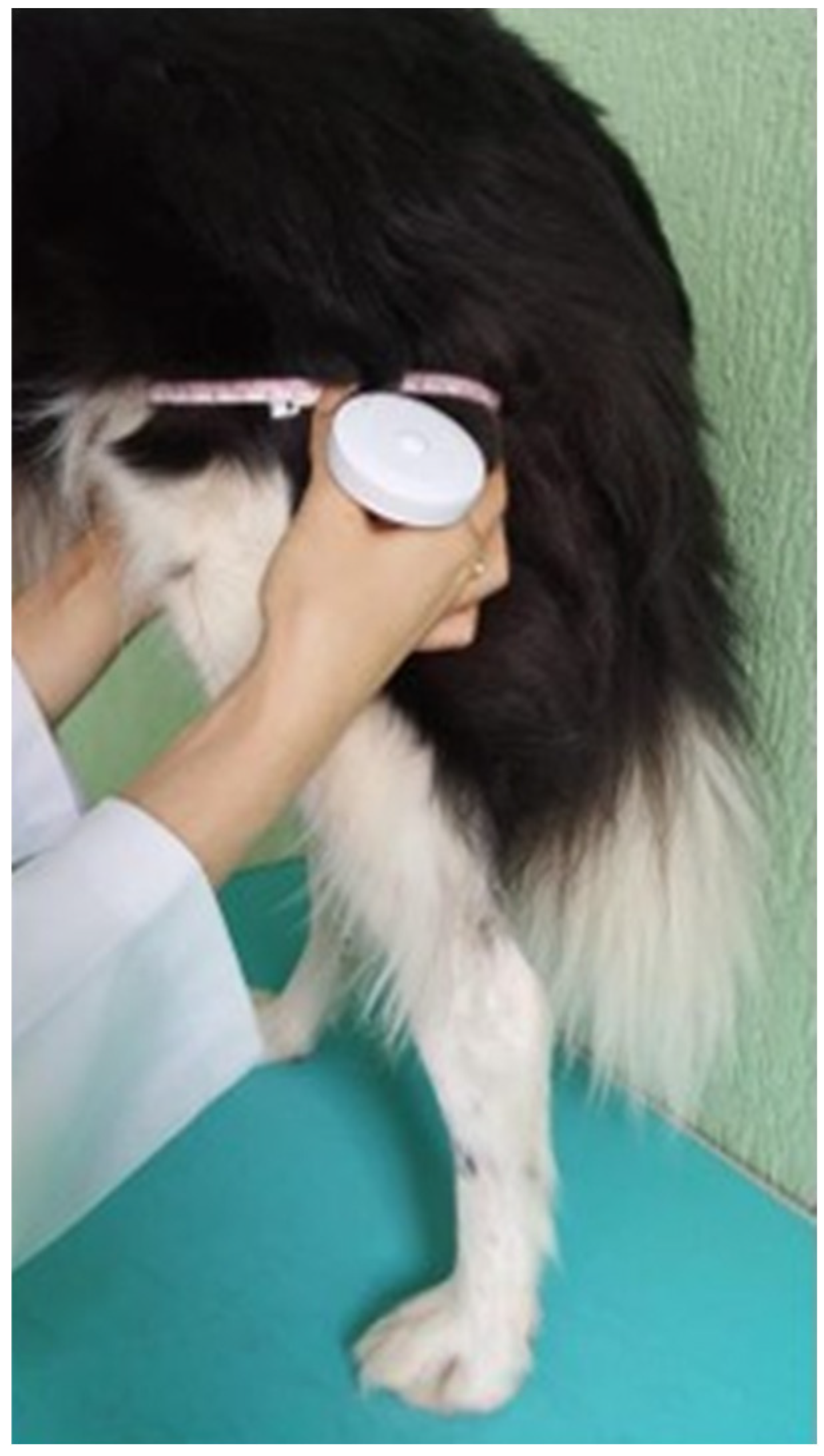
Circumference measurements of the thigh were 28 cm (right) and 29 cm (left). Circumference measurements were taken with a Gulick (Figure 6) tape measure, which accurately tracks muscle mass circumference values over the course of treatment. The device must always be positioned in the same location. The balls in the spring tension area become visible when tension is applied to the tape measure, ensuring that a constant tension is maintained during measurements. The Gulick was consistently positioned at 70% of femoral length, and measurements were performed with the patient in a standing position.
Figure 7.
Illustration of canine hip goniometry in flexion (A) and extension (B) [57].
Figure 7.
Illustration of canine hip goniometry in flexion (A) and extension (B) [57].
2.4. Methods
The treatment plan comprised 20 physiotherapy sessions utilizing the ACTIVet PRO® laser (Multi Radiance Medical, Solon, OH, USA) for pain and osteoarthritis management, aquatic treadmill for muscular reinforcement, Additionally, Alevica® 1 tablet per day for 60 days (Palmitoylethanolamide (30%), mono- and diglycerides of fatty acids esterified with organic acids (diglyceride of benzoic acid), sodium pyrophosphate, yeast, lupin flour, sodium chloride, sunflower seed oil, magnesium salts of organic acids (stearate)) was administered for pain control, and Condrostress® 1 tablet per day for 60 days (Egg membrane (10.8%), mono- and diglycerides of fatty acids esterified with organic acids (benzoic acid), sodium pyrophosphate, inactivated yeast, lupin protein flour, sodium chloride, vegetable oils (sunflower seeds), magnesium salts of organic acids (stearic acid) Nutritional additive: Vitamin E (42916 mg/kg). Coloring agent: 2a104 (2500 mg/kg). was prescribed to enhance cartilage hydration and synovial fluid viscosity in degenerative diseases such as coxofemoral dysplasia.
Rehabilitation sessions included super-pulsed laser therapy and aquatic treadmill exercises administered twice weekly for two months, followed by once-weekly sessions for the final two weeks. The primary goals of laser therapy were to expedite healing, control inflammation and pain, and alleviate muscle contractures. Specific laser treatments were targeted at pain in the thoracolumbar region, chronic pain at the surgical site, and pectineus muscle contracture.
Super-pulsed laser therapy, also known as cold laser therapy, represents a revolutionary advancement in technology. This therapy utilizes a technology called ‘super pulsing,’ which offers a safer and more effective treatment compared to traditional methods, such as Class IV lasers, due to its lack of thermal output (classified as Class 1). This innovative super-pulsing technology facilitates deeper tissue penetration than lasers of the same wavelength and power output. The absence of heat production enhances patient comfort, eliminates the need for specialized rooms or equipment, and allows the laser to be maintained in a single position for optimal treatment. Continuous movement of the laser is unnecessary. The scope of this case report is to assess whether this technology can be used over metallic implants without causing overheating or burns.
Laser treatment aimed to control pain in the thoracolumbar spine was performed in all sessions at 1000 Hz for 5 minutes, in direct contact, with the emitter moved slowly (1 cm per second) across the thoracolumbar region to stimulate endorphin release. For chronic pain and inflammation at the surgical site, treatment was applied during the first two weeks once pain subsided, at 50 Hz around the joint where the femoral head would be, in direct contact without trichotomy, scanning for 10 minutes. To manage pectineus muscle contracture, treatment was applied in all sessions at 1000-3000 Hz in static mode, with the laser in direct contact with the muscle, also without trichotomy, for 5 minutes. All applications utilized the pulsed mode with both red and infrared lights, using the dome probe
Hydrotherapy sessions progressed through different water levels: hip joint level during the first two weeks, totaling 4 sessions (two sessions per week) of 10 minutes each; mid-femur level during the subsequent sessions, totaling 4 sessions (two sessions per week) of 15 minutes each; and knee-deep water for the remaining sessions, totaling 12 sessions (two sessions per week) of 20 minutes each. Significant improvements were noted in muscle contracture, range of motion, and weight-bearing in the hind limbs after the fourth session
Physiotherapy continued for two months to restore muscle mass, improve joint amplitude, and achieve total pain control. Condrostress® was administered indefinitely to enhance the quality of the remaining joints, given that the coxofemoral joint no longer exists.
3. Results
In the last evaluation, the patient exhibited no signs of pain and demonstrated ease in sitting and standing. The post-surgery treatment effectively restored the patient’s function and quality of life. Radiographic assessment revealed no developmental signs of osteoarthritis and an increase in muscle mass. Thigh perimetry measurements showed improvement, with the right thigh measuring 30 cm and the left thigh measuring 31 cm.
In this specific case, the owner reported substantial improvement, indicating that the patient is no longer hesitant to walk, sit, or stand
And in the measurements [13]
Goniometry of the left coxofemoral joint pre-treatment: flexion 48º extension 158º (normal flexion 50º extension 162º) Post treatment flexion 50º extension 160º
Goniometry of the right coxofemoral joint pre-treatment: flexion 47º extension 157º (normal flexion 50º extension 162º) Post treatment flexion 50º extension 159º
Pre-treatment perimeter left thigh 28 cm left thigh 29 cm
Post treatment Right thigh perimetry 30 cm; left thigh 31 cm.
The change in angulation was not very large but was significant, as was the change in perimeter measurements. This small alteration was sufficient to observe clinical improvement and almost complete physiological recovery in goniometry (when compared to the normal values referred to in the publications) [52,56].
4. Discussion
The evolution of rehabilitation has brought forth substantial scientific evidence supporting the effectiveness of non-surgical interventions for treating hip dysplasia, particularly in cases where there is no joint luxation. This case report underscores the necessity of rehabilitative measures even following corrective surgery, given that surgical techniques such as osteotomy, denervation, or prosthesis placement do not address soft tissue alterations (e.g., muscles, joint capsule). Consequently, it is essential to integrate pre- and post-surgery physiotherapy into all hip dysplasia treatment protocols to facilitate enhanced and accelerated patient recovery.
5. Conclusions
Recent studies demonstrate the efficacy of various physical therapeutic modalities in the conservative or post-surgical treatment of CFD [28,32,48]. Physiatry exerts biological effects through physical mechanisms, eliciting biochemical responses in the body. Laser therapy is recommended for its analgesic effects [12,39,40], and low-impact exercises on a hydro treadmill [17,18,20,47] are beneficial for individuals with osteoarthritis.
Physical exercises on an aquatic treadmill contribute to weight control, a crucial factor in therapeutic management to avoid joint overload [21,24]. The physical properties of water decrease the relative weight of the animal due to buoyancy, and the increased density challenges the muscles more than air [20,42]. These properties make underwater treadmills and swimming ideal exercises for animals with arthropathy, providing greater energy expenditure and less joint impact [11], and are particularly important in the multimodal treatment of canine obesity in dogs with osteoarthritis [11,21,42,56].
Regular treadmill exercise enhances the release of beta-endorphins, improves self-esteem, and aids in pain relief in humans [11]. Further studies are necessary to elucidate the effects of aquatic physical exercises on dogs in relation to the release of endogenous substances and overall well-being. Nonetheless, the thermal effect of heated water is known to promote relaxation and improve joint mobility in dogs [48,49].
For these reasons, we began the treatment with water set at the level of the coxofemoral joint, where there is greater buoyancy and less overload, with the intention of improving the range of motion and pain control, as the workload on the longissimus dorsi and gluteus medius muscles decreases with increased water depth in the underwater treadmill (UWT). Subsequently, the water level was adjusted to mid-femur, creating greater joint overload, and finally, to knee-depth water to increase resistance and enhance muscle mass gain [48,49]
Laser therapy is effective in controlling joint pain and chronic disease in both humans [12,15,16] and dogs [38,39] Its analgesic effects occur through photo biomodulation of the inflammatory response, increasing cellular ATP synthesis and transforming prostaglandins and prostacyclin, thereby exerting anti-edematous and anti-inflammatory effects [16]. Laser therapy also increases the release of endorphins, providing relief from non-inflammatory pain [45]. Importantly, these therapies pose minimal risk or contraindication, as the use of super pulsed laser does not result in burns or abrasions on the patient’s skin and does not require shaving. Despite the presence of metallic material at the subcutaneous level (the lead pellets from the weapon with which the patient was injured), the use of super pulsed laser did not cause heating or reflection (as evidenced by significant anti-inflammatory improvement). Therefore, we can use super pulsed laser even if the patient has plates, pins, or other metallic implants without contraindications.
To promote pain relief, improve joint movement amplitude, and increase muscle mass through physiatry modalities, it is essential to employ appropriate assessment methods for each parameter. In this case report, subjective assessments by the owner, goniometry, and perimetry of the coxofemoral joint and associated muscles were utilized [22,44,45,46].
Based on our findings, we conclude that hydrotherapy and laser therapy offer effective means to address muscle contractures, joint extension, and support strength, ultimately enhancing the quality of life for our patients.
To further elucidate these benefits, continued studies and comparative analyses with cases of hip dysplasia, with or without surgical intervention, are warranted [27,28,29].
Informed Consent Statement
Informed consent was obtained from all subjects involved in the study.
Acknowledgments
Thank you for the support from the University of Teramo and for the support and assistance from Multi Radiance. I thank the reviewers from Multi Radiance for their valuable assistance in reviewing this manuscript.
Conflicts of Interest
The authors declare no conflicts of interest.
References
- Bettini CM, et al. Incidence of coxofemoral dysplasia in Border Collie dogs. Unipar Veterinary and Zoological Sciences Archive. 2007;10(1):21-25.
- McLaughlin Junior R, Tomlinson J. Radiographic diagnosis of canine hip dysplasia. Veterinary medicine. 1996;91(2):36-47.
- Minto BW, et al. Clinical evaluation of acetabular denervation in dogs with coxofemoral dysplasia treated at the FMVZ Veterinary Hospital - Botucatu - SP. Veterinary and Animal Sciences. 2012;19(1):91-98.
- Mueller M, et al. Effects of radial shock wave therapy on limb function of dogs with osteoarthritis of the hip. Veterinary record. 2007;160(1):762–765.
- Morgan JP, Wind A, Davidson AP. Elbow dysplasia. In: Hereditary diseases of bones and joints in dogs: osteochondrosis, hip dysplasia, elbow dysplasia. Hanover: Schlütersche; 2000. p. 41-94.
- Mostafa AA, Berry CR, Nahla MA. Quantitative evaluation of hip morphology to improve the identification of coxofemoral dysplasia in German Shepherd dogs. Am J Vet Res. 2023 Feb;7:1-10. [PubMed]
- Piermattei, D. Arthroplasty for excision of the coxofemoral joint in dogs and cats. Vet Comp Orthop Traumatol. 2011;24(1):89. [PubMed]
- Rocha FPC, et al. Coxofemoral dysplasia in dogs. Revista Cientí Eletrônica de Medicina Veterinária. 2008;4(11):1-7.
- Off W, Matis U. Arthroplasty for excision of the coxofemoral joint in dogs and cats. Results of clinical, radiographic and gait analysis of the Department of Surgery, Faculty of Veterinary Medicine, Ludwig-Maximilians-University of Munich, Germany. Vet Comp Orthop Traumatol. 2010;23(5):297-305. [PubMed]
- Virag Y, Gumpenberger M, Tichy A, Lutonsky C, Peham C, Bockstahler B. Center of pressure and reaction forces of the soil in Labradors and Golden Retrievers with and without coxofemoral dysplasia at 4, 8 and 12 months of age. Front Vet Sci. 2022 Dec 22;9:1087693. PMCID: PMC9814507. [PubMed]
- Reusing MSO, Villanova Júnior JA, Weber SH. Efeitos terapêuticos do exercício em esteira aquática e da laserterapia de baixa intensidade em cães com displasia coxofemoral = Therapeutic effects of underwater treadmill exercise and low-level laser therapy in dogs with hip dysplasia. 2019. Available at: link. Accessed July 16, 2023.
- Ali Q, et al. Low Energy Laser Effects in Relieving Pain in Patients with Knee Osteoarthritis. International Journal of Medicine and Public Health. 2018;2(1):6-10.
- Baker S, et al. Comparison of four commercial devices to measure limb circumference in dogs. Veterinary and Comparative Orthopaedics and Traumatology. 2010;23(6):406-410.
- Barr A, Denny H, Gibbs C. Clinical hip dysplasia in growing dogs: the long-term results of conservative management. Journal of Small Animal Practice. 1987;28(4):243-252.
- Bender T, et al. The effect of physical therapy on beta-endorphin levels. European journal of applied physiology. 2007;100(4):371-382.
- Bjordal JM, et al. A systematic review of low level laser therapy with location-specific doses for pain from chronic joint disorders. Journal of Physiotherapy. 2003;49(2):107-116.
- Bockstahler B, et al. Pelvic limb kinematics and surface electromyography of the vastus lateralis, biceps femoris, and gluteus medius muscle in dogs with hip osteoarthritis. Veterinary Surgery. 2012;41(1):54-62.
- Bockstahler BA, et al. Hind limb kinematics during therapeutic exercises in dogs with osteoarthritis of the hip joints. American Journal of Veterinary Research. 2012;73(9):1371-1376.
- Brown DC, et al. Ability of the canine brief pain inventory to detect response to treatment in dogs with osteoarthritis. Journal of the American Veterinary Medical Association. 2008;233(8):1278-1283.
- Cartlidge, H. Hydrotherapy for the osteoarthritic dog: why might it help and is there any evidence? The Veterinary Nurse. 2015;6(10):600-606.
- Chauvet A, et al. Incorporation of exercise, using an underwater treadmill, and active client education into a weight management program for obese dogs. Canadian Veterinary Journal. 2011;52(5):491.
- Collard F, et al. Canine hip denervation: Comparison between clinical outcome and gait analysis. Revue de Medecine Veterinaire. 2010;161(6):277.
- Crestani MV, Telöken MA, Gusmão PDF. Impacto femoroacetabular: uma das condições precursoras da osteoartrose do quadril. Rev Bras Ortop. 2006;41(8):285-293.
- Denning WE, Bressel E, Dolny DG. Underwater treadmill exercise as a potential treatment for adults with osteoarthritis. International Journal of Aquatic Research and Education. 2010;4(1):9.
- Diniz, R. Hidroterapia. In: Lopes RS, Diniz R, editors. Fisiatria em Pequenos Animais. São Paulo: Editora Inteligente; 2018. p. 156-162.
- Doust S, et al. Preliminary study of the hip dysplasia incidence based on clinical and radiographical examination in large breed dogs referred to veterinary teaching hospital of Ferdowsi University of Mashhad. Journal of Veterinary Research. 2018;73(1).
- Duff R, Campbell J. Long-term results of excision arthroplasty of the canine hip. The Veterinary Record. 1977;101(10):181-184.
- Edge-Hughes, L. Hip and sacroiliac disease: selected disorders and their management with physical therapy. Clinical Techniques in Small Animal Practice. 2007;22(4):183-194.
- Ginja M, et al. Diagnosis, genetic control and preventive management of canine hip dysplasia: a review. The Veterinary Journal. 2010;184(3):269-276.
- Greshake RJ, Ackerman N. Ultrasound evaluation of the coxofemoral joints of the canine neonate. Veterinary Radiology & Ultrasound. 1993;34(2):99-104.
- Gulick Tape Measure. (n.d.). VBS Group. Available online: https://vbsgroup-shop.eu/en/diagnosis-veterinary-equipment/59-gulick-tape.html (accessed on 4 August 2024).
- Gusi N, et al. Exercise in waist-high warm water decreases pain and improves health-related quality of life and strength in the lower extremities in women with fibromyalgia. Arthritis Care & Research. 2006;55(1):66-73.
- Harper, T.A. Conservative Management of Hip Dysplasia. Veterinary Clinics: Small Animal Practice. 2017;47(4):807-821.
- Henrigson B, Norberg I, Olssons SE. On the etiology and pathogenesis of hip dysplasia: a comparative review. Journal of Small Animal Practice. 1966;7(11):673-688.
- Honmura A, et al. Therapeutic effect of Ga-Al-As diode laser irradiation on experimentally induced inflammation in rats. Lasers in surgery and medicine. 1992;12(4):441-449.
- Jaegger G, Marcellin-Little DJ, Levine D. Reliability of goniometry in Labrador Retrievers. American journal of veterinary research. 2002;63(7):979-986.
- Kass P, Wallace L, Guffy M. Association between pelvic muscle mass and canine hip dysplasia. Journal of the American Veterinary Medical Association. 1997;210(10):1466-1473.
- Kyriazis, A. Canine hip dysplasia. Part I: Aetiopathogenesis & diagnostic approach. Hellenic Journal of Companion Animal Medicine. 2016;5(1):37.
- König HE, Liebich H-G. Anatomia dos Animais Domésticos-: Texto e Atlas Colorido. Artmed Editora; 2016.
- Lewis G, Holt S. The Effects of Low-level Laser Therapy on the Gait of the Osteoarthritic Canine Hindlimb. BVNA Congress 2018. 2018.
- Matera JM, Tatarunas AC, Oliveira SM. Uso do laser arseneto de gálio (904nm) após excisão artroplástica da cabeça do fêmur em cães. Acta Cirúrgica Brasileira. 2003;18(2):102-106.
- McCarthy G, et al. Randomised double-blind, positive-controlled trial to assess the efficacy of glucosamine/chondroitin sulfate for the treatment of dogs with osteoarthritis. The Veterinary Journal. 2007;174(1):54-61.
- Mendes S, Coutinho I, Rebelo P. Hidroterapia canina. Revista Portuguesa de Ciências Veterinárias. 2015;110(595-596):160-164.
- Millis DL, Levine D. The role of exercise and physical modalities in the treatment of osteoarthritis. Veterinary Clinics: Small Animal Practice. 1997;27(4):913-930.
- Morales J, et al. He-Ne Laser has no Effect on Cell Cycle Phases. Revista espanola de fisiologia. 1995;51(1):43-48.
- MVS Hospital for Pets. Hip Dysplasia. MVS Hospital for Pets. Available online: https://www.mvshospital.com/hip-dysplasia/ (accessed on 4 June 2024).
- Nielsen C, Pluhar G. Diagnosis and treatment of hind limb muscle strain injuries in dogs. Veterinary and Comparative Orthopaedics and Traumatology. 2005;18(4):247.
- Nganvongpanit, K., Boonchai, T., Taothong, O., & Sathanawongs, A. (2014). Physiological effects of water temperatures in swimming toy breed dogs. Kafkas Universitesi Veteriner Fakultesi Dergisi, 20(2), 213-218. [CrossRef]
- Parkinson, S., Wills, A. P., Tabor, G., & Williams, J. M. (2018). Effect of water depth on muscle activity of dogs when walking on a water treadmill. Comparative Exercise Physiology, 14(2), 1-12. [CrossRef]
- Pereira FC, et al. Antinociceptive effects of low-level laser therapy at 3 and 8 j/cm2 in a rat model of postoperative pain: possible role of endogenous Opioids. Lasers in surgery and medicine. 2017;49(9):844-851.
- Prankel, S. Hydrotherapy in practice. In practice. 2008;30(5):272.
- Petazzoni, M. (Ed.). (2008). Atlas of Clinical Goniometry and Radiographic Measurements of the Canine Pelvic Limb (2nd ed.). Merial.
- Raghuvir H, et al. Treatment of canine hip dysplasia: A review. J Anim Sci Adv. 2013;3(12):589-597.
- Remedios AM, Fries CL. Treatment of canine hip dysplasia: a review. The Canadian Veterinary Journal. 1995;36(8):503.
- Syrcle, J. Hip Dysplasia: Clinical Signs and Physical Examination Findings. Veterinary Clinics: Small Animal Practice. 2017;47(4):769-775.
- Reusing, M., Brocardo, M., Weber, S., & Villanova, J. Jr. (2020). Goniometric evaluation and passive range of joint motion in chondrodystrophic and nonchondrodystrophic dogs of different sizes. VCOT Open, 3, e66–e71.
- Reusing, M.S.O. (2019). Therapeutic Effects of Underwater Treadmill Exercise and Low-Level Laser Therapy in Dogs with Hip Dysplasia. Master’s Thesis, Pontifícia Universidade Católica do Paraná, School of Life Sciences, Graduate Program in Animal Science, Curitiba, Brazil.
- Tai G, Tai M, Zhao M. Electrically stimulated cell migration and its contribution to wound healing. Burns & trauma. 2018;6(1):20.
- Upariputti R, et al. Effect of interferential current therapy on ground reaction force in dogs with hip osteoarthritis: A randomized placebo-controlled cross-over clinical trial. The Thai Journal of Veterinary Medicine. 2018;48(1):111-116.
- Vince, K.J. Canine hip dysplasia: surgical treatment for the military working dog. Army Medical Department Journal. 2007;44-50.
- Wallace, L.J. Pectineus tendon surgery for the management of canine hip dysplasia. Veterinary Clinics of North America: Small Animal Practice. 1992;22(3):607-621.
- Weigel JP, Cartee RE, Marich KW. Preliminary study on the use of ultrasonic transmission imaging to evaluate the hip joint in the immature dog. Ultrasound in medicine & biology. 1983;9(4):371-378.
- Wong, E. Swim to Recovery: Canine Hydrotherapy Healing. Veloce Publishing Ltd.; 2011.
- Zink C, Carr BJ. What is a canine athlete? Canine sports medicine and rehabilitation. 2018;1-22.
- Zink C, Van Dyke JB. Canine sports medicine and rehabilitation. John Wiley & Sons; 2018.
|
Disclaimer/Publisher’s Note: The statements, opinions and data contained in all publications are solely those of the individual author(s) and contributor(s) and not of MDPI and/or the editor(s). MDPI and/or the editor(s) disclaim responsibility for any injury to people or property resulting from any ideas, methods, instructions or products referred to in the content. |
© 2024 by the authors. Licensee MDPI, Basel, Switzerland. This article is an open access article distributed under the terms and conditions of the Creative Commons Attribution (CC BY) license (https://creativecommons.org/licenses/by/4.0/).
