Submitted:
20 September 2024
Posted:
23 September 2024
Read the latest preprint version here
Abstract

Keywords:
1. Introduction
2. Material and Methods
2.1. Liposomal Preparation
2.2. Screening of the Antibacterial Activity
2.2.1. Agar Well-Diffusion Method
2.2.2. Minimum Inhibitory Concentration (MIC)
2.2.3. S. aureus Susceptibility
2.3. Cell Culture
2.3.1. Cell Viability Assay (MTT)
2.3.2. Cell Migration Assay
2.4. DPPH Assay
2.5. Statistical Analysis
3. Results and Discussion
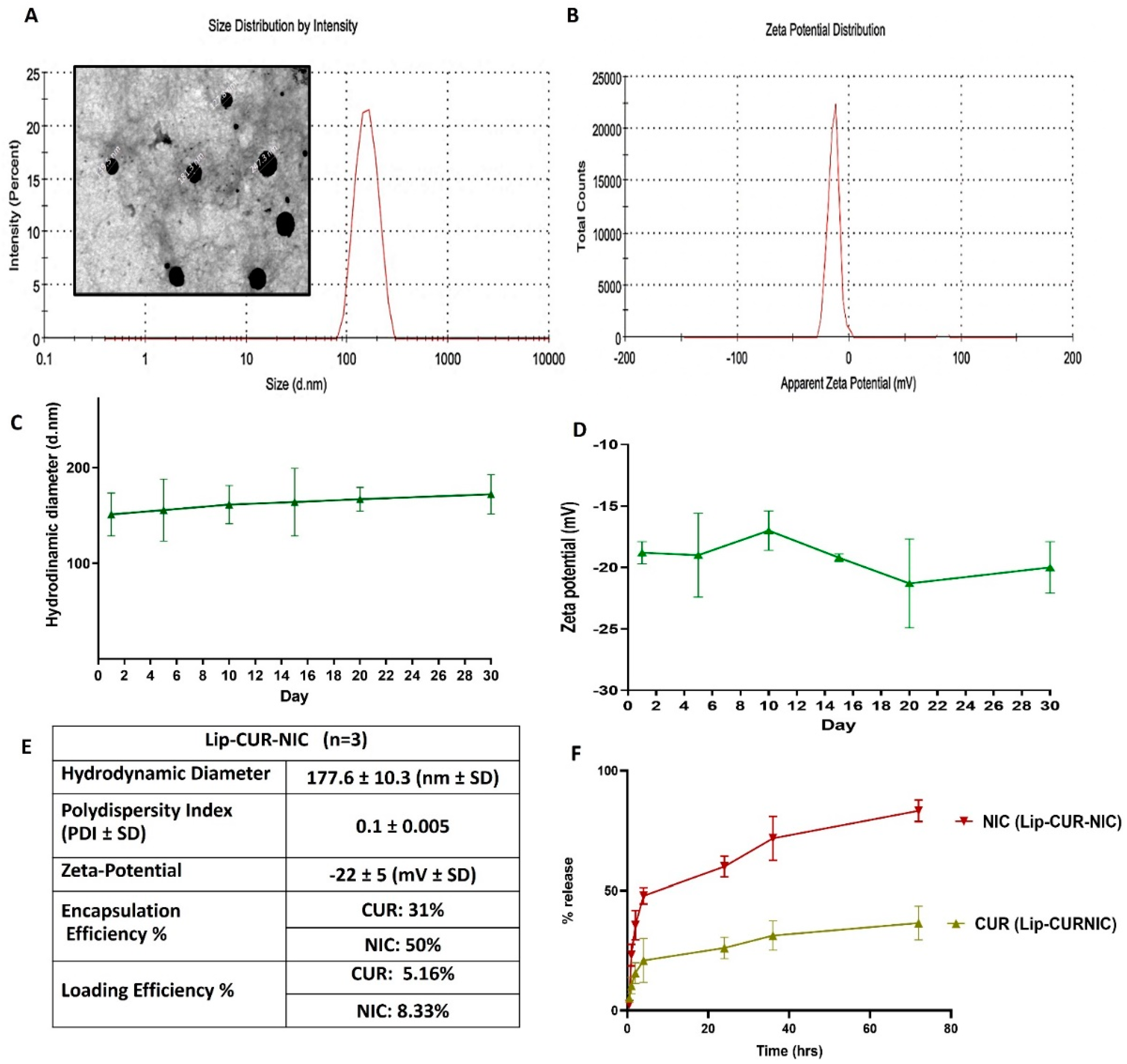
3.1. Liposomal Preparation and Characterization
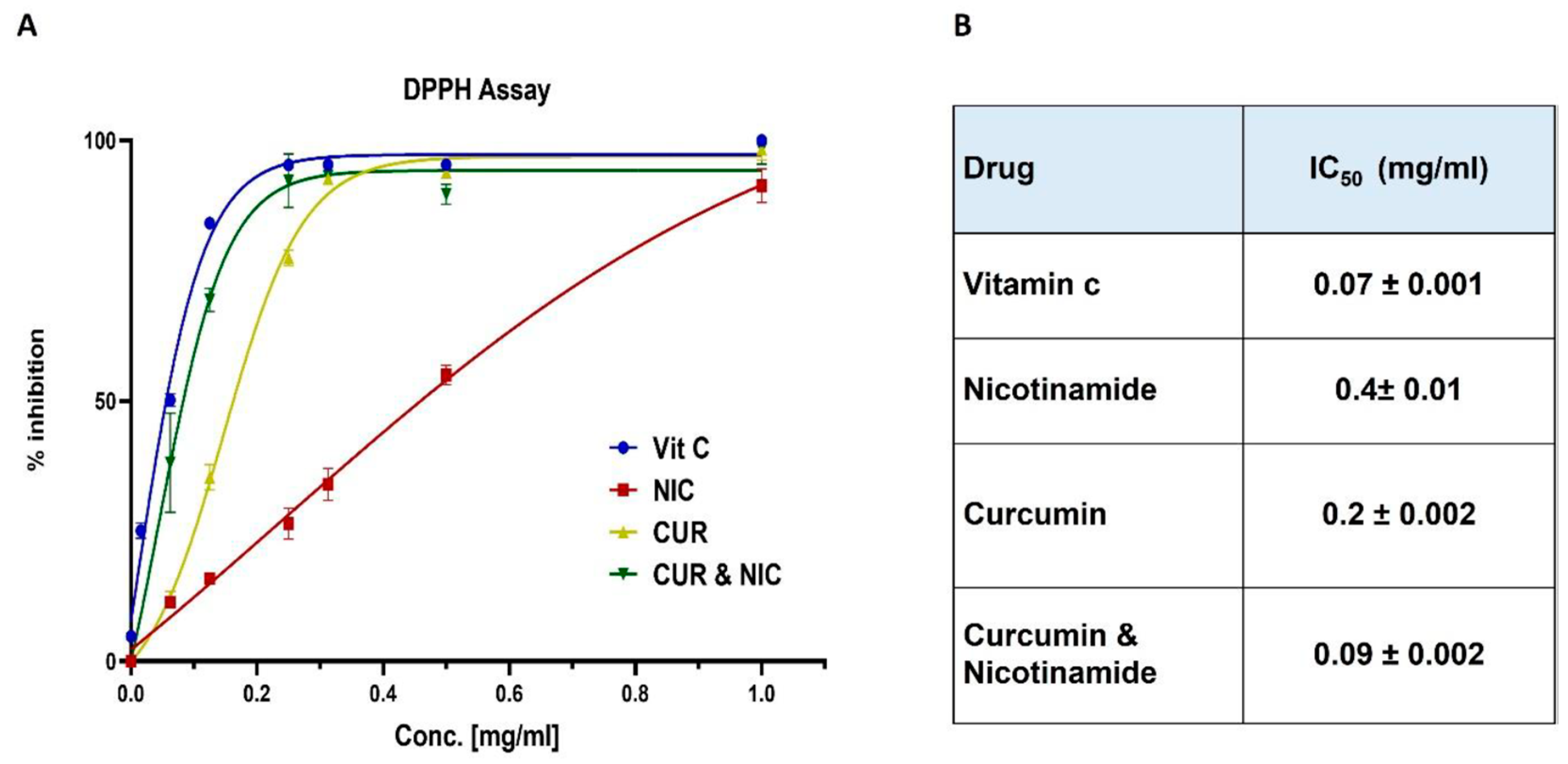
3.2. Antioxidant Activity and DPPH Assay
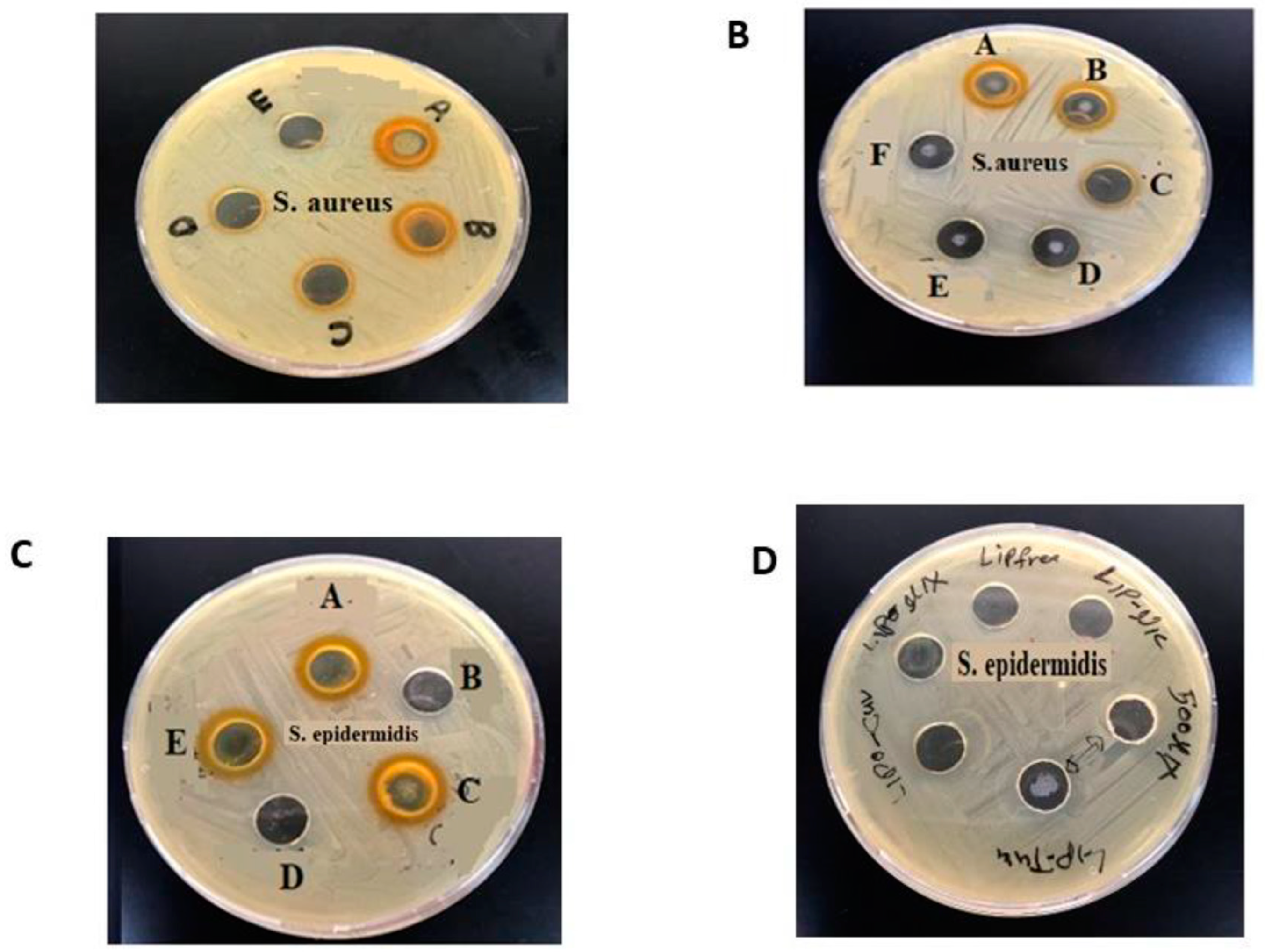
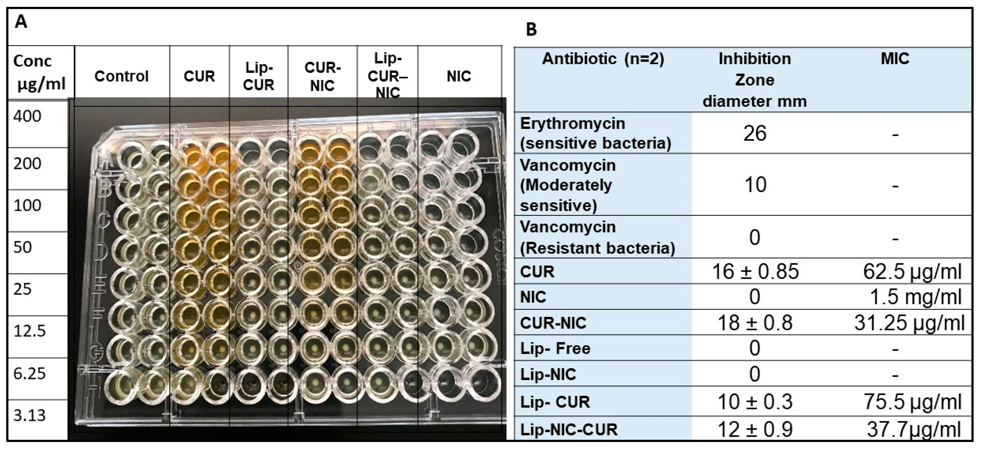
3.3. Antibacterial Activity of CUR, NIC and lip- CUR-NC
3.4. Cytotoxicity Study and Anticancer Activity
3.5. Migration Test (Scratch Assay)
4. Conclusion
References
- Sorrenti, V.; Burò, I.; Consoli, V.; Vanella, L. Recent Advances in Health Benefits of Bioactive Compounds from Food Wastes and By-Products: Biochemical Aspects. Int J Mol Sci 2023, 24. [Google Scholar] [CrossRef]
- Nsairat, H.; Lafi, Z.; Al-Sulaibi, M.; Gharaibeh, L.; Alshaer, W. Impact of nanotechnology on the oral delivery of phyto-bioactive compounds. Food Chemistry 2023, 424, 136438. [Google Scholar] [CrossRef]
- Lafi, Z.; Alshaer, W.; Ma’mon, M.H.; Zihlif, M.A.; Asha, N.Y.; Abdelnabi, H.; Awidi, A. A review Echinomycin: A Journey of Challenges. Jordan Journal of Pharmaceutical Sciences 2023, 16, 640–654. [Google Scholar] [CrossRef]
- Hammad, H.M.; Imraish, A.; Al-Hussaini, M.; Zihlif, M.; Harb, A.A.; Abu Thiab, T.M.; Lafi, Z.; Nassar, Z.D.; Afifi, F.U. Ethanol extract of Achillea fragrantissima enhances angiogenesis through stimulation of VEGF production. Endocrine, Metabolic & Immune Disorders-Drug Targets (Formerly Current Drug Targets-Immune, Endocrine & Metabolic Disorders) 2021, 21, 2035–2042. [Google Scholar] [CrossRef]
- Lafi, Z.M.; Irshaid, Y.M.; El-Khateeb, M.; Ajlouni, K.M.; Hyassat, D. Association of rs7041 and rs4588 polymorphisms of the vitamin D binding protein and the rs10741657 polymorphism of CYP2R1 with vitamin D status among Jordanian patients. Genetic testing and molecular biomarkers 2015, 19, 629–636. [Google Scholar] [CrossRef]
- Watkins, R.R.; Bonomo, R.A. Overview: Glosbal and Local Impact of Antibiotic Resistance. Infect Dis Clin North Am 2016, 30, 313–322. [Google Scholar] [CrossRef]
- Hussain, Y.; Alam, W.; Ullah, H.; Dacrema, M.; Daglia, M.; Khan, H.; Arciola, C.R. Antimicrobial Potential of Curcumin: Therapeutic Potential and Challenges to Clinical Applications. Antibiotics (Basel) 2022, 11. [Google Scholar] [CrossRef]
- Chinemerem Nwobodo, D.; Ugwu, M.C.; Oliseloke Anie, C.; Al-Ouqaili, M.T.S.; Chinedu Ikem, J.; Victor Chigozie, U.; Saki, M. Antibiotic resistance: The challenges and some emerging strategies for tackling a global menace. J Clin Lab Anal 2022, 36, e24655. [Google Scholar] [CrossRef]
- Laikova, K.V.; Oberemok, V.V.; Krasnodubets, A.M.; Gal’chinsky, N.V.; Useinov, R.Z.; Novikov, I.A.; Temirova, Z.Z.; Gorlov, M.V.; Shved, N.A.; Kumeiko, V.V. Advances in the understanding of skin cancer: ultraviolet radiation, mutations, and antisense oligonucleotides as anticancer drugs. Molecules 2019, 24, 1516. [Google Scholar] [CrossRef]
- Abd El-Hack, M.E.; de Oliveira, M.C.; Attia, Y.A.; Kamal, M.; Almohmadi, N.H.; Youssef, I.M.; Khalifa, N.E.; Moustafa, M.; Al-Shehri, M.; Taha, A.E. The efficacy of polyphenols as an antioxidant agent: An updated review. International Journal of Biological Macromolecules 2023, 126525. [Google Scholar] [CrossRef]
- Munef, A.; Lafi, Z.; Shalan, N. Investigating anti-cancer activity of dual-loaded liposomes with thymoquinone and vitamin C. Therapeutic Delivery 2024, 15, 267–278. [Google Scholar] [CrossRef]
- AlSaleh, A.; Shahid, M.; Farid, E.; Kamal, N.; Bindayna, K. Synergistic antimicrobial effect of ascorbic acid and nicotinamide with rifampicin and vancomycin against SCCmec type IV methicillin-resistant Staphylococcus aureus (MRSA). Access Microbiol 2023, 5. [Google Scholar] [CrossRef]
- Simmons, J.D.; Peterson, G.J.; Campo, M.; Lohmiller, J.; Skerrett, S.J.; Tunaru, S.; Offermanns, S.; Sherman, D.R.; Hawn, T.R. Nicotinamide Limits Replication of Mycobacterium tuberculosis and Bacille Calmette-Guérin Within Macrophages. J Infect Dis 2020, 221, 989–999. [Google Scholar] [CrossRef]
- Murray, M.F. Nicotinamide: an oral antimicrobial agent with activity against both Mycobacterium tuberculosis and human immunodeficiency virus. Clinical infectious diseases 2003, 36, 453–460. [Google Scholar] [CrossRef]
- Starr, P. Oral Nicotinamide Prevents Common Skin Cancers in High-Risk Patients, Reduces Costs. Am Health Drug Benefits 2015, 8, 13–14. [Google Scholar]
- Allen, N.C.; Martin, A.J.; Snaidr, V.A.; Eggins, R.; Chong, A.H.; Fernandéz-Peñas, P.; Gin, D.; Sidhu, S.; Paddon, V.L.; Banney, L.A. Nicotinamide for Skin-Cancer Chemoprevention in Transplant Recipients. New England Journal of Medicine 2023, 388, 804–812. [Google Scholar] [CrossRef]
- Tosti, G.; Pepe, F.; Gnagnarella, P.; Silvestri, F.; Gaeta, A.; Queirolo, P.; Gandini, S. The Role of Nicotinamide as Chemo-Preventive Agent in NMSCs: A Systematic Review and Meta-Analysis. Nutrients 2024, 16, 100. [Google Scholar] [CrossRef]
- Surjana, D.; Halliday, G.M.; Damian, D.L. Role of nicotinamide in DNA damage, mutagenesis, and DNA repair. Journal of nucleic acids 2010, 2010. [Google Scholar] [CrossRef]
- Salech, F.; Ponce, D.P.; Paula-Lima, A.C.; SanMartin, C.D.; Behrens, M.I. Nicotinamide, a poly [ADP-ribose] polymerase 1 (PARP-1) inhibitor, as an adjunctive therapy for the treatment of Alzheimer’s disease. Frontiers in Aging Neuroscience 2020, 12, 255. [Google Scholar] [CrossRef]
- Lafi, Z.; Hiba, T.; Hanan, A. An updated assessment on anticancer activity of screened medicinal plants in Jordan: Mini review. Journal of Pharmacognosy and Phytochemistry 2020, 9, 55–58. [Google Scholar] [CrossRef]
- Lafi, Z.; Aboalhaija, N.; Afifi, F. Ethnopharmacological importance of local flora in the traditional medicine of Jordan:(A mini review). Jordan Journal of Pharmaceutical Sciences 2022, 15, 132–144. [Google Scholar] [CrossRef]
- Lafi, Z.; Alshaer, W.; Ma'mon, M.H.; Zihlif, M.; Alqudah, D.A.; Nsairat, H.; Azzam, H.; Aburjai, T.; Bustanji, Y.; Awidi, A. Aptamer-functionalized pH-sensitive liposomes for a selective delivery of echinomycin into cancer cells. RSC Advances 2021, 11, 29164–29177. [Google Scholar] [CrossRef]
- Alshaer, W.; Zraikat, M.; Amer, A.; Nsairat, H.; Lafi, Z.; Alqudah, D.A.; Al Qadi, E.; Alsheleh, T.; Odeh, F.; Alkaraki, A. Encapsulation of echinomycin in cyclodextrin inclusion complexes into liposomes: in vitro anti-proliferative and anti-invasive activity in glioblastoma. RSC advances 2019, 9, 30976–30988. [Google Scholar] [CrossRef]
- Allateef, A.; Shalan, N.; Lafi, Z. Anticancer activity of liposomal formulation co-encapsulated with coumarin and phenyl butyric acid. Journal of Applied Pharmaceutical Science 2024. [Google Scholar] [CrossRef]
- Weinstein, M.P.; Lewis, J.S. The clinical and laboratory standards institute subcommittee on antimicrobial susceptibility testing: background, organization, functions, and processes. Journal of clinical microbiology 2020, 58, e01864–01819. [Google Scholar] [CrossRef]
- Grimmig, R.; Babczyk, P.; Gillemot, P.; Schmitz, K.-P.; Schulze, M.; Tobiasch, E. Development and evaluation of a prototype scratch apparatus for wound assays adjustable to different forces and substrates. Applied Sciences 2019, 9, 4414. [Google Scholar] [CrossRef]
- De Leo, V.; Milano, F.; Mancini, E.; Comparelli, R.; Giotta, L.; Nacci, A.; Longobardi, F.; Garbetta, A.; Agostiano, A.; Catucci, L. Encapsulation of Curcumin-Loaded Liposomes for Colonic Drug Delivery in a pH-Responsive Polymer Cluster Using a pH-Driven and Organic Solvent-Free Process. Molecules 2018, 23. [Google Scholar] [CrossRef]
- Zhang, Q.; Li, J.; Zhong, H.; Xu, Y. The mechanism of nicotinamide on reducing acute lung injury by inhibiting MAPK and NF-κB signal pathway. Mol Med 2021, 27, 115. [Google Scholar] [CrossRef]
- Harush-Frenkel, O.; Bivas-Benita, M.; Nassar, T.; Springer, C.; Sherman, Y.; Avital, A.; Altschuler, Y.; Borlak, J.; Benita, S. A safety and tolerability study of differently-charged nanoparticles for local pulmonary drug delivery. Toxicology and Applied Pharmacology 2010, 246, 83–90. [Google Scholar] [CrossRef]
- Shajari, M.; Rostamizadeh, K.; Shapouri, R.; Taghavi, L. Eco-friendly curcumin-loaded nanostructured lipid carrier as an efficient antibacterial for hospital wastewater treatment. Environmental Technology & Innovation 2020, 18, 100703. [Google Scholar] [CrossRef]
- Stavri, M.; Piddock, L.J.; Gibbons, S. Bacterial efflux pump inhibitors from natural sources. J Antimicrob Chemother 2007, 59, 1247–1260. [Google Scholar] [CrossRef]
- Teow, S.Y.; Ali, S.A. Synergistic antibacterial activity of Curcumin with antibiotics against Staphylococcus aureus. Pak J Pharm Sci 2015, 28, 2109–2114. [Google Scholar]
- Mun, S.H.; Joung, D.K.; Kim, Y.S.; Kang, O.H.; Kim, S.B.; Seo, Y.S.; Kim, Y.C.; Lee, D.S.; Shin, D.W.; Kweon, K.T.; et al. Synergistic antibacterial effect of curcumin against methicillin-resistant Staphylococcus aureus. Phytomedicine 2013, 20, 714–718. [Google Scholar] [CrossRef]
- Zhou, Z.; Pan, C.; Lu, Y.; Gao, Y.; Liu, W.; Yin, P.; Yu, X. Combination of erythromycin and curcumin alleviates Staphylococcus aureus induced osteomyelitis in rats. Frontiers in cellular and infection microbiology 2017, 7, 379. [Google Scholar] [CrossRef]
- Wang, Y.; Lu, Z.; Wu, H.; Lv, F. Study on the antibiotic activity of microcapsule curcumin against foodborne pathogens. Int J Food Microbiol 2009, 136, 71–74. [Google Scholar] [CrossRef]
- Gunes, H.; Gulen, D.; Mutlu, R.; Gumus, A.; Tas, T.; Topkaya, A.E. Antibacterial effects of curcumin: An in vitro minimum inhibitory concentration study. Toxicol Ind Health 2016, 32, 246–250. [Google Scholar] [CrossRef]
- Shailendiran, D.; Pawar, N.; Chanchal, A.; Pandey, R.P.; Bohidar, H.B.; Verma, A.K. Characterization and Antimicrobial Activity of Nanocurcumin and Curcumin. Proceedings of 2011 International Conference on Nanoscience, Technology and Societal Implications, 8-10 Dec. 2011; pp. 1–7. [Google Scholar]
- Hu, P.; Huang, P.; Chen, M.W. Curcumin reduces Streptococcus mutans biofilm formation by inhibiting sortase A activity. Arch Oral Biol 2013, 58, 1343–1348. [Google Scholar] [CrossRef]
- Izui, S.; Sekine, S.; Maeda, K.; Kuboniwa, M.; Takada, A.; Amano, A.; Nagata, H. Antibacterial Activity of Curcumin Against Periodontopathic Bacteria. J Periodontol 2016, 87, 83–90. [Google Scholar] [CrossRef]
Disclaimer/Publisher’s Note: The statements, opinions and data contained in all publications are solely those of the individual author(s) and contributor(s) and not of MDPI and/or the editor(s). MDPI and/or the editor(s) disclaim responsibility for any injury to people or property resulting from any ideas, methods, instructions or products referred to in the content. |
© 2024 by the authors. Licensee MDPI, Basel, Switzerland. This article is an open access article distributed under the terms and conditions of the Creative Commons Attribution (CC BY) license (http://creativecommons.org/licenses/by/4.0/).


