Preprint
Article

Essential Oils as an Antifungal Alternative to Control Fusarium spp., Penicillium spp., Trichoderma spp. and Aspergillus spp.

Altmetrics

Downloads

129

Views

69

Comments

0

This version is not peer-reviewed

Submitted:

27 August 2024

Posted:

28 August 2024

You are already at the latest version

Alerts
Abstract
Essential oils oregano (Origanum vulgare), rosemary (Salvia rosmarinus), clove (Syzygium aromaticum), thyme (Thymus), cinnamon (Cinnamomum verum), basil (Ocimum basilicum), have antifungal properties. This study evaluated its ability to inhibit growth of isolated fungi and purified obtained from the rot of the banana peel, for the purpose of combating or inhibiting fungal growth in bananas (Musa paradisiaca). The methodology consists in preparing the dilutions to inoculate on PDA medium (Potato Dextrose Agar) amended with chloramphenicol to re-inoculate until obtaining the isolated and purified fungi of Trichoderma spp., Aspergillus spp., Penicillium spp. and Fusarium spp., which are characterized macroscopically and microscopically, subsequently, the growth is analyzed in vivo and the inhibition percentage in vitro. The results of the analysis in vivo report that the fungi in order of severity from greatest to least, was Penicillium spp., Trichoderma spp, Fusarium spp. y Aspergillus spp., while that the analysis in vivo reports that the rosemary and the basil does not inhibit fungal growth, while that the clove inhibit a 800 ppm, the cinnamon to 400 ppm and the oregano to 200 ppm to the four fungi. -2077601433541Citation: To be added by editorial staff during production. Academic Editor: Firstname Lastname Received: date Revised: date Accepted: date Published: date Copyright: © 2024 by the authors. Submitted for possible open access publication under the terms and conditions of the Creative Commons Attribution (CC BY) license (https://creativecommons.org/licenses/by/4.0/).00Citation: To be added by editorial staff during production. Academic Editor: Firstname Lastname Received: date Revised: date Accepted: date Published: date Copyright: © 2024 by the authors. Submitted for possible open access publication under the terms and conditions of the Creative Commons Attribution (CC BY) license (https://creativecommons.org/licenses/by/4.0/).
Keywords: 
Subject: Biology and Life Sciences  -   Agricultural Science and Agronomy

1. Introduction

Banana (Musa paradisiaca), a food of great importance in tropical regions and in many developing countries, plays an essential role in serving as a pillar for economic growth and social development of local communities, for its ability to maintain consistent production throughout the year [1]. Over the past decade, countries such as Ecuador, the Philippines and Costa Rica have excelled in their significant banana export activity, while the European Union, the United States and Russia position as the main importers, followed by markets such as the Middle East, Eastern Europe, Africa and Asia.[2].
Ecuador’s banana production is essential to its economy and food security [3]. It contributes significantly to employment and represents approximately 2% of total PIB and 35% of agricultural PIB. The main producing provinces are Guayas, El Oro and Los Ríos, highlighting El Oro for its quality, while Guayas and Los Ríos for the major producers [2,4]. In production, various factors, both external and internal, affect the quality of the fruit during the post-harvest phase. External factors include environmental conditions such as relative humidity and temperature and among internal factors, metabolic changes and the presence of fungal pathogens are highlighted [3].
It is important to emphasize that fungal pathogens, in particular diseases associated with Fusarium spp., Penicillium spp., Trichoderma spp., y Aspergillus spp., represent the main causes of banana wilt [5]. These diseases manifest as a reduction in the firmness of the superficial tissues in the areas of the rachis and the banana crown, accompanied by changes in leaf coloration and cracks at the base of the pseudo stem [6]. This causes internal rot in the fingers of the banana, eventually resulting in the crown rot and, in more severe cases, the death of the fruit.
Today, there is a growing preference for organically produced fruits, free of toxic residues. Therefore, the search for new alternatives to combat fungal pathogen rot is of vital importance [7]. One of the most promising and natural solutions are essential oils that have demonstrated their ability to prolong the life of the fruit, provide fungitoxic effects and increase resistance to post-harvest diseases.
Essential oils of oregano and thyme share similar volatile compounds [8], such as the carvacrol, thymol and p-cymene, the rosemary contains alpha-pinene, 1.8 cineol, camphor and verbenone [9,10]. Clove oil, is characterized by its content of eugenol, acetyl eugenol and α and β caryophyllenes and the basil contains estragole, linalool, eugenol and methyl cinnamate [9], while cinnamon has cinnamic aldehyde and eugenol and is used as a bioactive compound in coatings [11].
The main objective of this study was to investigate the potential of essential oils oregano, thyme, clove, basil, rosemary and cinnamon as antifungal agents [12], for the control of Fusarium spp., Penicillium spp., Trichoderma spp., y Aspergillus spp. Effectiveness in antifungal activity with low environmental impact, their use can be a notable alternative to replace synthetic fungicides in the postharvest disease control [13], keeping intact the physico-chemical and organoleptic properties of the treated products.
Fusarium is a genus known as sabrophite, filamentous fungus, has a variety of species and ability to adapt to different environmental conditions, affects the banana causing vascular wilt, infecting the conducting vessels of the bananas by blocking the transport of water and nutrients in the plant as a result decreases the quality and quantity of fruits produced [14]. In Ecuador is generally found the species oxysporum, verticillioides and solani that are studied to innovate management and control strategies to avoid damage to the banana [15].
This genus grows at a temperature of approximately 37°C and is known as opportunistic because it can cause diseases [16]. Fusarium have mycelium that are networks of filaments (hyphae), conidia (asexual and sexual spores or ascospores), because of their variety of species, the morphological characterization can have multiple differences. In the microscope, the phialide is generally thin, with a bottle – shaped that can be simple or branched; short or long, they can have different characteristics depending on their species. Therefore, DNA sequencing is required to identify the species [17].
Penicillium is a common fungus that can survive in different habitats and some species have antagonistic activity against pathogens that can cause deterioration of the fruit in post-harvest [18]. On the surface of the banana are commonly species such as citrinum, expansum and digitatum, mandarin essential oil was used to prevent the growth of Penicillium by the presence of Carvacrol on the P. digitatum [19].
The presence of citrinum can cause stains and discoloration on the peel of the banana, affecting its appearance and quality, expansum can affect the bananas during storage, causing rotting and loss of firmness, while digitatum is more common in citrus fruits but can affect bananas if are stored in wet and warm storage conditions, causing rotting and stains on the peel [20]. To prevent the presence of this fungus should be handled properly during the storage and transport of the fruit.
Trichoderma is a genus of filamentous, cosmopolitan fungi commonly found in soil, in decomposing organic matter and in other environments [21,22]. It has the capacity of adaptation and production of metabolites, enzymes and compounds that by its mechanism of action of antibiotics, microparasitism, production of secondary metabolites, competition for space and nutrients, it is classified as a biocontrol agent against phytopathogenic fungi [23]. India distributes certified species of T. asperellum, T. atroviride, T. gamsii, T. hamatum, T. harzianum, T. polysporum, T. virens, and T. viride and with genetic engineering made significant improvements to apply in industrial processes [21].
Some macroscopic characteristics are that their colony grows rapidly and can be white, green, yellow or even orange, the texture can vary from cottony to velvety or granular, depending on the medium, the species, the age of the colony and the growth conditions. They can produce branched conidiophores with verticils, the conidia are small and can form in chains or clusters, being oval or cylindrical shape and may be unicellular or multicellular, usually have a characteristic odor, which is not considered a easily distinguishable macroscopic feature [24].
Aspergillus is another genus that affects bananas during storage and transportation, they are found in diverse environments and among the morphological characteristics, the conidiophores, are long and slender structures that emerge from the substrate surface of the substrate to produce asexual spores called conidia, which are asexual spores that are produced at the tip of the conidiophores and are generally spherical or oval and are grouped in a structure called sporangiophores [25].
Colonies of Aspergillus are usually green, gray, white or black, depending on the species and the medium of growth, have a cottony or velvety appearance, the hyphae are septate (divided by cross-walls) and branched, these structures form the network of the mycelium that grows on the substrate. In some species, conidiophores may be branched and have a brush-like or pencil – like appearance, in some cases, conidiophores may end up in a spherical or vesicle – shaped structure that produces conidia on its surface [25].
Species such as A. niger are known for their ability to produce enzymes that decompose cellulose and other plant components that can contribute to the decomposition of fruit tissues, A. flavus can produce toxins called aflatoxins, which are dangerous to human health and can contaminate agricultural products if storage conditions are not adequate, while A. parasiticus is similar to A. fravus, this species can also produce aflatoxin and affect the quality and safety of bananas [26].

2. Materials and Methods

In this research it was used Musa Paradisiaca, harvested Ecuadorian banana, treaties and considerations for export. A sample of bananas was taken and exposed to ambient conditions of approximately 25°C temperature and 95% relative humidity until signs of rot were observed. Bananas showed at least 50% of the typical symptoms of dirt and the presence of fungi in the banana peel [27].

2.1. Isolation and Purification of Microorganisms

To the isolation and purification of microorganisms, 39 grams of PDA (Potato Dextrose Agar) were dissolved in one liter of distilled water, to sterilize the medium and amend it with 0.5 g/L of chloramphenicol to prevent the growth of bacteria [28]. The medium is poured into sterilized Petri dishes to wait for it to solidify, seal and store.
A sample of 30 diseased bananas was considered to take approximately 20 grams of visibly affected peel, then it is submerged in distilled water and manually agitated for 2 minutes to remove epiphytes microorganisms. Subsequently, the tissue was rinsed twice with sterile water which was then removed [29].
The contaminated tissues of each fruit were distributed into 10 Erlenmeyer flasks, each containing 200 mL of aqueous solution at 0.05% (v/v) of Tween 80 [27], considering as the solution used for serial dilutions. Four dilution series are obtained at 0.1% of the initial solution to shake in the vortex for approximately 1 minute [30].
Was taken 0.1 mL of each dilution was inoculated onto Petri dishes with PDA medium and incubated at a temperature of 25 °C for periodic observation of the growth of the inoculated fungi [31].
For the isolation of the fungi, a colony was selected with a sterile loop, and weekly subcultures are performed on solid medium PDA until a purified fungus is obtained [31], stored at 25°C in the incubator and finally, it was carried macroscopic and microscopic characterization.

2.2. Macroscopic Identification

For the identification of the causative agents affecting the banana, the upper and lower surfaces of the Petri dishes were observed macroscopically, considering the morphological similarity obtained through direct comparisons. The pure cultures were examined in triplicate over a period of two weeks after inoculation with the isolated pathogens [32]. Their macroscopic characteristics were recorded and compared with bibliographic information from books and descriptive guides of fungal morphology to identify the genus of the pathogen [29,33]. Aspects such as colony shape, elevation, edges and appearance of pure fungi were considered.

2.3. Microscopic Identification

To visualize the microscopic and specialized structures, a piece of adhesive tape was used to collect the aerial mycelium of the fungus and it was mounted on a microscope slide [29]. The plate was examined using a compound microscope with a 40X and 60X objective lens, this analysis was performed in triplicate [13]. The evaluation was based on the observation of hyphae, mycelium, spores and other microscopic structures present.

2.4. In Vivo Fungal Activity In Vivo

After the macroscopic and microscopic characterization, the in vivo antifungal activity of the pathogens was assessed of pathogens isolate and purified of the banana peel; Trichoderma spp., Aspergillus spp., Penicillium spp. and Fusarium spp., that had between 7 – 10 storage days [34]. The inhibition index was analyzed in 20 samples using concentration inoculum 106 conidia/mL, based on measurement of fungal growth diameter in the fruit to determine the severity of described pathogens [14].
The fungal growth diameter was measured weekly for up to 5 weeks after inoculation. To classify the severity, all purified pathogens were considered [34].

2.5. In Vitro Antifungal Activity with Essential Oils

Antifungal activity evaluated in vitro using essential oils of oregano (Origanum vulgare), rosemary (Salvia rosmarinus), clove (Syzygium aromaticum), thyme (Thymus), cinnamon (Cinnamomum verum) and basil (Ocimum basilicum), obtained by steam-drawn distillation, available in a store [34]. Solutions were prepared with 100 mL of PDA containing concentrations of 200, 400, 600, 800 y 1000 ppm of each essential oil, including an essential oil-free control [34,35]. The solutions were solidified to inoculate the pathogens and incubated to 25°C, to identify the most effective concentration that inhibits growth.
The visualization of the cultures in Petri dishes was performed in quadruplicate and evaluated every 48 hours to determine the percentage of inhibition and the effectiveness of the essential oils used [35]. The experimental design was 6 x 5 mixed factor, where the study variable consisted of the 6 types of essential oil and the concentration of the essential oil with five different levels. The response variable was the inhibition percentage, which allowed to identify the most effective oil to inhibit the growth of the pathogen.

3. Results

Isolation and purification of microorganisms are crucial for macroscopic and microscopic characterization, allowing for accurate identification and classification of their physical characteristics and detailed microscopic features, facilitating understanding of their potential applications.

3.2. Macroscopic Identification

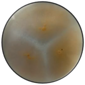
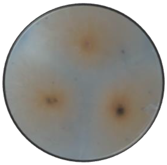
The Table 1 presents a macroscopic comparison of the fungi isolated from the banana peels, considering appearance, the shape of the colony, elevation, edge, texture, surface, color of the front and back.

3.3. Microscopic identification

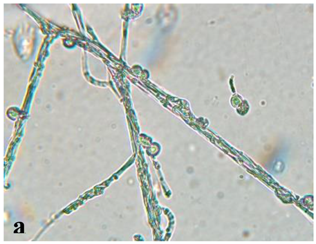
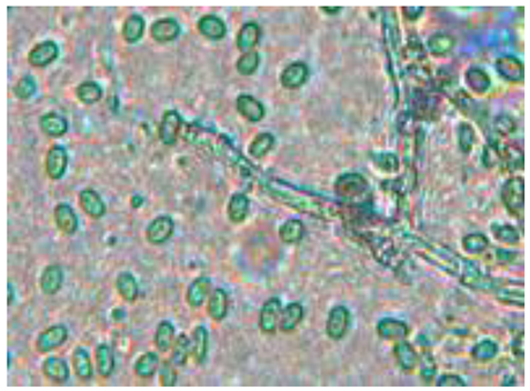
Table 2 describes the hyphae, mycelium, spores and appearance of microorganisms.

3.4. In Vivo Fungal Activity

The growth of the fungi is shown in the Figure 1.

3.5. In Vitro Antifungal Activity with Essential Oils

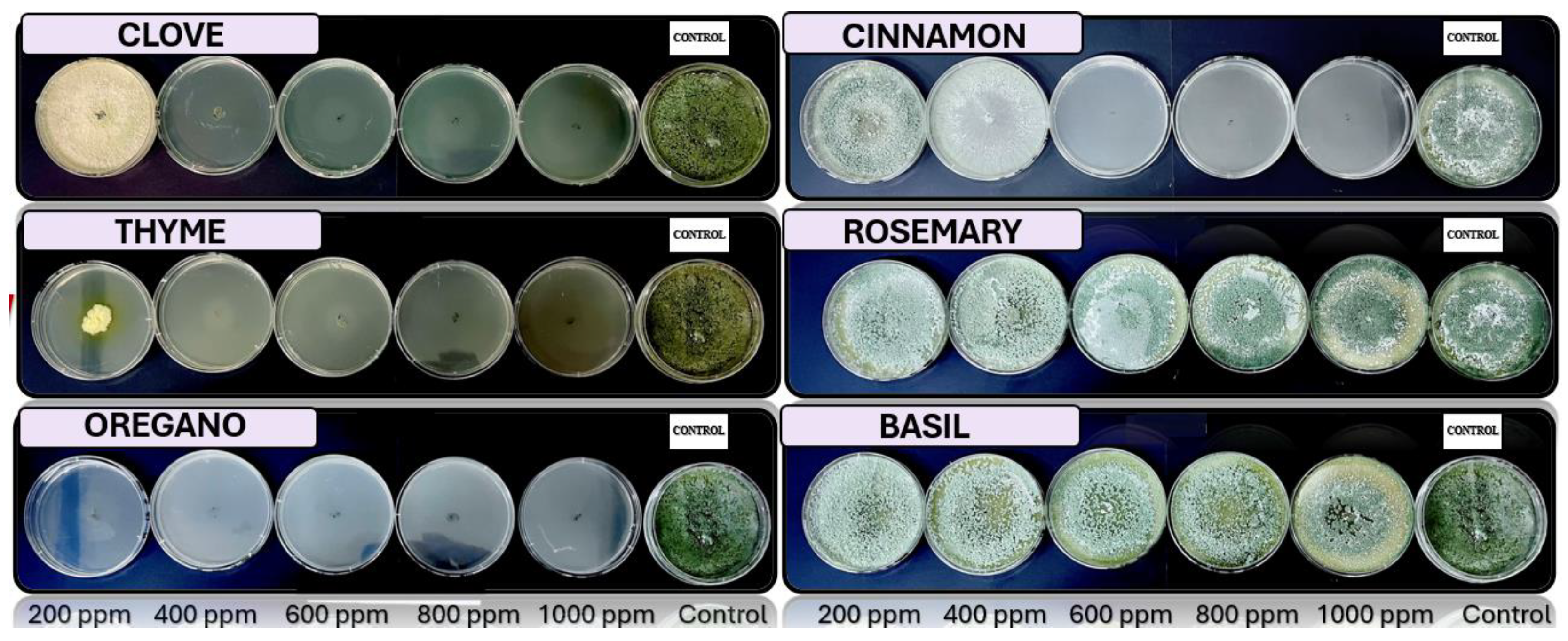
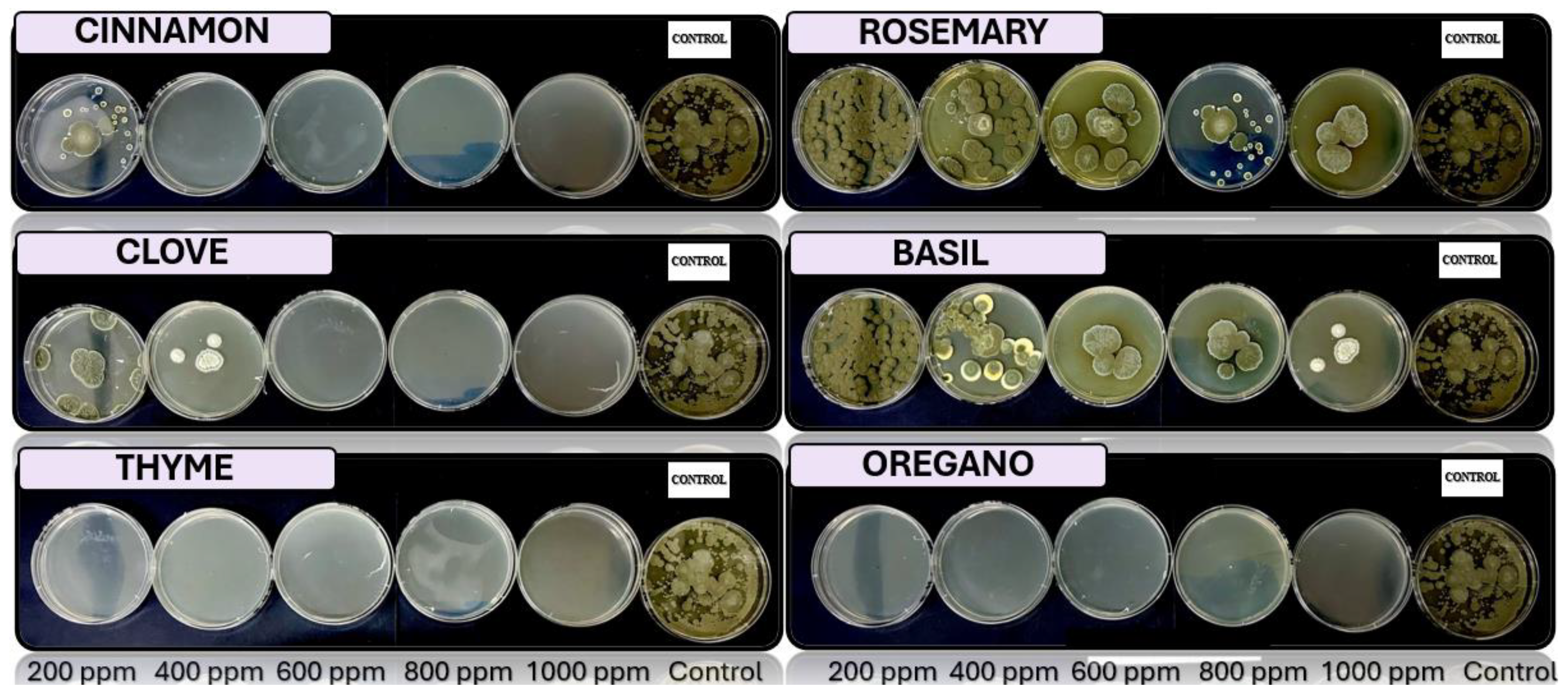
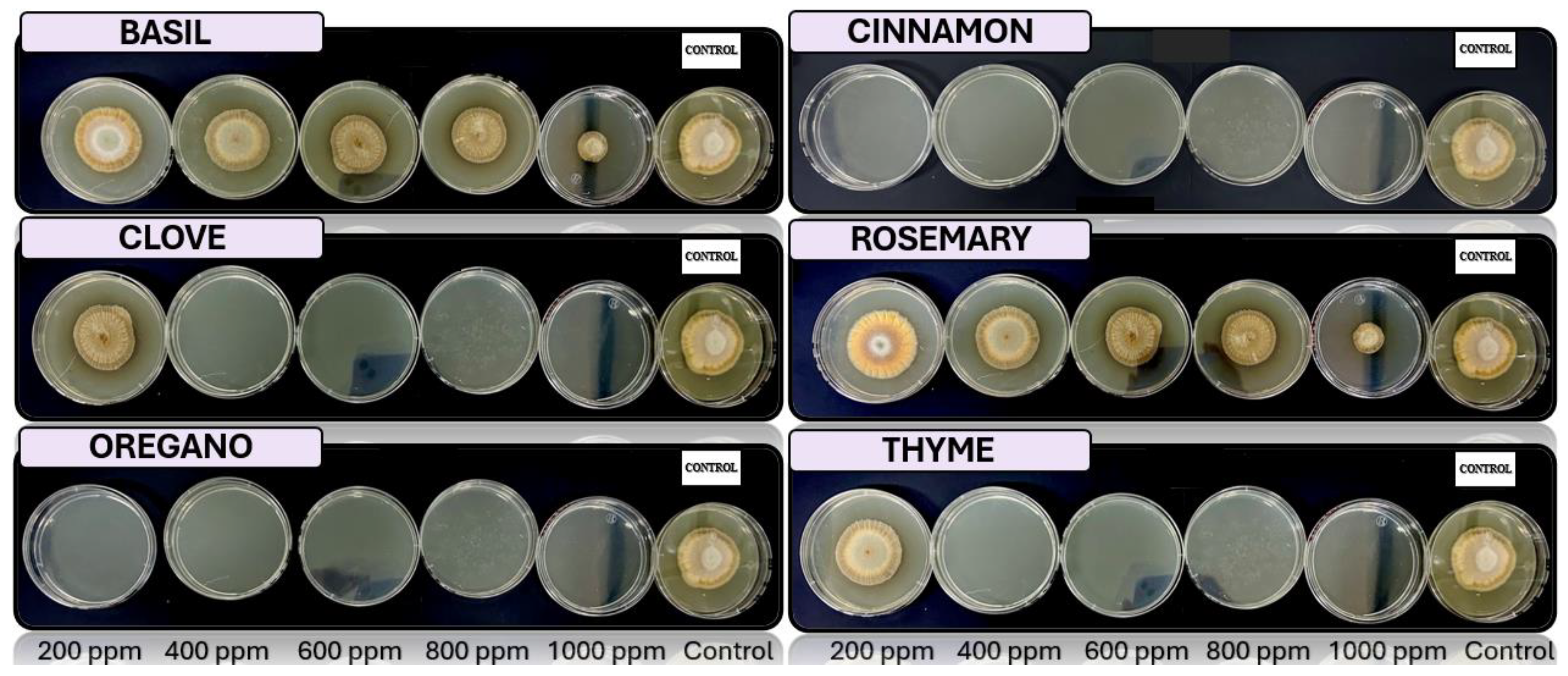
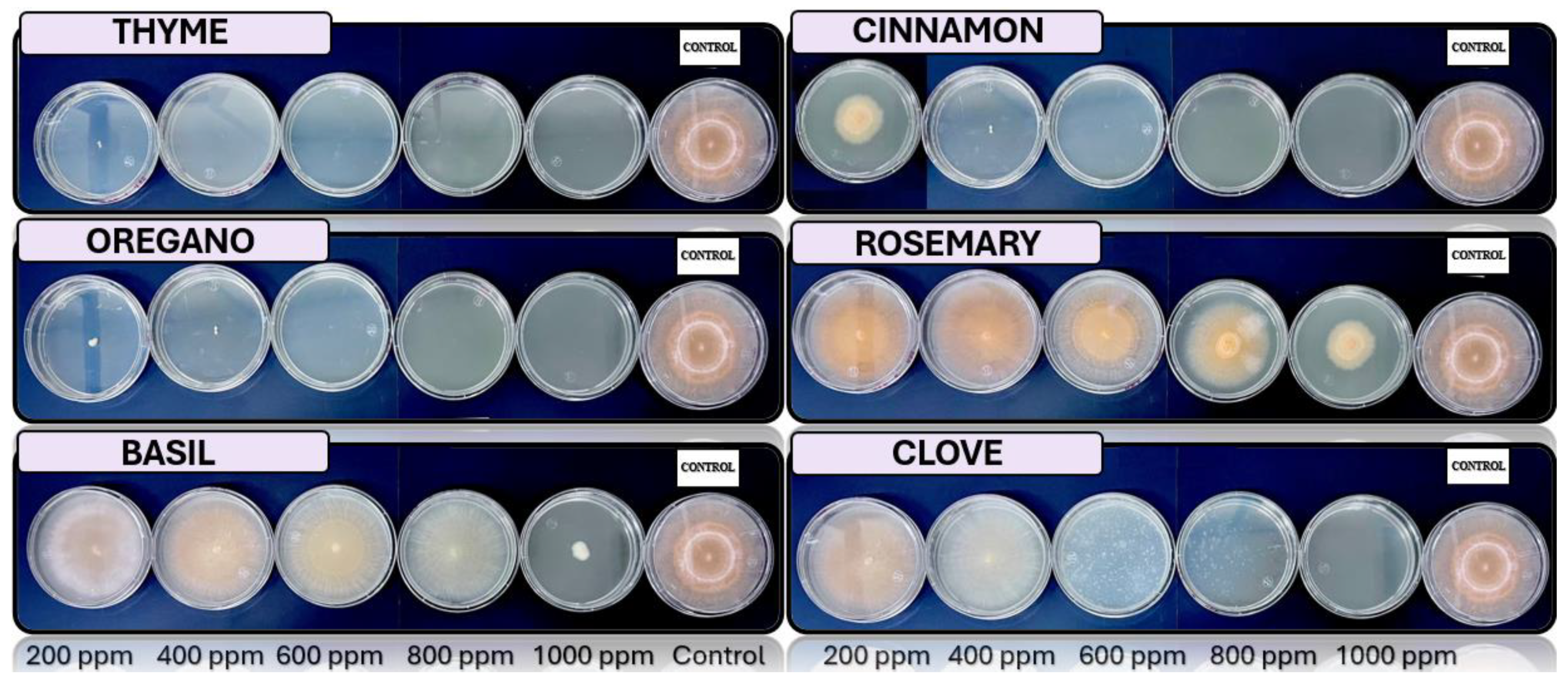
Figure 2, Figure 3, Figure 4 and Figure 5 show the growths in vitro de Fusarium spp., Penicillium spp. Trichoderma spp. and Aspergillus spp. in medium PDA with 200 ppm, 400 ppm, 600 ppm, 800 ppm and 1000 ppm of oregano, basil, cinnamon rosemary, thyme and clove.

4. Discussion

4.1. Macroscopic Identification

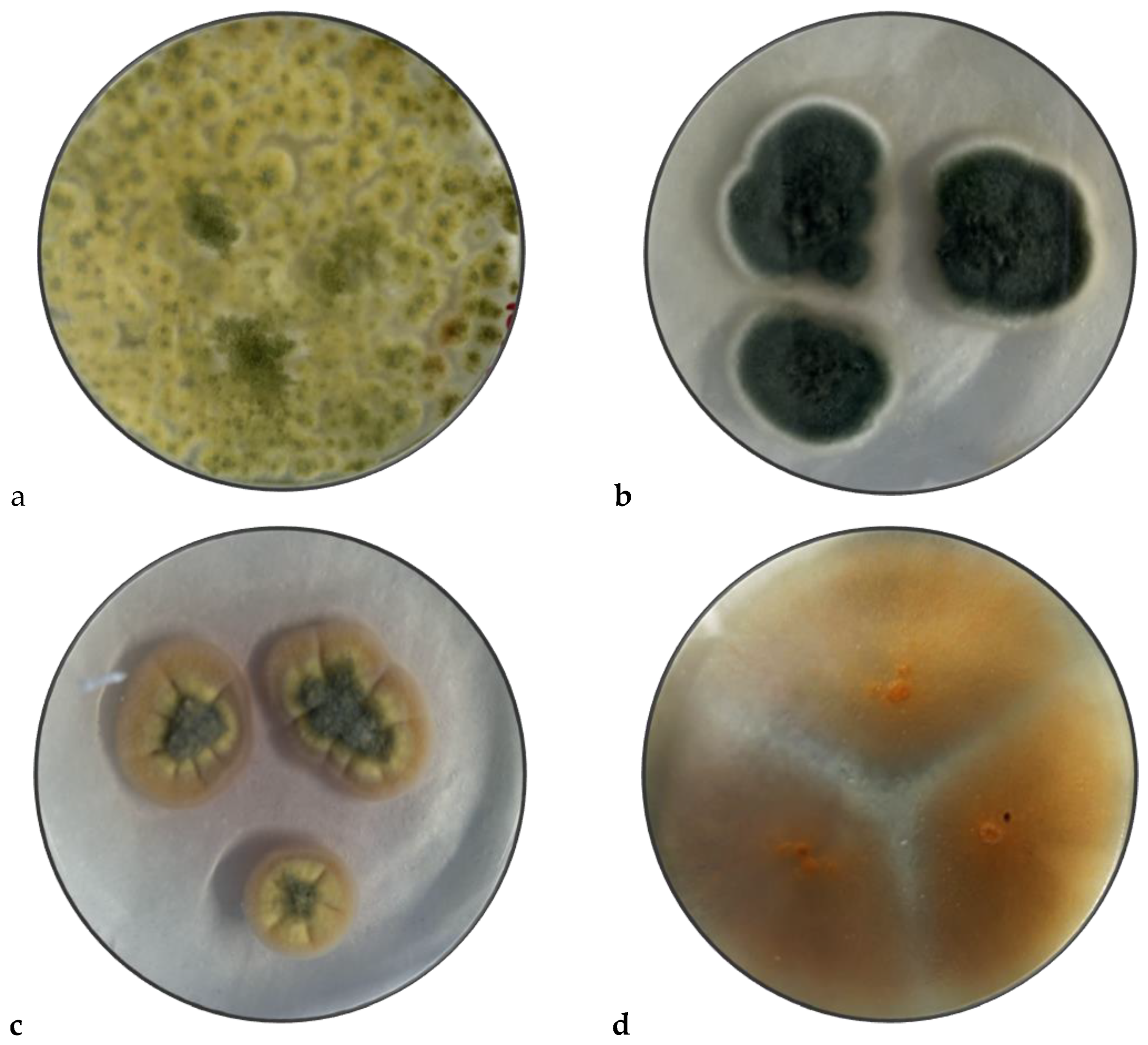
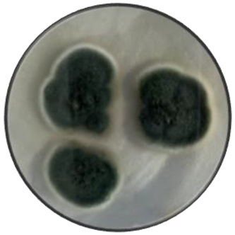
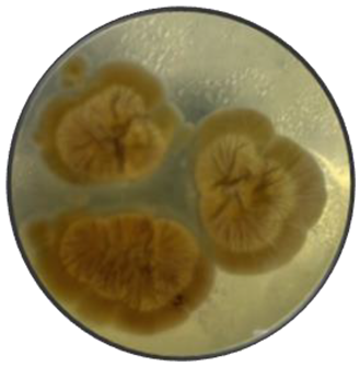
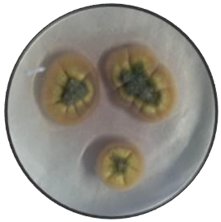
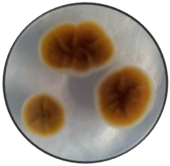
The fungi isolated and purified initially on selective medium with chloramphenicol to prevent bacterial growth are observed in Figure 6. These fungi were obtained from visibly affected banana peel and were stored on PDA medium at approximately 25°C.
When studying the species of Trichoderma spp. it is observed that they have features such as the green, its shape is circular with rough surface, regular edges and slightly elevated [36], Penicillium spp. has green colonies with white edge, flat surface, powdery with a smooth edge [18], Aspergillus spp. has a yellow – green coloring, rugged surface with wavy edges, with cratiform elevation [26] and Fusarium spp. has an orange color, flat surface, cottony with regular edge [16].

4.2. Microscopic Identification

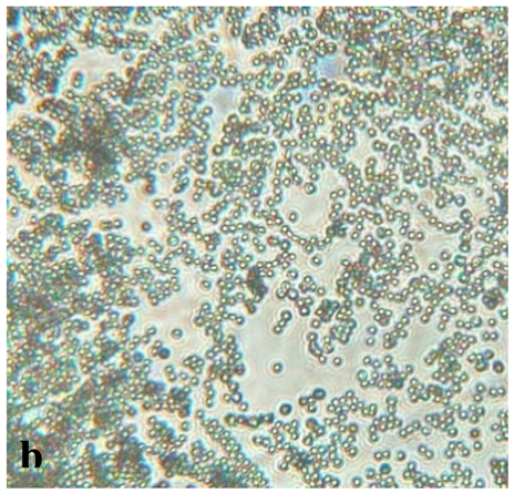
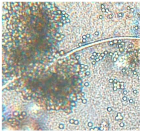
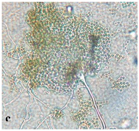
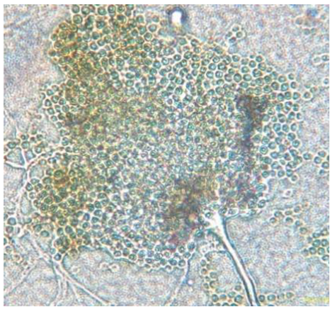
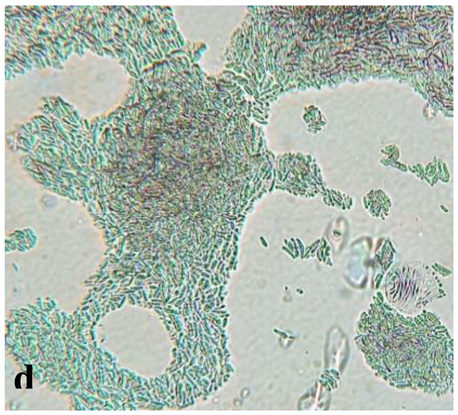
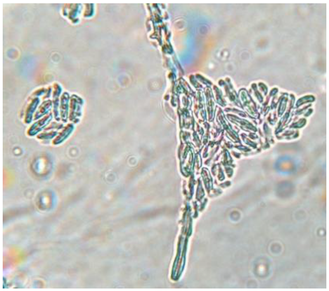
Table 3 presents the analysis of the aerial mycelium of Trichoderma spp., Aspergillus spp., Penicillium spp., and Fusarium spp. using an optical microscope. The evaluation focused on the characteristics of hyphae, mycelium, spores, and overall structure. The results are consistent with the conidium in an ellipsoidal shape for species T. asperellum, T. harzianum, T. koningii [37], with septate and thin hyphae with ellipsoid conidium that are grouped. Penicillium spp. features septate hyphae of variable length that form a structure resembling a plume. Aspergillus spp. also has septate hyphae but is distinguished by conidiophores bearing long, ellipsoid conidia and a characteristic plumose structure. Fusarium spp. is noted for its fusiform conidia, cylindrical shape, and arrangement in chains or clusters.
Trichoderma spp. has conidia clustered on simple conidiophores. Penicillium spp. features conidiophores that terminate in a characteristic plumose structure, with conidia arranged in short chains. Aspergillus spp. is distinguished by a prominent columella and more organized, plumose structures, with conidia emerging from the columella. In contrast, Fusarium spp. presents fusiform conidia in simpler and less organized chains.
The colonies of the Trichoderma spp. are usually green and fast-growing, with fuzzy or cottony texture, regular and slightly elevated edges, while the genus Penicillium spp. have colonies from white to green or blue, velvety surface and structure resembles a ‘brush’, with defined edges. Aspergillus spp. has colonies with a range of colors, which are typically rough with a texture that may resemble a tuft, with regular or slightly wavy edges, whereas the colonies of Fusarium spp. are pink, red or orange, with cottony or velvety texture, diffuse edges and flat or slightly elevated.

4.3. In Vivo Fungal Activity

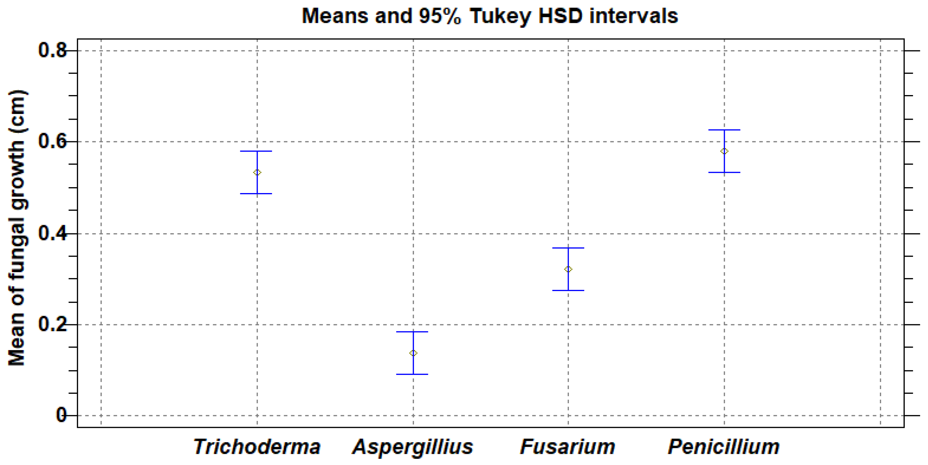
The inhibition rate was evaluated by measuring fungal growth from 20 inoculations of each fungus to assess the severity of the pathogens. Figure 7 shows the analysis of means, considering the 95% Tukey HSD, with the result of the severity of the fungi on Musa Paradisiaca, from highest to lowest, are: Penicillium spp., Trichoderma spp., Fusarium spp. and Aspergillus spp.

4.4. In Vitro Antifungal Activity with Essential Oils

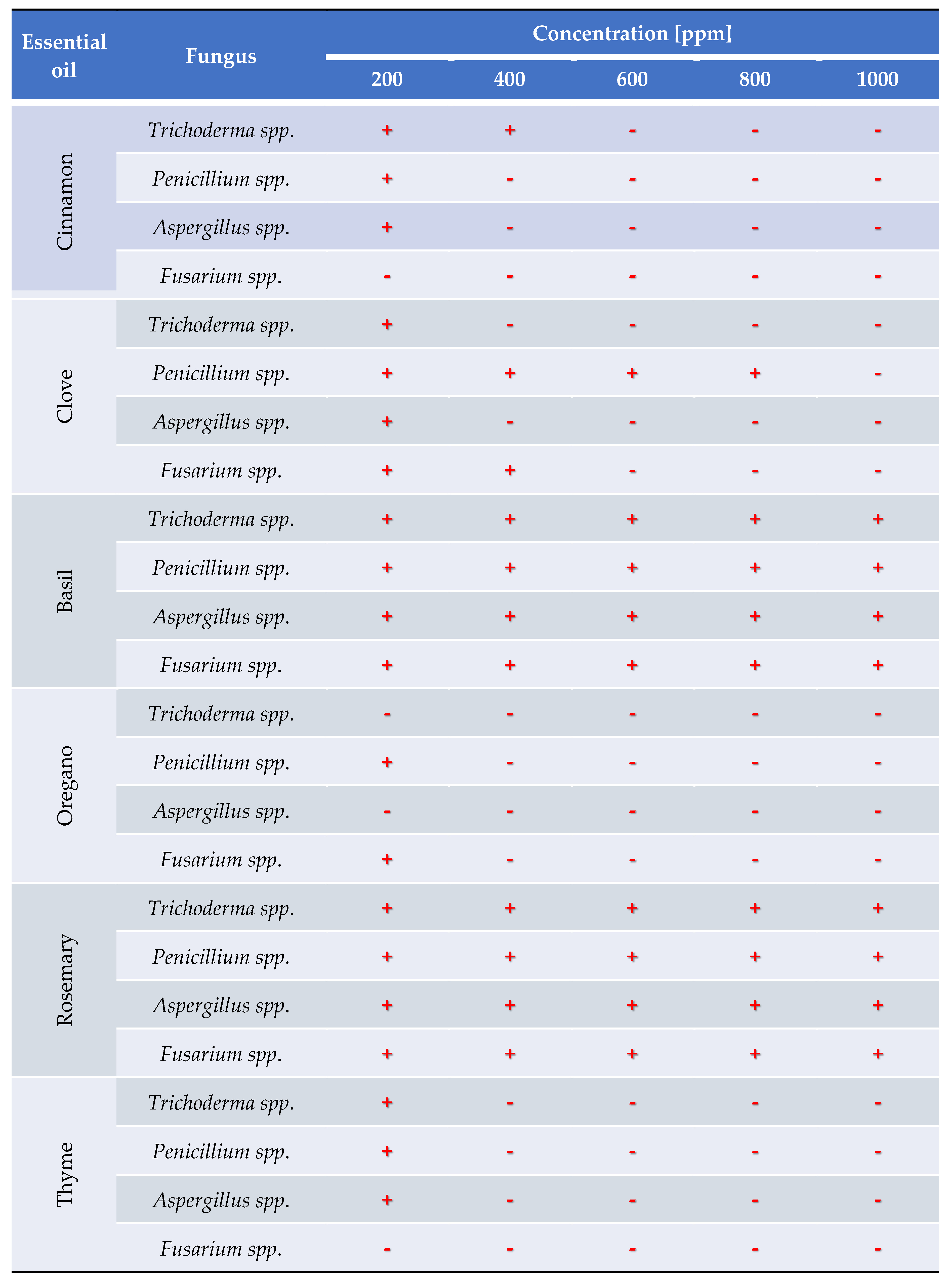
In the evaluation of antifungal activity in vitro considering the use essential oils of oregano (Origanum vulgare), rosemary (Salvia rosmarinus), clove (Syzygium aromaticum), thyme (Thymus), cinnamon (Cinnamomum verum), basil (Ocimum basilicum), with concentration of 200, 400, 600, 800, 1000 ppm is report in the Table 3.

5. Conclusions

The characterization of the strains by order of severity identified Penicillium spp., Trichoderma spp., Fusarium spp., and Aspergillus spp. In the in vitro analysis of essential oil effectiveness, oregano and thyme were effective at 400 ppm, cinnamon at 600 ppm, and clove at 1000 ppm. Basil and rosemary did not inhibit the growth of the analyzed pathogens. Specifically, Penicillium spp. was controlled with 400 ppm of cinnamon, oregano, thyme, and 1000 ppm of clove.
Trichoderma spp. was inhibited by 200 ppm of oregano, 400 ppm of clove and thyme, and 600 ppm of cinnamon. Fusarium spp. was effectively managed with 200 ppm of cinnamon and thyme, and 400 ppm of oregano. Aspergillus spp. was controlled by 200 ppm of oregano, and 400 ppm of cinnamon, clove, and thyme.
At 400 ppm of oregano and thyme essential oils, 600 ppm of cinnamon, and 1000 ppm of clove, the growth of all four analyzed fungi was inhibited.

Author Contributions

All authors listed have significantly contributed to the development and writing of this article. All authors have read and agreed to the published version of the manuscript.”.

Acknowledgments

The authors acknowledge the support from Departamento de Ciencia Ambientales y Biotecnología – Escuela Politécnica Nacional y FICAYA – Universidad Técnica del Norte.

Conflicts of Interest

The funders had no role in the design of the study, in the collection, analysis, or interpretation of data, in the writing of the manuscript, or in the decision to publish the results.

References

  1. Galan V, Rangel A, Lopez J, Hernandez JBP, Sandoval J, Rocha HS. Propagación del banano: técnicas tradicionales, nuevas tecnologías e innovaciones. Rev Bras Frutic 2018;40. [CrossRef]
  2. Capa Benítez LB, Alaña Castillo TP, Benítez Narváez RM. Importancia de la producción de banano orgánico. Caso: Provincia de El Oro, Ecuador. Revista Universidad y Sociedad 2016;8:64–71.
  3. Ruiz Medina MD, Ruales J. Postharvest Alternatives in Banana Cultivation. 2024. [CrossRef]
  4. Mata Anchundia D, Suatunce Cunuhay P, Poveda Morán R. Análisis económico del banano orgánico y convencional en la provincia Los Ríos, Ecuador. Avances 2021;23:419–30.
  5. Aguilar-Anccota R, Arévalo-Quinde CG, Morales-Pizarro A, Galecio-Julca M, Aguilar-Anccota R, Arévalo-Quinde CG, et al. Hongos asociados a la necrosis de haces vasculares en el cultivo de banano orgánico: síntomas, aislamiento e identificación, y alternativas de manejo integrado. Scientia Agropecuaria 2021;12:249–56. [CrossRef]
  6. Guzmán O. El nemátodo barrenador (Radopholus similis [COBB] Thorne) del banano y plátano 2011;1.
  7. Castellanos D, Algecira N, Villota C. Aspectos relevantes en el almacenamiento de banano en empaques con atmósferas modificadas. 2011 2011;12:114–34.
  8. Barrera Necha LL, García Barrera LJ. Actividad antifúngica de aceites esenciales y sus compuestos sobre el crecimiento de Fusarium sp. aislado de papaya ( Carica papaya). Revista Científica UDO Agrícola 2008;8:33–41.
  9. López Luengo MT. El romero. Planta aromática con efectos antioxidantes. Offarm 2008;27:60–3.
  10. Flores-Villa E, Sáenz-Galindo A, Castañeda-Facio AO, Narro-Céspedes RI, Flores-Villa E, Sáenz-Galindo A, et al. Romero (Rosmarinus officinalis L.): su origen, importancia y generalidades de sus metabolitos secundarios. TIP Revista especializada en ciencias químico-biológicas 2020;23. [CrossRef]
  11. Ruiz M, Ávila J, Ruales J. Diseño de un recubrimiento comestible bioactivo para aplicarlo en la frutilla (Fragaria vesca) como proceso de postcosecha 2016;17:276–87.
  12. Características de los aceites esenciales de vegetales y las implicaciones de sus interacciones bioquímicas con los componentes de alimentos en la eficiencia de los procesos de conservación | TecnoCultura 2023.
  13. Mesa V a. M, Marín P, Ocampo O, Calle J, Monsalve Z, Mesa V a. M, et al. Fungicidas a partir de extractos vegetales: una alternativa en el manejo integrado de hongos fitopatógenos. RIA Revista de Investigaciones Agropecuarias 2019;45:23–30.
  14. Jeri R, H C. Consideraciones epidemiológicas para el manejo de la marchitez por Fusarium (Fusarium oxysporum f. sp. cubense) del banano en la región central del Perú 2012.
  15. Magdama F, Monserrate-Maggi L, Serrano L, García Onofre J, Jiménez-Gasco M del M. Genetic Diversity of Fusarium oxysporum f. sp. cubense, the Fusarium Wilt Pathogen of Banana, in Ecuador. Plants 2020;9:1133. [CrossRef]
  16. Tapia C, Amaro J. Género Fusarium. Revista Chilena de Infectología 2014;31:85–6. [CrossRef]
  17. Ortiz E, Riascos D, Angarita M, Castro O, Rivera C, Romero D, et al. Tópicos taxonómicos para el estudio del género Fusarium 2020;33:61–6.
  18. Srinivasan R, Prabhu G, Prasad M, Mishra M, Chaudhary M, Srivastava R. Penicillium. In: Amaresan N, Senthil Kumar M, Annapurna K, Kumar K, Sankaranarayanan A, editors. Beneficial Microbes in Agro-Ecology, Academic Press; 2020, p. 651–67. [CrossRef]
  19. Velásquez MA, Álvarez RM, Tamayo PJ, Carvalho CP. Evaluación in vitro de la actividad fungistática del aceite esencial de mandarina sobre el crecimiento de Penicillium sp. Ciencia y Tecnología Agropecuaria 2014;15:7–14. [CrossRef]
  20. Smith J, Henderson R. Mycotoxins and Animal Foods. 1er ed. Glasgow, Scotland: CRC Press; 1991.
  21. Schuster A, Schmoll M. Biology and biotechnology of Trichoderma | Applied Microbiology and Biotechnology 2010. https://link.springer.com/article/10.1007/s00253-010-2632-1 (accessed August 4, 2024). [CrossRef]
  22. Brotman Y, Kapuganti JG, Viterbo A. Trichoderma. Current Biology 2010;20:R390–1. [CrossRef]
  23. Hernández-Melchor DJ, Ferrera-Cerrato R, Alarcón A, Hernández-Melchor DJ, Ferrera-Cerrato R, Alarcón A. Trichoderma: Importancia agrícola, biotecnológica y sistemas de fermentación para producir biomasa y enzimas de interés industrial. Chilean Journal of Agricultural & Animal Sciences 2019;35:98–112. [CrossRef]
  24. Harman GE, Kubicek CP. Trichoderma And Gliocladium: Basic Biology, Taxonomy and Genetics. Vienna, Austria: CRC Press; 2002.
  25. Chang PK, Horn BW, Abe K, Gomi K. Aspergillus: Introduction. Encyclopedia of Food Microbiology: Second Edition, Elsevier Inc.; 2014, p. 77–82. [CrossRef]
  26. Rokas A. Aspergillus. Current Biology 2013;23:R187–8. [CrossRef]
  27. Vargas-Fernández JP, Wang-Wong A, Muñoz-Fonseca M, Vargas-Fernández JP, Wang-Wong A, Muñoz-Fonseca M. Microorganismos asociados a la enfermedad conocida como pudrición suave del fruto de banano (Musa sp.) y alternativas de control microbiológicas y químicas a nivel in vitro *. Agronomía Costarricense 2022;46:61–76. [CrossRef]
  28. Salazar E, Hernández R, Tapia A, Gómez-Alpízar L. Identificación molecular del hongo Colletotrichum spp., aislado de banano (Musa spp) de la altura en la zona de Turrialba y determinación de su sensibilidad a fungicidas poscosecha. Agronomía Costarricense 2012;36:53–68.
  29. Morales R, Henríquez G. Aislamiento e identificación del moho causante de antracnosis en musa paradisiaca l. (plátano) en cooperativa san carlos, el salvador y aislamiento de mohos y levaduras con capacidad antagonista. Crea Ciencia Revista Científica 2021;13:84–94. [CrossRef]
  30. Tortora GJ, Funke BR, Case CL. Introducción a la microbiología. Ed. Médica Panamericana; 2007.
  31. Vanegas J Santamaría, González N Comba, Mancilla XC Pérez. Manual de Microbiología General: Principios Básicos de Laboratorio. Editorial Tadeo Lozano; 2014.
  32. Suárez Contreras LY. Identificación molecular de aislamientos de Moniliophthora roreri en huertos de cacao de Norte de Santander, Colombia. Acta Agronómica 2016;65:51–7. [CrossRef]
  33. Aguilar Armijos JS. Identificación del hongo fitopatógeno Phoma spp. aislado a partir de plantas de uvilla (Physalis peruviana L.) en localidades de zona norte y centro-norte de la serranía ecuatoriana 2020.
  34. Aguilar-Anccota R, Apaza-Apaza S, Maldonado E, Calle-Cheje Y, Rafael-Rutte R, Montalvo K, et al. Control in vitro e in vivo de Thielavipsis paradoxa y Collettrichum musae cn biofungicidas en frutos de banano orgánico. Manglar 2024;21:57–63. [CrossRef]
  35. Aquino-Martínez JG, Vázquez-García LM, Reyes-Reyes BG. Biocontrol in vitro e in vivo de Fusarium oxysporum Schlecht. f. sp. dianthi (Prill. y Delacr.) Snyder y Hans. con Hongos Antagonistas Nativos de la Zona Florícola de Villa Guerrero, Estado de México. Revista mexicana de fitopatología 2008;26:127–37.
  36. Cayotopa-Torres J, Arevalo L, Pichis-García R, Olivera-Cayotopa D, Rimachi-Valle M, Kadir KJMD. New cadmium bioremediation agents: Trichodermaspecies native to the rhizosphere of cacao trees. Scientia Agropecuaria 2021;24:155–60. [CrossRef]
  37. Savín-Molina J, Hernández-Montiel LG, Ceiro-Catasú W, Ávila-Quezada GD, Palacios-Espinosa A, Ruiz-Espinoza FH, et al. Caracterización morfológica y potencial de biocontrol de especies de Trichoderma aisladas de suelos del semiárido. Revista mexicana de fitopatología 2021;39:435–51. [CrossRef]
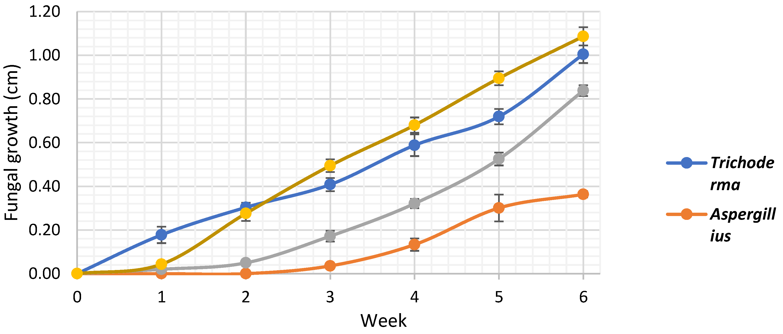
Figure 1. Fungal growth (cm) for 6 weeks in 20 banana samples inoculated with Trichoderma spp., Penicillium spp., Aspergillus spp., and Fusarium spp., stored at 13°C and 95% relative humidity.
Preprints 116433 g001
Figure 2. In vitro growth analysis of Trichoderma spp. on PDA medium with basil, cinnamon, clove, oregano, rosemary, and thyme essential oils at 200, 400, 600, 800, and 1000 ppm, stored at 25°C (n=4).
Preprints 116433 g002
Figure 3. In vitro growth analysis of Penicillium spp. on PDA medium with basil, cinnamon, clove, oregano, rosemary, and thyme essential oils at 200, 400, 600, 800, and 1000 ppm, stored at 25°C (n=4).
Preprints 116433 g003
Figure 4. In vitro growth analysis of Aspergillus spp. on PDA medium with basil, cinnamon, clove, oregano, rosemary, and thyme essential oils at 200, 400, 600, 800, and 1000 ppm, stored at 25°C (n=4).
Preprints 116433 g004
Figure 5. In vitro growth analysis of Fusarium spp. on PDA medium with basil, cinnamon, clove, oregano, rosemary, and thyme essential oils at 200, 400, 600, 800, and 1000 ppm, stored at 25°C (n=4).
Preprints 116433 g005
Figure 6. Fungi isolated and purified from banana peel rot, inoculated on selective medium (PDA with chloramphenicol), and stored on PDA at a temperature of 25°C approx. in the incubator, (a) strain of Trichoderma spp., (b) Penicillium spp., (c) Aspergillus spp. and (d) Fusarium spp.
Preprints 116433 g006
Figure 7. Mean diameter (cm) of fungal decay circles in banana samples inoculated with Trichoderma spp., Penicillium spp., Aspergillus spp., and Fusarium spp. for 6 weeks, stored at 13°C and 95% HR (n=20).
Preprints 116433 g007
Table 1. Macroscopic characterization of (a) Trichoderma spp, (b) Penicillium spp. (c) Aspergillus spp. and (d) Fusarium spp. considering appearance of upper side and lower side.
Fungus Macroscopic characteristics
Upper side Lower side Shape Appearance Elevation Shore Surface Color
Top Bottom
 a
Preprints 116433 i001
Preprints 116433 i002 Circulate too spherical Cottony Flat, slightly elevated, convex Entire, slightly undulating Smooth, rough and granular White, green or yellow Color of the fungal colony, yellow
 b
Preprints 116433 i003
Preprints 116433 i004 Variable Cottony, dusty Convex and crateriform Irregular and wavy Smooth and rough Green and white Beige and yellow
 c
Preprints 116433 i005
Preprints 116433 i006 Variable Velvety, Cottony and dusty Flat, elevate and umbonate Irregular and wavy Smooth and rough Green, yellow and brown Pale yellow
 d
Preprints 116433 i007
Preprints 116433 i008 Cerebriform Cottony Flat Irregular Smooth Orange Orange
Table 2. Microscopic characterization of (a) Trichoderma spp., (b) Penicillium spp., (c) Aspergillus spp. and (d) Fusarium spp. observed under the microscope.
Fungus Microscopic characteristics
40 X 60 X Hyphae Mycelium Spores Appearance
Preprints 116433 i009 Preprints 116433 i010  
Septate
Dense mycelium, often branching Conidium (asexual), of an ellipsoid shape Conidium scattered around of the hypha
Preprints 116433 i011 Preprints 116433 i012  
Septate, branching
Sporulation structures similar to a tuft Conidium (asexual) in a circular shape and in clusters Conidium often on a conidiophore head
Preprints 116433 i013 Preprints 116433 i014  
Septate, branching
Mycelium profusely branching, often dense Structure with numerous conidia (asexual) that appear like a plumose structure Cluster of conidia with hyphae
Preprints 116433 i015 Preprints 116433 i016  
Septate, branching
Form a complex and extensive network in the culture medium Elongated and fusiform conidium forming chains Enlarged macroconidium with septets and sections
Table 3. Evaluation of antifungal activity in vitro of Trichoderma spp, Penicillium spp. Aspergillus spp. and Fusarium spp. use essential oil of oregano, rosemary, clove, thyme, cinnamon and basil.
Preprints 116433 i017
Disclaimer/Publisher’s Note: The statements, opinions and data contained in all publications are solely those of the individual author(s) and contributor(s) and not of MDPI and/or the editor(s). MDPI and/or the editor(s) disclaim responsibility for any injury to people or property resulting from any ideas, methods, instructions or products referred to in the content.
Copyright: This open access article is published under a Creative Commons CC BY 4.0 license, which permit the free download, distribution, and reuse, provided that the author and preprint are cited in any reuse.
Prerpints.org logo

Preprints.org is a free preprint server supported by MDPI in Basel, Switzerland.

Subscribe

© 2024 MDPI (Basel, Switzerland) unless otherwise stated