1. Introduction
Metal-organic frameworks (MOFs) are attractive structured composites of metal ions or clusters that assemble with multifunctional organic ligands via robust chemical bonds and supramolecular interactions. These assemblies create periodic polymeric networks with organic and inorganic components, spanning two or three dimensions of space (2D or 3D) [
1]. Besides, the MOF functionalization without altering their structure could be achieved by post-synthesis modifications, allowing unique properties and improved reactivities [
2]. In recent years, MOFs have experienced rapid advancement, showing potential applications in diverse fields such as gas adsorption/separation, controlled drug release, catalysis, and luminescence. This progress allows the rational design of materials with tailored electrochemical and optical properties based on carefully selecting and combining metals and ligands. In general, the properties of MOFs are intimately linked to their structural features, encompassing compositions and architectures [
3].
Laboratory analyses typically involve numerous time-consuming steps following specific protocols, utilizing expensive and often large equipment that requires handling by qualified professionals in secure environments. The concept of lab-on-a-chip was introduced to address these challenges and reduce the overall cost of analysis [
4]. These miniaturized analytical platforms, typically constructed from materials such as quartz, glass, or polymers, have paved the way for innovation [
5]. Additionally, alternative materials such as paper-based analytical devices (PADs) have emerged, offering a promising avenue for developing analytical biosensors. PADs provide several advantages. Firstly, they leverage the ubiquity and affordability of paper, which can be locally sourced from renewable and recyclable resources [
6,
7,
8]. On the other hand, paper is easy to store, transport, and handle and lends itself well to printing, coating, and impregnating.
Moreover, its high surface area to volume ratio, compatibility with proteins and biomolecules, and excellent filtration properties make it ideal for various analytical applications [
9]. Furthermore, the porous structure of paper enables lateral flow assays, chromatographic separations, and the development of microfluidic devices. Additionally, paper is biodegradable and can be disposed of through incineration. Consequently, PADs emerge as biocompatible and disposable analytical tools, requiring minimal sample and reagent consumption while offering straightforward operation [
9].
In recent decades, biosensors have attracted considerable interest in both the scientific and industrial sectors [
10,
11,
12,
13,
14,
15]. These devices cleverly integrate biology, physics, chemistry, and engineering knowledge, offering relevant and practical solutions to contemporary analytical challenges [
10,
11,
12,
13,
14,
15]. However, several works have been reported on biosensors based on MOFs platform for biomolecules immobilization, but only a few examples based on PADs have been explored [
16,
17,
18,
19,
20,
21,
22,
23,
24,
25]. Ortiz-Gómez et al. (2017) [
18] developed a microfluidic paper-based analytical device (μPAD) for glucose determination using a supported MIL-101 (Fe) MOF acting as a peroxidase mimic. In that same sense, Wei et al. (2021) [
19] reported an electrochemical sensor based on Co-MOF modified carbon cloth/paper applied to quantitative glucose detection. Also, Hassanzadeh et al. (2019) [
20] presented a chemiluminescence PAD to estimate total phenolic content based on a H
2O
2−rhodamine b−Co-MOF. In the present year, Catalá-Icardo et al. (2024) [
21] fabricated MIL-53(Al)-modified PADs for the efficient extraction of neonicotinoid insecticides. Moreover, Chang et al. (2024) [
22] proposed
colorimetric PADs based on two-dimensional (2D) MOF nanozyme for dichlorophen assay. On the other hand, Feng et al. (2024) [
23]
reported a paper-based electrochemiluminescence biosensor for detecting pathogenic bacteria Staphylococcus aureus. This biosensor was constructed using porous Zn-MOFs to form Ru(bpy)3+2 functionalized MOF nanoflowers (MOF-5 (Ru) NFs) [
23]
. Besides, Guan et al. (2024) [
24]
fabricated a hydrogel-based colorimetric
PADs platform for the visual colorimetric assay of creatinine using CdTe@UiO-66-PC-Cu MOF. Without a doubt, PADs biosensors based on MOFs platform for biomolecule immobilization open exciting doors for the development of analytical devices.
In the present work, we synthesized and characterized three amino-functionalized MOFs platform (UiO-66-NH2 (Zr), MIL-125-NH2 (Ti), and MIL-101-NH2 (Fe)) and employed them to develop sensitive, portable, PADs for monitoring ALP enzyme activity by laser-induced fluorescence.
2. Materials and Methods
2.1. Reagents and Materials
All the used reagents had analytical grade and were purchased without further purification treatment. 2-aminoterephthalic acid (H2ATA), titanium isopropoxide (Ti(OiPr)4), N,N'-dimethylformamide (DMF), methanol (MeOH), zirconium tetrachloride (ZrCl4) and 2-amino-1,4-benzenedicarboxylic acid were purchased from Merck. Iron(III) chloride hexahydrate (FeCl3.6H2O, 97%), dopamine hydrochloride, and Tris-HCl were purchased from J&K Scientific Ltd, 4-methylumbelliferyl phosphate (4-MUP) from Fluka and alkaline phosphatase (ALP) enzyme from Sigma-Aldrich. Hydrogen peroxide (H2O2) 30% (v/v), glutaraldehyde (GLU, 5% aqueous solution), and acetone were purchased from Merck. Filter paper N° 1 was provided by Whatman. All buffer solutions were prepared with milli-Q water.
2.2. Equipment
All pH measurements were made with an Orion Research Inc. (Orion Research Inc., Cambridge, MA, USA) Model EA 940 equipped with a glass combination electrode (Orion Research Inc.). Scanning electron microscopy LEO 1450VP (SEM). The paper microzones were printed with a Xerox ColorQube 8870 printer. The system for detecting fluorescence signals was constructed using LED operated a 430 nm, which is the source of fluorescent excitation. The sample was placed in such a way that its surface is at 45° from the LED and has a focal length of 30 cm. The fluorescent radiation was detected with the assembly's optical axis perpendicular to the device's plane. The light was collected with a microscope objective (10:1, NA 0.30, working distance 6 mm, PZO, Poland) mounted on a microscope body (BIOLAR L, PZO). A fiber-optic collection bundle was mounted on a sealed housing at the end of the microscope lens, which was connected to a QE65000-FL scientific-grade spectrometer (Ocean Optics, USA). The entire assembly was covered with a large box to eliminate spurious light. The powder X-ray diffraction (PXRD) plots were obtained with a Rigaku - Ultima IV type II XRD diffractometer. The best counting statistics were achieved using a scanning step of 0.05◦ between 2◦ and 50◦ Bragg angles with an exposure time of 5 s per step. The PXRD of all powdered samples were compared with the respective simulated patterns. Infrared (FTIR) spectroscopic measurements were obtained in a Spectrum 65 FTIR spectrometer Perkin Elmer in a region from 4000 to 400 cm−1.
2.3. Synthesis of Amino-MOFs
The synthesis of MIL-125-NH2 (Ti) was carried out following the previously reported procedure [
10] with some modifications in the amounts of the components: 0.54 g of H
2ATA (0.294.10
-3 mol.L
-1) and 0.315 mL of Ti(OiPr)
4 (1.05.10
-3 mol.L
-1) were mixed in 24 mL of DMF solution and 6 mL of MeOH solution. The mixture was stirred for 30 min, then transferred to a 45 mL Parr reactor coated internally with teflon and heated to 150°C for 72 h. After that, a pale-yellow powder was obtained. The solid underwent a two-step washing process with continuous stirring at 500 rpm: initially with 45 mL of a DMF solution, followed by 20 mL of a MeOH solution. Afterward, the solid was dried at 75°C in an oven for 48 h. The resulting powder was reduced in particle size through manual grinding for 90 min in an agate mortar. Then, a 0.5% w/v suspension of MIL-125-NH
2 (Ti) in 0.1 mol.L
-1 PBS buffer solution pH 7.2 was prepared and sonicated for 60 min. The
UiO-66-NH2 (Zr) synthesis was carried out following the previously described steps [
11] with some modifications in the amounts of the components: 0.288 g of anhydrous ZrCl
4 (1.23x10
-3 mol.L
-1) and 0.209 g (1.92 x10
-3 mol.L
-1) of H
2ATA were dissolved in 60 mL of DMF and left stirring for 30 min. Subsequently, the solution was transferred to a 45 mL Teflon-lined Parr reactor and brought to 120°C for 24 h. After that, it was cooled to room temperature, and the resulting light brown powder was washed three times with DMF solution and finally dried at 70°C for 24 h. A 0.1 mol.L
-1 pH 7.2 PBS buffer solution containing 0.5 % w/v of UiO-66-NH2 (Zr) was prepared for analytical measurements. The synthesis of MIL-101-
NH2 (Fe), the steps previously described [
12] were followed with some modifications. 0.504 g of anhydrous FeCl
3.6H
2O (2x10
-3 mol.L
-1) and 0.181 g (1x10
-3 mol.L
-1) of H
2ATA were mixed in 15 mL of DMF under constant stirring for 30 min. The mixture was transferred into a 120 mL stainless steel Parr autoclave reactor internally lined with Teflon, which was hermetically sealed and heated at 120°C for 24 h. After cooling to room temperature, the resulting dark brown powder was washed with constant agitation using 20 mL of DMF for 24 h. Finally, the product was dried at 75°C for 48 h. Before employing the product, a 0.1 mol.L
-1 PBS buffer solution pH 7.2 containing 0.5% w/v of the
MIL-101-_NH2 (Fe) was prepared.
2.4. Biosensor Design
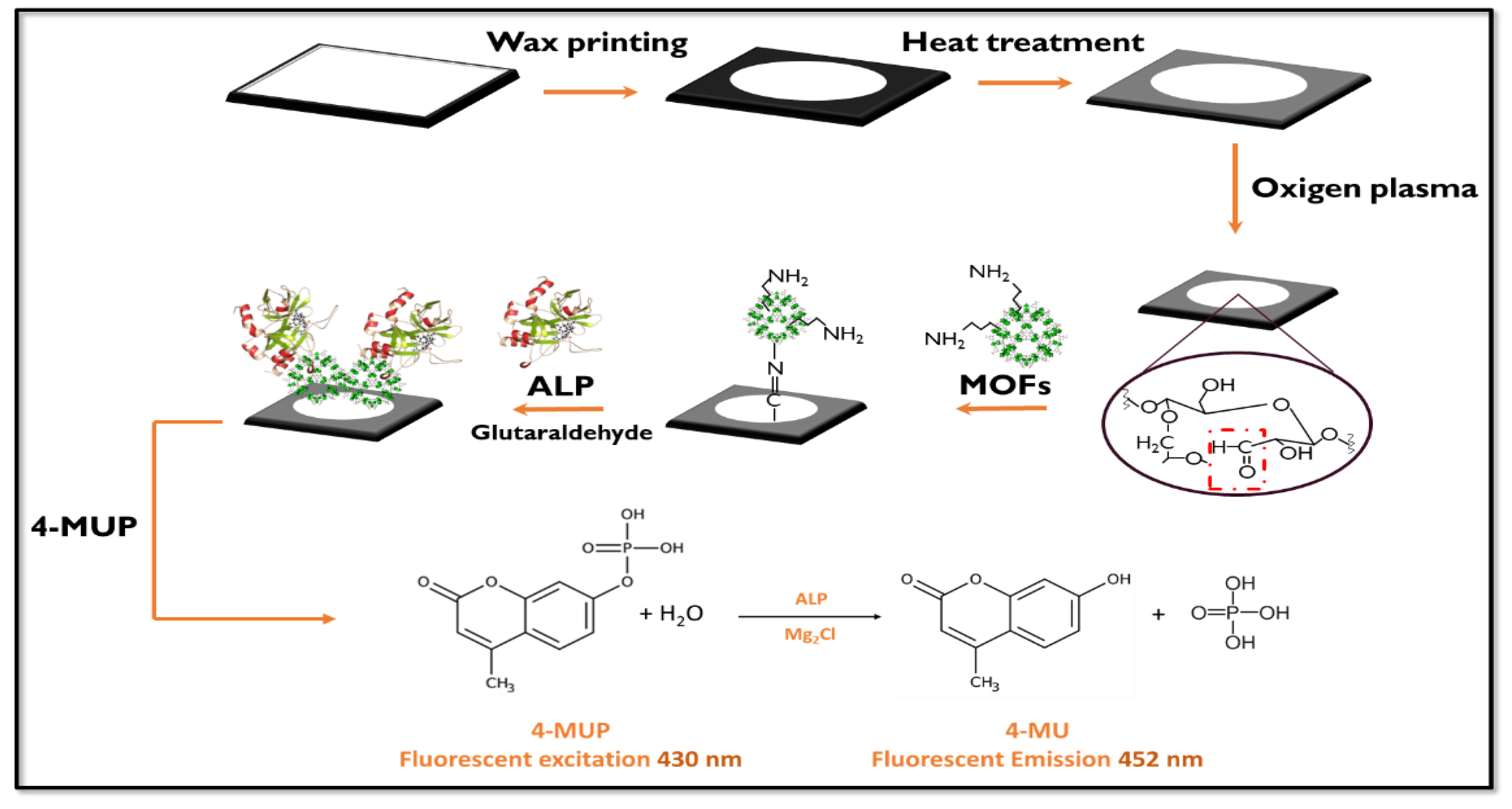
Whatman No.1 filter paper was used to design and construct the immunosensor. A 6 mm diameter hydrophobic containment barrier was delimited circularly on a microzone of this paper by wax stamping, using Corel Draw 9 graphic software and the Xerox ColorQube 8870 wax printer. The cut papers were then placed on a hot plate at 80°C to homogenize the walls forming the hydrophobic wax barrier.
The papers were subjected to a constant flow of oxygen plasma for 3 min to generate as many aldehyde groups as possible on their surface by oxidation of the cellulose to have a more significant reaction surface to modify the papers.
2.5 Paper Modification
Suspensions of UiO-66-NH2 (Zr), MIL-125-NH2 (Ti), and MIL-101-NH2 (Fe) were prepared at a concentration of 0.5 % wv-1 in 0.1 mol.L-1 PBS buffer solution pH 7.2 and dispersed under sonication for 15 min. When the suspensions were completely homogenized, 20 µL of each was placed on the different pre-treated papers by drop casting method and left in a humid chamber for 30 min. During this time, the aldehyde groups on the cellulose surface linked to the amino groups of the mentioned MOFs. After 30 min, the papers were washed three times with 0.1 mol.L-1 PBS buffer solution pH 7.2 and dried under N2 flow.
2.6 Enzyme Immobilization
To achieve covalent bonds between the amino groups of the immobilized MOFs on the paper with the amino groups present in the ALP, a cross-linking agent solution glutaraldehyde (GLU) was employed. This procedure consists of several steps: First, the papers were immersed for 30 min in a 0.5 % w/v GLU acetone solution at pH 8. After being washed three times with 0.1 mol.L-1 pH 7.2 PBS buffer solution and dried, 20 µL of 0.1% w/v ALP solution in 0.1 mol.L-1 PBS buffer solution pH 7.2 was added and incubated at 37°C for 10 min. Subsequently, they were washed three times with PBS buffer solution to remove all excess protein that was not bound to the modified paper support. This ensures that what is on the support remains covalently bound after the various impregnations and immobilizations.
Figure 1.
Schematic representation of fluorescent paper-based biosensor construction showing the modification of the paper surface and the alkaline phosphatase determination procedure.
Figure 1.
Schematic representation of fluorescent paper-based biosensor construction showing the modification of the paper surface and the alkaline phosphatase determination procedure.
2.7. Study of the Enzymatic Response by Laser-Induced Fluorescence
An optical system was constructed using a single frequency 430 nm DPSS laser (Cobolt ZoukTM, USA) operated at 10 mW that served as the fluorescence excitation source to detect enzyme activity and test the paper biosensor. The laser was focused on the detection channel at 90◦ geometry with respect to the surface.
The relative fluorescence signal, which corresponded to the ALP, was measured in situ on the modified paper device. The fluorescence of 4-MUP was measured using excitation at 430 nm and emission at 452 nm. DEA buffer (0,1 mol.L-1 diethanolamine, 50x10-3 mol.L-1 KCl, 1x10⁻³ mol.L-1 MgCl2, pH 9.6) was used to prepare the 4-MUP solution. Finally, 5 µl of substrate solution (2.5x10⁻³ mol.L-1 4-MUP in DEA buffer solution, pH 9.6) was injected into the modified paper, and LIF measured the enzyme product.
The paths of the reflected beams were meticulously arranged to prevent collisions with other components of the device cell, thereby minimizing the risk of photobleaching. Fluorescent radiation was detected along the optical axis of the array. An optical fiber collection system was installed within a sealed housing linked to the QE65000-FL scientific-grade spectrophotometer (Ocean Optics, Inc., USA). The entire system was enclosed within a black box during measurements to eliminate spurious light interference. These assessments were conducted for both a blank assay, where the enzyme was directly immobilized onto unmodified paper, and assays, where the enzyme was immobilized onto papers modified with three amino-functionalized MOFs.
3. Results and Discussion
3.1. Elemental Characterization of MOFs
SEM-EDS
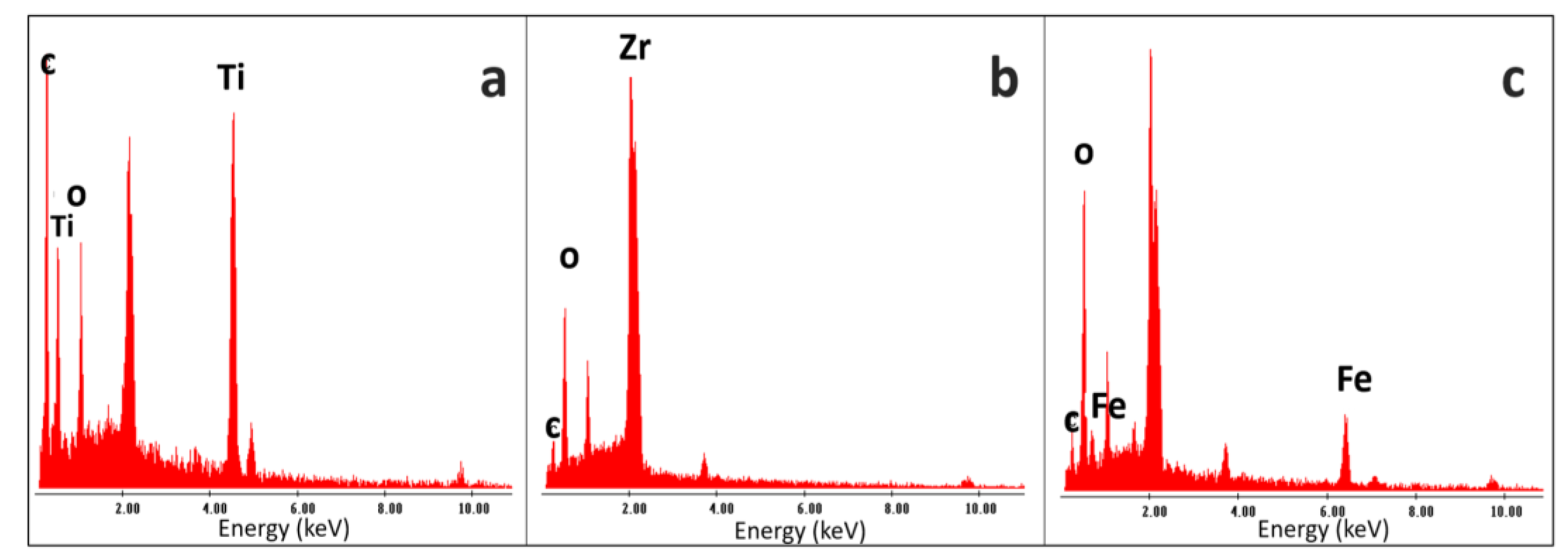
The synthesized MOFs were characterized by SEM and EDS analysis to get information regarding the particle size and semi-quantitative elemental percentage. As shown in
Figure 2 a), MIL-125-NH
2 (Ti) particles exhibit a spherical morphology with an average size of around 0.6 ± 0.2 µm. Moreover, the EDS data indicate the presence of high lines from titanium metal ions and signals from C and O belonging to the organic backbone (
Figure 3(a)).
In
Figure 2(b), the particles of UiO-66-NH
2 (Zr) showed a flat morphology, with an average size of 0.53 ± 0.2 µm, confirming the elemental composition of the metal Zr and the ligand components C and O (
Figure 3 b)). Finally, besides
Figure 2(c), the MIL-101-NH2 (Fe) particles exhibited a spherical shape, with a uniform average size of 0.26 ± 0.06 µm. Also, EDS analysis revealed characteristic X-ray lines corresponding to the iron along with O and C from the ligands (
Figure 3 c)).
The paper supports, with and without modifications, were also characterized by SEM and EDS techniques, as illustrated in
Figure 4 and
Figure 5. In the unmodified paper, the cellulose fibers are shown in their original state provided by the manufacturer, with characteristic EDS peaks confirming their C and O composition (see
Figure 5 a)).
Figure 4(b) shows the impregnated paper with MIL-101-NH
2 (Fe), revealing visible particles deposited on the support and confirming this observation through an X-ray line spectrum (see
Figure 5 b)). In
Figure 4(c), the MOF UiO-66-NH
2 (Zr) is observed impregnating the cellulose support, with visible particles deposited and elemental composition that confirms the presence of cellulose and mesoporous elements (see
Figure 5 c)). Finally,
Figure 4 d) shows cellulose paper impregnated with MIL-125-NH
2 (Ti), demonstrating the presence of elements through composition analysis (see
Figure 5 c)) and a uniform dispersion of particles throughout the paper.
3.2. DRX
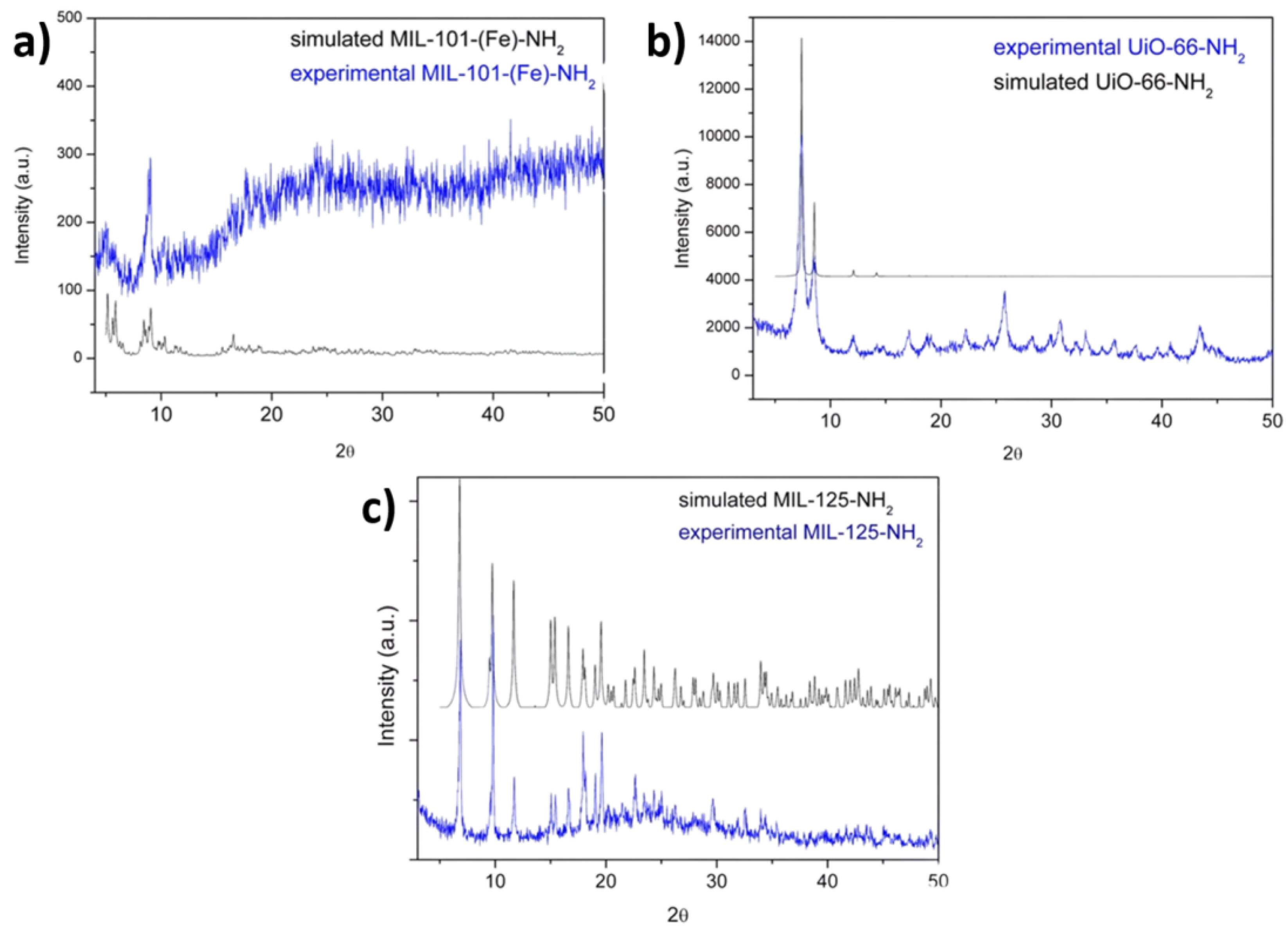
Using powder PXRD, experimental diffractograms were compared with the corresponding simulated powder plots (the latter were obtained using .cif files extracted from the literature [
10,
11,
12]). In MIL-101-NH
2 (Fe), the presence of a single phase could be verified. However, lower intensities are observed in the diffraction peaks, possibly due to a lower crystallinity related to the synthesis method (see
Figure 6 a)). This MOF crystallized in the cubic space group Fd-3m (No. 227). Performing the same analysis for the UiO-66-NH
2 (Zr) phase, a coincidence is also observed between the experimental pattern and its corresponding theoretical one (see
Figure 6 b)). The compound with the formula crystallized in the cubic space group Fm-3m.
Figure 6 c) shows the comparison between the theoretical and experimental diffractograms of MIL-125-NH
2 (Ti), concluding in a broad coincidence between the diffraction peaks, confirming the presence of a single pure phase. It is found that this MOF was indeed crystallized in the orthorhombic space group I4/mmm (N° 139).
MIL-101-NH
2-Fe with chemical composition {[Fe
3(μ
3-O)(μ
4-BDCNH
2)
3(H
2O)
2Cl] used in the present study belongs to a group of materials called MIL-101 (where MIL = Matériaux de l'Institut Lavoisier) [
25]. The crystal structure of MIL-101(Fe)-NH
2 is built from 2-ATA molecules as linkers and Fe(III) ions as nodes. Fe(III) ions are arranged in a trigonal planar cluster with composition [Fe
3(μ
3-O)(μ
2-COO)
6(H
2O)
2Cl] (COO = carboxylate) and a trigonal prismatic secondary building unit (
Figure 7 a)) [
26]. Four building unit clusters are bridged with six 2-ATA anions under the formation of a supertetrahedron unit. The final crystal structure of MIL-101-NH
2 (Fe) contains different cages with different sizes: a microporous cage with a free diameter for entrance windows close to 8.6 Å and two different mesoporous cages with a diameter close to 29 Å and 34 Å, respectively (see
Figure 7 b)).
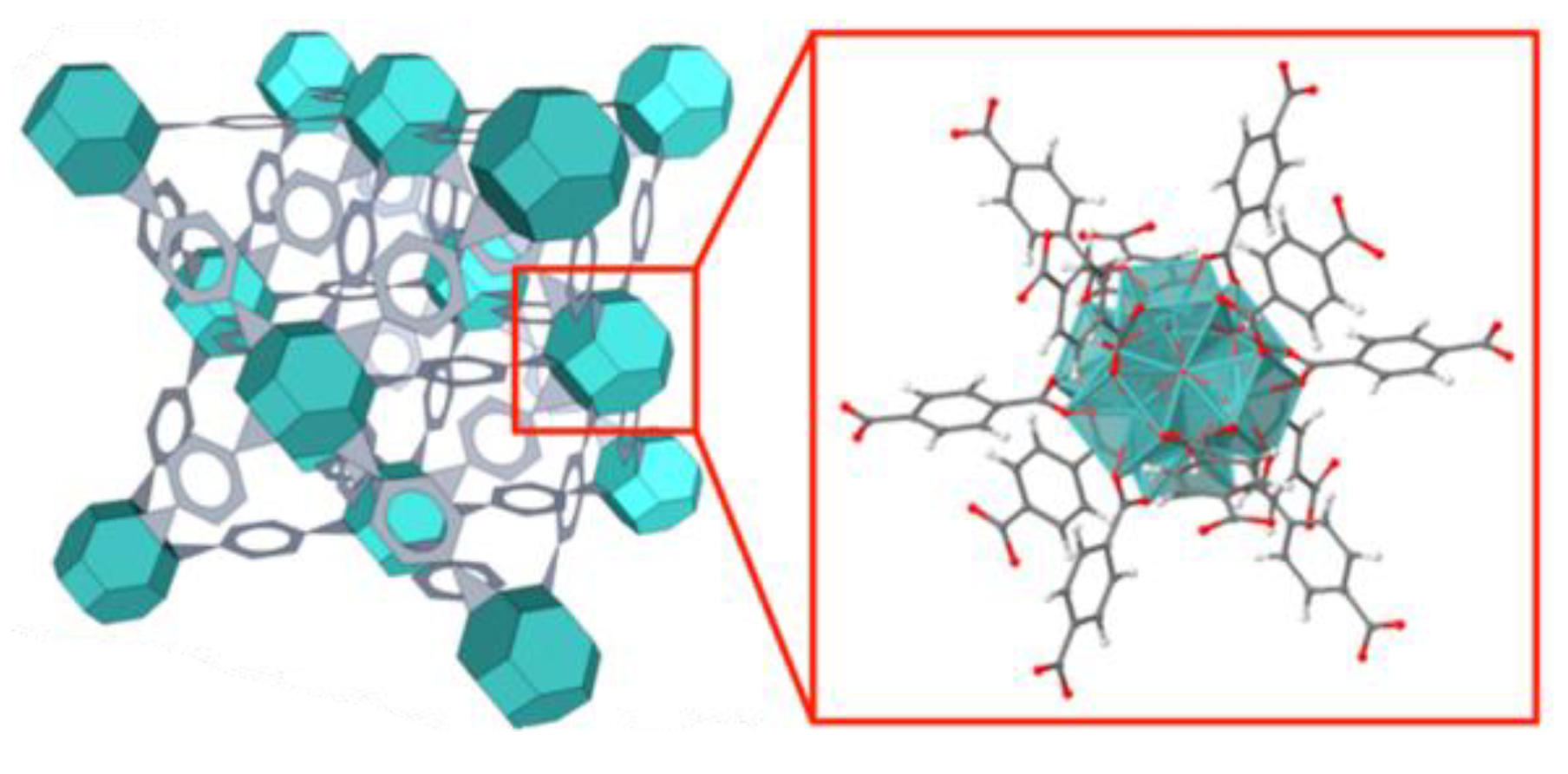
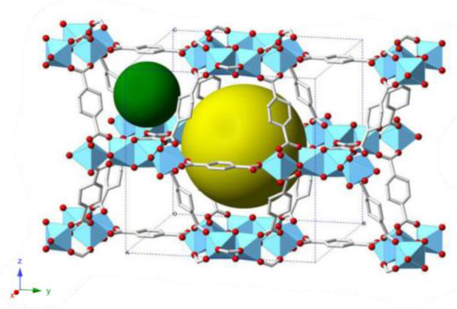
The most stable form of UiO-66-NH
2 (Zr) consists in a face-centered-cubic structure containing an fm−3m symmetry with a lattice parameter of 20.7 Å. It contains two separate cages, a tetrahedron cage of 7.5 Å, another octahedron cage of 12 Å, and a pore aperture of 6 Å [
27] (see
Figure 8).
MIL-125-NH
2-(Ti), comprising of [Ti
8O
8(OH)
4-(2-ATA)
6] building units, and its crystal structure is established from the cyclic octamers constructed from octahedral titanium units present at the edge or corner [
28]. These octamers are associated with 12 other cyclic octamers, giving rise to a porous 3-D quasi-cubic tetragonal structure (see
Figure 9). Also, the structure has two types of cages, an octahedral (12.5 Å) and a tetrahedral (6 Å) cage, accessible through narrow triangular windows of ca. 6 Å.
3.3. Characterization of ALP Immobilization on the Support
FTIR
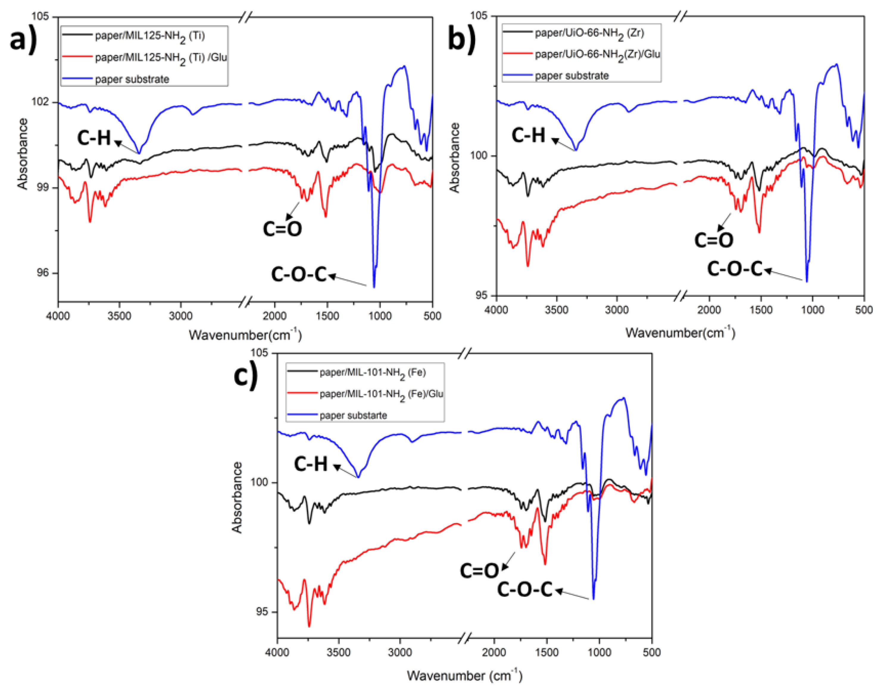
Another method employed to confirm the immobilization processes conducted on the paper substrate was Fourier Transform Infrared (FTIR) characterization.
The spectra provided below (see
Figure 10) depict a comparison between the unaltered paper (shown in blue spectra) and the same paper modified with MIL-125-NH
2 (Ti), UiO-66-NH
2 (Zr), and MIL-101-NH
2 (Fe) - (black traces), followed by the subsequent stage where glutaraldehyde molecules were attached, leaving exposed terminal aldehyde groups (depicted in red spectra), for all three cases.
By the FTIR technique, it is possible to analyze the principal vibrational modes of the cellulose fibers and their interactions with the MOF particles. The paper exhibits the C-H stretching modes of cellulose located at 2850 cm
-1 and a prominent band attributed to the vibrations of the C-O-C groups within the β-glucopyranose ring of cellulose at 1066 cm
-1. This band decreases significantly upon modifying the paper with MOFs, suggesting a robust covalent interaction between these bonds and the amino groups functionalizing the materials [
9]. The following chemical reaction illustrates the bonding between the amino groups of MOFs and the aldehyde groups of cellulose.
In all cases where the paper support is modified with different MOFs, the presence of solvent trapped in its pores is evident at 3625 cm-1. Finally, there is an increase in the intensity of the carbonyl band (C=O stretching) centered at 1745 cm-1 when glutaraldehyde is bound to the MOFs.
3.4. Optimization of Experimental Parameters
To carry out the elemental parameter optimization tests, the following considerations were taken into account: adjust the MOF concentration for the paper support modification, adjust the ALP enzyme concentration, optimize the 4-MUP substrate concentration, determine the reaction time, and identify the optimum pH for enzymatic activity.
For the ALP enzyme, the substrate used was 4-MUP. After enzymatic action, the substrate produces 4-methylumbelliferone (4-MU), a fluorescent compound emitting at 452 nm.
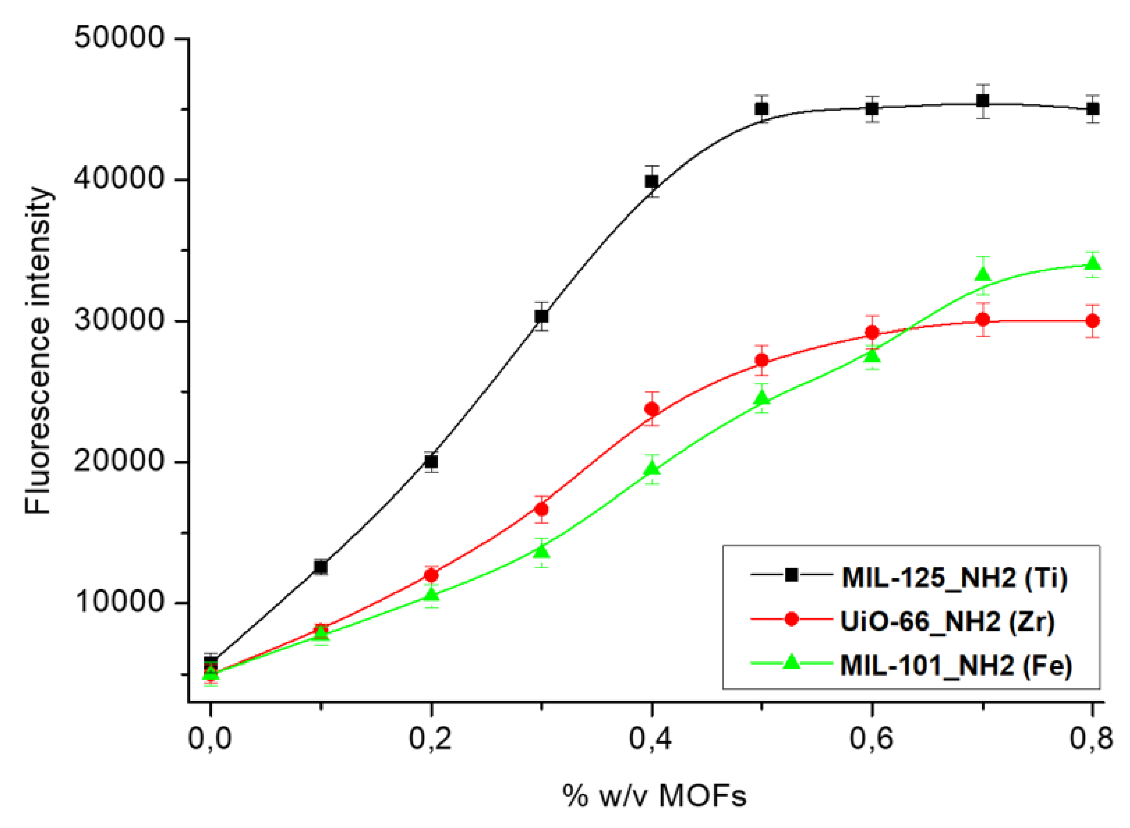
Besides, to optimize the concentration of the different used MOFs for modifying the paper support, dispersions of the three MOFs were prepared in 1 mol.L-1 PBS buffer solution pH 7.2, in a concentration range of 0 to 0.8 % w/v. The ALP enzyme was immobilized at 0.1 % w/v in 0.1 mol.L-1 PBS buffer pH 7.2 for each modified paper with the different MOF concentrations. Finally, the ALP response was monitored using 5 µl of fluorescent substrate solution (2.5x10⁻³ mol.L-1 4-MUP in DEA buffer solution (0,1 mol.L-1 diethanolamine, 50x10-3 mol.L-1 KCl, 1x10⁻³ mol.L-1 MgCl2) pH 9.6) in each paper microzone. Subsequently, the enzyme product was measured by LIF.
The obtained signal from the enzymatic response was greater when the microzones of the paper were modified with different concentrations of MIL-125-NH
2 (Ti), which demonstrates that there is an increase in the immobilization surface due to the incorporation of MIL-125-NH
2 (Ti) in comparison to UiO-66-NH
2 (Zr) and MIL-101-NH
2 (Fe). On the other hand, when higher concentrations of MIL-125-NH
2 (Ti) than 0.5% w/v were used, a system saturation was observed, so this was selected as the concentration limit for all subsequent tests (see
Figure 11).
In light of these results highlighting the differential response of the modified papers with MIL-125-NH2 (Ti)/ALP compared to those with UiO-66-NH2 (Zr)/ALP, MIL-101-NH2 (Fe)/ALP, it was decided to continue with the optimizations of experimental variables using the MOF MIL-125-NH2 (Ti) as a proposal for the best surface to apply to the development of biosensors.
According to the literature, MIL-101-NH
2 (Fe) exhibits a higher BET surface area of 3528 m
2.g
-1 and a pore volume of 1.48 cm
3. g
-1 [
26] respect to those values from MIL-125-NH
2 (Ti) (1469 m
2.g
-1 and a pore volume of 0.6 cm
3. g
-1) [
28] and UiO-66-NH
2 (Zr) (822 m
2.g
-1 and a pore volume of 0.23 cm
3.g
-1 ) [
29]. Nevertheless, MIL-125-NH2 (Ti) exhibits better immobilization performance, which could be reasonably attributed to a lower pore volume and, consequently, lower water occupancy.
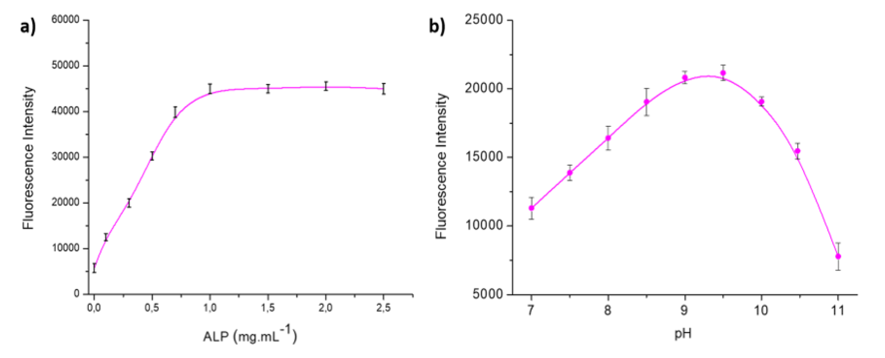
Optimizing the concentration of ALP employed for immobilization was a critical parameter to evaluate. Therefore, a comprehensive study was conducted within the 0.1 to 2.5 mg.mL-1 concentration range.
Figure 12 a) illustrates that the fluorescence intensity increased proportionally with the concentration of immobilized ALP, reaching a peak at 1 mg mL
-1. Beyond this concentration, the intensity plateaued, indicating saturation. Consequently, 1 mg.mL
−1 of ALP emerged as the optimal concentration for immobilization on paper modified with 0.5 % w/v MOF MIL-125-NH
2 (Ti).
The impact of pH on the enzymatic response was also evaluated by adjusting the pH of the substrate solution in a range of 7 to 11. The enzyme activity exhibited its maximum value when the DEA buffer solution was at a pH of 9.6, as shown in
Figure 12 b). Therefore, pH 9.6 was selected as the optimal pH for all subsequent experimental measurements.
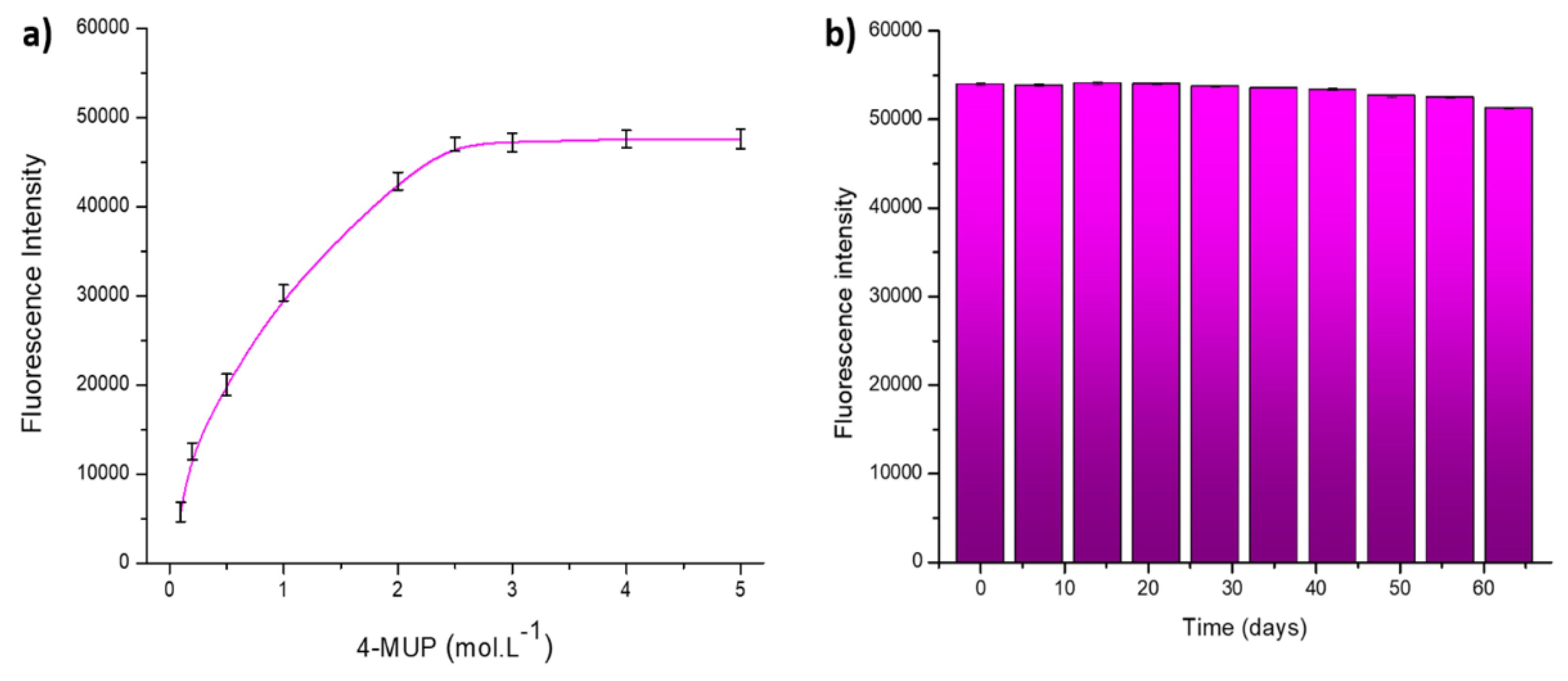
The impact of 4-MUP concentration on the enzymatic response of ALP was investigated within the range of 0.1x10
-3–5.0x10
-3 mol.L
-1 in DEA buffer solution at pH 9.6. The optimal concentration of 4-MUP, which produces the highest enzymatic response, was determined to be 2.5x10
-3 mol.L
-1, as shown in
Figure 13 a). Consequently, this concentration was adopted for all subsequent experimental measurements.
Long-term stability testing of the system was performed by freeze-drying the MIL-125-NH
2 (Ti)/ALP modified paper devices and storing them at 4°C, protected from light. Measurements were taken every 7 days over 2 months, as illustrated in
Figure 13 b). In particular, the fluorescent signal response remained practically constant after 45 days, with only a 5 % decrease observed after 60 days. This result shows the sensor's great stability.
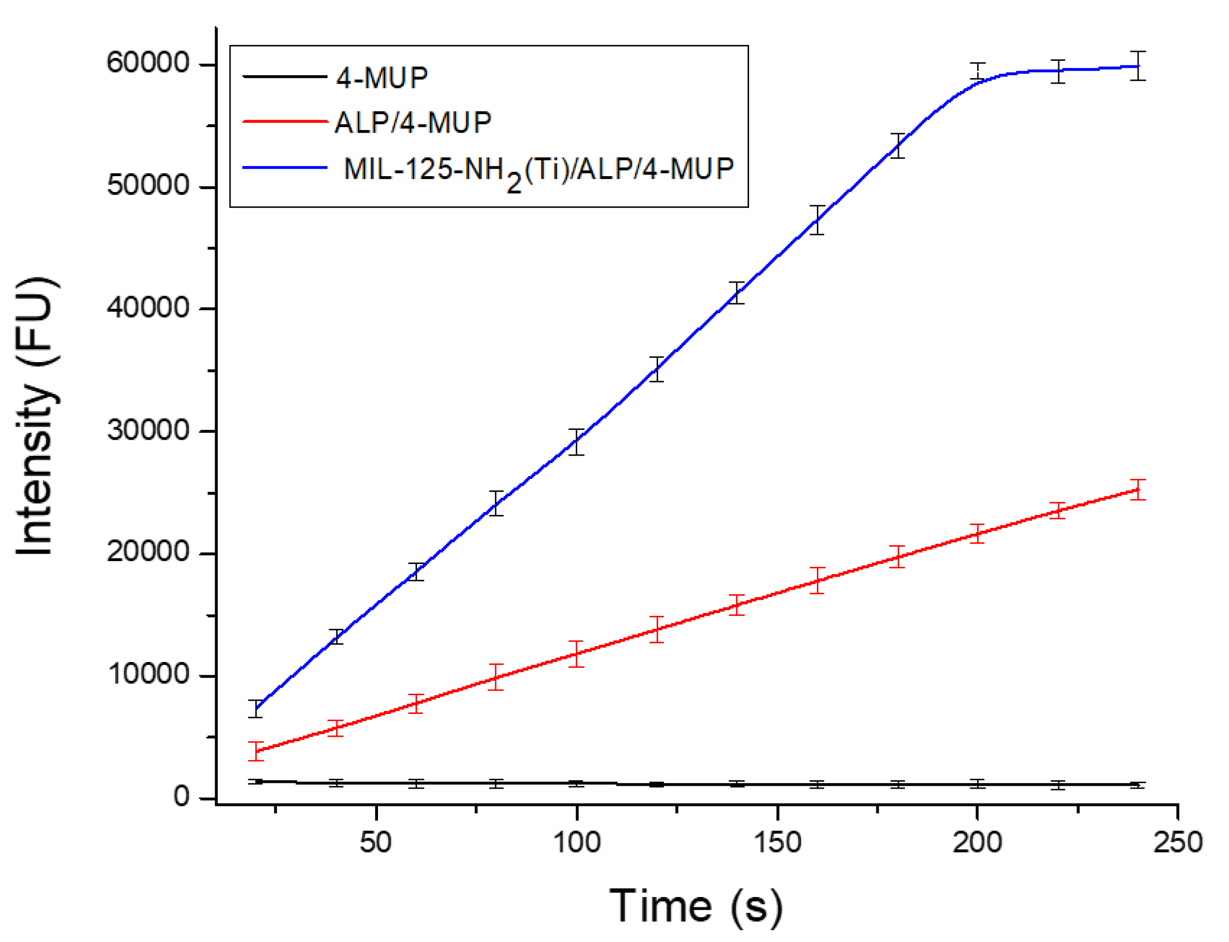
Reaction time is also a crucial parameter to define, especially in applications that demand fast and reliable analysis. This study used MIL-125-NH2 (Ti)/ALP/4-MUP modified papers and analyzed them at various incubation times ranging from 20 to 240 seconds. The fluorescence intensity progressively increased with longer incubation periods until reaching 180 s. Beyond this point, a saturation effect became evident. Consequently, the optimal reaction time was determined to be 180 s (see
Figure 14). Finally, a summary of the experimental working experiments is depicted in
Table 1.
4. Conclusions
In the present work, MOFs MIL-125-NH2 (Ti), UiO-66-NH2 (Zr), and MIL-101-NH2 (Fe) were synthesized, characterized, and successfully used for the modification of paper surfaces to development of PADs biosensors. It could be concluded that the MIL-125-NH2 (Ti) MOF significantly increased the effective surface area for the immobilization of biomolecules in comparison to UiO-66-NH2 (Zr) and MIL-101-NH2 (Fe). This feature was confirmed by immobilizing the ALP enzyme and monitoring its activity by laser-induced fluorescence, giving rise to a higher signal intensity for the same concentrations of the three dispersions and modifying the paper support. Based on this, we have chosen MIL-125-NH2 (Ti) for further optimization trials to conclude a reliable and reproducible platform suitable for biomolecule detection. Consequently, the MIL-125-NH2(Ti)/ALP modified papers showed excellent stability and reproducibility, demonstrating the potential use of this new modified platform for developing biosensors for the rapid detection of various bioanalytes of interest.
Author Contributions
Conceptualization, S.V.P., G.E.G. and M.A.F-B.; methodology, S.V.P., G.E.G. and M.A.F-B.; validation, S.V.P., G.R.T., A.B.S., M.D.R. and G.A.M.; formal analysis, S.V.P., G.E.G., M.D.R. and M.A.F.-B.; resources, S.V.P., G.R.T., A.B.S., M.D.R. and G.A.M.; writing—original draft preparation, S.V.P., G.E.G. and M.A.F-B.; writing—review and editing, S.V.P., G.E.G., G.R.T., A.B.S., G.A.M. and M.A.F-B.; supervision, G.E.G. and M.A.F-B.; project administration, G.E.G., G.A.M. and M.A.F-B.; funding acquisition, G.A.M. and M.A.F-B. All authors have read and agreed to the published version of the manuscript.
Funding
Funding is acknowledged from the Universidad Nacional de San Luis (PROICO 02-2220), from Agencia Nacional de Promoción Científica y Tecnológica (AGENCIA, FONCyT, Argentina, PICT-2021-GRF-TII-0390, PICT-2021-GRF-TI-0136, PICT-2020-2347 and PICT-2020-2369), from Consejo Nacional de Investigaciones Científicas y Técnicas (CONICET, Argentina, PIP11220200100033CO), from Ministerio de Ciencia, Tecnología e Innovación, Argentina, Proyectos de Redes Federales de Alto Impacto: NANOQUIMISENS (IF-2023-85161983-APN-DNOYPI#MCT) and from Agencia Nacional de Investigación y Desarrollo (ANID) (ANID/FOVI220003)
Institutional Review Board Statement
Not applicable.
Informed Consent Statement
Not applicable.
Data Availability Statement
The complete data of this work can be obtained upon request from the corresponding author.
Acknowledgments
This work was supported by Universidad Nacional de San Luis, Instituto de Química de San Luis (INQUISAL); Consejo Nacional de Investigaciones Científicas y Técnicas (CONICET); Agencia Nacional de Promoción Científica y Tecnológica (FONCYT) (PICT-BID).
Conflicts of Interest
The authors declare no conflict of interest.
References
- Batten, S.R.; Champness, N.R.; Chen, X.M.; Garcia-Martinez, J.; Kitagawa, S.; Öhrström, L.; O'Keeffe, M.; Suh, M.P.; Reedijk, J. Terminology of Metal-Organic Frameworks and Coordination Polymers (IUPAC Recommendations 2013). Pure and Applied Chemistry 2013, 85, 1715–1724. [Google Scholar] [CrossRef]
- Long, J.R.; Yaghi, O.M. The Pervasive Chemistry of Metal-Organic Frameworks. Chemical Society Reviews 2009, 38, 1213–1214. [Google Scholar] [CrossRef] [PubMed]
- Xu, Q.; Kitagawa, H. MOFs: New Useful Materials – A Special Issue in Honor of Prof. Susumu Kitagawa. Advanced Materials 2018, 30, 1–3. [Google Scholar] [CrossRef]
- Figeys, D.; Pinto, D. Lab-on-a-Chip: A Revolution in Biological and Medical Sciences. Analytical Chemistry 2000, 72, 330–335. [Google Scholar] [CrossRef] [PubMed]
- Lu, R.; Shi, W.; Jiang, L.; Qin, J.; Lin, B. Rapid Prototyping of Paper-Based Microfluidics with Wax for Low-Cost, Portable Bioassay. Electrophoresis 2009, 30, 1497–1500. [Google Scholar] [CrossRef]
- Cate, D.M.; Adkins, J.A.; Mettakoonpitak, J.; Henry, C.S. Recent Developments in Paper-Based Microfluidic Devices. Analytical Chemistry 2015, 87, 19–41. [Google Scholar] [CrossRef]
- Martinez, A.W.; Phillips, S.T.; Whitesides, G.M.; Carrilho, E. Diagnostics for the Developing World: Microfluidic Paper-Based Analytical Devices. Analytical Chemistry 2010, 82, 3–10. [Google Scholar] [CrossRef] [PubMed]
- Wang, S.; Ge, L.; Song, X.; Yan, M.; Ge, S.; Yu, J.; Zeng, F. Simple and Covalent Fabrication of a Paper Device and Its Application in Sensitive Chemiluminescence Immunoassay. Analyst 2012, 137, 3821–3827. [Google Scholar] [CrossRef]
- Pelton, R. Bioactive Paper Provides a Low-Cost Platform for Diagnostics. TrAC - Trends in Analytical Chemistry 2009, 28, 925–942. [Google Scholar] [CrossRef]
- Hendon, C.H.; Tiana, D.; Fontecave, M.; Sanchez, C.; D'Arras, L.; Sassoye, C.; Rozes, L.; Mellot-Draznieks, C.; Walsh, A. Engineering the Optical Response of the Titanium-MIL-125 Metal-Organic Framework through Ligand Functionalization. Journal of the American Chemical Society 2013, 135, 10942–10945. [Google Scholar] [CrossRef]
- Ding, L.; Shao, P.; Luo, Y.; Yin, X.; Yu, S.; Fang, L.; Yang, L.; Yang, J.; Luo, X. Functionalization of UiO-66-NH2 with Rhodanine via Amidation: Towarding a Robust Adsorbent with Dual Coordination Sites for Selective Capture of Ag(I) from Wastewater. Chemical Engineering Journal 2020, 382, 123009. [Google Scholar] [CrossRef]
- Shan, Y.; Xu, C.; Zhang, H.; Chen, H.; Bilal, M.; Niu, S.; Cao, L.; Huang, Q. Polydopamine-Modified Metal–Organic Frameworks, NH2-Fe-MIL-101, as PH-Sensitive Nanocarriers for Controlled Pesticide Release. Nanomaterials 2020, 10, 1–13. [Google Scholar] [CrossRef] [PubMed]
- Pashazadeh-Panahi, P.; Belali, S.; Sohrabi, H.; Oroojalian, F.; Hashemzaei, M.; Mokhtarzadeh, A.; de la Guardia, M. Metal-Organic Frameworks Conjugated with Biomolecules as Efficient Platforms for Development of Biosensors. TrAC - Trends in Analytical Chemistry 2021, 141, 116285. [Google Scholar] [CrossRef]
- Sohrabi, H.; Salahshour Sani, P.; Orooji, Y.; Majidi, M.R.; Yoon, Y.; Khataee, A. MOF-Based Sensor Platforms for Rapid Detection of Pesticides to Maintain Food Quality and Safety. Food and Chemical Toxicology 2022, 165, 113176. [Google Scholar] [CrossRef]
- Osman, D.I.; El-Sheikh, S.M.; Sheta, S.M.; Ali, O.I.; Salem, A.M.; Shousha, W.G.; EL-Khamisy, S.F.; Shawky, S.M. Nucleic Acids Biosensors Based on Metal-Organic Framework (MOF): Paving the Way to Clinical Laboratory Diagnosis. Biosensors and Bioelectronics 2019, 141, 111451. [Google Scholar] [CrossRef]
- Dai, C.; Gan, Y.; Qin, J.; Ma, L.; Liu, Q.; Huang, L.; Yang, Z.; Zang, G.; Zhu, S. An Ultrasensitive Solid-State ECL Biosensor Based on Synergistic Effect between Zn-NGQDs and Porphyrin-Based MOF as "on-off-on" platform. Colloids and Surfaces B: Biointerfaces 2023, 226, 113322. [Google Scholar] [CrossRef]
- Li, M.; Zhang, G.; Boakye, A.; Chai, H.; Qu, L.; Zhang, X. Recent Advances in Metal-Organic Framework-Based Electrochemical Biosensing Applications. Frontiers in Bioengineering and Biotechnology 2021, 9, 1–8. [Google Scholar] [CrossRef]
- Ortiz-Gómez, I.; Salinas-Castillo, A.; García, A.G.; Álvarez-Bermejo, J.A.; de Orbe-Payá, I.; Rodríguez-Diéguez, A.; Capitán-Vallvey, L.F. Microfluidic Paper-Based Device for Colorimetric Determination of Glucose Based on a Metal-Organic Framework Acting as Peroxidase Mimetic. Microchimica Acta 2018, 185, 1–8. [Google Scholar] [CrossRef]
- Wei, X.; Guo, J.; Lian, H.; Sun, X.; Liu, B. Cobalt Metal-Organic Framework Modified Carbon Cloth/Paper Hybrid Electrochemical Button-Sensor for Nonenzymatic Glucose Diagnostics. Sensors and Actuators B: Chemical 2021, 329, 129205. [Google Scholar] [CrossRef]
- Hassanzadeh, J.; Al Lawati, H.A.J.; Al Lawati, I. Metal-Organic Framework Loaded by Rhodamine b as a Novel Chemiluminescence System for the Paper-Based Analytical Devices and Its Application for Total Phenolic Content Determination in Food Samples. Analytical Chemistry 2019, 91, 10631–10639. [Google Scholar] [CrossRef]
- Catalá-Icardo, M.; Gómez-Benito, C.; Martínez-Pérez-Cejuela, H.; Simó-Alfonso, E.F.; Herrero-Martínez, J.M. Green Synthesis of MIL53(Al)-Modified Paper-Based Analytical Device for Efficient Extraction of Neonicotinoid Insecticides from Environmental Water Samples. Analytica Chimica Acta 2024, 1316. [Google Scholar] [CrossRef] [PubMed]
- Chang, J.; Hu, R.; Zhang, J.; Hou, T.; Li, F. Two-dimensional metal-organic framework nanozyme-mediated portable paper-based analytical device for dichlorophen assay. Biosensors and Bioelectronics 2024, 255, 116271. [Google Scholar] [CrossRef]
- Feng, Q,; Wang, C. ; Miao, X.; Wu, M. A novel paper-based electrochemiluminescence biosensor for non-destructive detection of pathogenic bacteria in real samples. Talanta 2024, 267, 125224. [Google Scholar] [CrossRef]
- Guan, J.; Xiong, Y.; Wang, M.; Liu, Q.; Chen, X. A novel functionalized CdTe@MOFs based fluorometric and colorimetric biosensor for dual-readout assay of creatinine. Sensors and Actuators B: Chemical 2024, 399, 134842. [Google Scholar] [CrossRef]
- Lebedev, O.I.; Millange, F.; Serre, C.; Van Tendeloo, G.; Férey, G. First direct imaging of giant pores of the metal−organic framework MIL-101. Chem. Mater. 2005, 26, 6525. [Google Scholar] [CrossRef]
- Capková, D. , Almáši, M., Kazda, T., Čech, O., Király, N., Čudek, P.,... & Hornebecq, V. Metal-organic framework MIL-101 (Fe)–NH2 as an efficient host for sulphur storage in long-cycle Li–S batteries. Electrochimica Acta 2020, 354, 136640. [Google Scholar] [CrossRef]
- Winarta, J. , Shan, B., Mcintyre, S. M., Ye, L., Wang, C., Liu, J., & Mu, B. A decade of UiO-66 research: a historic review of dynamic structure, synthesis mechanisms, and characterization techniques of an archetypal metal–organic framework. Crystal Growth & Design 2019, 20, 1347–1362. [Google Scholar] [CrossRef]
- Kim, S. N. , Kim, J., Kim, H. Y., Cho, H. Y., & Ahn, W. S. Adsorption/catalytic properties of MIL-125 and NH2-MIL-125. Catalysis today. [CrossRef]
- Cao, Y. , Zhang, H., Song, F., Huang, T., Ji, J., Zhong, Q.,... & Xu, Q. UiO-66-NH2/GO composite: synthesis, characterization and CO2 adsorption performance. Materials 2018, 11, 589. [Google Scholar] [CrossRef]
Figure 2.
SEM image characterization: a) MIL-125-NH2 (Ti), b) UiO-66-NH2 (Zr) and c) MIL-101-NH2 (Fe).
Figure 2.
SEM image characterization: a) MIL-125-NH2 (Ti), b) UiO-66-NH2 (Zr) and c) MIL-101-NH2 (Fe).
Figure 3.
EDS spectra characterization: a) MIL-125-NH2 (Ti), b) UiO-66-NH2 (Zr) and c) MIL-101-NH2 (Fe).
Figure 3.
EDS spectra characterization: a) MIL-125-NH2 (Ti), b) UiO-66-NH2 (Zr) and c) MIL-101-NH2 (Fe).
Figure 4.
SEM image characterization: a) unmodified paper surface, b) paper surface modified with amino-functionalized MIL-101-NH2 (Fe), c) paper surface modified with amino-functionalized UiO-66-NH2 (Zr), d) paper surface modified with amino-functionalized MIL-125-NH2 (Ti).
Figure 4.
SEM image characterization: a) unmodified paper surface, b) paper surface modified with amino-functionalized MIL-101-NH2 (Fe), c) paper surface modified with amino-functionalized UiO-66-NH2 (Zr), d) paper surface modified with amino-functionalized MIL-125-NH2 (Ti).
Figure 5.
EDS spectra characterization: a) unmodified paper surface, b) paper surface modified with amino-functionalized MIL-101-NH2 (Fe), c) paper surface modified with amino-functionalized UiO-66-NH2 (Zr), d) paper surface modified with amino-functionalized MIL-125-NH2 (Ti).
Figure 5.
EDS spectra characterization: a) unmodified paper surface, b) paper surface modified with amino-functionalized MIL-101-NH2 (Fe), c) paper surface modified with amino-functionalized UiO-66-NH2 (Zr), d) paper surface modified with amino-functionalized MIL-125-NH2 (Ti).
Figure 6.
Experimental XRD pattern of a) MIL-101-NH2 (Fe), b) UiO-66-NH2 (Zr), and c) MIL-125-NH2 (Ti) (blue lines) compared to the simulated diffractograms.
Figure 6.
Experimental XRD pattern of a) MIL-101-NH2 (Fe), b) UiO-66-NH2 (Zr), and c) MIL-125-NH2 (Ti) (blue lines) compared to the simulated diffractograms.
Figure 7.
a) Secondary building unit and b) the formed cages in MIL-101-NH2 (Fe).
Figure 7.
a) Secondary building unit and b) the formed cages in MIL-101-NH2 (Fe).
Figure 8.
A schematic structure of UiO-66-NH2 (Zr). The face-center-cubic UiO-66 structure type is composed of the metal node (aqua) and ligand (gray) with an atomic representation of the node and 12-connected 2-ATA linkers.
Figure 8.
A schematic structure of UiO-66-NH2 (Zr). The face-center-cubic UiO-66 structure type is composed of the metal node (aqua) and ligand (gray) with an atomic representation of the node and 12-connected 2-ATA linkers.
Figure 9.
A schematic structure of MIL-125-NH2-(Ti) shows the two cages types.
Figure 9.
A schematic structure of MIL-125-NH2-(Ti) shows the two cages types.
Figure 10.
FTIR study of paper modification with a) MIL-125-NH2 (Ti) and subsequent stage with GLU; b) UiO-66-NH2 (Zr) and subsequent stage with GLU; c)MIL-101-NH2 (Fe) and subsequent stage with GLU.
Figure 10.
FTIR study of paper modification with a) MIL-125-NH2 (Ti) and subsequent stage with GLU; b) UiO-66-NH2 (Zr) and subsequent stage with GLU; c)MIL-101-NH2 (Fe) and subsequent stage with GLU.
Figure 11.
Optimization of impregnated MOFs concentration for paper support modification. .
Figure 11.
Optimization of impregnated MOFs concentration for paper support modification. .
Figure 12.
a) Optimization of the ALP concentration used for immobilization; b) Optimization of the optimal pH range of the enzymatic activity.
Figure 12.
a) Optimization of the ALP concentration used for immobilization; b) Optimization of the optimal pH range of the enzymatic activity.
Figure 13.
a) Optimization of 4-MUP concentration; b) Stability tets of the modified paper support.
Figure 13.
a) Optimization of 4-MUP concentration; b) Stability tets of the modified paper support.
Figure 14.
Fluorescence response at different incubation times of MIL-125-NH2 (Ti)/ALP/4-MUP by modifying the paper support.
Figure 14.
Fluorescence response at different incubation times of MIL-125-NH2 (Ti)/ALP/4-MUP by modifying the paper support.
Table 1.
Summary of optimal working conditions.
Table 1.
Summary of optimal working conditions.
|
Disclaimer/Publisher’s Note: The statements, opinions and data contained in all publications are solely those of the individual author(s) and contributor(s) and not of MDPI and/or the editor(s). MDPI and/or the editor(s) disclaim responsibility for any injury to people or property resulting from any ideas, methods, instructions or products referred to in the content. |
© 2024 by the authors. Licensee MDPI, Basel, Switzerland. This article is an open access article distributed under the terms and conditions of the Creative Commons Attribution (CC BY) license (http://creativecommons.org/licenses/by/4.0/).

