Preprint
Article

Unveiling the Metabolic Trajectory of Pig Feces across Different Ages and Senescence

Altmetrics

Downloads

63

Views

25

Comments

0

A peer-reviewed article of this preprint also exists.

Submitted:

01 September 2024

Posted:

02 September 2024

You are already at the latest version

Alerts
Abstract
Porcine metabolomics research has been extensively conducted, primarily focusing on conventional cohorts or animal models. However, the composition and functions of fecal metabolites in pigs at various ages, particularly in the elderly, remain poorly understood. In the present study, an untargeted metabolomics approach was employed to analyze the fecal metabolomes of pigs at three distinct age stages, with the aim of elucidating age-associated changes in metabolite composition and functionality under standardized rearing conditions. Through the untargeted metabolomic analysis of fecal samples from young (one year), middle-aged (four years), and elderly (eight years) pig cohorts, a variety of age-related metabolites were identified. For example, L-methionine sulfoxide was observed to increase with age, while cytidine-5-monophosphate exhibited a concomitant gradual decrease during the aging process. These metabolites were found to undergo alterations across multiple biological pathways, including energy metabolism, pyrimidine metabolism, lipid metabolism, and amino acid metabolism. Furthermore, the age-dependent metabolic changes were associated with known senescence biomarkers, offering new insights into the mechanistic understanding of aging in pigs.
Keywords: 
Subject: Biology and Life Sciences  -   Animal Science, Veterinary Science and Zoology

1. Introduction

Aging is a natural and exceedingly intricate biological phenomenon governed by an interplay of genomics, transcriptomics, proteomics, metabolomics, as well as dietary habits, lifestyle, and environmental factors[1]. The burgeoning elderly population poses an escalating public health issue on a global scale, with its proportion rapidly increasing and projected to reach two billion by 2050 for individuals aged 60 and above[2]. Moreover, the health status of the aging population is deteriorating, leading to a surge in the prevalence of age-related diseases, particularly in China, where it is estimated that by 2040, there will be approximately 402 million elderly individuals[2]. The recent decline in fertility and mortality rates has catalyzed a swift aging of the population, raising concerns about the health and quality of life of the elderly, and presenting significant challenges to healthcare systems. Aging is a complex biological process involving the interaction of various molecular and cellular mechanisms, such as theories of programmed longevity, free radical immunity, wear and tear, cross-linking, endocrine, and basal metabolism to describe the aging process[3,4]. Despite significant advances in aging research over the past decades, a comprehensive understanding of metabolic changes during the aging process remains limited. In recent years, an increasing number of researchers have begun to utilize omics technologies to study normal aging due to their high-throughput characteristics in proteomics, genomics, transcriptomics, and metabolomics[5,6,7,8].Metabolomic as a branch of systems biology, offers a powerful tool for the study of aging by quantifying all metabolites within a biological system[9].
In the field of agricultural science, understanding the aging process in animal models such as pigs is of significant importance for comprehending aging and age-related diseases, as well as optimizing breeding management strategies to enhance production efficiency and meat quality[10]. Pigs as an important agricultural animal model with physiological and anatomical characteristics similar to humans, serve as an ideal subject for studying aging-related metabolic changes[11]. With the rapid development of metabolomics technologies, especially the application of high-resolution mass spectrometry and nuclear magnetic resonance techniques, studying the metabolic changes in pigs at the molecular level has become feasible[12,13].The aim of this study is to utilize metabolomics methods to analyze the metabolite profiles of pigs of different age groups to identify metabolic changes associated with aging. By conducting non-targeted metabolomics analysis on blood and tissue samples from young, middle-aged, and elderly pigs, we aim to reveal age-related metabolites and their changes in biological pathways. This information will not only aid in understanding the aging mechanisms in pigs but also provide a reference for aging research in other species, including humans.
In this study, we initially collected feces samples from pigs of three different age groups: (1) one year old, (2) four years old, and (3) eight years old[14]. Subsequently, non-targeted metabolomics analysis was performed using liquid chromatography-mass spectrometry (LC-MS) techniques. Through data analysis, we identified a series of age-related metabolites and observed significant metabolic changes in multiple biological pathways, including pyrimidine metabolism, vitamin metabolism, and amino acid metabolism, which play key roles in the aging process. Furthermore, we found that these metabolic changes are associated with known aging markers such as oxidative stress, inflammation, and cellular damage. These findings suggest that the changes of fecal metabolites can serve as a powerful tool to uncover molecular changes during the aging process in pigs, leading to the development of new biomarkers and therapeutic targets for age-related diseases. It may also provide new targets for the development of strategies to delay aging or improve human health and animal welfare.

2. Materials and Methods

2.1. Study Design and Sample Collection

A cohort of 45 pigs was enrolled in this study, with fecal samples collected from each individual. The pigs were categorized into three age groups, comprising 15 pigs each: one-year-old, approximately four years old, and approximately eight years old. This study design facilitated the collection of comprehensive and representative biological samples across various age points, which is essential for investigating the complexities of the aging process.

2.2. Chemicals and Regents

Sigma Aldrich provided Ammonium acetate (NH4AC), Merck provided Acetonitrile, and Fisher provided ammonium hydroxide (NH4OH) and methanol. Thermo Fisher provided MS-grade methanol, MS-grade acetonitrile, and HPLC-grade 2-propanol. Sigma provided HPLC-grade formic acid and HPLC-grade ammonium formate. All chemicals used in this study were of high purity and quality, ensuring the reliability and reproducibility of the results.

2.3. Metabolomics Study of Feces Samples

Untargeted metabolomic profiles of samples were measured using a combination of two UHPLC-MS/MS methods, capturing metabolites in both positive and negative ionization modes. The raw data files were processed with Compound Discoverer 3.1 (CD3.1, Thermo Fisher) to perform peak alignment, peak picking, and metabolite quantification. The primary parameters used in this analysis included a retention time tolerance of 0.2 minutes, an actual mass tolerance of 5 ppm, a signal intensity tolerance of 30%, a signal-to-noise ratio of 3, and a minimum intensity threshold of 100,000. Peak intensities were normalized to the total spectral intensity and utilized to predict molecular formulas based on additive ions, molecular ion peaks, and fragment ions. These peaks were then matched with the mzCloud (https://www.mzcloud.org/), HMDB, mzVault, and MassList databases for untargeted metabolomic analysis, and with the LipidMaps and LipidBlast databases for lipidomic analysis. Consequently, accurate qualitative and relative quantitative results were obtained. Partial least squares discriminant analysis (PLS-DA) was employed to elucidate the metabolic changes among the groups using the R package ropls. Metabolites demonstrating a variable importance in projection (VIP) greater than 1 and a corrected p-value (Wilcoxon test) less than 0.05 were selected for enrichment analysis. The enrichment pathways of the differential plasma metabolite profiles between any two groups of pigs were analyzed using MetaboAnalyst 5.0 (http://www.metaboanalyst.ca).

2.4. Statistical Analysis

All statistical analyses were conducted using the R platform (version 4.0). For comparisons among multiple groups, a one-way ANOVA was employed to evaluate differences among the three groups. Only indices that demonstrated significant differences were further assessed using the Mann–Whitney U test with Bonferroni correction as a post-hoc analysis between each pair of groups. Adjusted p-values less than 0.05 were considered statistically significant. Error bars represent the mean ± standard error (SE).

3. Results

3.1. The Overall Characteristics of Feces Metabolomics

In metabolomics studies utilizing mass spectrometry, quality control (QC) samples are routinely employed to ensure the reliability and high quality of the acquired metabolomics data. Although QC samples are theoretically identical, systematic errors are often introduced during the processes of sample extraction, detection, and analysis. These errors can lead to variations among QC samples; the smaller the variations, the higher the method stability and data quality.
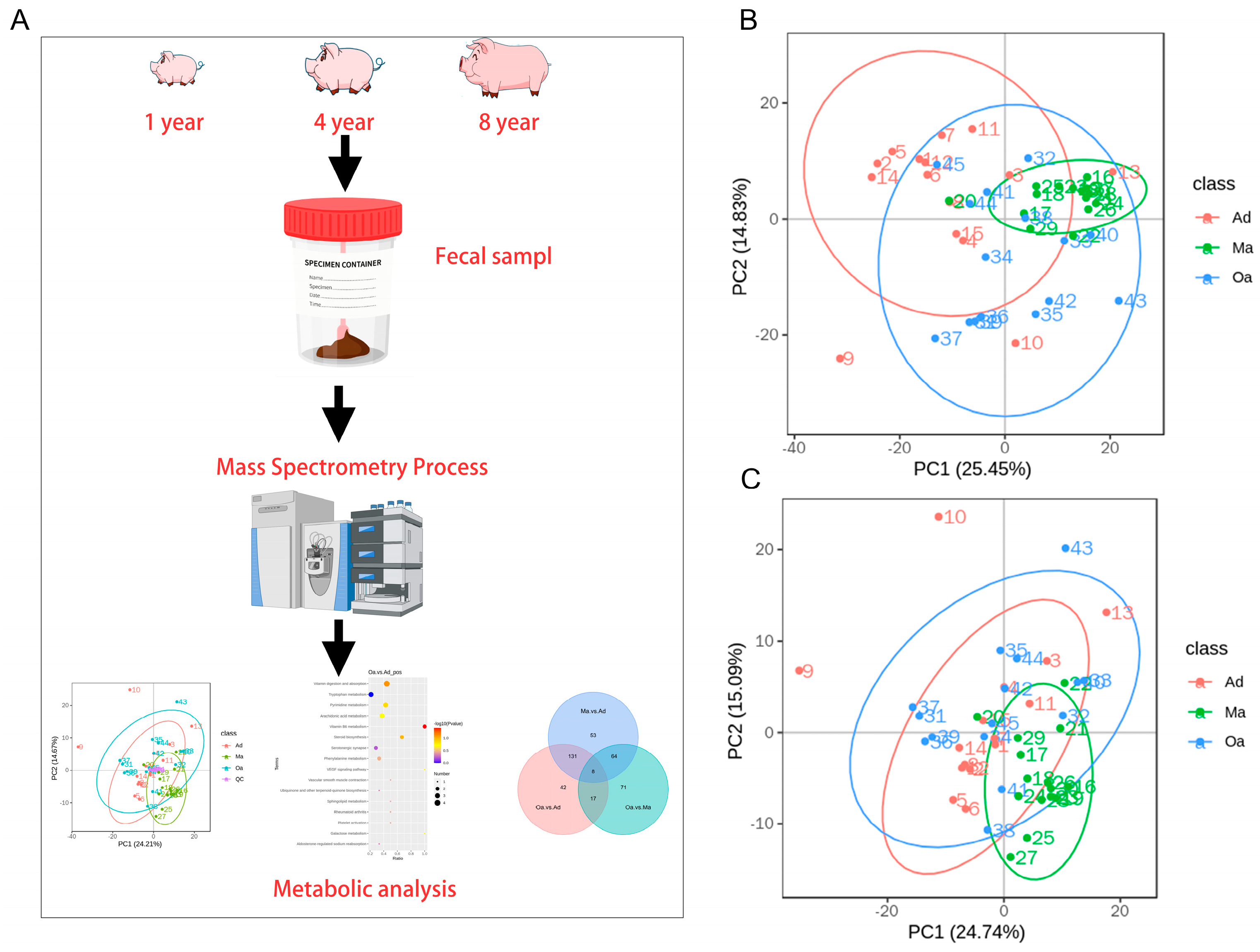
In the present study, the primary aim is to investigate the trajectory of changes in fecal metabolic profiles across various age stages in pigs, specifically at one year of age (Ad), four years of age (Ma), and eight years of age (Oa) (Figure 1A). Principal component analysis (PCA) revealed a significant separation among the three age groups, indicating distinct metabolic profiles associated with aging. Additionally, the analysis highlighted a tight clustering of the QC samples, which suggests a high degree of stability and repeatability in the detection process. This dense distribution of QC samples reinforces the reliability of the data obtained (Figure 1B,C).

3.2. The Trajectory of Dynamic Changes of Fecal Metabolites Associated with Aging

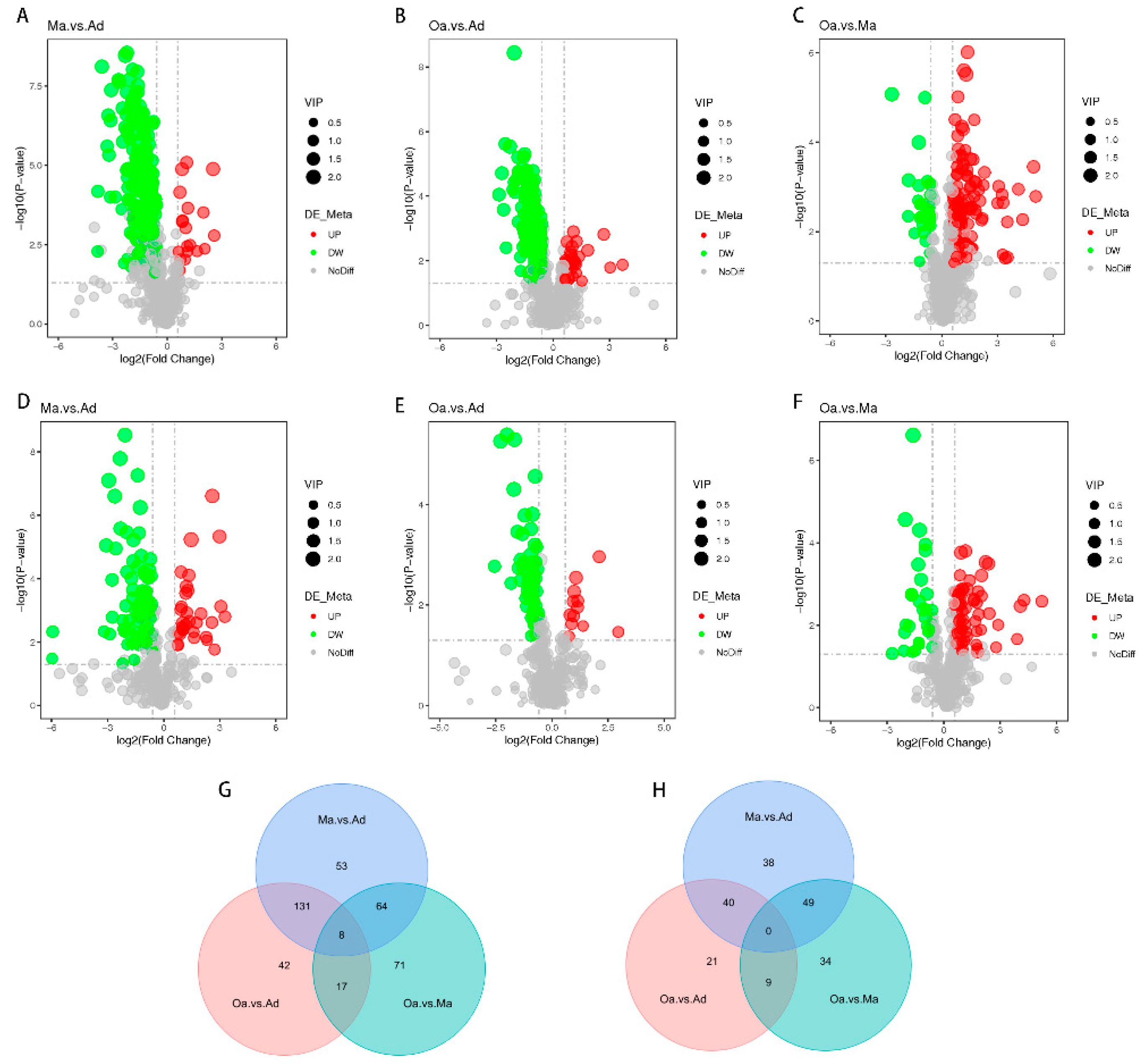
In the differential metabolomics analyses, a substantial number of metabolites were identified as significantly distinct among the swine in the middle-aged (Ma, four years old) group compared to the one-year-old (Ad) group, the elderly (Oa, eight years old) group compared to the Ad group, and the Oa group compared to the Ma group. Volcano plots indicated that a total of 383, 268, and 225 differentially expressed metabolites were detected in these comparisons, respectively (Tables S1–S3). Among these metabolites, the upregulated counts were 333, 225, and 60, while the downregulated counts were 50, 43, and 192, respectively (Figure 2A–F).
In positive ion mode, pairwise comparisons between the three groups revealed 53, 42, and 71 group-specific differential metabolites for the Ma vs. Ad, Oa vs. Ad, and Oa vs. Ma comparisons, respectively. Additionally, eight common differential metabolites were identified across these comparisons (Figure 2G). In contrast, a total of 38, 21, and 34 group-specific differential metabolites were identified in negative ion mode; however, no overlapping differential metabolites were found in this mode (Figure 2H). The eight common differential metabolites identified include: 4-(acetylamino)phenyl 3-chlorobenzoate, amoxicillin, 1-(4-nitrophenyl)piperidine, cyanidin, 4-guanidinobutanoic acid, ethyl 5-methoxy-2-methyl-1-phenyl-1H-indole-3-carboxylate, 5-(tert-butyl)-2-methyl-N-(4-nitrophenyl)-3-furamide, and valine.

3.3. Screening of Fecal Metabolites Intimately Linked to Aging

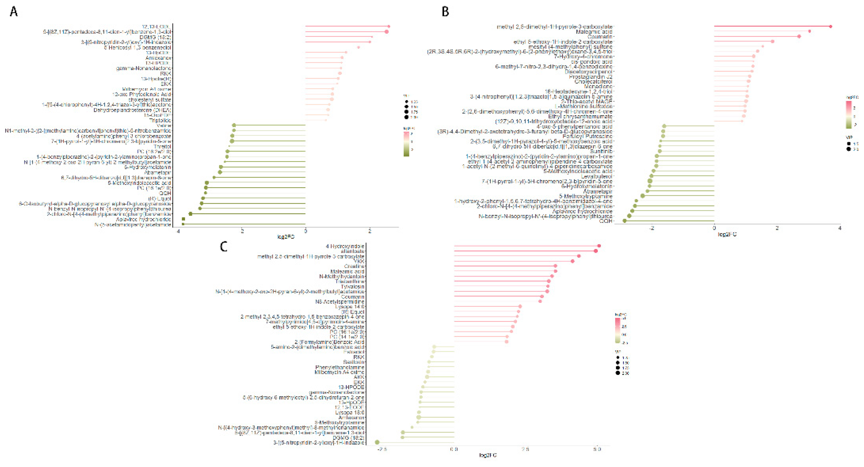
Differential metabolites were identified based on three key parameters: Variable Importance in Projection (VIP), fold change (FC), and p-value. To qualify as differential metabolites, a VIP value greater than 1 and a p-value less than 0.05 were required. As a result, metabolites were selected as differentially expressed according to the criteria of a p-value below 0.05 and a VIP value exceeding 1, with FC values either greater than 1 or less than 1. In pairwise comparisons among the one-year-old (Ad), four-year-old (Ma), and eight-year-old (Oa) groups, the top 20 upregulated and downregulated metabolites exhibiting the highest fold changes were selected for analysis. In the comparison between the Ma and Ad groups, 12,13-EODE demonstrated the highest level of upregulation, while N-(5-acetamidopentyl) acetamide showed the most significant downregulation (Figure 3A). In the comparison between the Oa and Ad groups, methyl 2,5-dimethyl-AH-pyrrole-3-carboxylate exhibited the greatest upregulation, whereas QQH was the most pronounced downregulated metabolite (Figure 3B). Finally, in the comparison between the Oa and Ma groups, 4-hydroxyindole was identified as the most upregulated metabolite, while 3-(G-nitropyridin-2-yl)oxy-1H-indazole exhibited the highest level of downregulation (Figure 3C). Collectively, these findings highlight distinct metabolic alterations associated with aging and underscore the potential relevance of these metabolites in understanding the biochemical changes that occur across different life stages in pigs.
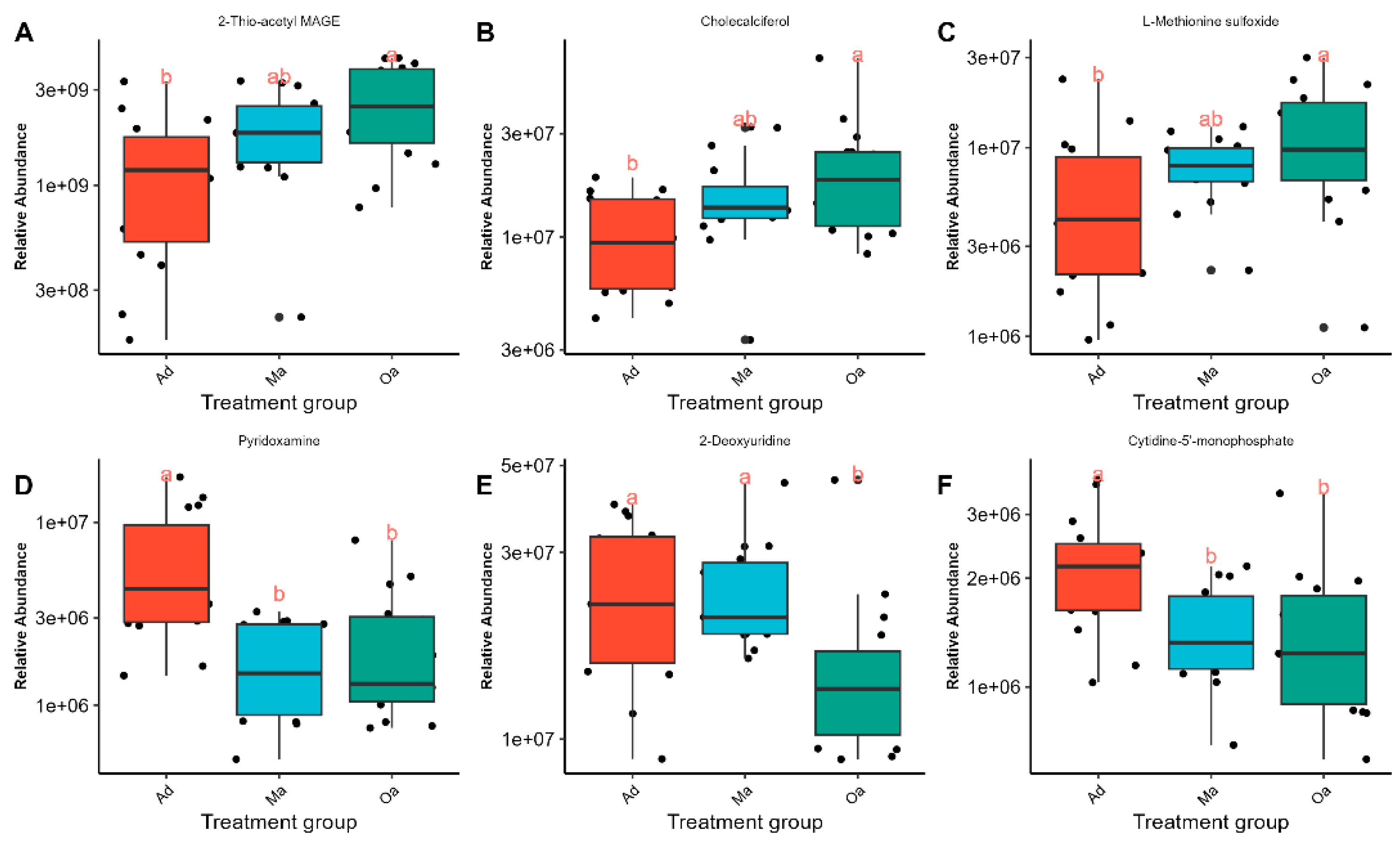
As the age of the pig population increases, 2-Thio-acetyl MAGE, Cholecalciferol, and L-Methionine sulfoxide have been observed to exhibit an increasing trend (Figure 4A–C), while Pyridoxamine, 2-Deoxyuridine, and Cytidine-5-monophosphate have shown a decreasing trend (Figure 4D–F). The identification of these down-regulated metabolites may provide insights into metabolic pathways that become less active over time. Together, these visual representations facilitate a clearer understanding of the dynamic metabolic shifts that accompany the aging process in pigs, underscoring the importance of both up-regulated and down-regulated metabolites in age-related research.

3.4. KEGG Enrichment Analysis Based on Differential Metabolites

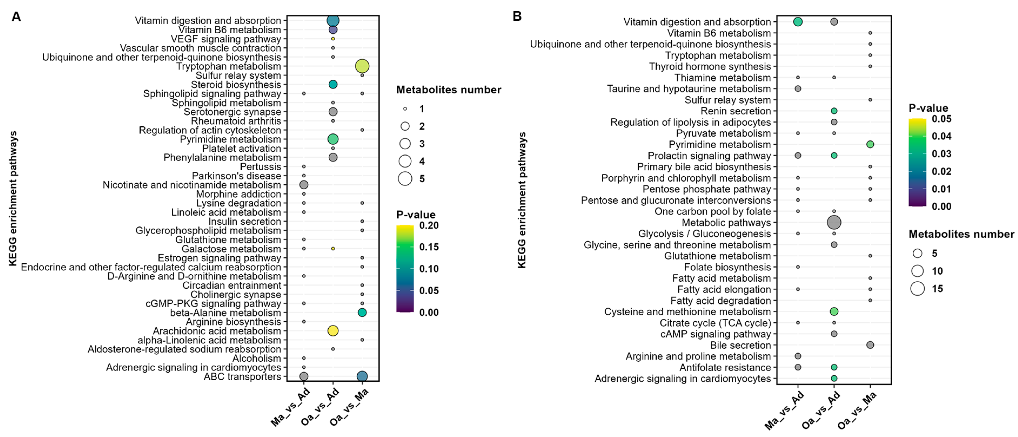
In the comparative analysis between the Ma (four years old) and Ad (one year old) groups, a total of 15 metabolic pathways were identified. These pathways predominantly included those related to vitamin digestion and absorption, taurine and hypotaurine metabolism, prolactin signaling, antifolate resistance, sphingolipid signaling, lysine degradation, linoleic acid metabolism, galactose metabolism, D-arginine and D-ornithine metabolism, the cGMP-PKG signaling pathway, and adrenergic signaling in cardiomyocytes.
When comparing the Oa (eight years old) group with the Ad (one year old) group, 12 metabolic pathways were identified, including Metabolic pathways, Cysteine and methionine metabolism, Renin secretion, Regulation of lipolysis in adipocytes, Prolactin signaling pathway, cAMP signaling pathway, Antifolate resistance, Adrenergic signaling in cardiomyocytes, Vitamin digestion and absorption, Pyrimidine metabolism, Vitamin B6 metabolism, and Steroid biosynthesis.
In the comparison between the Oa (eight years old) and Ma (four years old) groups, three metabolic pathways were identified, including Pyrimidine metabolism, ABC transporters, and beta-Alanine metabolism (Figure 5A,B).
Furthermore, the analysis revealed that Pyrimidine metabolism and Metabolic pathways are significantly differential profiles common to all three groups (Tables 4–6). Pyrimidine metabolism plays a crucial role in cell division and DNA synthesis and is closely associated with the aging process. As age progresses, the rate of cellular division slows, and the capacity for DNA repair declines, which can affect the normal progression of pyrimidine metabolism. Abnormalities in pyrimidine metabolism may lead to the accumulation of abnormal metabolites within cells, potentially impacting cellular function and even leading to cell death.

4. Discussion

This study leverages metabolomics to uncover the aging related metabolomics and identify differences in metabolites composition among pigs aged 1 (Ad), 4 (Ma), and 8 years old (Oa). The aim is to identify specific biomarkers of the aging process and understand their underlying mechanisms, thereby formulating strategies to promote healthy aging and potentially prevent or treat age-related diseases[15]. Results indicate that metabolites such as Cholecalciferol (Vitamin D), L-Methionine sulfoxide, Pyridoxamine, and Cytidine-5-monophosphate exhibit monotonic changes, either upregulated or downregulated, with increasing age. KEGG enrichment analysis revealed pathways such as Vitamin digestion and absorption, Cysteine and methionine metabolism, Vitamin B6 metabolism, and Pyrimidine metabolism as the dominant differential metabolic pathways.
Cholecalciferol, also known as Vitamin D, plays a multitude of crucial roles in the human body, including regulation of calcium and phosphorus metabolism and bone health[16]. Adequate levels of Vitamin D are essential for maintaining bone strength and preventing osteoporosis. As age advances, the body's requirement for Vitamin D increases, as it helps maintain skeletal health, and deficiency is associated with an increased risk of osteoporosis and fractures in the elderly[17]. Previous studies have suggested that Vitamin D may be related to various physiological processes, including immune function, cell growth and differentiation, and endocrine regulation, all of which are closely related to the aging process[18]. Therefore, maintaining appropriate Vitamin D levels may help delay certain aspects of aging, such as the decline in muscle strength and cognitive function[19]. Aging is a natural and inevitable process of molecular and cellular damage accumulation, leading to functional deficits in cells, tissues, and entire organs, thereby weakening the entire body[20]. With the decline in overall immune capacity during aging, the relative number of senescent immune cells decreases[21], reducing immune surveillance, such as the detection and destruction of tumor cells by cytotoxic T cells[22]. Individuals with low immune function have a significantly higher risk of cancer than those with high immune function[23]. The cancer-protective effect of adequate Vitamin D status may be primarily related to the ability to maintain a high level of immune capacity[24]. Additionally, Souraya et al. reported that Vitamin D can enhance immunity during the aging process by partially blocking p38 MAPK signaling, thereby inhibiting the production of SASP (senescence-associated secretory phenotype) in senescent cells[25]. Our study found that Cholecalciferol also shows an increasing trend with age, which is opposite to human aging, suggesting that its sufficiency is an essential component of healthy aging, not only helping to maintain the good condition of bones and skeletal muscles but also contributing to the homeostasis of the immune system, consistent with the views of Fantini et al.[26].
L-Methionine sulfoxide (MetO) is a form of protein that has undergone a change in function and structure due to oxidation by reactive oxygen species (ROS) and hydrogen peroxide when exposed to methionine and methionyl residues under physiological or pathological conditions. In mammals, oxidative stress is usually accompanied by an inflammatory response, generating ROS and other molecules that cause tissue damage, report indicated L-Methionine sulfoxide can promote the initiation of inflammatory responses[27]. Lee et al. proposed that the reduction of L-Methionine sulfoxide promotes mitophagy, which is involved in the process of mitophagy[28]. Excessive ROS can damage mitochondria, and mitophagy can remove damaged mitochondria, protecting cells from apoptosis. Furthermore, the role of L-Methionine sulfoxide in health and disease has been studied in various organisms from bacteria to mammals, including humans. For example, in mammals, elevated levels of MetO are involved in the expression of markers related to neurodegenerative diseases[29], mental health disorders[30], hearing loss[31], cystic fibrosis lung disease[32], macular degeneration[33], cardiovascular diseases[34], liver and kidney toxicity[35], and cancer[36]. Our study shows that L-Methionine sulfoxide also exhibits an increasing trend with age. Catanesi et al. found that a diet rich in L-Methionine may demonstrate therapeutic potential for human aging and age-related diseases, protecting neurons from oxidative imbalance and maintaining mitochondrial function[37]. Therefore, we believe that L-Methionine sulfoxide increases the risk of neurodegenerative diseases, including AD, and negatively regulates mitophagy, thereby accelerating cellular aging and apoptosis[38].
Aging and age-related diseases, such as diabetes, atherosclerosis, and neurodegenerative diseases, are characterized by an increase in oxidative chemical modification of tissue proteins[39]. Glyoxal or advanced glycation end-products (AGEs) are produced by secondary modification of proteins by carbohydrate oxidation products and are associated with the severity of diabetic complications in the kidneys, retina, and blood vessels[40]. Onorato et al. provided insights into the mechanism of action of Pyridoxamine as an AGEs inhibitor and suggested that Pyridoxamine may help inhibit the increased chemical modification of tissue proteins in diabetes, atherosclerosis, and other chronic diseases, thereby alleviating age-related diseases[41]. Additionally, Pyridoxamine reduces superoxide radicals produced by H2O2, possibly through a scavenging mechanism and reducing lipid peroxidation in cells[42]. Our data indicate that Pyridoxamine shows a declining trend with age, thereby increasing the likelihood of diseases such as diabetes during the aging process. The reduction in its content increases the production of free radicals, accelerating the aging process.
According to the KEGG analysis based on differential metabolites, pyrimidine metabolism is a significant differential profile common to all three groups. Cytidine-5-monophosphate, a product in its metabolic process, is a nucleotide within the cell that participates in many important biochemical processes, including nucleic acid synthesis, phospholipid metabolism, and phosphorylation reactions[43], playing an important role in the immunity of some adults, infants, and young mammals[44]. Notably, Nakagawara et al. reported that Cytidine-5-monophosphate enhances the expression of PGC-1α in mouse C2C12 cells and promotes the formation of myotubes[45], and PGC-1α can protect skeletal muscles and alleviate the occurrence of muscle atrophy[46], which may suggest a molecular mechanism for muscle atrophy during the aging process in humans or animals. During the aging process, a series of changes occur in cellular metabolism and function, which may affect the level and role of Cytidine-5-monophosphate. However, there is currently no direct evidence to suggest that Cytidine-5-monophosphate itself promotes or delays the aging process. Our study indicates that Cytidine-5-monophosphate consistently shows a declining trend with age, suggesting its potentiality as a biomarker involved in the biological aging process, although its specific mechanism remains to be investigated.
5. Conclusions

In summary, this study has unveiled metabolic variations across different age groups in pigs through metabolomic analysis. Metabolites such as Cholecalciferol, L-Methionine sulfoxide, and Pyridoxamine, which exhibit consistent monotonic increases or decreases with age, provide new insights into the aging process in pigs. Moreover, we propose that the pyrimidine metabolic pathway and its product, Cytidine-5-monophosphate, could serve as novel biomarkers for aging-related research.
These findings hold significant implications not only for the livestock industry but also contribute valuable information to the study of human aging. Future research should delve deeper into the regulatory mechanisms of these metabolic changes to comprehensively understand their roles in aging and age-related diseases.

Supplementary Materials

The following supporting information can be downloaded at: Preprints.org, Table S1: Differential metabolites between Ma and Ad group from positive and negative ion mode were merged; Table S2: Differential metabolites between Oa and Ad group from positive and negative ion mode were merged; Table S3: Differential metabolites between Oa and Ma group from positive and negative ion mode were merged; Table S4: KEGG enrichment pathways based on the merged differential metabolites between Ma and Ad group; Table S4: KEGG enrichment pathways based on the merged differential metabolites between Oa and Ad group; Table S4: KEGG enrichment pathways based on the merged differential metabolites between Oa and Ma group.

Author Contributions

Conceptualization, M.H. and C.Q.; methodology, M.H. and C.L.; software, M.H. and C.L.; validation, M.H., C.Q.; formal analysis, R.D. and S.W.; investigation, R.D. and S.W.; resources, C.Q. and M.H.; data curation, C.Q.; writing—original draft preparation, C.Q. and C.L.; writing—review and editing, M.H.; visualization, C.L. and M.H.; supervision, M.H.; project administration, C.Q. and M.H.; funding acquisition, C.Q. and M.H.. All authors have read and agreed to the published version of the manuscript.

Funding

This work was supported by National Natural Science Foundation of China (82302568, 32260138), Anhui Provincial National Science Foundation (2308085QH283), the grant from the Science and Technology Planning Project of Hainan Province (SQKY2022-0015), and Seed Laboratory of Yazhou Bay, Hainan Province (B22C11209).

Institutional Review Board Statement

The animal study protocol was approved by the the Institutional Animal Care and Use Committee at the Experimental Animal Center of Hainan Academy of Agricultural Science (HNXMSY-20210503). The study was conducted in accordance with the local legislation and institutional requirements.

Data Availability Statement

Data are available on reasonable request.

Conflicts of Interest

The authors declare no conflicts of interest.

References

  1. Adav SS, Wang Y. Metabolomics Signatures of Aging: Recent Advances. Aging and disease. 2021;12(2):646-661. [CrossRef]
  2. Wang H, Chen H. Aging in China: Challenges and Opportunities. China CDC Wkly. 2022;4(27):601-602. [CrossRef]
  3. Viña J, Borrás C, Miquel J. Theories of ageing. IUBMB Life. 2007;59(4-5):249-254. [CrossRef]
  4. Jin, K. Jin K. Modern Biological Theories of Aging. Aging Dis. 2010;1(2):72-74.
  5. Wilhelm M, Schlegl J, Hahne H, et al. Mass-spectrometry-based draft of the human proteome. Nature. 2014;509(7502):582-587. [CrossRef]
  6. Wang J, Zhang S, Wang Y, Chen L, Zhang XS. Disease-aging network reveals significant roles of aging genes in connecting genetic diseases. PLoS Comput Biol. 2009;5(9):e1000521. [CrossRef]
  7. Glass D, Viñuela A, Davies MN, et al. Gene expression changes with age in skin, adipose tissue, blood and brain. Genome Biol. 2013;14(7):R75. [CrossRef]
  8. Yu Z, Zhai G, Singmann P, et al. Human serum metabolic profiles are age dependent. Aging Cell. 2012;11(6):960-967. [CrossRef]
  9. Fang W, Chen S, Jin X, Liu S, Cao X, Liu B. Metabolomics in aging research: aging markers from organs. Front Cell Dev Biol. 2023;11:1198794. [CrossRef]
  10. Zhao M, Zhang J, Liu F, et al. Effects of Different Feed Additives on Intestinal Metabolite Composition of Weaned Piglets. Metabolites. 2024;14(3):138. [CrossRef]
  11. Lunney JK, Van Goor A, Walker KE, Hailstock T, Franklin J, Dai C. Importance of the pig as a human biomedical model. Sci Transl Med. 2021;13(621):eabd575. [CrossRef]
  12. Zhang A, Sun H, Wu X, Wang X. Urine metabolomics. Clin Chim Acta. 2012;414:65-69. [CrossRef]
  13. Bouatra S, Aziat F, Mandal R, et al. The human urine metabolome. PLoS One. 2013;8(9):e73076. [CrossRef]
  14. Tohyama S, Kobayashi E. Age-Appropriateness of Porcine Models Used for Cell Transplantation. Cell Transplant. 2019;28(2):224-228. [CrossRef]
  15. López-Otín C, Blasco MA, Partridge L, Serrano M, Kroemer G. Hallmarks of aging: An expanding universe. Cell. 2023;186(2):243-278. [CrossRef]
  16. Bouillon R, Suda T. Vitamin D: calcium and bone homeostasis during evolution. Bonekey Rep. 2014;3:480. [CrossRef]
  17. Maradit Kremers H, Grossardt BR, Miller AR, et al. Fracture Risk Among Living Kidney Donors 25 Years After Donation. JAMA Netw Open. 2024;7(1):e2353005. [CrossRef]
  18. Janubová M, Žitňanová I. The effects of vitamin D on different types of cells. Steroids. 2024;202:109350. [CrossRef]
  19. Zeqaj I, Piffero R, Calzaducca E, Pirisi M, Bellan M. The Potential Role of Vitamin D Supplementation in Cognitive Impairment Prevention. CNS Neurol Disord Drug Targets. 2024;23(5):628-637. [CrossRef]
  20. Guo J, Huang X, Dou L, et al. Aging and aging-related diseases: from molecular mechanisms to interventions and treatments. Signal Transduct Target Ther. 2022;7:391. [CrossRef]
  21. Aw D, Silva AB, Palmer DB. Immunosenescence: emerging challenges for an ageing population. Immunology. 2007;120(4):435-446. [CrossRef]
  22. Grivennikov SI, Greten FR, Karin M. Immunity, Inflammation, and Cancer. Cell. 2010;140(6):883-899. [CrossRef]
  23. Ahuja SK, Manoharan MS, Lee GC, et al. Immune resilience despite inflammatory stress promotes longevity and favorable health outcomes including resistance to infection. Nat Commun. 2023;14:3286. [CrossRef]
  24. Carlberg C, Velleuer E. Vitamin D and Aging: Central Role of Immunocompetence. Nutrients. 2024;16(3):398. [CrossRef]
  25. Souraya S, Fantecelle CH, Laphanuwat P, et al. Vitamin D3 inhibits p38 MAPK and senescence-associated inflammatory mediator secretion by senescent fibroblasts that impacts immune responses during ageing. Aging Cell. 2024;23(4):e14093. [CrossRef]
  26. Fantini C, Corinaldesi C, Lenzi A, Migliaccio S, Crescioli C. Vitamin D as a Shield against Aging. Int J Mol Sci. 2023;24(5):4546. [CrossRef]
  27. Moskovitz J, Smith A. Methionine sulfoxide and the methionine sulfoxide reductase system as modulators of signal transduction pathways: a review. Amino Acids. 2021;53(7):1011-1020. [CrossRef]
  28. Lee SH, Lee S, Du J, et al. Mitochondrial MsrB2 serves as a switch and transducer for mitophagy. EMBO Mol Med. 2019;11(8):e10409. [CrossRef]
  29. Oien DB, Moskovitz J. Genetic regulation of longevity and age-associated diseases through the methionine sulfoxide reductase system. Biochim Biophys Acta Mol Basis Dis. 2019;1865(7):1756-1762. [CrossRef]
  30. Walss-Bass C, Soto-Bernardini MC, Johnson-Pais T, et al. Methionine sulfoxide reductase: a novel schizophrenia candidate gene. Am J Med Genet B Neuropsychiatr Genet. 2009;150B(2):219-225. [CrossRef]
  31. Kwon TJ, Cho HJ, Kim UK, et al. Methionine sulfoxide reductase B3 deficiency causes hearing loss due to stereocilia degeneration and apoptotic cell death in cochlear hair cells. Hum Mol Genet. 2014;23(6):1591-1601. [CrossRef]
  32. Chandler JD, Margaroli C, Horati H, et al. Myeloperoxidase oxidation of methionine associates with early cystic fibrosis lung disease. Eur Respir J. 2018;52(4):1801118. [CrossRef]
  33. Sreekumar PG, Hinton DR, Kannan R. Methionine sulfoxide reductase A: Structure, function and role in ocular pathology. World J Biol Chem. 2011;2(8):184-192. [CrossRef]
  34. Rose AH, Hoffmann PR. Selenoproteins and cardiovascular stress. Thromb Haemost. 2015;113(3):494-504. [CrossRef]
  35. Noh MR, Kim KY, Han SJ, Kim JI, Kim HY, Park KM. Methionine Sulfoxide Reductase A Deficiency Exacerbates Cisplatin-Induced Nephrotoxicity via Increased Mitochondrial Damage and Renal Cell Death. Antioxid Redox Signal. 2017;27(11):727-741. [CrossRef]
  36. He Q, Li H, Meng F, et al. Methionine Sulfoxide Reductase B1 Regulates Hepatocellular Carcinoma Cell Proliferation and Invasion via the Mitogen-Activated Protein Kinase Pathway and Epithelial-Mesenchymal Transition. Oxid Med Cell Longev. 2018;2018:5287971. [CrossRef]
  37. Catanesi M, Brandolini L, d’Angelo M, et al. L-Methionine Protects against Oxidative Stress and Mitochondrial Dysfunction in an In Vitro Model of Parkinson’s Disease. Antioxidants (Basel). 2021;10(9):1467. [CrossRef]
  38. Chandran S, Binninger D. Role of Oxidative Stress, Methionine Oxidation and Methionine Sulfoxide Reductases (MSR) in Alzheimer’s Disease. Antioxidants. 2024;13(1). [CrossRef]
  39. Thorpe SR, Baynes JW. Role of the Maillard Reaction in Diabetes Mellitus and Diseases of Aging. Drugs & Aging. 1996;9(2):69-77. [CrossRef]
  40. Baynes JW, Thorpe SR. Role of oxidative stress in diabetic complications: a new perspective on an old paradigm. Diabetes. 1999;48(1):1-9. [CrossRef]
  41. Onorato JM, Jenkins AJ, Thorpe SR, Baynes JW. Pyridoxamine, an Inhibitor of Advanced Glycation Reactions, Also Inhibits Advanced Lipoxidation Reactions: MECHANISM OF ACTION OF PYRIDOXAMINE*. Journal of Biological Chemistry. 2000;275(28):21177-21184. [CrossRef]
  42. Kannan K, Jain SK. Effect of vitamin B6 on oxygen radicals, mitochondrial membrane potential, and lipid peroxidation in H2O2-treated U937 monocytes. Free Radical Biology and Medicine. 2004;36(4):423-428. [CrossRef]
  43. Teng F, Wang L, Hu M, Tao Y. Cell-free regeneration of ATP based on polyphosphate kinase 2 facilitates cytidine 5’-monophosphate production. Enzyme and Microbial Technology. 2023;165:110211. [CrossRef]
  44. Hawkes JS, Gibson RA, Roberton D, Makrides M. Effect of dietary nucleotide supplementation on growth and immune function in term infants: a randomized controlled trial. Eur J Clin Nutr. 2006;60(2):254-264. [CrossRef]
  45. Nakagawara K, Takeuchi C, Ishige K. 5′-CMP and 5′-UMP alleviate dexamethasone-induced muscular atrophy in C2C12 myotubes. Biochem Biophys Rep. 2023;34:101460. [CrossRef]
  46. Huang Y, Chen K, Ren Q, et al. Dihydromyricetin Attenuates Dexamethasone-Induced Muscle Atrophy by Improving Mitochondrial Function via the PGC-1α Pathway. Cellular Physiology and Biochemistry. 2018;49(2):758-779. [CrossRef]
Figure 1. The overall changes in metabolomics among the pigs in the Ad, Ma, and Oa groups. (A) A schematic representation of the sample preparation process and the steps involved in metabolomic profiling and analysis is provided. (B-C) Principal Component Analysis (PCA) was conducted based on the relative abundances of metabolites across the three groups, presented in positive ion mode (B) and negative ion mode (C).
Preprints 116901 g001
Figure 2. (A-E) Volcano plots highlight the fecal metabolites that were increased (red) or decreased (green) in the comparison group of Ma versus to Ad, Oa versus to Ad, and Oa versus to Ma in positive ion mode and negative mode, with FDR < 0.05, log2 fold change (FC) >0.25 or <−0.25. (G) The Venn diagram illustrated the intersection based on significant differential fecal metabolites derived from the comparison of any two groups, and revealing that 8 metabolites were present in the differential results of all 3 pairwise comparisons in positive ion mode. (H) none intersection was found in negative ion mode.
Preprints 116901 g002
Figure 3. A detailed examination of the differences in metabolite profiles across various age groups. Panel (A) presents a stem plot comparing metabolites between the middle-aged (Ma) and one-year-old (Ad) groups, highlighting significant metabolic variations that may reflect age-related physiological changes. Panel (B) displays a similar comparison between the elderly (Oa) and one-year-old (Ad) groups, further elucidating the metabolic alterations that occur as pigs transition into later stages of life. Finally, panel (C) illustrates the differences in metabolites between the elderly (Oa) and middle-aged (Ma) groups, offering insights into the metabolic pathways that may be uniquely affected during this critical period of aging.
Preprints 116901 g003
Figure 4. A panel of metabolites changes associated with aging. Panels (A-C) display box plots illustrating the up-regulated metabolites observed with advancing age. These metabolites demonstrate significant increases, highlighting their potential role in the physiological adaptations that occur as pigs age. Conversely, panels (D-F) depict box plots for down-regulated metabolites, which exhibit notable decreases in concentration with age.
Preprints 116901 g004
Figure 5. Illustration of the results of a metabolic pathway analysis conducted based on metabolites that exhibited differences between pairwise groups in both positive ion mode (A) and negative ion mode (B). The size of the circles is indicative of the degree of pathway enrichment, with larger circles representing pathways that are more significantly enriched. Additionally, the color of the circles reflects the statistical significance of each pathway, providing a visual representation of the relevance of these metabolic alterations across the age groups analyzed. This dual-mode analysis enhances the understanding of the intricate metabolic changes associated with aging in pigs, facilitating the identification of key pathways that may warrant further investigation in future studies.
Preprints 116901 g005
Disclaimer/Publisher’s Note: The statements, opinions and data contained in all publications are solely those of the individual author(s) and contributor(s) and not of MDPI and/or the editor(s). MDPI and/or the editor(s) disclaim responsibility for any injury to people or property resulting from any ideas, methods, instructions or products referred to in the content.
Copyright: This open access article is published under a Creative Commons CC BY 4.0 license, which permit the free download, distribution, and reuse, provided that the author and preprint are cited in any reuse.
Prerpints.org logo

Preprints.org is a free preprint server supported by MDPI in Basel, Switzerland.

Subscribe

© 2024 MDPI (Basel, Switzerland) unless otherwise stated