Preprint
Article

Composite Dynamic Double Network Hydrogels with Rapid Self-healing, Stretchable, Moldable and Antibacterial Properties based on PVA/ε-Poly-L-lysine/Hyaluronic Acid

Altmetrics

Downloads

107

Views

31

Comments

0

A peer-reviewed article of this preprint also exists.

Submitted:

02 September 2024

Posted:

03 September 2024

You are already at the latest version

Alerts
Abstract
Self-healing, stretchable and moldable hydrogels have a great potential application in tissue engineering and soft robotics. Despite great success in reported hydrogels, it is still a great challenge to construct the moldable hydrogels with an ultrafast self-healing performance. Herein, the dynamic double network (DN) composite hydrogels (PBLH) with ultrafast self-healing, stretchable and moldable were successfully constructed by poly (vinyl alcohol) (PVA), borate, ε-poly-L-lysine (EPL) and hyaluronic acid (HA) based on an efficient one-pot method. Fourier transform infrared spectroscopy, X-Ray diffraction and rheological measurements confirmed the formation of dynamic double networks among PVA, B, EPL and HA through the cross-linking of dynamic borate bonds, electrostatic interaction and hydrogen bonding. Having fabricated the dynamic double network structure, the damage gap of the composite hydrogels can heal within 1 min, presenting an excellent self-healing ability. Simultaneously, the DN hydrogels can be molded to various shapes and the length of the DN hydrogels can be stretched to 15 times than original length. In addition, the DN hydrogels exhibited an excellent antibacterial property against Staphylococcus aureus (S. aureus) and Escherichia coli (E. coli). Our results illustrated that the DN composite hydrogels not only retain the advantages of traditional hydrogels, but also possess ultrafast self-healing, outstanding stretchable and antibacterial properties, presenting a prospective candidate for constructing biomedical materials.
Keywords: 
Subject: Chemistry and Materials Science  -   Physical Chemistry

1. Introduction

Hydrogels are unique macromolecular materials with high water content and unique three-dimensional networks [1], presenting an extensive applications in the fields of wound dressing [2], tissue engineering scaffolds [3], wearable electronics [4], drug delivery [5,6] and so forth. Such applications are attributed to their multiple properties, such as self-healing, stretchability, moldability, compressibility and flexibility. Among these properties, the self-healing ability have become a research hotspot [7,8,9]. Self-healing property is quite common in living creatures. An organism can automatically repair damages by activating the self-healing process, especially of human skin. In recent years, the more and more self-healing hydrogels have been exploited to avoid structural deterioration and loss of components, thus to prolong the life span of material. To date, the polymers, such as (PVA) [10,11,12,13], hyaluronic acid (HA) [14,15], chitosan [16] and agarose (Agr) [17], are common to construct self-healing hydrogels due to their outstanding biocompatibility, nontoxicity, accessible and their tunable physical properties. Generally, the chemical cross-linking and non-covalent combining are main patterns for fabricating self-healing hydrogels. The specific approaches consist of dynamic covalent bonds (e.g., imine, acylhydrazone, borate ester, disulfide, etc.) [18,19,20], host-guest interaction (e.g., β-cyclodextrin) [21] and physical interactions (e.g., hydrogen bonding, ionic bonding) etc [22,23]. However, the mechanical strength and self-healing ability of the traditional single network hydrogels is generally poor, which somewhat hinder the development of self-healing hydrogels for practical applications. To enhance the mechanical property and self-healing ability of hydrogels, the double network (DN) have been exploited due to the controllable mechanical performance.
In recent years, double network (DN) hydrogels have attracted considerable attention in self-healing material fields. DN hydrogels, consisting of two intertwined crosslinked networks [24,25,26,27,28], can be prepared via different preparation methods, such as chemically-chemically crosslinked, hybrid physically-chemically and physically-physically crosslinked. Among these DN hydrogels, the DN hydrogels, constructing by dynamic covalent bonds and physical interactions, demonstrate an extraordinary physical and mechanical properties [29,30]. Generally, one of the networks for constructing DN hydrogels is by using the cross-linking reaction between of PVA/borate. Note that the network is fabricated by the formation of dynamic borate ester bonds between two diol units and borate ions [31]. For instance, Jiang et al. fabricated an ultrafast self-healing hydrogel by cross-linking agarose/PVA/borax double networks through dynamic boron ester bonds [32]. Dong et al. developed a highly stretchable and self-repairing hydrogel based on PVA/borax, coupled with the hydrogen bonding of carboxy methyl cellulose sodium (CMC) [2], which significantly accelerated wound healing by reducing bacterial infections. Recently, antibacterial agents as another network were introduced to the soft PVA/borate network, and then constructing antibacterial DN hydrogels. Wang et al. designed the highly stretchable, moldable, rapid self-healing hydrogels with good antioxidant and antibacterial properties based on embedding CNF and TA into PVA and borax hydrogel networks, which provided a facile approach to fabricate a kind of multifunctional composite hydrogels [33].
Recently, cationic polypeptides ε-poly-L-lysine (EPL), containing 25-30 lysines, have been applied to develop for antibacterial materials, wearable material, drug/gene carriers and so on, owing to their controllable antibacterial properties and high biocompatibility [34,35]. For example, Kim et al. reported a method for generating multinucleated colonies by chemically modifying single-walled carbon nanotubes via poly-L-lysine [36]. In addition, a photocurable hydrogel based on ε-poly-L-lysine (EPL) composite was fabricated in situ by photocuring crosslinking reaction using glycidyl methacrylate, and then complexed with tannic acid (TA) to improve the mechanical stability and antibacterial performance of the EPL hydrogels [37]. However, the chemical modifications of EPL derivatives generally involves multiple and complex chemical synthesis steps, which issue limit their application in constructing the antibacterial materials.
In this work, we successfully fabricated the double networks (DN) composite hydrogels based on PVA, borate, EPL and HA, which the introduction of EPL into the network just via simply mixing. As shown in Scheme 1, one of the two networks was formed by PVA, borate and HA through multiple dynamic borate eater cross-linking. Another network was fabricated by forming strong hydrogen bonding and electrostatic interactions, which the EPL acted as a cross-linker. The intertwined of the two networks not only enhanced the mechanical strength of composite hydrogels, but also endowed the excellent self-healing, stretchable and moldable properties. Moreover, these properties and the rheological behaviors, morphology of composite hydrogels were evaluated. Simultaneously, the antibacterial performance of the formed hydrogels were assessed by monitor the viability of S. aureus and E. coli under scrutiny. All the findings indicate that the DN composite hydrogels with multiple properties paves a promising prospect for biomedical materials.

2. Results

2.1. Fabrication of the Dynamic Double Network (DN) Hydrogels

Recently, the DN hydrogels have drawn more and more attentions due to their outstanding mechanical property and self-healing ability. In this study, DN composite hydrogels were successfully fabricated by employing the PVA, HA, borate and EPL through one-pot method. The borate, EPL and HA were introduced into the colorless and transparent PVA solutions with a certain concentration under agitation and heating, then the pearl white DN hydrogels formed. The resulting DN hydrogels were confirmed via inverted-tube method [38], as shown in Figure 1a. The primary network was formed by cross-linking between PVA and borate. Borate, acting as a cross-linking agent, could react with the diol structure of PVA and further form dynamic covalent borate bonds. We systematically investigated the states of the formed hydrogels with different composition, founding PB and PBH hydrogels show flow property after inversion for 1 h, as depicted in Figure S1. This phenomenon illustrate these hydrogels system with weak mechanical properties, which restrict their applications in the fields of biological medicine. Thus, incorporating another network may be a promising strategy to resolve these limitations. In this system, EPL was employed as a candidate to fabricate the second network. The introduced EPL cross-linked with the original polymer network via hydrogen bonds and electrostatic interaction (Figure 1b), resulting in the networks intertwined and then the DN hydrogels (PBLH) formed. Notably, the formed PBLH hydrogels not flow under inverted-tube even for 12 h. It is demonstrated that the addition of the second network endows the composite hydrogels with strong mechanical property and structures [33].

2.2. Structure and Micromorphology of DN Hydrogels

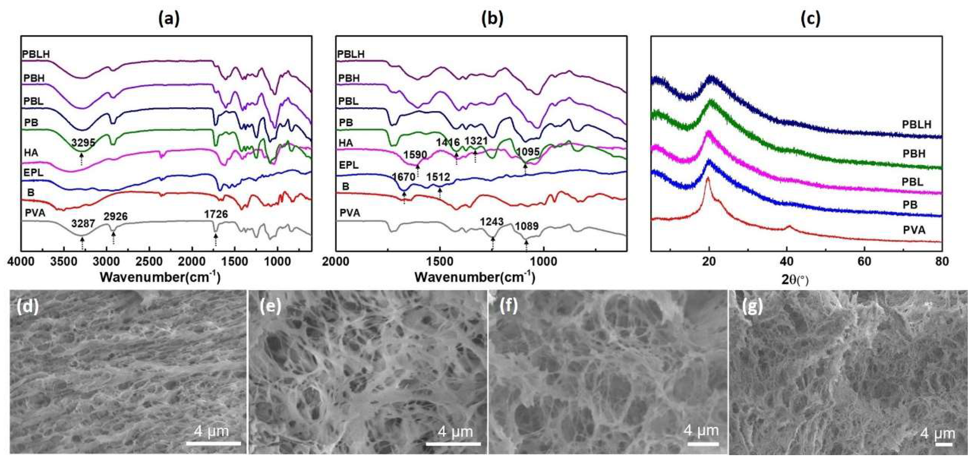
Fourier Transform Infrared spectroscopy (FTIR) was employed to investigate the functional groups and the interactions among the formed hydrogels. As shown in Figure 2, the characteristic peaks and changes of PVA, borate, HA, EPL and the resulting hydrogels in the spectra were involves illustrated. For neat PVA, the prominent absorption bands at around 1089, 2926 and 3287 cm−1 correspond to the stretching vibrations of C−O, C-H and O−H, respectively [33]. In the spectrum of EPL sample, the stretching vibrations of typical amide I, II absorption bands around appear at 1670 and 1512 cm−1 [39]. Besides, the band at 1590 cm−1 represent the C=O stretching in the spectrum of HA. After formed PB hydrogels, it was observed that the red-shift of the stretching vibrations of C−O and O−H to 1095 and 3295 cm−1. Furthermore, two new characteristic peaks at 1321 and 1416 cm−1 appeared, which attributed to the asymmetric stretching relaxation of B−O−C [32]. These results are strong evidence for confirming the formation of the dynamic borate ester bonds between borate and PVA chains. Clearly, after introduction of EPL and HA into PB system, the typical amide I, II absorption bands shifted to lower wavenumbers. As we all know, the intra- or intermolecular hydrogen bonding can reduce the force constants of the chemical bonds, and result in their vibrational frequencies shifting to lower wavenumbers [39]. Hence, these blue-shifts of the absorption bands prove the formation of H-bonding between EPL and HA.
X-ray diffraction (XRD) is a powerful tool for analyzing and identifying the crystalline and amorphous phase of materials. The sharp diffraction peaks represent a high degree of crystallinity, while broad diffraction peaks indicate a low degree of crystallinity. To explore the crystal structure changes before and after gelation, XRD diffraction patterns of hydrogels were employed. Obviously, as illustrated in Figure 2c, the XRD patterns of PVA have three diffraction peaks at 2θ = 19.6°, 2θ = 22.9°, and 2θ = 40.8°, which corresponding to the (101), (200), and (103) planes of PVA crystallites [33]. The stable crystal structures of PVA attribute to the PVA molecule containing a large number of hydroxyls as hydrogen bond sites. After crosslinking with borate (PB), the sharp diffraction peak at 2θ = 19.6° turned into a blunt peak and the diffraction peaks at 2θ = 19.6°, 2θ = 22.9° disappeared. The changes of the specific peaks of PVA crystallites probably attribute to the strong interactions between the -OH group of PVA and borate, which leading to the destruction of PVA crystallites structures [33,39]. Compare with PB hydrogels, the typical diffraction peaks of PBL, PBH and PBLH hydrogels changed slightly with addition of EPL and HA, further confirming the formation of the strong interactions between PVA, B, EPL and HA during the cross-linking and intertwinement process.
The cross-sectional microstructures of PB, PBL, PBH and PBLH hydrogels were characterized by using SEM, as shown in Figure 2d-g. Apparently, the different of cross-linking networks and pore sizes of the dried hydrogels can be observed. The PB hydrogel shows a well-defined denser porous network. Compared with PB hydrogel, a lot of larger pores emerge in the appearance of PBL or PBH hydrogels, while PBLH hydrogels with both EPL and HA exhibit a tighter network with thicker walls and smaller pores, which may attribute to the increased degree of cross-linking in DN hydrogel. Thus, the incorporation of the EPL and HA could form an additional network and further intertwine with the before networks, which is beneficial to enhance the mechanical and performance of the composite hydrogels.

2.3. Rheology and Self-Healing Properties of the DN Composite Hydrogels

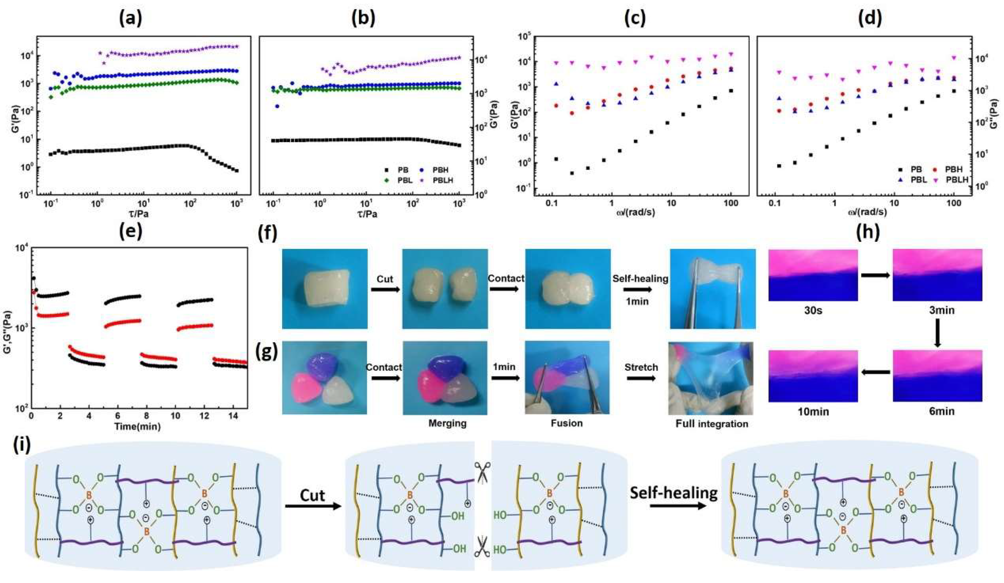
The mechanical properties of hydrogels play an important role in effect decision their suitability for specific applications. Rheology measurements usually used to characterize that performance of hydrogels. In the rheological diagram, the storage modulus (G′) reflect the energy storage, which represent the elastic properties (solid-like behavior) of samples, while loss modulus (G″) reflect and the energy loss, which represent the elastic properties (liquid-like behavior) of samples [40]. As can be seen from Figure 3a-b, the G′ of all hydrogels are higher than G″ over the entire region of strain tests, exhibiting the typical solid-like character of the composite hydrogels. When increasing of strain to about 100 Pa, the G′ and G″ of PB hydrogels began to decrease, and then transformed into a liquid-like appearance, which indicated the degree of the crosslinking networks of PB lower than other hydrogels. By incorporation of EPL and HA, both the maximum stress of G′ and G″ of the hydrogels increased, suggesting the formation of the new network and further enhancement of the mechanical property of the hydrogels [41]. In addition, in the frequency sweep test (Figure 3c-d), both G′ and G″ for all of hydrogels gradually improved with frequency increasing, exhibiting frequency-dependent behavior. As was reported, the dynamically crosslinked hydrogels generally demonstrate frequency dependent modulus, while the permanently crosslinked hydrogels show frequency-independent modulus [42,43]. Furthermore, the value of G′ is always bigger than that of G″ over the frequency tests range, and the gelation state can be maintained within range of 0.1-100 rad/s. Therefore, through analysis of the rheological studies, the intertwined networks of the composite hydrogels are dynamically.
Generally, the dynamic nature of networks usually endow the materials with dynamic performance. We systematically investigated the self-healing behaviors of the PBLH hydrogels by macroscopic and rheological recovery test. Initially, the self-repairing ability was measured by using the rheological test in different strains (2%-100%-2%-100%-2%-100%). As illustrated in Figure 3e, when the strain at 2% low strain, there is no apparent change in the values of G′ and G″, and the typical solid-like properties of hydrogels could be maintained (G′ > G″), while the networks of hydrogels are collapsed at the 100% high strain (G′ > G″). As the strain is restored to 2%, the G′ and G″ of the PBLH hydrogel instantly recover to the initial values immediately, revealing the hydrogel network structures were reconstructed [44]. Noticeably, the values of G′ and G″ nearly back to the original values even after three-interval oscillatory strain switching back and forth test, demonstrating the excellent self-healing ability of PBLH hydrogels. Subsequently, we further researched the self-healing behaviors of PBLH hydrogels by in situ and non in situ self-healing tests. As shown in Figure 3f, two pieces of unstained PBLH hydrogel contacted to each other, and the fractured hydrogels immediately healed into a whole after 1min without any external force. In addition, the two stained hydrogels and unstained hydrogel were gently put together, and then the three different hydrogels integrated into a new hybrid hydrogel rapidly. This hybrid hydrogel could maintain its integrity, even under stretching and picking up with tweezers, suggesting the PBLH composite hydrogels with good self-healing property. Furthermore, the self-healing processes of the healing surface between two stained hydrogels were further recorded by optical microscopy (Figure 3g). Apparently, with the passage of time, the gap between the two stained hydrogels decreased and even completely disappeared after10 min. Simultaneously, the dye molecules continuously spread across the fusion interfaces during the healing process, and eventually interacted with each other at the boundary, which testifying the dynamic networks of hydrogels [45]. Based on the overall results, we suspected that the extremely fast self-healing property of PBLH hydrogels may attribute to the synergistic effect of the dual dynamically reversible borate-diol bonds, electronic interaction and hydrogen bonds (Figure. 3i).

2.4. Stretchable, Shapeable and Adhesive Properties of PBLH Composite Hydrogels

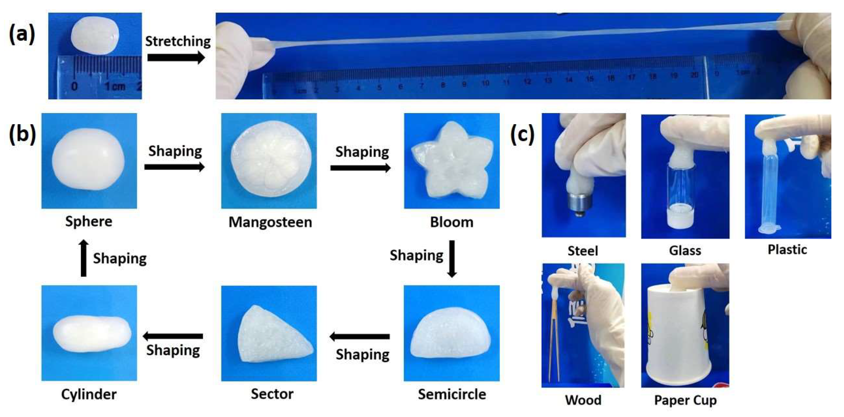
The suitable stretchable, shapeable and adhesive properties of materials are necessary to meet the requirements of different wounds. Herein, the stretchable, shapeable and adhesive performance of the PBLH hydrogels were evaluated by macroscopic tests. Interestingly, the PBLH composite hydrogels exhibit a high stretchable ability, which could be stretched to more than 15 times of that original length, and without any visible crack appeared even under twisting (Figure 4a and S2). Moreover, as can be seen from Figure 4b, the obtained composite hydrogels could be easily molded and remolded into a variety of shapes, including sphere, mangosteen, bloom, semicircle, sector and cylinder, suggesting that hydrogels with outstanding ductility and shapeable ability. In addition, the hydrogels exhibited good adhesion to the various different characters of objects, such as steel, glass, plastic, wood and paper cup. Overall, the excellent adjustable properties of PBLH composite hydrogels exhibit potential applications for biomedical engineering, such as wound dressings.

2.5. Antibacterial Activity of the Hydrogels

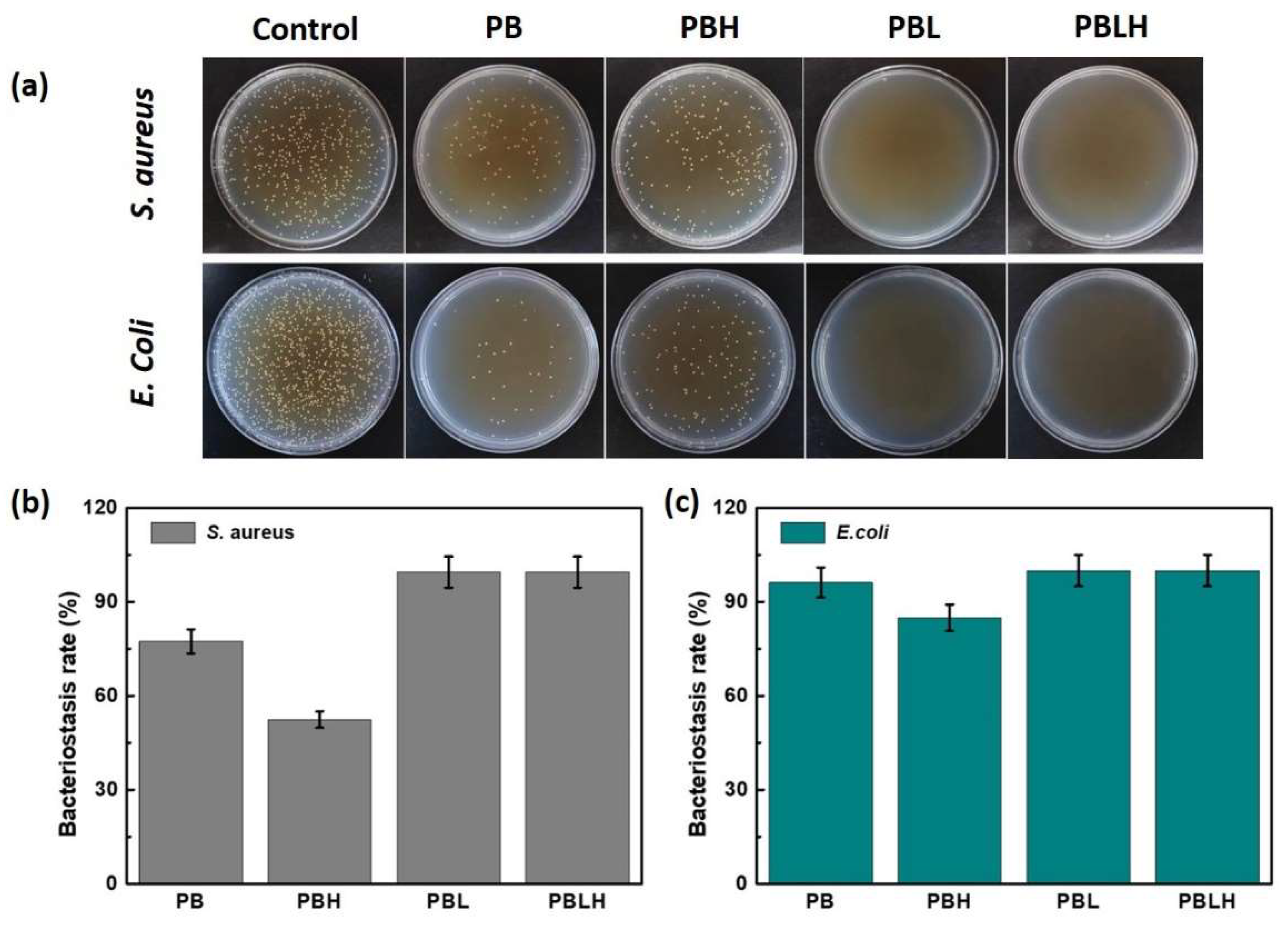
To evaluate the antibacterial performance of the fabricated hydrogels, S. aureus and E. coli were selected as the test bacteria. As shown in Figure 5a, in comparison with the control group, only a few or even none of bacteria survived from the agar plate tests treated by the composite hydrogels, suggesting the formed hydrogels with outstanding antibacterial property. Obviously, from the column chart of bacteriostasis rate (Figure 5b-c), the hydrogels show lower antibacterial activity against Gram-positive S. aureus than Gram-negative E. coli. In addition, the antibacterial performance of composite hydrogels was enhanced by the incorporation of the EPL. These results may attribute to the –NH and –NH2 groups of EPL, which can attach to the surface of bacteria and then destroy their cell membrane.

3. Materials and Methods

3.1. Materials

Poly(vinyl alcohol) (PVA, alcoholysis: 85.0-90.0 mol%, viscosity: 20.0-30.0 mPa), Hyaluronic acid (HA, molecular weight 80-100 KDa) and ε-poly-L-lysine (EPL, 98%, n=25-35) were supplied by Tianjin Xiensi Biochemical Technology Co., LTD. Borate (B, sodium tetraborate decahydrate, 99.5%) was purchased from Sinopharm Group Chemical reagent Co., LTD. All the materials were used as received without any purification. Deionized water was used throughout all the experiments.

3.2. Hydrogels Preparation

The hydrogels were prepared by using one-pot method, the detail of preparing process as follows: a certain amount of PVA was dissolved in hot deionized water and agitated 95 °C for a duration of 2 h, resulting in the formation of PVA solutions with a concentration of 10 wt%. Then, some amount of borate, HA and EPL were mixed in aqueous solution with agitating until the solid dissolved thoroughly. Finally, the white hydrogels were obtained (PBLH: 10.0 wt% PVA, 0.5 wt% borax, 4.0 wt% HA and 0.5 wt% EPL). With the same preparation steps, the hydrogels of PB (10.0 wt% PVA and 0.5 wt% borax), PBL (10.0 wt% PVA, 0.5 wt% borax and 0.5 wt% EPL), PBH (10.0 wt% PVA, 0.5 wt% borax and 4.0 wt% HA) were obtained.

3.3. Characterization

Fourier Transform Infrared spectroscopy (FTIR). FTIR spectra of hydrogels were obtained by using a Thermo Fisher Spectrum over a range of 4000-400 cm-1 with a resolution of 4 cm−1 d and total of 32 scans for each sample.
Rheological measurements. The rheological properties of the DN composite hydrogels were assessed using an Anton-Paar Rheometer. A core-plate system of C35/1° Ti L07116 with a plate diameter of 35 mm and core angle of 1°. All rheological tests were carried out at 25.0 °C. In dynamic oscillatory strain sweeping tests, fixing frequency at 1Hz, the shear rate over a range rates from 0.01 s-1 to 1000 s-1. In frequency sweeping tests, fixing the shear rate at 1 Hz, the frequency sweeping from 0.01 Hz to 100 Hz. In self-healing property tests, fixing frequency at 1Hz, time sweeps were employed to evaluate the self-healing capability of the hydrogels. The strain was initially set at 2% for 2.5 min, increased to 100% for 2.5 min, and then restored to 2% for an additional 2.5 min. The changes of storage modulus (G′) and loss modulus (G″) were assessed the self-healing capacity of the samples.
Scanning Electron Microscopy (SEM). To investigate the microstructures of the DN composite hydrogels, SEM was employed. Hydrogel sample was freeze-drying in a vacuum extractor for 24 h by freeze dryer. A small volume of xerogel was paste into silica wafers. Subsequently, the samples were subjected to gold plating and observation by using microscope.
Self-Healing Experiments. The two hydrogels were stained by methylene blue and eosin respectively. The unstained hydrogel and two stained hydrogels are contacted in air with no other stress or outside stimulus during the healing process. Subsequently, the gap between the two stained hydrogels at different times were observed by using microscope. All of the situations of the DN composite hydrogels were photographed.
Antibacterial Activity Evaluation. In vitro antibacterial activities of the hydrogels were evaluated against pathogenic representatives of Gram-positive bacteria Staphylococcus aureus (S. aureus) and Gram-negative bacteria Escherichia coli (E. coli). The hydrogels were put into bacteria culture tubes with liquid nutrient medium, which the bacteria with a certain concentration. The bacteria culture tubes were shocked at 37 °C for 12 h in shaker. Then, the liquid nutrient medium was transferred onto the surface of the Luria-Bertani (LB) plates. The LB plates were incubated at 37 °C for 12 h in an incubator. The colony formation unit of LB plates were measured, which reflected the antibacterial activity of hydrogels.

4. Conclusions

In summary, rapid self-healing, stretchable, moldable and antibacterial double network composite hydrogels were successfully prepared by Poly (vinyl alcohol) (PVA), Borate (B), ε-poly-L-lysine (EPL) and hyaluronic acid (HA) based on one-pot method. SEM images of the fabricated hydrogels exhibited networks with porous structures. FTIR confirmed the existence of the hydrogen bonding in the system. In addition, rheology measurements proved the dynamic property of the networks of hydrogels. Interestingly, the fabricated hydrogels shown outstanding rapid self-healing, stretchable and moldable performances, which could completely heal within 1 min and could be mold and remold to various shape. Meanwhile, the composite hydrogels displayed excellent antibacterial activity for Staphylococcus aureus (S. aureus) and Escherichia coli (E. coli). Overall, the dynamic double network hydrogels would lay the foundation of composite hydrogels in biomedical fields, especially for wound dressing and antibacterial materials.

Supplementary Materials

The following supporting information can be downloaded at the website of this paper posted on Preprints.org, Figure S1: The photographs of the states of the PB, PBH, PBL, PBLH hydrogels at initial and after certain hours; Figure S2: The photographs of the stretchable states of the PBLH composite hydrogels.

Author Contributions

N.S.: Conceptualization, Methodology, Project administration, Resources, Software, Writing—original draft. X.L.: Data curation, Formal analysis, Investigation, Writing—original draft. W.L.: Data curation, Formal analysis, Investigation, Writing—original draft. C.X.: Data curation, Formal analysis, Investigation. A.Z.: Formal analysis, Validation. P.S.: Writing—review & editing, Visualization, Supervision, Project administration, Funding acquisition. All authors have read and agreed to the published version of the manuscript.

Funding

P. Sun was funded by the Natural Science Foundation of Shandong Province (No. ZR2023QB052). N. Sun was funded by the National Natural Science Foundation of China (No. 22102118). This work is funded by the domestic visiting project of Shandong Second Medical University.

Institutional Review Board Statement

Not applicable.

Informed Consent Statement

Not applicable.

Data Availability Statement

Data are contained within the article and Supplementary Materials.

Conflicts of Interest

The authors declare no conflicts of interest.

References

  1. Yang, D. Recent Advances in Hydrogels. Chem. Mater. 2022, 34, 1987–1989. [Google Scholar] [CrossRef]
  2. Wang, W.; Yuan, Z.; Li, T.; Wang, Y.; Zhang, K.; Wu, J.; Zhang, S.; Yuan, F.; Dong, W. Rapid Preparation of Highly Stretchable and Fast Self-Repairing Antibacterial Hydrogels for Promoting Hemostasis and Wound Healing. ACS Appl. Bio Mater. 2024, 7, 394–405. [Google Scholar] [CrossRef]
  3. Dehghan-Niri, M.; Vasheghani-Farahani, E.; Eslaminejad, M. Tavakol, M.; Bagheri, F. Preparation of Gum Tragacanth/Poly (vinyl alcohol)/Halloysite Hydrogel using Electron Beam Irradiation with Potential for Bone Tissue Engineering. Carbohyd. Polym. 2023, 305, 120548. [Google Scholar] [CrossRef] [PubMed]
  4. Guo, W-Y. Ma, M-G. Choi, W.; S. Kohane, D. Hybrid Nanoparticle–Hydrogel Systems for Drug Delivery Depots and Other Biomedical Applications. ACS Nano 2024, 18, 22780–22792. [Google Scholar] [CrossRef] [PubMed]
  5. Toufanian, S.; Mohammed, J.; Winterhelt, E.; Lofts, A.; Dave, R.; K. Coombes, B.; Hoare, T. A Nanocomposite Dynamic Covalent Cross-Linked Hydrogel Loaded with Fusidic Acid for Treating Antibiotic-Resistant Infected Wounds. ACS Applied Bio Mater. 2024, 7, 1947–1957. [Google Scholar] [CrossRef] [PubMed]
  6. Yin, H.; Liu, F.; Abdiryim, T.; Liu, X. Self-Healing Hydrogels: From Synthesis to Multiple Applications. ACS Mater. Lett. 2023, 5, 1787–1830. [Google Scholar] [CrossRef]
  7. Ding, X.; Fan, L.; Wang, L.; Zhou, M.; Wang, Y.; Zhao, Y. Designing Self-healing Hydrogels for Biomedical Applications. Mater. Horiz. 2023, 10, 3929–3947. [Google Scholar] [CrossRef]
  8. Rammal, H.; GhavamiNejad, A.; Erdem, A.; Mbeleck, R.; Nematollahi, M.; Emir Diltemiz, S.; Alem, H.; Ali Darabi, M.; Ertas, Y.; J. Catersonm, E.; Ashammakhi, N.. Advances in Biomedical Applications of Self-healing Hydrogels. Mater. Chem. Front., 2021, 5, 4368–4400. [Google Scholar] [CrossRef]
  9. Sun, P.; Ren, S.; Liu, F.; Wu, A.; Sun, N.; Shi, L.; Zheng, L. Smart low molecular weight hydrogels with dynamic covalent skeletons. Soft Matter, 2018, 14, 6678–6683. [Google Scholar] [CrossRef]
  10. Luo, C.; Guo, A.; Zhao, Y.; Sun, X. A High Strength, Low Friction, and Biocompatible Hydrogel from PVA, Chitosan and Sodium Alginate for Articular Cartilage. Carbohyd. Polym. 2022, 286, 119268. [Google Scholar] [CrossRef]
  11. Cho, S.; Hwang, S.; Dongyeop, X.; Park, J. Recent Progress in Self-healing Polymers and Hydrogels based on Reversible Dynamic B-O Bonds: Boronic/Boronate Esters, Borax, and Benzoxaborole. J. Mater. Chem. A, 2021, 9, 14630–14655. [Google Scholar] [CrossRef]
  12. Shui, T.; Pan, M.; Li, A.; Fan, H.; Wu, J.; Liu, Q.; Zeng, H. Poly (vinyl Alcohol) (PVA)-Based Hydrogel Scaffold with Isotropic Ultratoughness Enabled by Dynamic Amine-Catechol Interactions. Chem. Mater. 2022, 34, 8613–8628. [Google Scholar] [CrossRef]
  13. Yang, X.; Wang, B.; Sha, D.; Liu, Y.; Liu, Z.; Shi, K.; Liu, W.; Yu, C.; Ji, X. PVA/Poly(hexamethylene guanidine)/Gallic Acid Composite Hydrogel Films and Their Antibacterial Performance. ACS Appl. Polym. Mater. 2021, 3, 3867–3877. [Google Scholar] [CrossRef]
  14. Zhang, X.; Ren, K.; Xiao, C.; Chen, X. Guanosine-driven Hyaluronic Acid-based Supramolecular Hydrogels with Peroxidase-like Activity for Chronic Diabetic Wound Treatment. Acta Biomater. 2023, 172, 206–217. [Google Scholar] [CrossRef] [PubMed]
  15. Guo, H.; Huang, S.; Xu, A.; Xue, W. Injectable Adhesive Self-Healing Multiple-Dynamic-Bond Crosslinked Hydrogel with Photothermal Antibacterial Activity for Infected Wound Healing. Chem. Mater. 2022, 34, 2655–2671. [Google Scholar] [CrossRef]
  16. Li, Q.; Zhang, S.; Du, R.; Yang, Y.; Liu, Y.; Wan, Z.; Yang, X. Injectable Self-Healing Adhesive Natural Glycyrrhizic Acid Bioactive Hydrogel for Bacteria-Infected Wound Healing. ACS Appl. Mater. Interfaces 2023, 15, 17562–17576. [Google Scholar] [CrossRef]
  17. Zhang, Z.; Wang, X.; Wang, Y.; Hao, J. Rapid-Forming and Self-Healing Agarose-Based Hydrogels for Tissue Adhesives and Potential Wound Dressings. Biomacromolecules. 2018, 19, 3980–3988. [Google Scholar] [CrossRef]
  18. Li, Q.; Zhang, S.; Du, R.; Yang, Y.; Liu, Y.; Wan, Z.; Yang, X. Injectable Self-Healing Adhesive Natural Glycyrrhizic Acid Bioactive Hydrogel for Bacteria-Infected Wound Healing. ACS Appl. Mater. Interfaces 2023, 15, 17562–17576. [Google Scholar] [CrossRef]
  19. Fan, L.; He, Z.; Peng, X.; Xie, J; Su, F. ; Wei, D-X. Zheng, Y.; Yao, D. Injectable, Intrinsically Antibacterial Conductive Hydrogels with Self-Healing and pH Stimulus Responsiveness for Epidermal Sensors and Wound Healing. ACS Appl. Mater. Interfaces 2021, 13, 53541–53552. [Google Scholar] [CrossRef]
  20. Yan, B.; Huang, J.; Han, L.; Gong, L.; Li, L; N. Israelachvili, J.; Zeng, H. Duplicating Dynamic Strain-Stiffening Behavior and Nanomechanics of Biological Tissues in a Synthetic Self-Healing Flexible Network Hydrogel. ACS Nano 2017, 11, 11074–11081. [Google Scholar] [CrossRef]
  21. Li, P.; Zong, H.; Li, G.; Shi, Z.; Yu, X.; Zhang, K.; Xia, P.; Yan, S.; Yin, J. Building a Poly(amino acid)/Chitosan-Based Self-Healing Hydrogel via Host–Guest Interaction for Cartilage Regeneration. ACS Biomater. Sci. Eng. 2023, 9, 4855–4866. [Google Scholar] [CrossRef]
  22. Narasimhan, B. ; W. Dixon, A.; Mansel, B.; Taberner, A.; Mata, J.;Malmström, J. Hydrogen Bonding Dissipating Hydrogels: The Influence of Network Structure Design on Structure-Property Relationships. J. Colloid Interf. Sci. 2023, 630, 638–653. [Google Scholar]
  23. Wang, L.; Gao, G.; Zhou, Y.; Xu, T.; Chen, J.; Wang, R.; Zhang, R.; Fu, J. Tough, Adhesive, Self-Healable, and Transparent Ionically Conductive Zwitterionic Nanocomposite Hydrogels as Skin Strain Sensors. ACS Appl. Mater. Interfaces 2019, 11, 3506–3515. [Google Scholar] [CrossRef] [PubMed]
  24. Xu, X.; Victor Jerca, V.; Hoogenboom, R. Bioinspired Double Network Hydrogels: from Covalent Double Network Hydrogels via Hybrid Double Network Hydrogels to Physical Double Network Hydrogels. Mater. Horiz., 2021, 8, 1173–1188. [Google Scholar] [CrossRef] [PubMed]
  25. B. Rodell, C.; N. Dusaj, N.; B. Highley, C.; A. Burdick, J. Injectable and Cytocompatible Tough Double-Network Hydrogels through Tandem Supramolecular and Covalent Crosslinking. Adv. Mater. 2016, 20160226. [Google Scholar]
  26. Yin, Y.; Gu, Q.; Liu, X.; Liu, F.; Julian McClement, D. Double network hydrogels: Design, fabrication, and application in biomedicines and foods. Adv Colloid Interface Sci. 2023, 320, 102999. [Google Scholar] [CrossRef]
  27. C. Ollier, R.; Xiang, Y.; M. Yacovelli, A.; J. Webber, M. Biomimetic Strain-Stiffening in Fully Synthetic Dynamic-Covalent Hydrogel Networks. Chem. Sci., 2023, 14, 4796–4805. [Google Scholar] [CrossRef]
  28. Rezanejade Bardajee, G.; Ghadimkhani, R.; Jafarpour, F. A Biocompatible Double Network Hydrogel based on Poly (acrylic acid) Grafted onto Sodium Alginate for Doxorubicin Hydrochloride Anticancer Drug Release. Int. J. Biol. Macromol. 2024, 260, 128871. [Google Scholar]
  29. Wang, Y.; Zhang, Y.; Ren, P.; Yu, S.; Cui, P.; B. Nielsen, C.; Abrahams, I.; Briscoe, J.; Lu, Y. Versatile and Recyclable Double-Network PVA/Cellulose Hydrogels for Strain Sensors and Triboelectric Nanogenerators under Harsh Conditions. Nano Energy 2024, 125, 109599. [Google Scholar] [CrossRef]
  30. Li, L.; Wu, P.; Yu, F.; Ma, J. Double Network Hydrogels for Energy/Environmental Applications: Challenges and Opportunities. J. Mater. Chem. A, 2022, 10, 9215–9247. [Google Scholar] [CrossRef]
  31. Xu, X.; Victor Jerca, V.; Hoogenboom, R. Bioinspired Double Network Hydrogels: from Covalent Double Network Hydrogels via Hybrid Double Network Hydrogels to Physical Double Network Hydrogels. Mater. Horiz., 2021, 8, 1173–1188. [Google Scholar] [CrossRef] [PubMed]
  32. Chen, W-P.; Hao, D-Z.; Hao, W-J.; Guo, X-L.; Jiang, L. Hydrogel with Ultrafast Self-Healing Property Both in Air and Underwater. ACS Appl. Mater. Interfaces 2018, 10, 1258–1265. [CrossRef] [PubMed]
  33. Ge, W.; Cao, S.; Shen, F.; Wang, Y.; Ren, J.; Wang, X. Rapid Self-healing, Stretchable, Moldable, Antioxidant and Antibacterial Tannic Acid-cellulose Nanofibril Composite Hydrogels. Carbohyd. Polym. 2019, 224, 115147. [Google Scholar] [CrossRef] [PubMed]
  34. Teng, J.; Zhao, W.; Zhang, S.; Yang, D.; Liu, Y.; Huang, R.; Ma, Y.; Jiang, L.; Wei, H.; Zhang, J.; Chen, J. Injectable Nanoparticle-Crosslinked Xyloglucan/ε-Poly-L-lysine Composite Hydrogel with Hemostatic, Antimicrobial, and Angiogenic Properties for Infected Wound Healing. Carbohyd. Polym. 2024, 336, 122102. [Google Scholar] [CrossRef]
  35. Xu, Q.; Dai, X.; Yang, L.; Liu, X.; Li, Y.; Gao, F. ε Polylysine-Based Macromolecules with Catalase-Like Activity to Accelerate Wound Healing by Clearing Bacteria and Attenuating Inflammatory Response. ACS Biomater. Sci. Eng. 2022, 8, 5018–5026. [Google Scholar] [CrossRef]
  36. Lee, J-H. ; Kwon, H-K.; Shin, H-J.; Nam, G-H.; Kim, J-H.; Cho, S. Quasi-Stem Cells Derived from Human Somatic Cells by Chemically Modified Carbon Nanotubes. ACS Appl. Mater. Interfaces 2018, 10, 8417–8425. [Google Scholar] [CrossRef]
  37. Meng, Z.; He, Y.; Wang, F.; Hang, R.; Zhang, X.; Huang, X.; Yao, X. Enhancement of Antibacterial and Mechanical Properties of Photocurable ε Poly L lysine Hydrogels by Tannic Acid Treatment. ACS Appl. Bio Mater. 2021, 4, 2713–2722. [Google Scholar] [CrossRef]
  38. Wei, P.; Duan, Y.; Wang, C.; Sun, P.; Sun, N. Co-Assembled Supramolecular Organohydrogels of Amphiphilic Zwitterion and Polyoxometalate with Controlled Microstructures. Molecules 2024, 29, 2286. [Google Scholar] [CrossRef]
  39. Dehghan-Niri, M.; Vasheghani-Farahani, E.; Eslaminejad, M.; Tavakol, M.; Bagheri, F. Preparation of Gum Tragacanth/Poly (vinyl alcohol)/Halloysite Hydrogel using Electron Beam Irradiation with Potential for Bone Tissue Engineering. Carbohyd. Poym. 2023, 305, 120548. [Google Scholar] [CrossRef]
  40. Zhang, X.; Xu, J.; Lang, C.; Qiao, S.; An, G.; Fan, X.; Zhao, L.; Hou, C.; Liu, J. Enzyme-Regulated Fast Self-Healing of a Pillararene-Based Hydrogel. Biomacromolecules 2017, 18, 1885–1892. [Google Scholar] [CrossRef]
  41. Guo, H.; Huang, S.; Xu, A.; Xue, W. Injectable Adhesive Self-Healing Multiple-Dynamic-Bond Crosslinked Hydrogel with Photothermal Antibacterial Activity for Infected Wound Healing. Chem. Mater. 2022, 34, 2655–2671. [Google Scholar] [CrossRef]
  42. Huang, J.; Wu, C.; Yu, X.; Li, H.; Ding, S.; Zhang, W. Biocompatible Autonomic Self-healing PVA-TA Hydrogel with High Mechanical Strength, Macromol. Chem. Phys. 2021, 222, 2100061. [Google Scholar]
  43. Si, R.; Wang, Y.; Yang, Y.; Javeed, A.; Chen, J.; Han, B. Dynamic Dual-Crosslinking Antibacterial Hydrogel with Enhanced Bio-adhesion and Self-healing Activities for Rapid Hemostasis in Vitro and in Vivo. Mater. Des. 2023, 233, 112244. [Google Scholar] [CrossRef]
  44. Peng, Y-Y.; Cheng, Q.; Wang, W.; Wu, M.; Diaz-Dussan, D.; Kumarc, P.; Narain, R. Multi-Responsive, Injectable, and Self-healing Hydrogels based on Benzoxaborole-Tannic Acid Complexation. Polym. Chem. 2021, 12, 5623–5630. [CrossRef]
  45. Yang, X.; Liu, G.; Peng, L.; Guo, J.; Tao, L.; Yuan, J.; Chang, C.; Wei, Y.; Zhang, L. Highly Efficient Self-Healable and Dual Responsive Cellulose-Based Hydrogels for Controlled Release and 3D Cell Culture. Adv. Funct. Mater. 2017, 1703174. [Google Scholar] [CrossRef]
Scheme 1. Schematic diagram of the mechanism of dynamic double network (DN) composite hydrogel through dynamic borate eater reaction and physical cross-linking.
Preprints 117044 sch001
Figure 1. The photographs of the states of the hydrogels (a); schematic illustration of the interactions of double network (DN) hydrogels between all of components, including dynamic borate eater reaction and physical cross-linking (b).
Preprints 117044 g001
Figure 2. The FTIR spectra of pure PVA, B, EPL, HA and the formed hydrogels of PB, PBL, PBH, PBLH (a, b); XRD patterns of PVA, PB, PBL, PBH, PBLH (c); SEM images of PB, PBL, PBH, PBLH, respectively (d-g). Scale bar = 4 μm.
Preprints 117044 g002
Figure 3. Dynamic oscillatory strain sweep tests (from 0.1 Pa to 1000 Pa) (a, b) and Frequency sweep tests (from 0.1 rad/s to 100 rad/s) (c, d) of the hydrogels. G’ and G’’ of the PBLH hydrogels under alternate low (2%) and high (100%) strain at a fixed frequency of 1 Hz (e). Images of the in situ (unstained hydrogels) and (f) non-in situ (stained hydrogels) methods were used to observe self-healing properties (g). The hydrogels were stained by methylene blue and eosin, respectively. Photographs of the self-healing process of PBLH hydrogels by microscope (h). Schematic diagram of the self-healing mechanism of PBLH hydrogels (i).
Preprints 117044 g003
Figure 4. Photographs of the stretchable (a) shapeable (b) and adhesive properties of PBLH composite hydrogels.
Preprints 117044 g004
Figure 5. Digital photos of the growth of S. aureus and E. coli cultured in vitro on agar plates under different conditions (a). Bacteriostasis rate against S. aureus (b) and E. coli (c).
Preprints 117044 g005
Disclaimer/Publisher’s Note: The statements, opinions and data contained in all publications are solely those of the individual author(s) and contributor(s) and not of MDPI and/or the editor(s). MDPI and/or the editor(s) disclaim responsibility for any injury to people or property resulting from any ideas, methods, instructions or products referred to in the content.
Copyright: This open access article is published under a Creative Commons CC BY 4.0 license, which permit the free download, distribution, and reuse, provided that the author and preprint are cited in any reuse.
Prerpints.org logo

Preprints.org is a free preprint server supported by MDPI in Basel, Switzerland.

Subscribe

© 2024 MDPI (Basel, Switzerland) unless otherwise stated