Preprint
Review

Carcinogenesis of the Human Scalp. An Immunometabolic Centered View

Altmetrics

Downloads

124

Views

93

Comments

0

A peer-reviewed article of this preprint also exists.

Submitted:

04 September 2024

Posted:

05 September 2024

Read the latest preprint version here

Alerts
Abstract
The human scalp is a common site of skin cancer in humans, with non-melanoma skin cancer being exceedingly common. In this review, two dermatologists with extensive experience in cutaneous oncology will discuss unique features of the epidemiology of cancer of the scalp. Clinical observations on these common skin cancers lead to insight into the pathogenesis and potential prevention and treatment of cutaneous scalp neoplasia. Our hypothesis is that the presence of hair protects against the development of skin cancer, but not by serving as a physical shield, but rather by providing a continuous IL-17 biased immunosurveillance. Loss of hair allow release from immunosurveillance, resulting in expansion of neoplastic cells towards skin cancer. Both hair follicles and metabolic changes in stroma allow permissiveness for tumor promotion.
Keywords: 
Subject: Medicine and Pharmacology  -   Dermatology

Introduction

The scalp is an exceedingly common site for basal cell, squamous cell carcinoma and occasional melanomas. Benign lesions, including seborrheic keratoses and blue nevi are also commonly found on the scalp. A major difference between the scalp and other common sun exposed areas for skin cancer is that the scalp is covered with hair during periods of peak sun exposure during youth. Carcinomas on the scalp are more common in men than women, but may become evident in women with sparse hair. Androgenetic alopecia is also common in men, beginning in the 20-30 year old age group, and becoming more evident with age. Women too lose hair, but not usually to the same extent as men. The incidence of scalp carcinomas is also higher in men than in women [1,2]. Androgenetic alopecia is thought to be mediated by dihydrotestosterone, and this has led to the use of dihydrotestosterone blockers as a common treatment for alopecia [3].
Two major differences exist in the scalp of elderly men vs elderly women. The first is the presence of hair. While elderly women experience hair thinning, frank alopecia is uncommon. The second change is the stroma. Stromal changes may be responsible for miniaturization of hair follicles, and stromal changes associated with miniaturization may also favor the development of actinic keratosis and nonmelanoma skin cancer [3,4].
Scalp stroma is subject to aging as is every other tissue. A candidate for aging in scalp stroma is Sirt3, a mitochondrial deacetylase which stimulates mitochondrial biogenesis [5,6]. TGF beta lowers Sirt3 levels, promotes fibrosis, and is decreased in fibrotic diseases such as scleroderma. Of interest, the scleroderma dermis does not support hair growth, and a similar mechanism of fibrotic stroma may decrease both hair growth and favor the development of cutaneous neoplasia [7,8].

Interplay of Genetics and Skin Carcinogenesis

Actinic keratoses are exceedingly common lesions on the scalp, and a known precursor to cutaneous squamous cell carcinoma. Genetic studies have implicated mutations in several genes in both actinic keratoses and cutaneous squamous cell carcinomas. These include mutations in p53 resulting in gain of function, activating mutations in phosphoinositol-3 kinase p85, and inactivating mutations in Notch 1 and 2. Genetic predisposition to actinic keratoses has been linked to IRF4, a transcription factor which links immunity to pigmentation, as well as pigmentation associated genes such as tyrosinase [9,10,11,12,13]. Current knowledge does not suggest a unique mutational profile of basal cell carcinoma of the scalp, with scalp and non-scalp basal cell carcinomas both having mutations in the Patch/Sonic hedgehog pathway.

IRF4- the Locus between Hair Growth and Inflammation

In previous studies, we have shown that normal skin has a tonic IL-12 mediated immunosurveillance, and an intact barrier function leads to a tonic production of IL-12, which suppresses both Th2 and Th17 mediated inflammation. Clinical observations suggest that the presence of hair skews the epidermis to a slight IL-17 predominance, which results in an increased frequency of IL-17 mediated inflammatory disorders, such as psoriasis, hidradenitis suppurativa and seborrheic dermatitis in hair bearing areas [9,10,11].
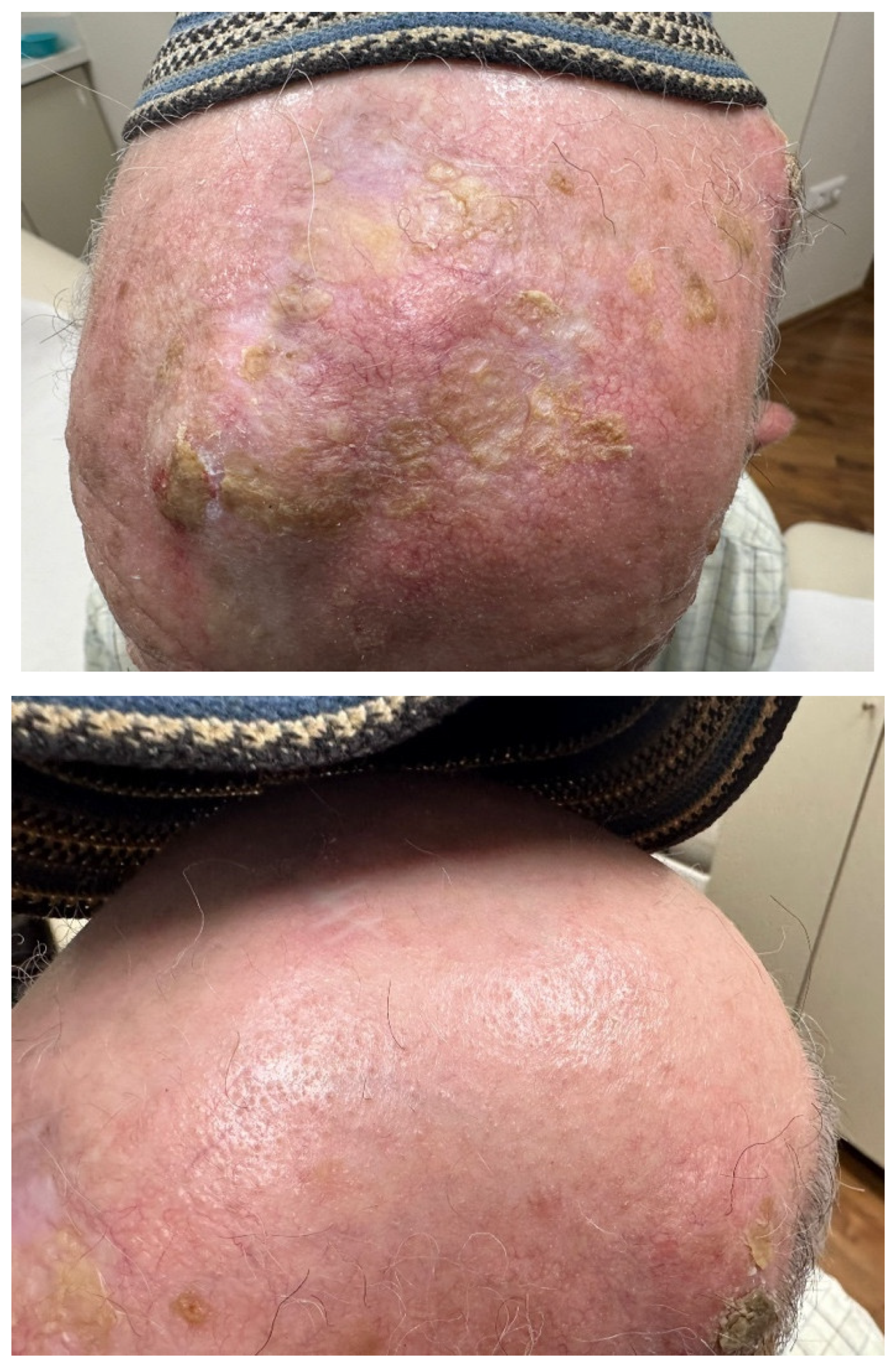
IRF4 is a transcription factor that plays a role in pigmentation and IL-17 mediated inflammation [9,10,11,12,13]. IRF4 has been associated with several human pigmentation traits, including hair graying and hair loss, and IRF4 has also been shown to bind to the MITF (microphthalmia) promoter, which is the master transcriptional switch in melanocytes. The presence of IRF4 in hair may explain in part the presence of scalp psoriasis [9,10,11]. Interestingly, scalp inflammation is often localized to hair bearing areas, and is absent in areas of alopecia (Figure 1). Additional sun protective measures, such as lifelong wearing of hats, may be protective of UV induced carcinogenesis (Figure 2).
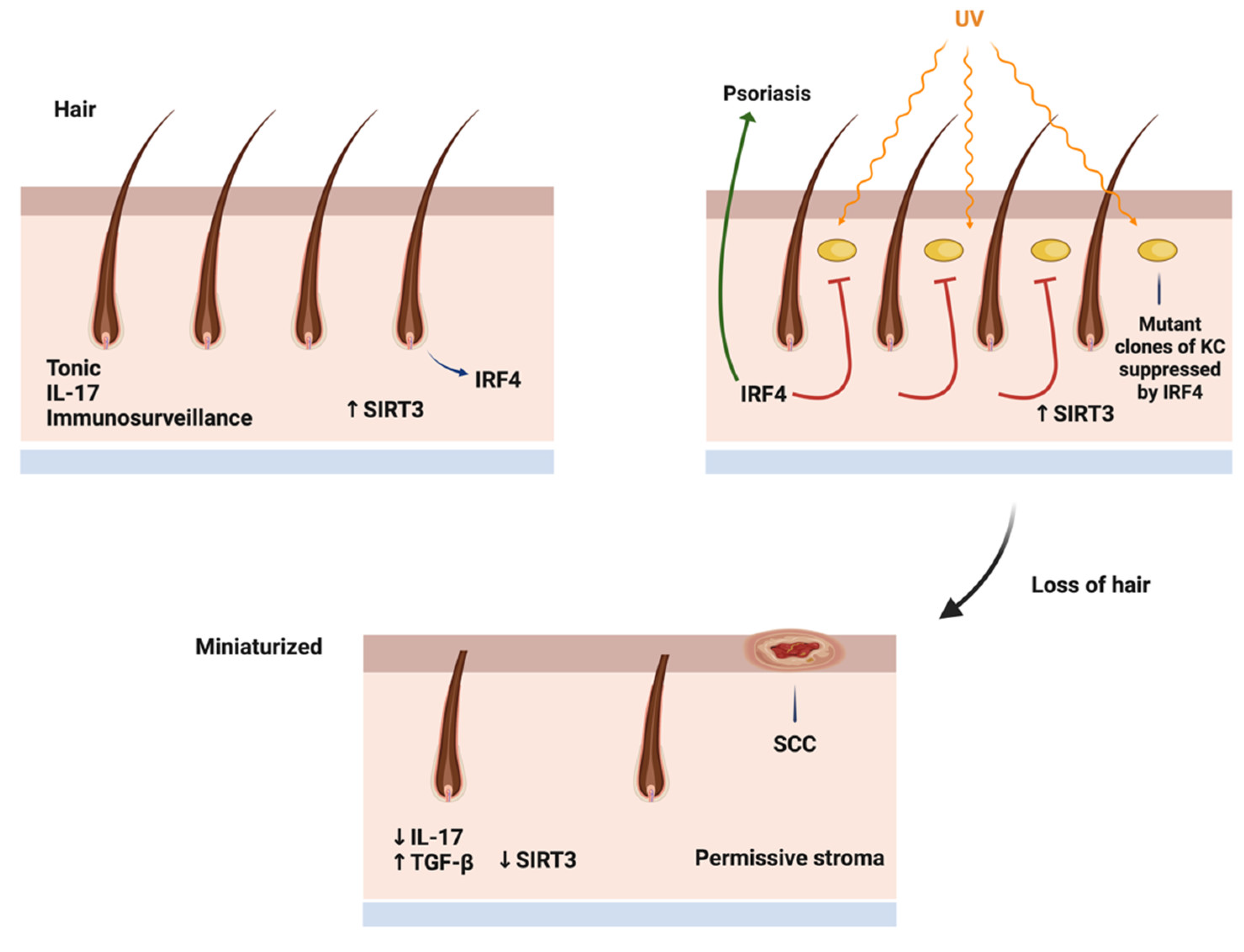
Decreased IRF4 leads to hair graying, which in turn may lead to alopecia and loss of the immune protection against nonmelanoma skin cancer [3,7]. Loss of hair likely diminishes the presence of IL-17, resulting in promotion of pre-existing UV mutant cells into clinically evident tumors (Figure 3).

Actinic Keratosis and Cutaneous Squamous Cell Carcinoma

Actinic keratosis (AK) and cutaneous squamous cell carcinoma (cSCC) are exceedingly common lesions that occur on the scalp. AK is a known precursor to cSCC, with a small but significant number of AK progressing to cSCC. For this reason, destruction of AK by cryotherapy and treatment with topical agents, such as fluorouracil, are among the most common cause of visits of elderly patients to dermatologists. AK and cSCC have long been known to be caused by UVB, and have a high tumor mutation burden, with mutation of p53 being among the most common. A recent extensive study of Thomson et al. [14] demonstrated additional mutations in tumor suppressor genes, such as Notch1, Notch2, FAT1 KMTC2 and HMCN1. In this study, the only dominant oncogenic mutation was in the phisphoinositol-3-kinase p85B. Our group was the first group to demonstrate that phosphoinositol-3 kinase inhibition in vivo led to decreased tumor growth in angiosarcoma, a tumor also associated with impaired p53 function.
Of note, TGFBR2 is downregulated in the progression from normal skin to AK to cSCC. This may represent an adaptation to scalp stroma, which becomes more rich in TGFb with aging.

Melanoma

The scalp is not an uncommon site for melanoma. Melanomas of the scalp have a distinct mutational profile compared to melanomas in other sites. Braf mutation is one of the most common driver mutations in melanoma, but the scalp has a different profile of Braf mutations, with V600K being more common that V600E mutations, and V600K appear to be more associated with chronic skin damage than V600E mutations [15,16,17]. Melanomas also appear to be more common in hair bearing areas than nonmelanoma skin cancers of he scalp, indicting that immunosurveillance may play less of a role for scalp melanoma than for nonmelanoma skin cancer [1,2,18]. Finally, mutations in other drivers are seen in scalp melanomas, including Rac1 and GNA11 [1,19,20,21]. An exceptional response was observed in a scalp melanoma with metastases in a patient treated with surgical debulking, gentian violet and imiquimod, with no recurrence for over 2 years until the patient died of congestive heart failure [22]. Scalp melanomas thus appear in two clinical scenarios, one of chronic sun exposure and hair loss, and one of sun protection and retention of hair, occurring at a younger age [1].

Angiosarcoma

Angiosarcoma is a rare malignancy of endothelial cells, and the scalp is the most common site of cutaneous angiosarcoma. This tumor has a high propensity for distant metastases, and the prognosis is poor. Complete excision is often difficult due to large primary size and skip areas. In the largest study of angiosarcoma performed to date, angiosarcoma of the scalp has a high tumor mutation burden and UV signature compared to other sites. Mutation of p53 has also been observed nearly universally in scalp angiosarcoma [23]. The high tumor mutation burden of scalp angiosarcoma has suggested a role of immunotherapy, and some exceptional responses have been observed [23,24,25].

IL-17, the Double Edged Sword

We have proposed that hair derived IL-17 may play a protective role against the development of skin cancer. Others have shown that IL-17 can promote other cancers [26,27]. How do we reconcile these findings. A recent study demonstrated that the presence of wild type p53 is required for the tumor promoting activity of IL-17 [27]. We have previously shown that tumors with mutant p53 signal differently than tumors with wild-type p53. The majority of basal and squamous cell carcinomas have defects/mutations in p53, while the majority of melanomas have wild type 53. This may explain in part why hair may be more protective against nonmelanoma skin cancer than melanoma.

Metabolic Parameters of the Aging Scalp

The major features of the aging scalp are loss of hair, manifesting in baldness. The aging associated alopecia is primarily a nonscarring alopecia, characterized by miniaturization of hair follicles, often associated with some degree of fibrosis [3,7]. The loss of longer hair likely diminishes the tonic IRF-4-IL-17 axis of the skin, allowing for decreased immunosurveillance of UV mutated keratinocytes and melanocytes. Hair requires ATP to maintain its complex, differentiates structure, and this requires optimal metabolism in both hair follicle keratinocytes and stromal support cells. As the scalp ages, there is decreased respiratory capacity in both hair generating cells and stroma, and the IRF-4-IL-17 axis is replaced with a TGF-beta low Sirt3 axis [8]. Low Sirt3 fibroblasts are associated with tumor stroma, and may play a support role in tumor progression. The clinical observation of increased skin cancers with low hair density reflects aging metabolism. The takeaway lessons from carcinogenesis of the scalp are the following. First, the hair does not provide as good a barrier to UV mutagenesis as commonly assumed. Wearing of hats or other protective measures may prevent scalp carcinogenesis. Second, the hair likely provides an immunological barrier to the development of skin cancer on the scalp, and interventions that increase IL-17 may be immunopreventive of scalp skin cancers. Third, metabolic stromal aging, mediated in part by dihydrotestosterone-reactive oxygen signaling, modifies the scalp into a less supportive environment for hair. At the same time increased TGF-beta, which decreases the mitochondrial enzyme Sirt3, provides a permissive stroma that allows progression of mutated keratinocytes and melanocytes to frank malignancy. Sirt3 has been shown to be protective of cochlear hair cells [5,6], and Sirt3 activators like honokiol and methyl-honokiol have been shown to be beneficial in murine models of alopecia [28,29]. Pharmacologic interventions which maintain hair may also be preventive of scalp carcinogenesis.
Conclusion: The appreciation of the scalp as a distinct immunometabolic site is beginning to be appreciated. A novel candidate for treatment of alopecia, a small molecule mitochondrial pyruvate transport inhibitor, promotes hair growth in mice and is being currently evaluated in human clinical trials (NCT06393452). The proposed mechanism of action of this compound is activation of lactate dehydrogenase (LDH), which activates hair stem cell growth. Of interest, Sirt3 also activates LDH by deacetylation. It is not known whether the activity of mitochondrial pyruvate transport inhibitors are Sirt3 dependent or independent [30,31].
Finally, the understanding of scalp carcinogenesis has public health implications. It is generally assumed that the presence of hair is protective against carcinogenesis, and therefore additional measures for sun protection are not needed. The clinical and scientific evidence suggests that hair does not prevent UV induced mutations, but may impede tumor progression until hair is lost. The presence of hair provides tonic IL-17 immunosurveillance, and a stroma which may restrain tumor progression. Thus additional measures to protect the scalp, ie hats, and agetns that prevent hair loss may be preventive of future scalp carcinogenesis.

References

  1. Licata, G.; Scharf, C.; Ronchi, A.; Pellerone, S.; Argenziano, G.; Verolino, P.; Moscarella, E. Diagnosis and Management of Melanoma of the Scalp: A Review of the Literature. Clin. Cosmet. Investig. Dermatol. 2021, 14, 1435–1447. [Google Scholar] [CrossRef] [PubMed]
  2. Porto, A. C.; Blumetti, T. P.; Calsavara, V. F.; Torrezan, G. T.; Andrade de Paula, C. A.; Lellis, R.; Duprat Neto, J. P.; Carraro, D. M.; Braga, J. C. T. A Cross-Sectional Study of Clinical, Dermoscopic, Histopathological, and Molecular Patterns of Scalp Melanoma in Patients with or without Androgenetic Alopecia. Sci. Rep. 2022, 12, 15096. [Google Scholar] [CrossRef] [PubMed]
  3. Lee, J.H.; Choi, S. Deciphering the Molecular Mechanisms of Stem Cell Dynamics in Hair Follicle Regeneration. Exp. Mol. Med. 2024, 56, 110–117. [Google Scholar] [CrossRef] [PubMed]
  4. Williams, R.; Westgate, G. E.; Pawlus, A. D.; Sikkink, S. K.; Thornton, M. J. Age-Related Changes in Female Scalp Dermal Sheath and Dermal Fibroblasts: How the Hair Follicle Environment Impacts Hair Aging. J. Invest. Dermatol. 2021, 141, 1041–1051. [Google Scholar] [CrossRef] [PubMed]
  5. Zhao, C.; Liang, W.; Yang, Z.; Chen, Z.; Du, Z.; Gong, S. SIRT3-Mediated Deacetylation Protects Inner Hair Cell Synapses in a H2O2-Induced Oxidative Stress Model in Vitro. Exp. Cell Res. 2022, 418, 113280. [Google Scholar] [CrossRef]
  6. Liang, W.; Zhao, C.; Chen, Z.; Yang, Z.; Liu, K.; Gong, S. Sirtuin-3 Protects Cochlear Hair Cells Against Noise-Induced Damage via the Superoxide Dismutase 2/Reactive Oxygen Species Signaling Pathway. Front. Cell Dev. Biol. 2021, 9, 766512. [Google Scholar] [CrossRef]
  7. Sennett, R.; Rendl, M. Mesenchymal-Epithelial Interactions During Hair Follicle Morphogenesis and Cycling. Semin. Cell Dev. Biol. 2012, 23, 917–927. [Google Scholar] [CrossRef]
  8. Sundaresan, N. R.; Bindu, S.; Pillai, V. B.; Samant, S.; Pan, Y.; Huang, J. Y.; Gupta, M.; Nagalingam, R. S.; Wolfgeher, D.; Verdin, E.; et al. SIRT3 Blocks Aging-Associated Tissue Fibrosis in Mice by Deacetylating and Activating Glycogen Synthase Kinase 3β. Mol. Cell Biol. 2015, 36, 678–692. [Google Scholar] [CrossRef]
  9. Pośpiech, E.; Kukla-Bartoszek, M.; Karłowska-Pik, J.; Zieliński, P.; Woźniak, A.; Boroń, M.; Dąbrowski, M.; Zubańska, M.; Jarosz, A.; Grzybowski, T.; et al. Exploring the Possibility of Predicting Human Head Hair Greying from DNA Using Whole-Exome and Targeted NGS Data. BMC Genomics 2020, 21, 538. [Google Scholar] [CrossRef]
  10. Mudter, J.; Yu, J.; Zufferey, C.; Brüstle, A.; Wirtz, S.; Weigmann, B.; Hoffman, A.; Schenk, M.; Galle, P.R.; Lehr, H.A.; et al. IRF4 Regulates IL-17A Promoter Activity and Controls RORγt-Dependent Th17 Colitis In Vivo. Inflamm. Bowel Dis. 2011, 17, 1343–1358. [Google Scholar] [CrossRef]
  11. Ni, A.; Chen, H.; Wu, Y.; Wang, Z. Expression of IRF-4 and IBP in Skin Lesions of Patients with Psoriasis Vulgaris. J. Huazhong Univ. Sci. Technol. [Med. Sci.] 2012, 32, 287–290. [Google Scholar] [CrossRef] [PubMed]
  12. Alam, M.S.; Gaida, M.M.; Ogawa, Y.; Kolios, A.G.A.; Lasitschka, F.; Ashwell, J.D. Counter-Regulation of T Cell Effector Function by Differentially Activated p38. J. Exp. Med. 2014, 211, 1257–1270. [Google Scholar] [CrossRef] [PubMed]
  13. Nakao, M.; Miyagaki, T.; Sugaya, M.; Sato, S. Exacerbated Imiquimod-Induced Psoriasis-Like Skin Inflammation in IRF5-Deficient Mice. Int. J. Mol. Sci. 2020, 21, 3681. [Google Scholar] [CrossRef]
  14. Thomson, J.; Bewicke-Copley, F.; Anene, C.A.; Gulati, A.; Nagano, A; Purdie, N; Inman, G.J.; Proby, C. M.; Leigh, I.M.; Harwood, C.A.; et al. The Genomic Landscape of Actinic Keratosis. J. Invest. Dermatol. 2021, 141, 1664–1674. [Google Scholar] [CrossRef] [PubMed]
  15. Nepote, A.; Avallone, G.; Ribero, S.; Cavallo, F.; Roccuzzo, G.; Mastorino, L.; Conforti, C.; Paruzzo, L.; Poletto, S.; Carnevale Schianca, F.; et al. Current Controversies and Challenges on BRAF V600K-Mutant Cutaneous Melanoma. J. Clin. Med. 2022, 11, 828. [Google Scholar] [CrossRef]
  16. Bauer, J.; Büttner, P.; Murali, R.; Okamoto, I.; Kolaitis, N. A.; Landi, M. T.; Scolyer, R. A.; Bastian, B. C. BRAF Mutations in Cutaneous Melanoma Are Independently Associated with Age, Anatomic Site of the Primary Tumor, and the Degree of Solar Elastosis at the Primary Tumor Site. Pigment Cell Melanoma Res. 2011, 24, 345–351. [Google Scholar] [CrossRef]
  17. Goto, K.; Yoshikawa, S.; Takai, T.; Tachibana, K.; Honma, K.; Isei, T.; Kukita, Y.; Oishi, T. Clinicopathologic and Genetic Characterization of Invasive Melanoma with BRAF V600K Mutation: A Study of 16 Cases. J. Cutan. Pathol. 2023, 50, 739–747. [Google Scholar] [CrossRef]
  18. Tas, F.; Erturk, K. Scalp Melanoma Is Associated with High Mitotic Rate and Is a Poor Prognostic Factor for Recurrence and Outcome. Melanoma Res. 2017, 27, 387–390. [Google Scholar] [CrossRef]
  19. Dai, J.; Tetzlaff, M. T.; Schuchter, L. M.; Elder, D. E.; Elenitsas, R. Histopathologic and Mutational Analysis of a Case of Blue Nevus-Like Melanoma. J. Cutan. Pathol. 2016, 43, 776–780. [Google Scholar] [CrossRef]
  20. Patel, S. P.; Kim, D. W.; Lacey, C. L.; Hwu, P. GNA11 Mutation in a Patient With Cutaneous Origin Melanoma: A Case Report. Medicine (Baltimore). 2016, 95, e2336. [Google Scholar] [CrossRef]
  21. Krauthammer, M.; Kong, Y.; Ha, B. H. , Evans, P.; Bacchiocchi, A.; McCusker, J. P.; Cheng, E.; Davis, M. J.; Goh, G.; Choi, M.; et al. Exome Sequencing Identifies Recurrent Somatic RAC1 Mutations in Melanoma. Nat. Genet. 2012, 44, 1006–1014. [Google Scholar] [CrossRef] [PubMed]
  22. Arbiser, J. L.; Bips, M.; Seidler, A.; Bonner, M. Y.; Kovach, C. Combination Therapy of Imiquimod and Gentian Violet for Cutaneous Melanoma Metastases. J. Am. Acad. Dermatol. 2012, 67, e81–e83. [Google Scholar] [CrossRef] [PubMed]
  23. Painter, C. A.; Jain, E.; Tomson, B. N.; Dunphy, M.; Stoddard, R. E.; Thomas, B. S.; Damon, A. L.; Shah, S.; Kim, D.; Gómez Tejeda Zañudo, J.; et al. The Angiosarcoma Project: Enabling Genomic and Clinical Discoveries in a Rare Cancer through Patient-Partnered Research. Nat. Med. 2020, 26, 181–187. [Google Scholar] [CrossRef] [PubMed]
  24. Boichard, A.; Wagner, M.J.; Kurzrock, R. Angiosarcoma Heterogeneity and Potential Therapeutic Vulnerability to Immune Checkpoint Blockade: Insights from Genomic Sequencing. Genome Med. 2020, 12, 61. [Google Scholar] [CrossRef]
  25. Chan, J. Y.; Lim, J. Q.; Yeong, J.; Ravi, V.; Guan, P.; Boot, A.; Tay, T. K. Y.; Selvarajan, S.; Md Nasir, N. D.; Loh, J. H.; et al. Multiomic Analysis and Immunoprofiling Reveal Distinct Subtypes of Human Angiosarcoma. J. Clin. Invest. 2020, 130, 5833–5846. [Google Scholar] [CrossRef]
  26. Yoshimura, N.; Kariya, R.; Shimada, M.; Tateyama, M.; Matsunaga, H.; Shibata, Y.; Tanimura, S.; Takata, K.; Arima, T.; Kawakami, J.; et al. The IL-17-IL-17RA Axis is Required to Promote Osteosarcoma Progression in Mice. Sci. Rep. 2023, 13, 21572. [Google Scholar] [CrossRef]
  27. Xu, B.; Guenther, J.F.; Pociask, D.A.; Wang, Y.; Kolls, J.K.; You, Z.; Chandrasekar, B.; Shan, B.; Sullivan, D.E.; Morris, G.F. Promotion of Lung Tumor Growth by Interleukin-17. Am. J. Physiol. Lung Cell Mol. Physiol. 2014, 307, L497–L508. [Google Scholar] [CrossRef]
  28. Li, S.; Chen, J.; Chen, F.; Wang, C.; Guo, X.; Wang, C.; Fan, Y.; Wang, Y.; Peng, Y.; Li, W. Liposomal Honokiol Promotes Hair Growth via Activating Wnt3a/β-Catenin Signaling Pathway and Down Regulating TGF-β1 in C57BL/6N Mice. Eur. J. Dermatol. 2011, 21, 1012–1014. [Google Scholar] [CrossRef] [PubMed]
  29. Kim, S. C.; Kang, J. I.; Kim, M. K.; Boo, H. J.; Park, D. B.; Lee, Y. K.; Kang, J. H.; Yoo, E. S.; Kim, Y. H.; Kang, H. K. The Hair Growth Promoting Effect of 4-O-Methylhonokiol. Eur. J. Dermatol. 2011, 21(6), 1012–1014. [Google Scholar] [CrossRef]
  30. Cui, Y.; Xu, D.; Shan, B.; et al. SIRT3 Enhances Glycolysis and Proliferation in SIRT3-Expressing Gastric Cancer Cells. PLoS One. 2015, 10, e0129834. [Google Scholar] [CrossRef]
  31. Liu, X.; Flores, A. A.; Situ, L.; Gu, W.; Ding, H.; Christofk, H. R.; Lowry, W. E.; Jung, M. E. Development of Novel Mitochondrial Pyruvate Carrier Inhibitors to Treat Hair Loss. J. Med. Chem. 2021, 64, 2046–2063. [Google Scholar] [CrossRef] [PubMed]
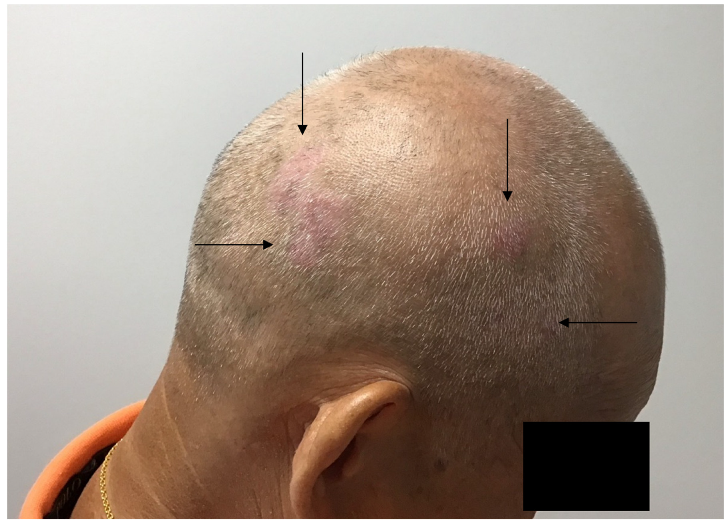
Figure 1. Psoriasis of the scalp is localized to hair bearing areas of the scalp (see arrows). Areas of pattern alopecia are spared from inflammation.
Preprints 117314 g001
Figure 2. Physical protection of scalp prevents carcinogenesis in areas of alopecia. This patient with extensive solar damage has squamous cell carcinoma on the scalp that is not protected by his headcovering (kipah), and clinically less solar damage on the covered area. The kipah is worn lifelong in males, and thus may have prevented UV carcinogenesis.
Preprints 117314 g002
Figure 3. Mechanism of immunometabolic regulation of scalp carcinogenesis. In the normal scalp, IRF4 transcription factor is highly expressed in hair follicles, and a stroma promotes healthy hair. UV carcinogenesis successfully causes mutation in hair bearing areas, but hair derived IL-17 and Sirt3 high stroma promotes hair growth and impedes tumor promotion. With hair loss, the tonic IRF4/IL-17 production is lost, and stroma becomes elevated in TGF beta and low in Sirt3, proving a permissive environment for tumor progression.
Preprints 117314 g003
Disclaimer/Publisher’s Note: The statements, opinions and data contained in all publications are solely those of the individual author(s) and contributor(s) and not of MDPI and/or the editor(s). MDPI and/or the editor(s) disclaim responsibility for any injury to people or property resulting from any ideas, methods, instructions or products referred to in the content.
Copyright: This open access article is published under a Creative Commons CC BY 4.0 license, which permit the free download, distribution, and reuse, provided that the author and preprint are cited in any reuse.
Prerpints.org logo

Preprints.org is a free preprint server supported by MDPI in Basel, Switzerland.

Subscribe

© 2024 MDPI (Basel, Switzerland) unless otherwise stated