Preprint
Article

Shedding Light on tRNA-Derived Fragment Repertoire in Facs Blood Cells

Altmetrics

Downloads

59

Views

60

Comments

0

A peer-reviewed article of this preprint also exists.

Submitted:

05 September 2024

Posted:

05 September 2024

You are already at the latest version

Alerts
Abstract
tRNA-derived fragments function as markers additionally to playing the role key of signalling molecules in a number of disorders.  It is known that the repertoire of these molecules differs greatly in different cell types and varies depending on the physiological condition. The aim of our research was to compare the pattern of tRF expression in the main blood cell types and to determine how the composition of these molecules changes during COVID-19-induced cytokine storm. Erythrocytes, monocytes, lymphocytes, neutrophils, basophils, and eosinophils from control donors and patients with severe COVID-19 were obtained by fluorescence sorting. We extracted RNA from FACS sorted cells and performed NGS of short RNAs. The composition of tRNA-derived fragments was analysed applying a semi-custom bioinformatic pipeline. In this study, we assessed the length and type distribution of TRFs and reported the 150 most prevalent TRF sequences across all cell types. Additionally we demonstrate a significant (p<0.05, fold change>16) change in the pattern of tRFs in erythrocytes (21 downregulated, 12 upregulated), monocytes (53 downregulated, 38 upregulated) and lymphocytes (49 upregulated) in patients with severe COVID-19. Thus, different blood cell types exhibit a significant variety of TRFs and react to cytokine storm by dramatically changing their differential expression patterns. We suppose that the observed phenomenon occurs due to the regulation of nucleotide modifications and alterations in activity of various Rnases.
Keywords: 
Subject: Biology and Life Sciences  -   Biochemistry and Molecular Biology

1. Introduction

tRNA-derived fragments (tRFs) are a relatively recently discovered class of small RNA molecules that are shown to be involved in the pathogenesis of various diseases, particularly cancer and viral infections. Processed from transfer RNAs, tRFs regulate gene expression through various mechanisms like miRNA-like silencing of target mRNAs [1,2,3,4].
There are several types of classified tRFs based on the cleavage site within the tRNA molecule: tRF-1: Cleaved from the 3' trailer sequence of pre-tRNAs. tRF-2: Cleaved from the D-loop of mature tRNAs. tRF-3: Cleaved from the 3' end of mature tRNAs. tRF-5: Cleaved from the 5' end of mature tRNAs. i-tRF: Internal tRFs derived from the internal region of mature tRNAs [5].
Different tissues and organs possess a different set of tRNA fragments [2]. Nevertheless, it has not been studied how the pattern of these molecules differs in different blood cells. Supposably, the repertoire of these molecules changes in correspondence with changes in the immunological state of patients. In particular, it is not clear how these molecules will change in blood cells during the cytokine storm induced by COVID-19.
It has been previously reported by Wu et. al. that tRFs were the most significantly affected small non-coding RNAs in nasopharyngeal swabs of COVID-19 patients, they had also observed that SARS-CoV-2-infected airway epithelial cells exhibit the same tendency [4]. In an attempt to additionally shed light on tRNA-derived fragments and their involvement in host-virus interactions it seems relevant to study tRFs in different pathophysiological circumstances. In the case of this study our aim was to reveal the differential expression of tRFs in fluorescence activated sorted cells of healthy control donors and SARS-CoV-2 infected patients, as well as to observe and characterise tRFs during viral infections.

2. Materials and Methods

2.1. Patients and Data Collection

Six healthy donors and five RT-PCR confirmed SARS-CoV-2 positive patients made up the two groups of research participants. Three patients were hospitalised to the critical care unit and two patients were moved to the infectious disease unit. Severe patients satisfied the following criteria for admission to the intensive care unit (ICU): body temperature ≥ 39 °C, respiration rate ≥ 30/min, and oxygen saturation (SpO2) ≤ 93%.

2.2. Cell Sorting

Сell sorting was performed on a MoFlow Astrios EQ device (Beckman Coulter). Erythrocytes were isolated using antibodies against CD235 protein (Beckman Coulter , IM2212U). To isolate leukocytes, erythrocytes were first lysed with VersaLyse lysing solution (Beckman Coulter). Granulocytes (neutrophils, basophils and eosinophils) were isolated using duraclone IM granulocyte antibody panel (B88651, Beckman Coulter). Lymphocytes and monocytes were isolated using antibodies CD45 (A79392 Beckman Coulter), CD16 (6607118 Beckman Coulter), CD14 (A70204 Beckman Coulter, USA). Cell sample purity for all cell populations was >95% according to flow cytometry data. A detailed description of cell sorting protocols is provided in the following article [6].

2.3. RNA Separation, Library Preparation and Next Generation Sequencing

RNA was isolated\extracted using ExtractRNA reagent (Evrogen) according to the provided protocol. After extraction, RNA was dissolved in 10 µl RNAse free water. The quality of the obtained RNA was checked on a TapeStation (Agilent). Only samples with RIN>5 were taken for sequencing. Short RNA libraries were prepared using the Small RNA Library Prep Kit (BGI, 1000006383). Sequencing was performed on a DNBSEQ-G400 instrument (BGI)

2.4. Bioinformatics

A general design of the bioinformatic processing pipeline is presented in Figure 1.

Read Quality Control

For quality control we applied FastQC [7]. All datasets qualified quality control standards. Each FastQ file contained between 17 and 27 million reads.

Adapter Trimming

Adapter trimming was executed using Trimmomatic [8]. Long clipping sequence was set as AGTCGGAGGCCAAGCGGTCTTAGGAAGACAA. We opted to use optimal run settings for SE (single-end) reads (TruSeq3-SE:2:30:10 LEADING:3 TRAILING:3 SLIDINGWINDOW:4:15 MINLEN:20) which allowed us to minimise execution time with no data loss. As an input, trimmomatic takes the adapter.fa file, in which our adapter sequence is registered. For each FastQ file from 50 to 68% of the input reads were marked as dropped after running Trimmomatic.

Read Mapping, Count and Normalisation

Full genome alignment and transriptome-based pseudoalignment methods are not applicable for small-RNA-seq identification due to tRNA cleavage events, which led us to utilising "BLAST"(Blast Local Alignment Search Tool) [9].
We implemented BLAST through “tRNAExplorer" [10], a Python-based pipeline, optimised for tRF-profile analysis. In order to utilise tRNAExplorer, we needed a *.bed genome annotation file as a reference, which we would BLAST our reads *.fa files against. While there is an option to generate a custom annotation file, we used an already compiled grch.38 database which came within the tRNAExplorer package. To run the pipeline, we created project directories with sample lists individually for each type of cells and custom configuration file, with data directories paths and launch options. While tRNAExplorer supports trimming and QC, we omitted these steps due to previous data processing. As an input each run of the pipeline takes all the *.fa files with read data from the specified directory, list of samples, and path to database *.bed file. We automated these runs with another custom Python script. As a final result of data processing we obtained *.csv and *.tsv text files, containing BLAST run results for every sample, cleavage sites data and a collective read count table for all provided samples with annotations.
This data was already sufficient for differential expression analysis and visualisation, but we applied another extra step, running a custom Python script which organises data and performs statistical tests.

2.5. Statistics

Wilcoxon–Mann–Whitney test was used to perform statistical analysis on the data. P-values less than 0.05 were considered statistically significant. The p-values for the RNAseq volcano graphics were log converted to log10 (1.31) for data visualisation.

3. Results

3.1. tRF Length Distribution in Main Cell Types

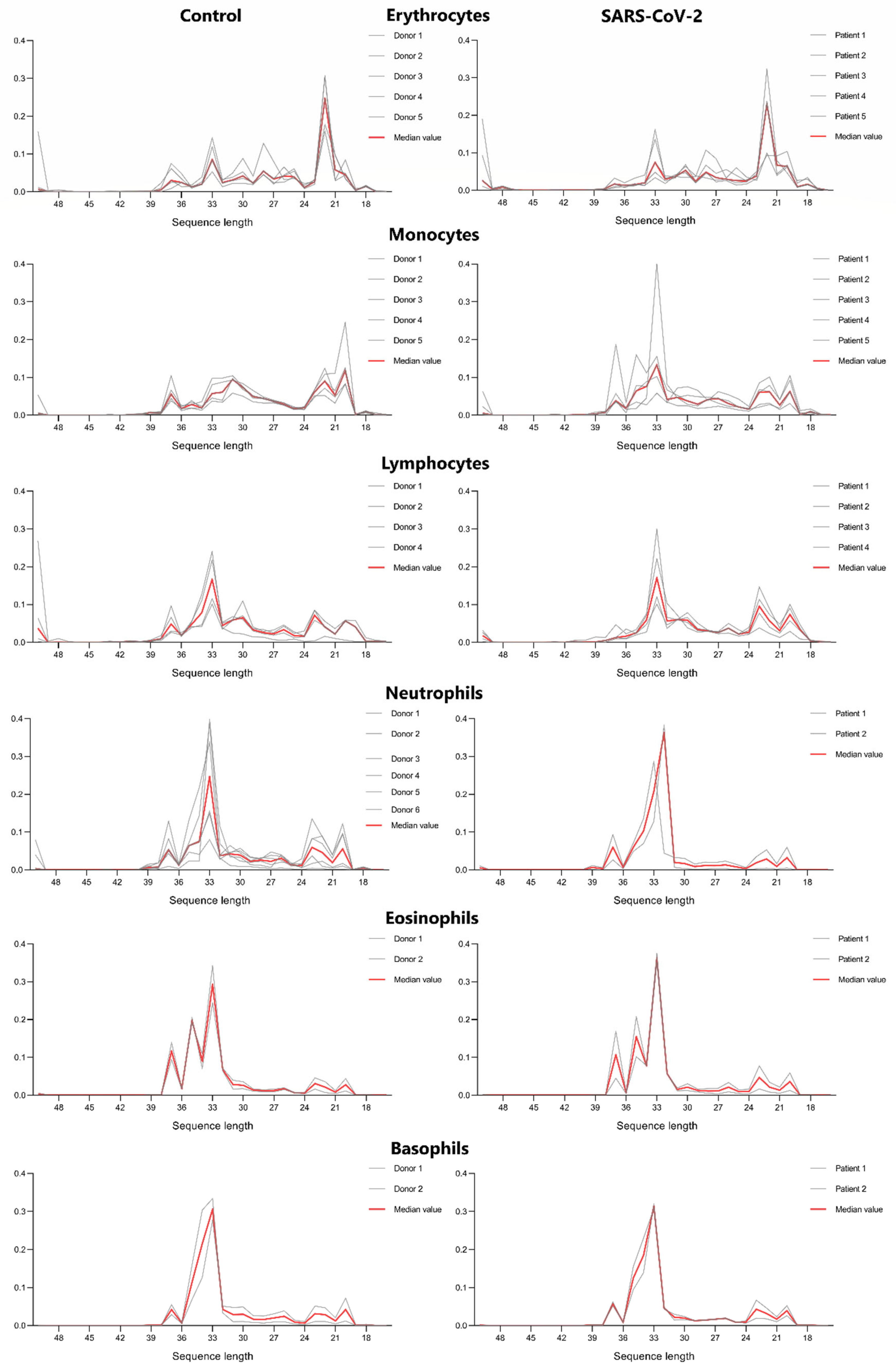
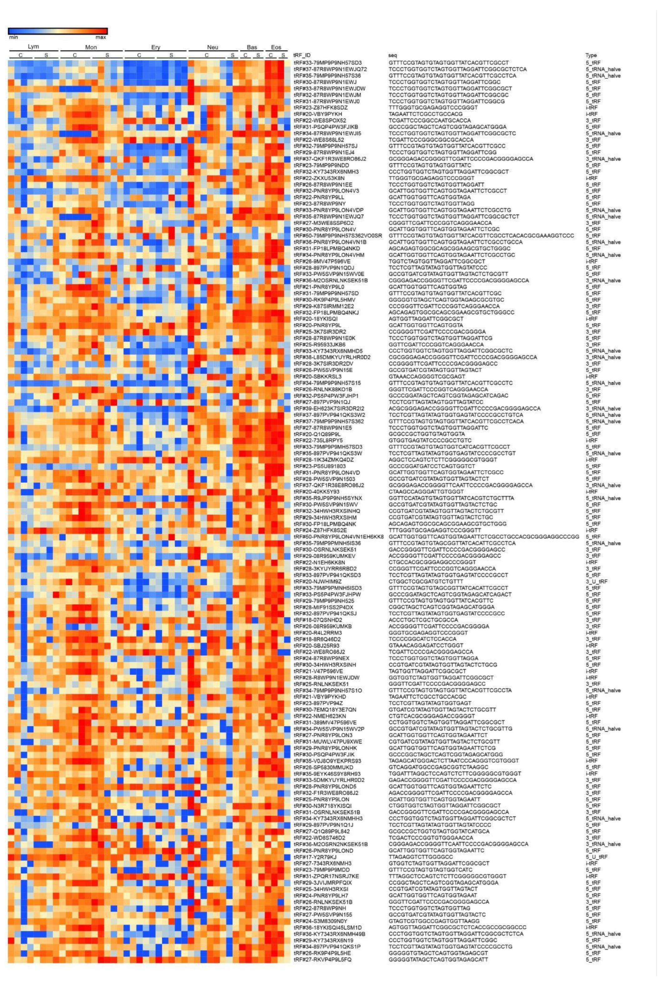
Analysing tRF length distribution in erythrocytes, monocytes, lymphocytes, neutrophils, basophils and eosinophils we observed a remarkable similarity between control and severe COVID-19 patient’s cells (Figure 2). This data is additionally supported by our heatmap, which demonstrated a distinguishable pattern of tRF expression depending on cell type. Erythrocytes are presented as the most distinguishable group of cells in terms of tRF length and tRF expression patterns, which may be due to the fact that the erythrocyte is an inactive cell in terms of synthetic processes.

3.2. tRF Type Composition in Main Cell Types

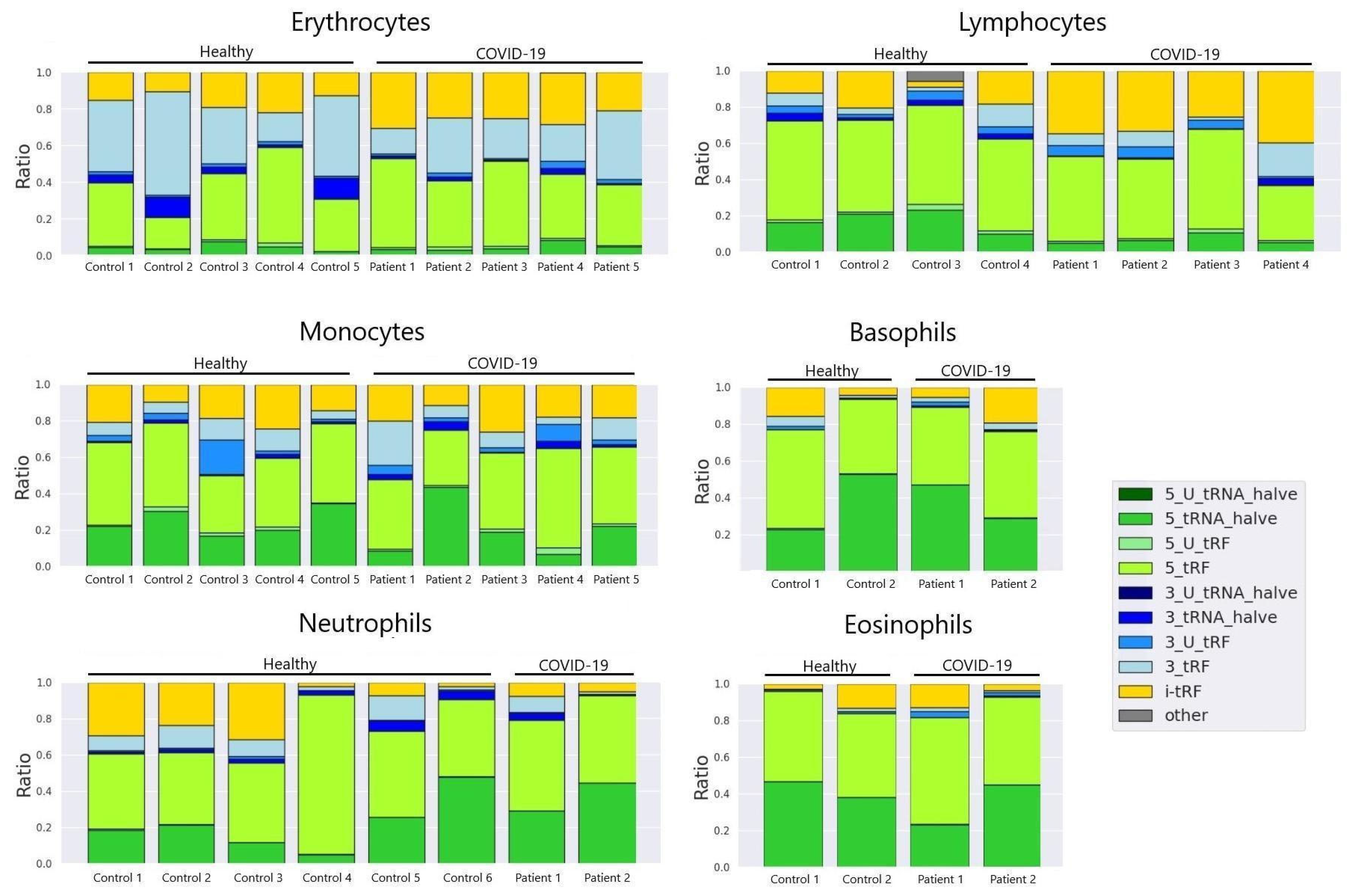
Analysing the main tRF types in sorted lymphocytes of control donors and severe COVID-19 patients we observed the prevalence of tRF-5 in 7 out of 8 samples. 1 severe COVID-19 patient exhibited dominating above all presence of i-tRF and tRF-3 types (Figure 3). It is worth noting that this patient had extremely high levels of IL-6 - 2 398 (pg/ml) on the same day that this sample was taken.

3.3. tRF Expression in Control Donors and Severe COVID-19 Patients

To compare tRF expression in different cell types we calculated the 150 most abundant tRFs based on RAW read counts, log normalised for data presentation. The heatmap demonstrates a relatively vivid frontier for erythrocytes (Figure 4).

3.4. tRF Differential Expression in Erythrocytes

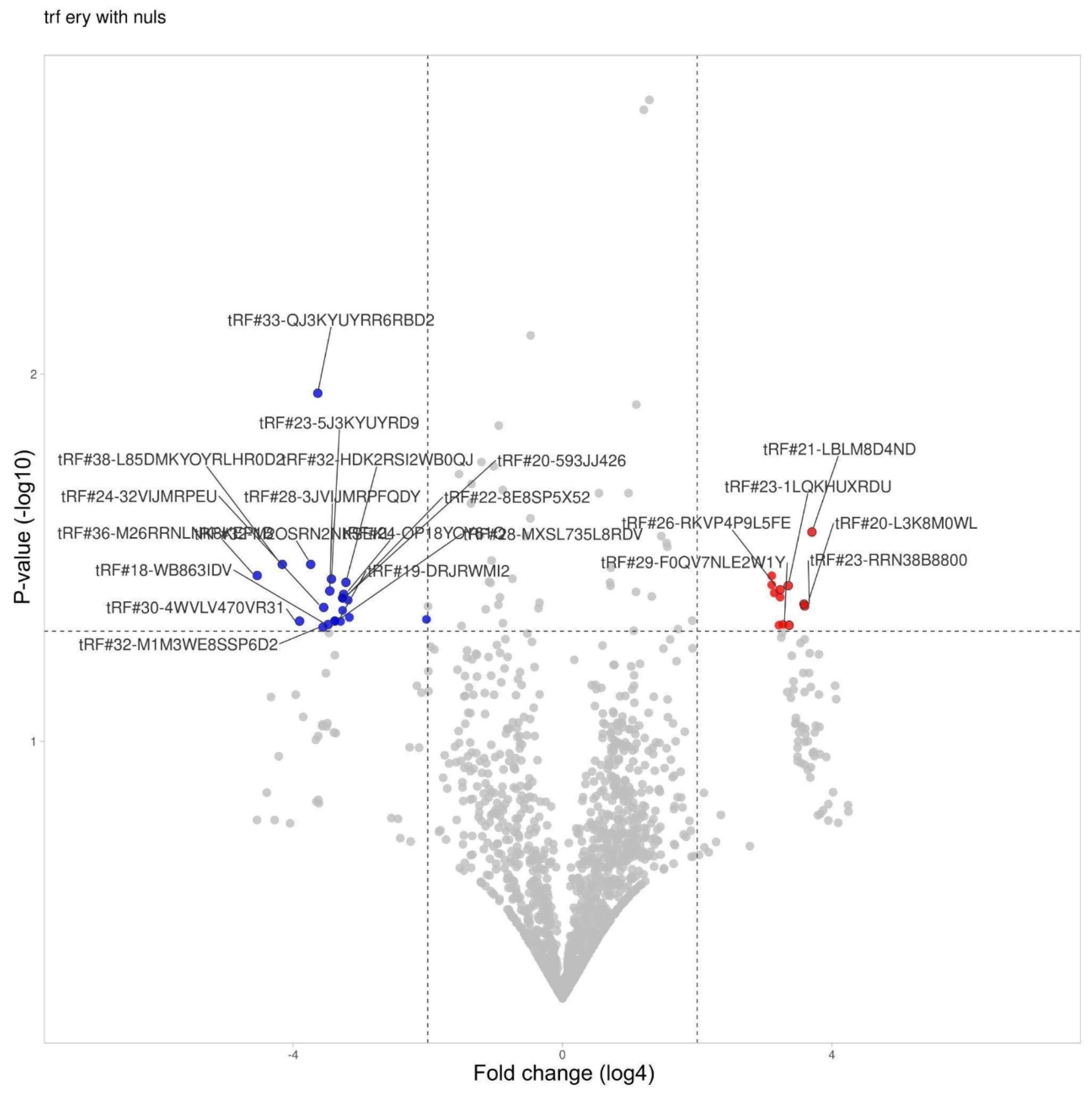
Analysing the expression of tRFs in sorted erythrocytes we observed 12 upregulated and 21 downregulated fragments (Figure 5, Table 1). tRF#33-QJ3KYUYRR6RBD2 stood out with significant downregulation, however, there is no data on the involvement of this fragment in any known biological processes.

3.5. tRF Differential Expression in Lymphocytes

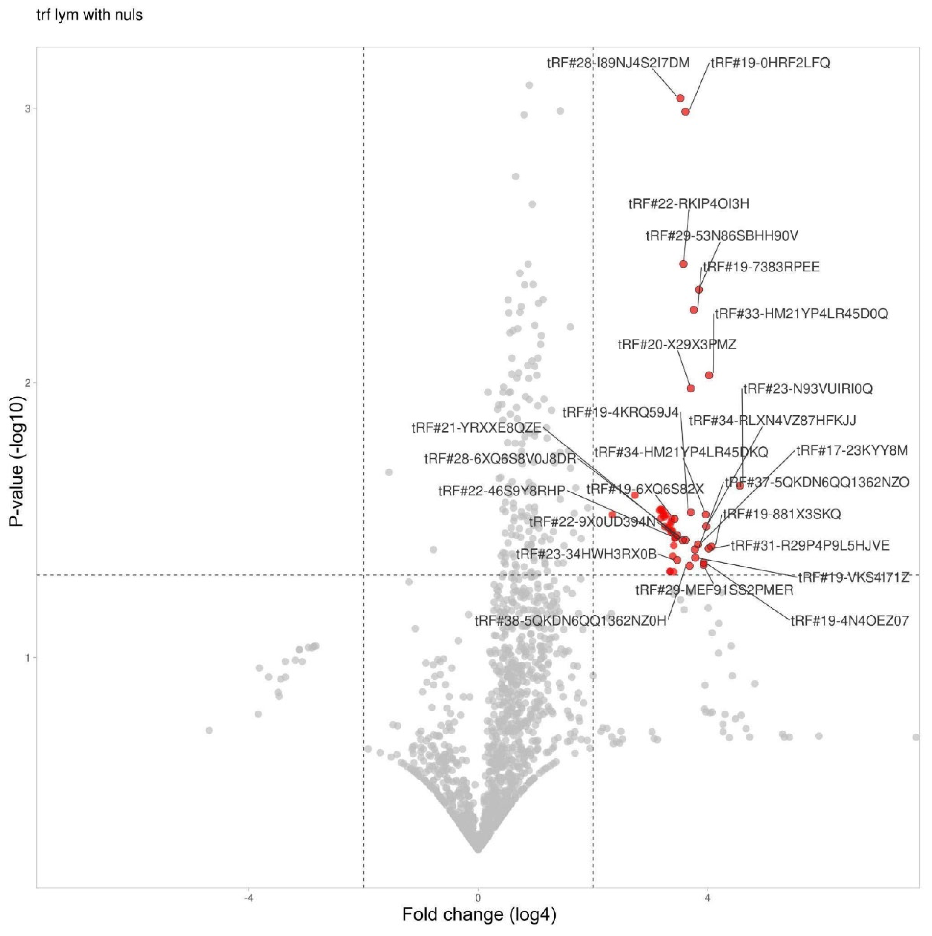
tRFs in lymphocytes demonstrated significant upregulation with 49 upregulated fragments, and zero downregulated, which vividly sets lymphocytes apart from other cells (Table 2). We applied log4 transformation for data presentation (Figure 6).

3.6. tRF Differential Expression in Monocytes

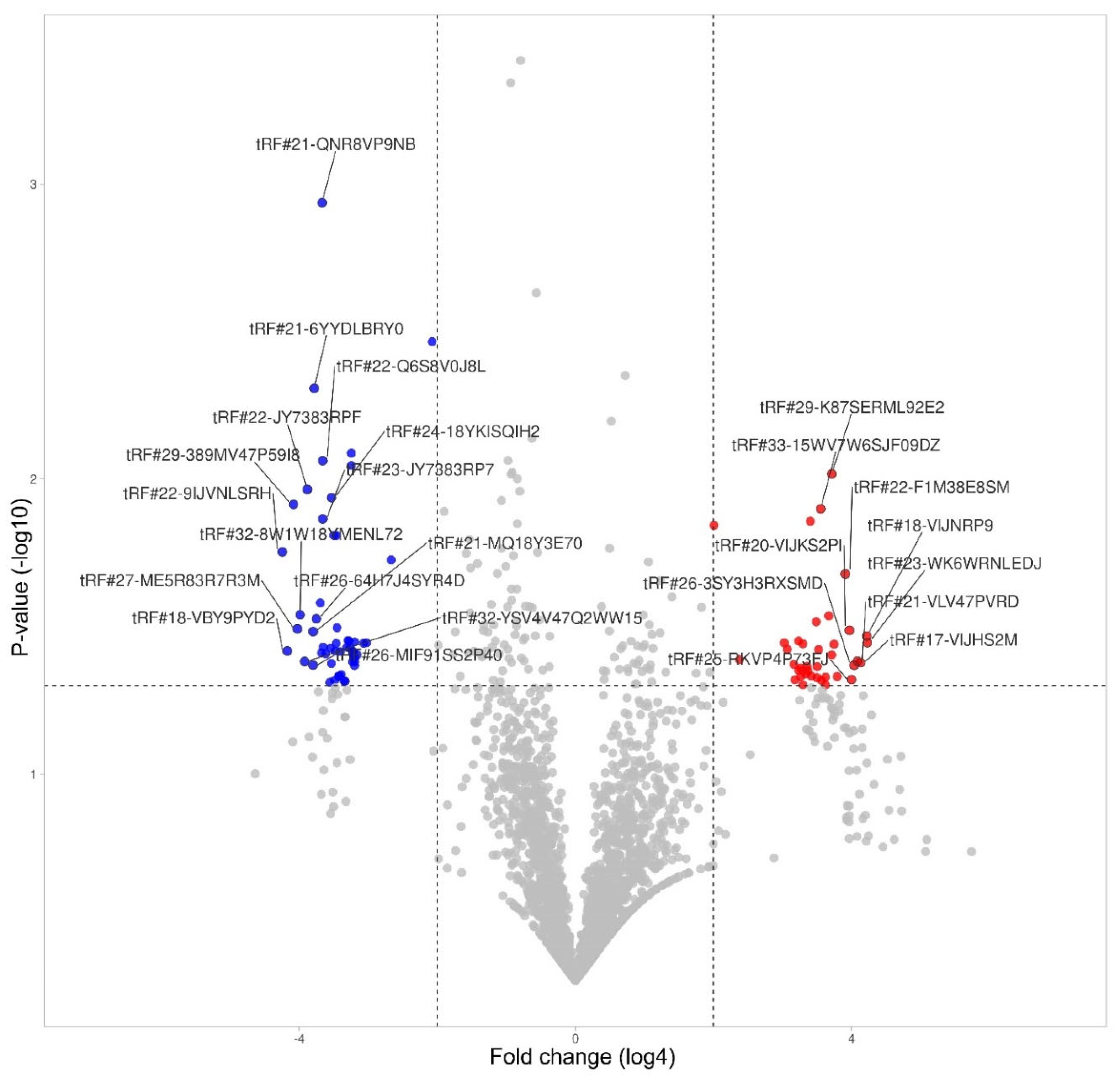
Monocytes were presented with the largest overall amount of significant hits. 38 upregulated tRFs and 53 downregulated (Figure 7, Table 3).

4. Discussion

This pilot study aimed at demonstrating a distinguishable tRF pattern for different sorted cell types. Given the fact that tRFs have been discovered relatively recently, research aimed at understanding their origin can help elucidate their biological purposes.
Analysing tRF length distribution in erythrocytes, monocytes, lymphocytes, neutrophils, basophils and eosinophils we observed a remarkable similarity between control and severe COVID-19 patient’s cells (Figure 2). This data is additionally supported by our heatmap (Figure 4), which demonstrated a distinguishable pattern of tRF expression depending on cell type. Erythrocytes are presented as the most distinguishable group of cells in terms of tRF length and tRF expression patterns, which may be due to the fact that the erythrocyte is an inactive cell in terms of synthetic processes.
Regarding tRF type composition, It has been previously noted that 3-tRFs are produced in response to various cellular stresses like oxidative stress, hypoxia, and viral infection [11]. Some 3-tRFs can inhibit viral replication by interfering with viral gene expression or packaging. They can act as signalling molecules to mediate stress responses [12,13].
Given the fact that IL-6 is a key mediator of the "cytokine storm" that leads to acute respiratory distress syndrome (ARDS) and multi-organ dysfunction in severe COVID-19 [14], it is assumable that such distribution of tRF types may be due the condition the patient underwent. Other cell types did not exhibit any significant alterations in tRF composition.
Overlooking tRF expression in control donors and severe COVID-19 patients, it is worth noting how the expression is increased in the majority of tRF in granulocytes (neutrophils, basophils, eosinophils). A similar tendency to increased expression is observed in monocytes. Granulocytes and monocytes both originate from CFU-GM (Colony Forming Unit–Granulocyte–Macrophage), also known as granulocyte–macrophage progenitor (GMP) [15].
The aforementioned suggests that tRFs may dominantly persist from the progenitor stages of cell development, and are only slightly modified by environmental or physiological factors.
Most knowledge today regarding tRFs is concentrated around cancer research [2,16,17,18].
In our study we encountered fragments which have been previously associated with several types of cancer.
Lymphocytic tRFs: It is noted that tRF#31-R29P4P9L5HJVE previously has been acknowledged as a marker for lung cancer prediction among smokers 10 years prior to being diagnosed [19].
tRF#19-VKS4I71Z has been mentioned as an abundant novel-trf in a 2016 study of RNA-seq data from human prostate tissue [20].
In a 2023 research dedicated to studying tRFs in cancer, a high level tRF#34-5QKDN6QQ1362HQ has been mentioned as a predictor of breast cancer improved survival [2].
Monocytic tRFs: tRF#22-WEK6S1852 in previous research was found to be significantly downregulated and associated with human malignant mesothelioma [16].
A 2019 colon cancer study revealed significantly differentially expressed tRFs between colon cancer tissues and peritumor tissues, whereas another upregulated tRF that we observed - tRF#22-9LON4VN11 was mentioned, demonstrating downregulation in colon cancer tissues with a log2 fold change of -1.26 [17].
Another fragment we observed - tRF#18-HSQSD2D2, downregulation of which has been previously associated with early-stage breast cancer [2].
In a study devoted to examining the dysregulation of different tRFs in chronic lymphocytic leukemia - tRF-20-RK9P4P9L was amongst the top 15 differentially expressed sRNAs in aggressive chronic lymphocytic leukemia vs. normal controls, with the linear fold change being −76.64 and −258.08, respectively. Samples were composed of CD5+/CD19+ B cells. It is worth noting that in our study, the same tRF was also significantly downregulated but only in CD14++ CD16− monocytes [18].
tRF#22-WEPSJR852 was found in peripheral blood of fibromyalgia patients in a dissertation dedicated to the search of morphological substrate to fibromyalgia [21].
Erythrocytes appeared to be the only cell type which showed differential expression of tRFs that have not previously been associated with any type of cancer or disease. The fact that the erythrocyte is an inactive cell in terms of synthetic processes may elucidate these findings, however, regarding different cell types, tRFs and their interrelations with the conditions mentioned earlier it is more likely to suppose a coincidence, rather than some significant finding.
Differences in the length, type, and composition of specific tRFs between different cells are supposedly due to the specificity of the tRNA cleavage system. The same can be said about changes that occur during cytokine storm. The regulation system of tRNA processing is still poorly understood.
Nonetheless, we can say that it occurs at several “levels”. First of all - the regulation of nucleotide modifications in certain molecules [22]. Another stage is dependent on the regulation of RNAses that cleave tRNAs at specific sites.
The main types of nucleases involved in tRF biogenesis are: Dicer - cleaves 5' ends of mature tRNAs. Angiogenin (ANG) - cleaves mature tRNAs at anticodon loops. RNase Z/ELAC2 - cleaves 3' trailer sequences of pre-tRNAs [11,23,24].
RNase P: excision of external transcribed spacer (ETS) and internal transcribed spacers (ITS) from pre-tRNA transcripts [25].
Regulation of these enzymes can occur at the level of transcription initiation, as well as post-translational modifications of the enzyme
It is also likely that tRNA cleavage is regulated by proteins that bind this molecule [26] and make certain sites inaccessible for the aforementioned process. Unfortunately, we cannot state by what mechanism the difference between tRFs in blood cells is regulated.
In summary, it should be noted that TRF profiles significantly differ in different types of blood cells and demonstrate dramatic differential expression (sometimes more than 500-fold) during cytokine storm. Such profound differences suggest a major role of tRNA-derived fragments in the functioning of blood cells.

5. Conclusions

The composition of tRFs in erythrocytes monocytes, lymphocytes, neutrophils, basophils and eosinophils was analysed on the basis of sRNA-SEQ. We demonstrate notable alterations in the length and types of these molecules in main blood cell populations. We additionally observed a significant change in the profile of tRFs in the erythrocytes, monocytes and lymphocytes of patients infected with SARS-CoV-2.

Supplementary Materials

The following supporting information can be downloaded at the website of this paper posted on Preprints.org. All RAW read data and nucleotide composition of tRFs are presented in two supplementary files: “Control cells” for control donors and “COVID cells” for patients with severe COVID-19.

Funding

This work was supported by St. Petersburg State University, project ID: 95412780.

Institutional Review Board Statement

This study was conducted according to the guidelines of the declaration of Helsinki and approved by the ethics committee.

Informed Consent Statement

Informed consent was obtained from all subjects involved in the study.

Conflicts of Interest

The authors declare no conflict of interest.

References

  1. Kim, V.N. Small RNAs: Classification, Biogenesis, and Function. Molecules and Cells 2005, 19, 1–15. [Google Scholar] [CrossRef] [PubMed]
  2. Fu, M.; Gu, J.; Wang, M.; Zhang, J.; Chen, Y.; Jiang, P.; Zhu, T.; Zhang, X. Emerging Roles of tRNA-Derived Fragments in Cancer. Molecular Cancer 2023, 22, 30. [Google Scholar] [CrossRef] [PubMed]
  3. Krishna, S.; Raghavan, S.; DasGupta, R.; Palakodeti, D. tRNA-Derived Fragments (tRFs): Establishing Their Turf in Post-Transcriptional Gene Regulation. Cell. Mol. Life Sci. 2021, 78, 2607–2619. [Google Scholar] [CrossRef]
  4. Wu, W.; Choi, E.-J.; Wang, B.; Zhang, K.; Adam, A.; Huang, G.; Tunkle, L.; Huang, P.; Goru, R.; Imirowicz, I.; et al. Changes of Small Non-Coding RNAs by Severe Acute Respiratory Syndrome Coronavirus 2 Infection. Front. Mol. Biosci. 2022, 9. [Google Scholar] [CrossRef]
  5. Yu, X.; Xie, Y.; Zhang, S.; Song, X.; Xiao, B.; Yan, Z. tRNA-Derived Fragments: Mechanisms Underlying Their Regulation of Gene Expression and Potential Applications as Therapeutic Targets in Cancers and Virus Infections. Theranostics 2021, 11, 461–469. [Google Scholar] [CrossRef] [PubMed]
  6. Kondratov, K.A.; Artamonov, A.A.; Nikitin, Y.V.; Velmiskina, A.A.; Mikhailovskii, V.Y.; Mosenko, S.V.; Polkovnikova, I.A.; Asinovskaya, A.Y.; Apalko, S.V.; Sushentseva, N.N.; et al. Revealing Differential Expression Patterns of piRNA in FACS Blood Cells of SARS-CoV−2 Infected Patients. BMC Medical Genomics 2024, 17, 212. [Google Scholar] [CrossRef]
  7. Andrews, S. FastQC: A Quality Control Tool for High Throughput Sequence Data. [Online] 2010. Available online at: http://www.bioinformatics.babraham.ac.uk/projects/fastqc/.
  8. Bolger, A.M.; Lohse, M.; Usadel, B. Trimmomatic: A Flexible Trimmer for Illumina Sequence Data. Bioinformatics 2014, 30, 2114–2120. [Google Scholar] [CrossRef]
  9. Altschul, S.F.; Gish, W.; Miller, W.; Myers, E.W.; Lipman, D.J. Basic Local Alignment Search Tool. J Mol Biol 1990, 215, 403–410. [Google Scholar] [CrossRef]
  10. Sun, Z.; Tan, J.; Zhao, M.; Peng, Q.; Zhou, M.; Zuo, S.; Wu, F.; Li, X.; Dong, Y.; Xie, M.; et al. Integrated Genomic Analysis Reveals Regulatory Pathways and Dynamic Landscapes of the tRNA Transcriptome. Sci Rep 2021, 11, 5226. [Google Scholar] [CrossRef]
  11. Zhang, S.; Yu, X.; Xie, Y.; Ye, G.; Guo, J. tRNA Derived Fragments:A Novel Player in Gene Regulation and Applications in Cancer. Front Oncol 2023, 13, 1063930. [Google Scholar] [CrossRef]
  12. Kumar, P.; Kuscu, C.; Dutta, A. Biogenesis and Function of Transfer RNA Related Fragments (tRFs). Trends Biochem Sci 2016, 41, 679–689. [Google Scholar] [CrossRef]
  13. Gong, M.; Deng, Y.; Xiang, Y.; Ye, D. The Role and Mechanism of Action of tRNA-Derived Fragments in the Diagnosis and Treatment of Malignant Tumors. Cell Communication and Signaling 2023, 21, 62. [Google Scholar] [CrossRef]
  14. McGonagle, D.; Sharif, K.; O’Regan, A.; Bridgewood, C. The Role of Cytokines Including Interleukin-6 in COVID-19 Induced Pneumonia and Macrophage Activation Syndrome-Like Disease. Autoimmun Rev 2020, 19, 102537. [Google Scholar] [CrossRef]
  15. Guzzi, N.; Cieśla, M.; Ngoc, P.C.T.; Lang, S.; Arora, S.; Dimitriou, M.; Pimková, K.; Sommarin, M.N.E.; Munita, R.; Lubas, M.; et al. Pseudouridylation of tRNA-Derived Fragments Steers Translational Control in Stem Cells. Cell 2018, 173, 1204–1216.e26. [Google Scholar] [CrossRef]
  16. Filetti, V.; La Ferlita, A.; Di Maria, A.; Cardile, V.; Graziano, A.C.E.; Rapisarda, V.; Ledda, C.; Pulvirenti, A.; Loreto, C. Dysregulation of microRNAs and tRNA-Derived ncRNAs in Mesothelial and Mesothelioma Cell Lines after Asbestiform Fiber Exposure. Sci Rep 2022, 12, 9181. [Google Scholar] [CrossRef]
  17. Xiong, W.; Wang, X.; Cai, X.; Xiong, W.; Liu, Y.; Li, C.; Liu, Q.; Qin, J.; Li, Y. Identification of tRNAderived Fragments in Colon Cancer by Comprehensive Small RNA Sequencing. Oncology Reports 2019, 42, 735–744. [Google Scholar] [CrossRef]
  18. Veneziano, D.; Tomasello, L.; Balatti, V.; Palamarchuk, A.; Rassenti, L.Z.; Kipps, T.J.; Pekarsky, Y.; Croce, C.M. Dysregulation of Different Classes of tRNA Fragments in Chronic Lymphocytic Leukemia. Proc Natl Acad Sci U S A 2019, 116, 24252–24258. [Google Scholar] [CrossRef]
  19. Umu, S.U.; Langseth, H.; Zuber, V.; Helland, Å.; Lyle, R.; Rounge, T.B. Serum RNAs Can Predict Lung Cancer up to 10 Years Prior to Diagnosis. eLife 2022, 11, e71035. [Google Scholar] [CrossRef]
  20. Olvedy, M.; Scaravilli, M.; Hoogstrate, Y.; Visakorpi, T.; Jenster, G.; Martens-Uzunova, E.S. A Comprehensive Repertoire of tRNA-Derived Fragments in Prostate Cancer. Oncotarget 2016, 7, 24766–24777. [Google Scholar] [CrossRef]
  21. Erbacher, C. Systemic and Local Mechanisms of Small Fiber Pathology in Female Patients with Fibromyalgia Syndrome, Universität Würzburg, 2023.
  22. Lyons, S.M.; Fay, M.M.; Ivanov, P. The Role of RNA Modifications in the Regulation of tRNA Cleavage. FEBS Letters 2018, 592, 2828–2844. [Google Scholar] [CrossRef]
  23. Kazimierczyk, M.; Wojnicka, M.; Biała, E.; Żydowicz-Machtel, P.; Imiołczyk, B.; Ostrowski, T.; Kurzyńska-Kokorniak, A.; Wrzesinski, J. Characteristics of Transfer RNA-Derived Fragments Expressed during Human Renal Cell Development: The Role of Dicer in tRF Biogenesis. International Journal of Molecular Sciences 2022, 23, 3644. [Google Scholar] [CrossRef]
  24. Liu, B.; Cao, J.; Wang, X.; Guo, C.; Liu, Y.; Wang, T. Deciphering the tRNA-Derived Small RNAs: Origin, Development, and Future. Cell Death Dis 2021, 13, 1–13. [Google Scholar] [CrossRef]
  25. Li, Z.; Stanton, B.A. Transfer RNA-Derived Fragments, the Underappreciated Regulatory Small RNAs in Microbial Pathogenesis. Front. Microbiol. 2021, 12. [Google Scholar] [CrossRef]
  26. Karanasios, E.; Simos, G. Building Arks for tRNA: Structure and Function of the Arc1p Family of Non-Catalytic tRNA-Binding Proteins. FEBS Letters 2010, 584, 3842–3849. [Google Scholar] [CrossRef]
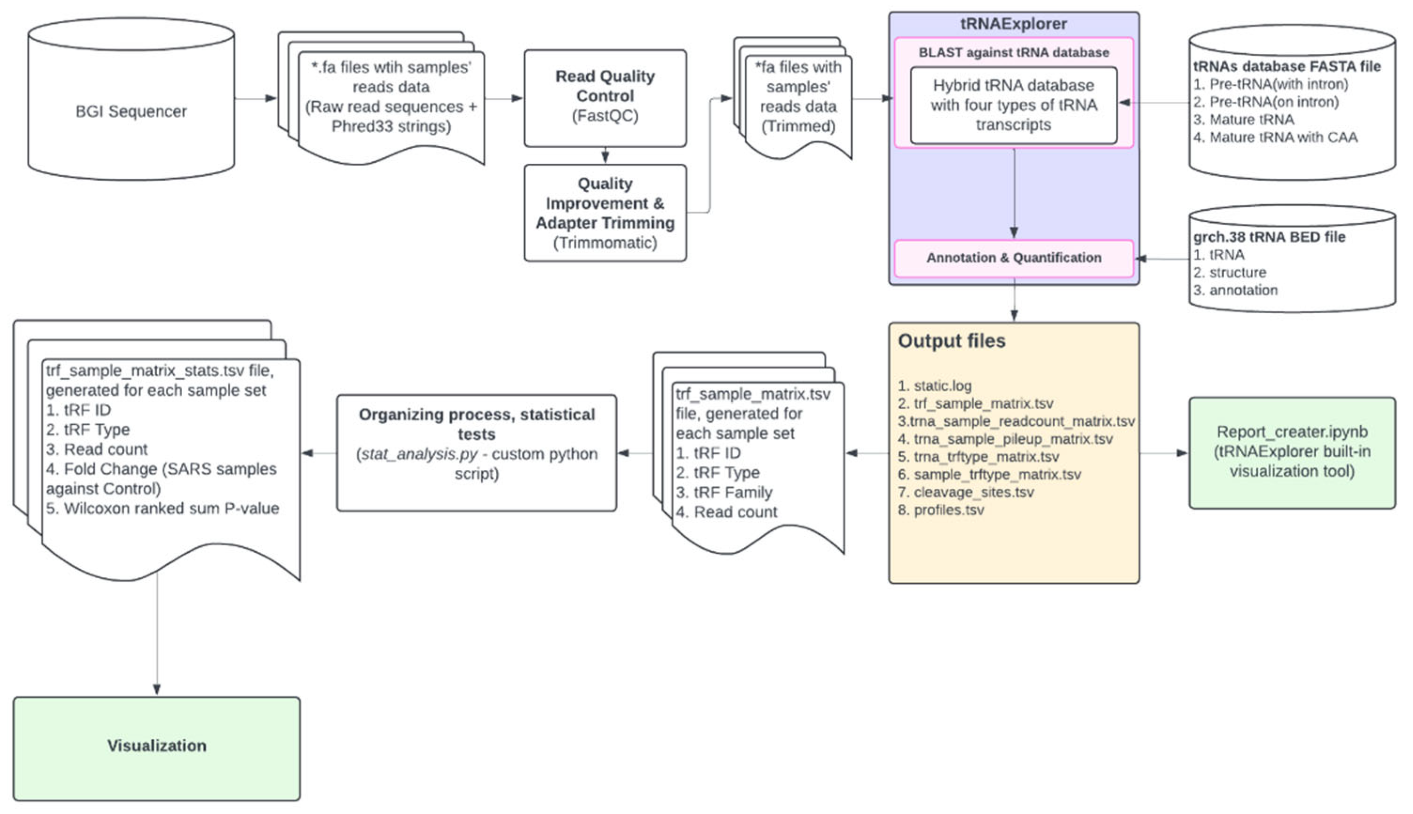
Figure 1. NGS data analysis pipeline.
Preprints 117397 g001
Figure 2. tRF length distribution in main cell types (red line represents median value).
Preprints 117397 g002
Figure 3. tRF type composition in main cell types (colour matches tRF types).
Preprints 117397 g003
Figure 4. tRF expression in control donors (C) and severe COVID-19 (S) patients. (Lym - lymphocytes, Mon - monocytes, Ery - erythrocytes, Neu - neutrophils, Bas - basophils, Eos - eosinophils).
Preprints 117397 g004
Figure 5. tRF differential expression in erythrocytes (For all volcano plots we applied log4 (instead of log2) for fold change presentation.).
Preprints 117397 g005
Figure 6. tRF differential expression in lymphocytes.
Preprints 117397 g006
Figure 7. tRF differential expression in monocytes.
Preprints 117397 g007
Table 1. List of differentially expressed tRNA-derived fragments in erythrocytes during severe COVID-19.
Table 21. Change Fold change P-value
tRF#21-LBLM8D4ND Increased 170 0,027
tRF#23-RRN38B8800 Increased 146 0,043
tRF#20-L3K8M0WL Increased 144 0,042
tRF#29-F0QV7NLE2W1Y Increased 106 0,048
tRF#23-1LQKHUXRDU Increased 104 0,038
tRF#21-SRRN2EK8B Increased 94 0,048
tRF#26-RKVP4P9L5FE Increased 88 0,039
tRF#17-M3WE8SN Increased 88 0,040
tRF#21-8PR9DM3WE Increased 86 0,048
tRF#23-6KK87SIRD4 Increased 78 0,039
tRF#33-73NK7F6Z2DNLDW Increased 74 0,035
tRF#31-79MP9P9MH57SD Increased 74 0,037
tRF#36-M26RRNLNK8KEP1B Decreased 536 0,035
tRF#38-L85DMKYOYRLHR0D2 Decreased 320 0,033
tRF#30-4WVLV470VR31 Decreased 224 0,047
tRF#32-M2OSRN2NKSEKL Decreased 178 0,033
tRF#33-QJ3KYUYRR6RBD2 Decreased 154 0,011
tRF#32-M1M3WE8SSP6D2 Decreased 138 0,049
tRF#24-32VIJMRPEU Decreased 136 0,043
tRF#18-WB863IDV Decreased 124 0,048
tRF#28-3JVIJMRPFQDY Decreased 120 0,039
tRF#23-5J3KYUYRD9 Decreased 116 0,036
tRF#19-DRJRWMI2 Decreased 108 0,047
tRF#28-MXSL735L8RDV Decreased 108 0,047
tRF#29-YP9LON4VN1EM Decreased 96 0,047
tRF#20-593JJ426 Decreased 92 0,041
tRF#22-8E8SP5X52 Decreased 92 0,041
tRF#20-PW5SVP9N Decreased 92 0,044
tRF#24-OP18YOY61Q Decreased 90 0,040
tRF#32-HDK2RSI2WB0QJ Decreased 86 0,037
tRF#21-1MQ8YUY60 Decreased 82 0,041
tRF#37-WYL1M3WE8S68L52 Decreased 80 0,046
tRF#28-3K76IR3DR2DV Decreased 16 0,047
Table 2. List of differentially expressed tRNA-derived fragments in lymphocytes during severe COVID-19.
tRF id Change Fold change P-value
tRF#23-N93VUIRI0Q Increased 557 0,024
tRF#19-881X3SKQ Increased 280 0,039
tRF#33-HM21YP4LR45D0Q Increased 265 0,009
tRF#31-R29P4P9L5HJVE Increased 263 0,040
tRF#34-RLXN4VZ87HFKJJ Increased 247 0,033
tRF#34-HM21YP4LR45DKQ Increased 245 0,030
tRF#29-MEF91SS2PMER Increased 233 0,046
tRF#19-4N4OEZ07 Increased 233 0,045
tRF#29-53N86SBHH90V Increased 207 0,005
tRF#17-23KYY8M Increased 203 0,039
tRF#19-VKS4I71Z Increased 190 0,043
tRF#37-5QKDN6QQ1362NZO Increased 188 0,040
tRF#19-7383RPEE Increased 183 0,005
tRF#19-4KRQ59J4 Increased 170 0,030
tRF#20-X29X3PMZ Increased 170 0,010
tRF#38-5QKDN6QQ1362NZ0H Increased 165 0,046
tRF#28-6XQ6S8V0J8DR Increased 150 0,037
tRF#19-0HRF2LFQ Increased 150 0,001
tRF#22-RKIP4OI3H Increased 143 0,004
tRF#22-9X0UD394N Increased 140 0,037
tRF#28-I89NJ4S2I7DM Increased 133 0,001
tRF#21-YRXXE8QZE Increased 123 0,036
tRF#23-34HWH3RX0B Increased 123 0,044
tRF#22-46S9Y8RHP Increased 118 0,037
tRF#19-P7M84I2Q Increased 115 0,037
tRF#19-6XQ6S82X Increased 115 0,031
tRF#20-1QKS3W2V Increased 113 0,049
tRF#19-MIF91S2H Increased 113 0,039
tRF#21-WRD81H93E Increased 110 0,043
tRF#21-9L5H52NL0 Increased 110 0,035
tRF#22-W60XY9BIQ Increased 108 0,031
tRF#23-R9J89O9N9 Increased 105 0,035
tRF#19-8NWE6WIZ Increased 105 0,035
tRF#28-Z3R918VBY9DV Increased 105 0,032
tRF#23-9M8O90Q4DZ Increased 103 0,049
tRF#22-Z3FJ6KEWH Increased 103 0,049
tRF#23-9N1QKS3WD1 Increased 103 0,033
tRF#22-7EMQ18Y31 Increased 98 0,034
tRF#21-7O3B1NR8E Increased 93 0,030
tRF#22-282K63ZNQ Increased 90 0,033
tRF#18-7383RP7 Increased 90 0,031
tRF#23-RPM8309M0F Increased 88 0,030
tRF#22-6LQ6S8V02 Increased 88 0,030
tRF#35-S4I7LZM3Q01M3K Increased 85 0,029
tRF#25-QSD2NSWWDZ Increased 83 0,031
tRF#23-1E6SF8WOD9 Increased 83 0,029
tRF#22-2EJ1OWZIQ Increased 80 0,029
tRF#34-5QKDN6QQ1362HQ Increased 44 0,026
tRF#35-5QKDN6QQ1362NZ Increased 25 0,030
Table 3. List of differentially expressed tRNA-derived fragments in monocytes during severe COVID-19.
tRF id Change Fold change P-value
tRF#23-WK6WRNLEDJ Increased 352 0,036
tRF#18-VIJNRP9 Increased 348 0,034
tRF#17-VIJHS2M Increased 306 0,042
tRF#21-VLV47PVRD Increased 288 0,041
tRF#26-3SY3H3RXSMD Increased 270 0,043
tRF#25-RKVP4P73FJ Increased 256 0,048
tRF#20-VIJKS2PI Increased 246 0,033
tRF#22-F1M38E8SM Increased 226 0,021
tRF#44-K8HJ83ML5F82NZD7HY Increased 192 0,047
tRF#25-RKILQ673FJ Increased 180 0,036
tRF#28-R1RXQ678Y2D8 Increased 172 0,039
tRF#29-K87SERML92E2 Increased 172 0,010
tRF#31-ZPQR16ZSIJ7KE Increased 162 0,029
tRF#22-WEK6S1852 Increased 152 0,047
tRF#23-K8HJ83MLDS Increased 152 0,050
tRF#18-VIJKS2DU Increased 140 0,048
tRF#33-15WV7W6SJF09DZ Increased 138 0,013
tRF#25-P21MNKYUPR Increased 132 0,038
tRF#38-Y2R79K3BEE4O3Q03 Increased 128 0,043
tRF#24-PS5U8918JP Increased 128 0,047
tRF#21-W60XY9BIE Increased 126 0,030
tRF#30-6Q46D6PUMZQZ Increased 114 0,046
tRF#22-9LON4VN11 Increased 112 0,014
tRF#34-IEWS7YRR50SRIZ Increased 106 0,045
tRF#18-HSQSD2D2 Increased 104 0,043
tRF#32-ZPQR17NSRJ7KQ Increased 102 0,046
tRF#26-R3HJ83RPFQE Increased 96 0,036
tRF#19-K876IR19 Increased 96 0,050
tRF#37-9EUK46S9Y8RH93Q Increased 92 0,044
tRF#20-9VWVEH93 Increased 92 0,047
tRF#28-WS3V2VR0PSDZ Increased 88 0,035
tRF#20-SVKKN27F Increased 88 0,044
tRF#32-FN5KYUSRYWRSJ Increased 82 0,048
tRF#27-WN1Q18Y3HRK Increased 80 0,042
tRF#20-3K7SIR3D Increased 70 0,038
tRF#22-WEPSJR852 Increased 66 0,036
tRF#21-MXSL73VLE Increased 27 0,041
tRF#25-QNR8ZP9LON Increased 16 0,014
tRF#22-9IJVNLSRH Decreased 360 0,018
tRF#18-VBY9PYD2 Decreased 326 0,038
tRF#29-389MV47P59I8 Decreased 288 0,012
tRF#27-ME5R83R7R3M Decreased 266 0,032
tRF#32-8W1W18YMENL72 Decreased 252 0,029
tRF#32-YSV4V47Q2WW15 Decreased 230 0,042
tRF#22-JY7383RPF Decreased 218 0,011
tRF#26-MIF91SS2P40 Decreased 194 0,043
tRF#21-MQ18Y3E70 Decreased 194 0,033
tRF#21-6YYDLBRY0 Decreased 190 0,005
tRF#26-64H7J4SYR4D Decreased 182 0,030
tRF#21-MUWLV47PE Decreased 168 0,026
tRF#21-R84QPYVMD Decreased 164 0,039
tRF#21-QNR8VP9NB Decreased 162 0,001
tRF#22-Q6S8V0J8L Decreased 160 0,009
tRF#23-JY7383RP7 Decreased 160 0,014
tRF#33-1MN0YU09FKRFD2 Decreased 158 0,037
tRF#22-9P9NH57SJ Decreased 150 0,039
tRF#23-RXSINHZ4DV Decreased 138 0,049
tRF#24-941QKS3WF8 Decreased 136 0,037
tRF#20-J87383RP Decreased 134 0,042
tRF#24-18YKISQIH2 Decreased 134 0,012
tRF#38-ML5F924ZDRJKW4DZ Decreased 128 0,016
tRF#21-9P4P9L5HE Decreased 126 0,048
tRF#24-Z3RQ18YJFH Decreased 124 0,038
tRF#23-J87383RP7 Decreased 122 0,036
tRF#20-RK9P4P9L Decreased 122 0,016
tRF#22-MQ18Y3E7M Decreased 120 0,032
tRF#28-RUPLQVNRDF0E Decreased 116 0,047
tRF#22-73H3RXPLM Decreased 114 0,046
tRF#24-94SX73V2KK Decreased 112 0,038
tRF#21-MIF91SS20 Decreased 110 0,046
tRF#24-04SXQ3V2KJ Decreased 104 0,048
tRF#21-9LV470JPD Decreased 102 0,048
tRF#20-1PSJPM17 Decreased 98 0,037
tRF#34-JY7383RPD9W1JV Decreased 96 0,035
tRF#24-YDLBRY73JL Decreased 94 0,035
tRF#21-3P47M26YB Decreased 92 0,037
tRF#22-6YR29P4PP Decreased 90 0,008
tRF#21-WB8689SVD Decreased 90 0,009
tRF#21-WLV47PU9E Decreased 88 0,039
tRF#21-N1EH6KK80 Decreased 88 0,042
tRF#18-SR99RHD2 Decreased 86 0,038
tRF#33-5F924ZDRJKW4DZ Decreased 86 0,039
tRF#22-5721V98B3 Decreased 84 0,036
tRF#26-MY73H3RXPL0 Decreased 84 0,040
tRF#22-J4S2I7L7M Decreased 84 0,042
tRF#28-RKVP4P9L5F0Q Decreased 84 0,043
tRF#26-94SL735FVI0 Decreased 80 0,039
tRF#18-6M0Y1MY Decreased 70 0,036
tRF#24-MY73H3RXII Decreased 66 0,036
tRF#33-1N3KYUSR681SD2 Decreased 40 0,019
tRF#25-R8VP9NFQFY Decreased 18 0,003
Disclaimer/Publisher’s Note: The statements, opinions and data contained in all publications are solely those of the individual author(s) and contributor(s) and not of MDPI and/or the editor(s). MDPI and/or the editor(s) disclaim responsibility for any injury to people or property resulting from any ideas, methods, instructions or products referred to in the content.
Copyright: This open access article is published under a Creative Commons CC BY 4.0 license, which permit the free download, distribution, and reuse, provided that the author and preprint are cited in any reuse.
Prerpints.org logo

Preprints.org is a free preprint server supported by MDPI in Basel, Switzerland.

Subscribe

© 2024 MDPI (Basel, Switzerland) unless otherwise stated