1. Introduction
Polarized Light Microscopy (PLM) is a fundamental optical technique for analyzing the optical properties and structures of anisotropic materials. Widely applied in fields such as mineralogy and biology, PLM facilitates the study of birefringent substances, including crystals, fibers, and biological tissues. Accurately determining optical properties through PLM allows for the identification of the molecular composition of a material [
1]. Additionally, PLM measurements enable for the precise determination of an object’s depth map.
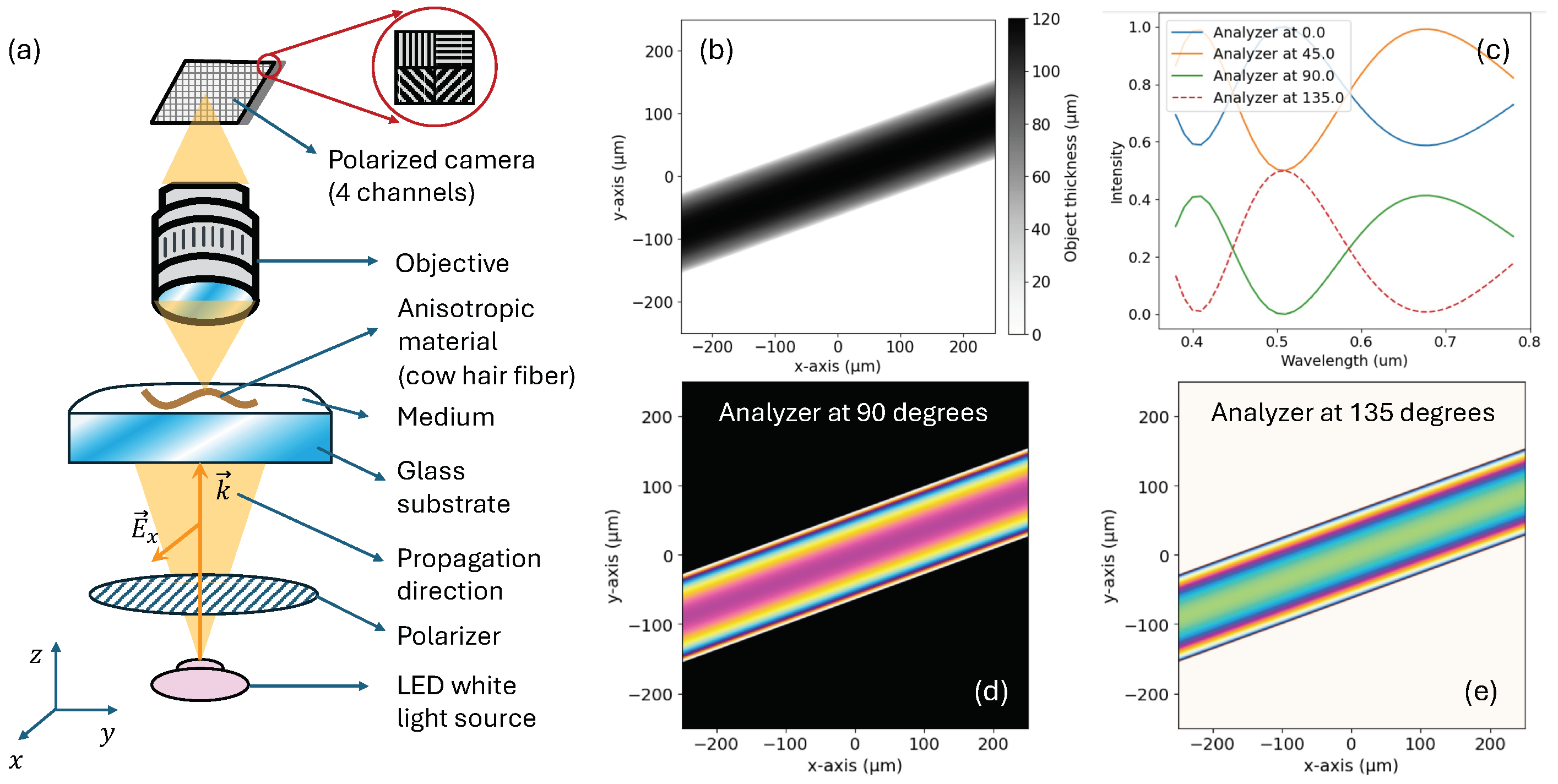
Figure 1a illustrates a basic experimental PLM setup [
2]: A normal incidence white-light beam passes through a linear polarizer before reaching the material. Within the sample, ordinary and extraordinary waves propagate at different velocities. When passing through an analyzer, the interference of the two waves produces the characteristic colors images at a sensor. A polarized camera captures multiple channels for different analyzer angles (typically 0, 45, 90, and 135 degrees). This angular variation allows for a precise determination of the optical path difference (OPD) between ordinary and extraordinary waves.
This work introduces two efficient methods for simulating polarized light microscopy (PLM) experiments: numerical and analytical. These simulations can enhance the imaging capabilities of conventional PLM techniques. Future research will focus on minimizing the mean square error between simulated data and actual measurements to determine optimal material properties and the 3D shape of objects.
2. Method
Uniaxial anisotropic materials have a specific orientation defined by an optical axis
, where
is the tilt angle of the material [
3]. In this work, we assume that the optical axis is perpendicular to the wave propagation direction along the
axis. This configuration can be manually set and commonly occurs in the study of many fibers, which often lie flat on the glass substrate (see Fig. 1a). This setup is particularly useful for distinguishing long hairs from different mammals.
The linear electric field
entering the material can be decomposed into two orthogonal components:
and
. The component perpendicular to the optical axis generates ordinary waves, characterized by the ordinary complex refractive index
. Here,
represents the speed of light in the material relative to the speed of light in a vacuum, and the extinction coefficient
measures light absorption. According to Lambert-Beer’s law, the intensity of a collimated beam is
, where
is the absorption coefficient [
4]. For materials that are nearly transparent in the visible spectrum
. Similarly, the electric field component parallel to the optical axis propagates with the extraordinary complex refractive index
, where
for our particular case of study.
The phase difference between ordinary and extraordinary waves [
5] is given by
, where
represents the birefringence and
the depth map at different locations. The analyzer in front of the sensor has a specific transmission axis, denoted by
. We need to consider the projection of both parallel and perpendicular electric fields along the
axis. These projections are expressed as the scalar values
and
. The final intensity at the sensor location is then calculated as
.
Although all these previous operations can be performed numerically, we have derived an equivalent analytical formula that enables a more efficient computation of the resulting image:
3. Results and Validation
We have carried out the wave propagation through the PLM system using a simulated sample of a single cow hair fiber [
6] with a certain tilt of 20 degrees with respect to the abscissas axis.
Figure 1b shows the simulated depth map used as an input for our models. The refractive indices were
and
, which define a slight positive birefringence
. The optical axis is along the longitudinal part of the fiber. The ordinary waves then occur when the E-field oscillates along the cross-wise direction of the fiber. We evaluated Equation (
1) for wavelengths from 380 to 780, with a step size of 10 nm. Following the procedure described in [
7], we converted a multi-wavelength intensity image into a simple sRGB image capture using a conventional sensor.
Figure 1c shows the raw intensity values for different wavelengths at the location
um. Two of the four output polarization channels captured at the simulated sensor are shown in
Figure 1d–e.
Several computational experiments have been performed to verify the validity of our methods. First, we empirically saw the equivalence of both the numerical and analytical approaches for multiple data within a reasonable range, . Then, we evaluated our models in some particular scenarios in which the ground-truth results are theoretically known. For example, when setting an isotropic material , the cross-polarized channel () correctly cancels out, the two polarized channels at cancel 50% of light, and the uncrossed polarization channel () transmits all the light, producing a uniform white constant image. If we now consider an anisotropic material with and with an optical axis , the resulting images exhibit the same behavior as isotropic materials. This is expected since only ordinary or extraordinary waves propagate in these orientations. Then, setting at different angles leads to the expected interference colors.
References
- Marquez, E.; Ballester, M.; Garcia, M.; Cintado, M.; Marquez, A.; Ruiz, J.; Fernández, S.; Blanco, E.; Willomitzer, F.; Katsaggelos, A. Complex dielectric function of H-free a-Si films: Photovoltaic light absorber. Materials Letters 2023, 345, 134485. [Google Scholar] [CrossRef]
- Frandsen, A.F. Polarized light microscopy. Technical report, 2016.
- Yeh, P.; Hendry, M. Optical waves in layered media. Physics Today 1990, 43, 77. [Google Scholar] [CrossRef]
- Márquez, E.; Blanco, E.; Mánuel, J.M.; Ballester, M.; García-Gurrea, M.; Rodríguez-Tapiador, M.I.; Fernández, S.M.; Willomitzer, F.; Katsaggelos, A.K. Mid-Infrared (MIR) Complex Refractive Index Spectra of Polycrystalline Copper-Nitride Films by IR-VASE Ellipsometry and Their FIB-SEM Porosity. Coatings 2023, 14, 5. [Google Scholar] [CrossRef]
- Hecht, E. Optics; Pearson Education India, 2012.
- Rowe, W.F. Identification of Natural Fibers. Handbook for the analysis of micro-particles in archaeological samples.
- Sørensen, B.E. A revised Michel-Lévy interference colour chart based on first-principles calculations. European Journal of Mineralogy 2013, 25, 5–10. [Google Scholar] [CrossRef]
|
Disclaimer/Publisher’s Note: The statements, opinions and data contained in all publications are solely those of the individual author(s) and contributor(s) and not of MDPI and/or the editor(s). MDPI and/or the editor(s) disclaim responsibility for any injury to people or property resulting from any ideas, methods, instructions or products referred to in the content. |
© 2024 by the authors. Licensee MDPI, Basel, Switzerland. This article is an open access article distributed under the terms and conditions of the Creative Commons Attribution (CC BY) license (http://creativecommons.org/licenses/by/4.0/).


