1. Body of Manuscript
Advances in artificial intelligence (AI) medical applications have surged within the field of dermatology. The integration of quantitative measures to assess and document skin conditions offers a standardized approach and allows for more objectivity in diagnosis and management of skin pathologies [
1,
6]. Facial analysis systems can provide an instantaneous numerical score for skin characteristics including facial redness, pigmentation, wrinkles, and sun damage [
1,
2,
3,
7]. Traditional AI devices available for dermatologic clinical practice have a bulky design [
4,
9]. Most devices currently in practice are not transportable as they require specialized equipment and are often expensive.
The transition to newer AI models may offer a convenient, portable, handheld approach that facilitates collection of data on facial characteristics, which aids providers in tracking patient progress and tailoring treatments [
1,
3,
8,
9]. The potential for more convenient, handheld solutions may also simplify facial skin data collection for patients. These emerging AI facial analysis devices can also provide more accessibility, particularly in remote or resource-limited settings [
10]. Despite these advances, newer AI systems still face challenges, with factors such as distance, lighting, and color correction impacting the accuracy of facial assessments [
8,
11]. These limitations can lead to inconsistent and unreliable facial analysis results [
12]. The objective of our study was to assess the effect of different lighting on facial characteristics’ scores produced by an iPad-based AI skin analysis system.
2. Materials and Methods
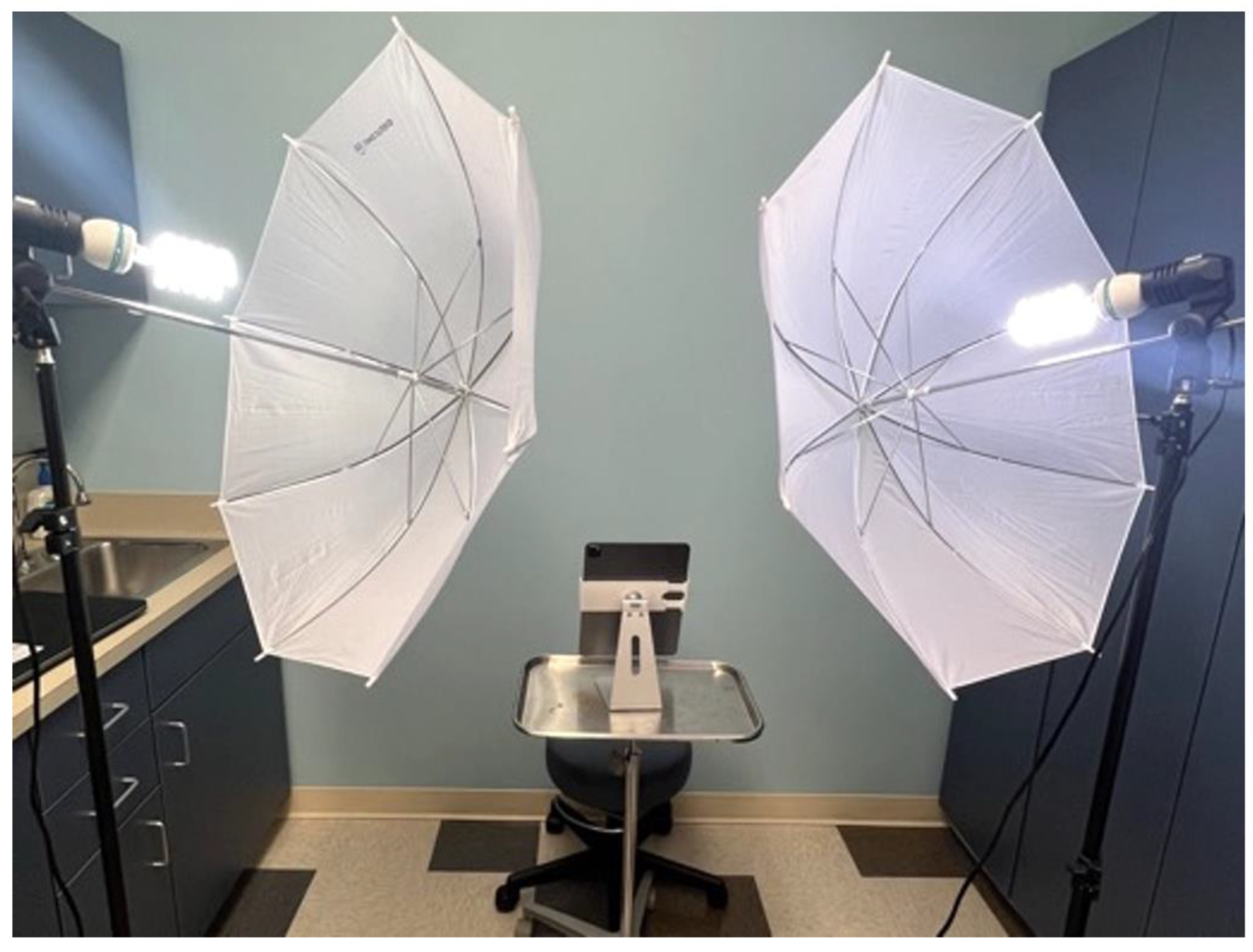
Upon institutional review board approval, 14 patients were recruited from Wake Forest Baptist Health’s dermatology clinic during the month of May 2024. Signed informed consent was obtained from all participants. Inclusion criteria included patients age ≥11 years, a Fitzpatrick skin type (FST) I-VI and the ability to remove any facial makeup, covering or glasses. An equal distribution of patients was recruited to fit two categories: FST I-III and FST IV-VI. Exclusion criteria included facial tattoos, sunburns, and significant facial scarring. Facial images were captured using an iPad-based AI skincare app (Perfect Corporation Skincare Pro Corporation) (
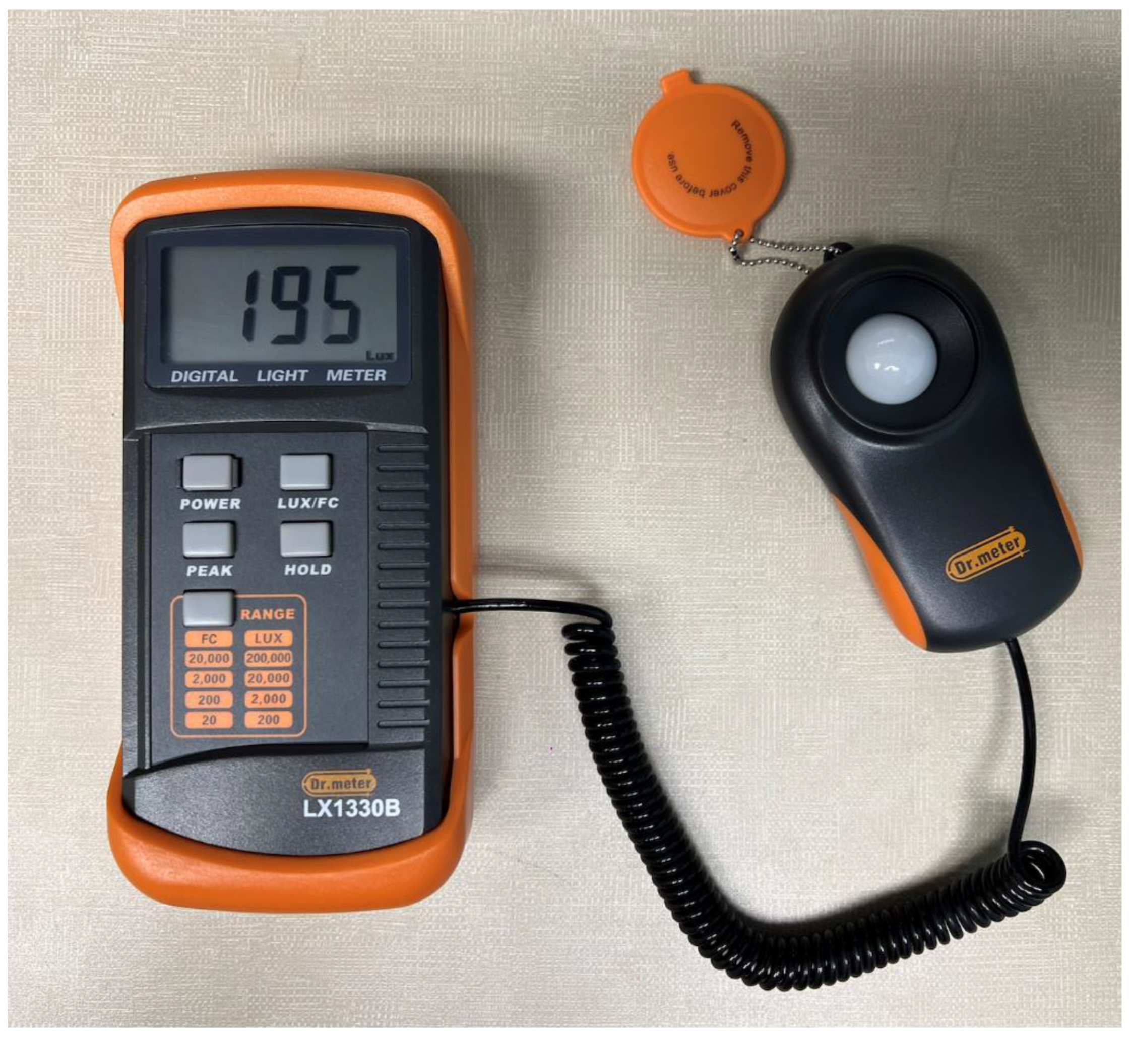
Figure 1). Lighting conditions were standardized using a digital illuminance meter; lux (LX) and foot-candle (FC) were recorded as units of light (
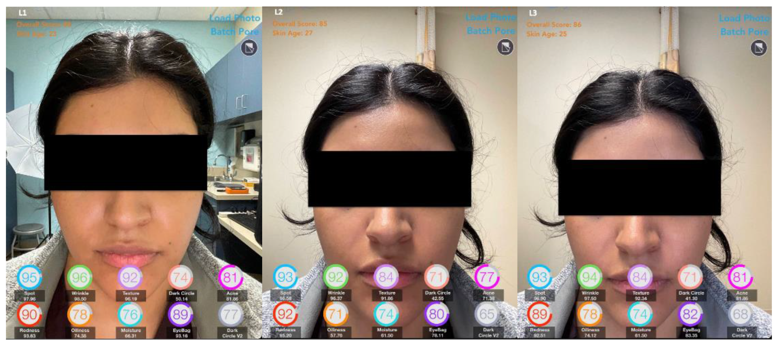
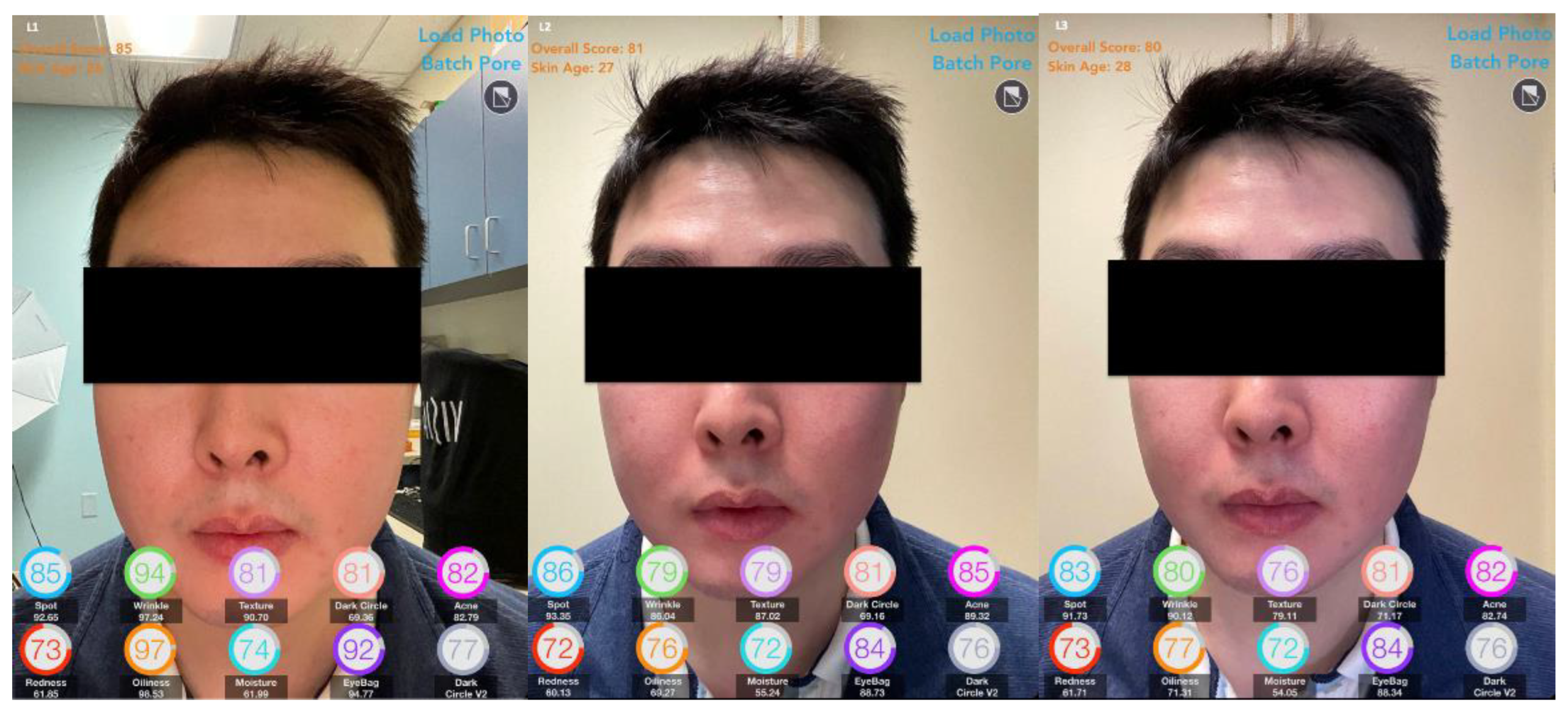
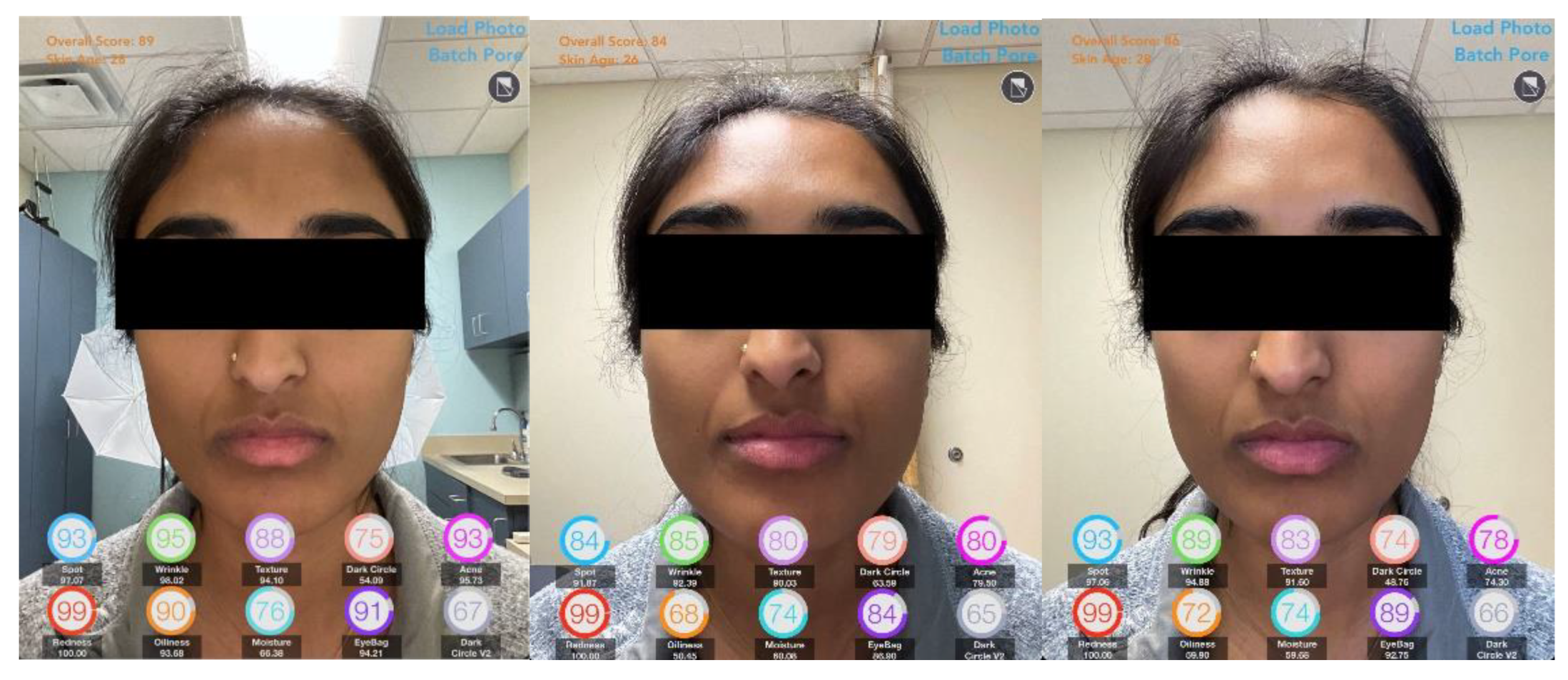
Figure 2). Three different lighting settings were used: below average lighting (L1: mean 161.64 LX/14.06 FC), standard room lighting (L2: mean 519 LX/48.61 FC), and enhanced studio lighting (L3: mean 650.57LX/61.15 FC) (
Figure 3,
Figure 4,
Figure 5 and
Figure 6). Raw scores for the following facial characteristics were recorded: spots, wrinkles, texture, dark circle, acne, redness, oiliness, moisture, eyebags, texture, eyes, forehead, lower, ex eyes, pores, radiance, firmness, upper lid, and lower lid. Higher raw scores indicated better skin health. Scores were compared between the three lighting conditions. Data collection and statistical analysis were performed using Excel and SPSS.
3. Results
The demographics of the participants were primarily Black (57%) females (71%) with a mean age of 41 years (range: 19-74). Participants were evenly distributed among FST; seven participants (50%) comprised both FST I-III and FST III-VI groups. Below average lighting had the highest raw scores for facial characteristics (79.67 ± 10.56). Mean raw scores were lowest for enhanced studio lighting conditions (74.43 ± 10.24) (Figure 7). The difference in scores produced by the different lighting conditions was not statistically significant (p=0.79) (Figure 7). Scores for L1, L2, and L3 settings had an intraclass correlation coefficient (ICC) and Cronbach's alpha measure of 0.893, 0.904, and 0.928, respectively (CI 95% 0.88-0.93, p<0.001), indicating excellent reliability.
4. Discussion
Compared to lower lighting, enhanced studio lighting allowed the AI facial app to detect more facial details, which correlated with lower raw scores. While studio lighting improved the level of detail captured, it was not significant, as similar results were obtained without additional lighting. This suggests that the new AI system's ability to assess facial characteristics may remain consistent across different lighting conditions. The application used in our study may serve as a cost-effective and convenient tool both within and outside clinical settings allowing enhanced quantitative analysis of facial characteristics. Its portability and accessibility make it highly adaptable for a range of uses across different environmental settings without impacting the reliability of the results.
The future direction of AI-based analysis tools in dermatology holds potential for innovation and expanded applications [
16,
17]. The perception of skin color, texture, and fine details were not given as much emphasis prior to the emergence of AI technology [
15,
18,
19,
20]. Environmental lighting conditions are an important factor in aiding dermatologic diagnosis as the AI-based analysis tools initially had complications differentiating nevus to melanoma [
12,
13]. Newer AI-based analysis models are being developed to differentiate between various shades of erythema in inflammatory dermatoses and to capture the texture of wrinkles and subtle pigmentation differences, which are areas that current AI models in cosmetic dermatology have yet to fully address [
14]. AI facial analysis systems must also ensure accuracy and reliability across various lighting conditions while considering the full spectrum of Fitzpatrick skin types, as differences in skin color tones can greatly influence the evaluation of pigmentation, erythema, and other key dermatologic characteristics.
9,18 AI-based analysis tools had lower performances in detecting dermatological manifestations of participants with darker Fitzpatrick skin types [
15]. Ensuring that these AI-analysis tools are standardized for diverse factors such as lighting conditions and skin tones can optimize accurate dermatologic assessments and provide equitable, accessible care to diverse patient populations.
Author Contributions
Shirley P. Parraga: Conceptualization, Shirley Parraga and Shailey Shah.; methodology, Shirley Parraga, Shailey Shah, and Robin Yi; software, Shirley Parraga; validation, Shirley Parraga, Shailey Shah, Robin Yi, and Steven Feldman; formal analysis, Shirley Parraga, Sarah Taylor, and Steven Feldman.; investigation, Shirley Parraga, Shailey Shah, Robin Yi, and Griffin Girard; resources, Shirley Parraga, Shailey Shah, Robin Yi; data curation, Shirley Parraga, Shailey Shah, Robin Yi; writing—original draft preparation, Shirley Parraga, Shailey Shah, Robin Yi, Sarah Taylor, and Steven Feldman; writing—review and editing, Sarah Taylor, and Steven Feldman; visualization, Steven Feldman; supervision, Shirley Parraga, Sarah Taylor, and Steven Feldman. All authors have read and agreed to the published version of the manuscript.
Funding
This research received no external funding.
Institutional Review Board Statement
The study was approved by the Institutional Review Board of Wake Forest University Health Sciences (IRB#00085220).
Informed Consent Statement
Informed consent was obtained from all subjects involved in the study.
Data Availability Statement
The original contributions presented in the study are included in the article/supplementary material, further inquiries can be directed to the corresponding author/s.
Conflicts of Interest
Steven Feldman has received research, speaking and/or consulting support from Eli Lilly and Company, GlaxoSmithKline/Stiefel, AbbVie, Janssen, Alovtech, vTv Therapeutics, Bristol-Myers Squibb, Samsung, Pfizer, Boehringer Ingelheim, Amgen, Dermavant, Arcutis, Novartis, Novan, UCB, Helsinn, Sun Pharma, Almirall, Galderma, Leo Pharma, Mylan, Celgene, Ortho Dermatology, Menlo, Merck & Co, Qurient, Forte, Arena, Biocon, Accordant, Argenx, Sanofi, Regeneron, the National Biological Corporation, Caremark, Teladoc, BMS, Ono, Micreos, Eurofins, Informa, UpToDate and the National Psoriasis Foundation. He is founder and part owner of Causa Research and holds stock in Sensal Health. The other authors have no conflicts to disclose.
References
- Oesch S, Vingan NR, Li X, Hoopman J, Akgul Y, Kenkel JM. A Correlation of the Glogau Scale With VISIA-CR Complexion Analysis Measurements in Assessing Facial Photoaging for Clinical Research. Aesthet Surg J. 2022;42(10):1175-1184. [CrossRef]
- Adatto MA, Adatto-Neilson RM. Facial treatment with acoustic wave therapy for improvement of facial skin texture, pores and wrinkles. J Cosmet Dermatol. 2020;19(4):845-849. [CrossRef]
- Campiche R, Trevisan S, Séroul P, et al. Appearance of aging signs in differently pigmented facial skin by a novel imaging system. J Cosmet Dermatol. 2019;18(2):614-627. [CrossRef]
- Henseler H. Assessment of the reproducibility and accuracy of the Visia® Complexion Analysis Camera System for objective skin analysis of facial wrinkles and skin age. GMS Interdiscip Plast Reconstr Surg DGPW. 2023;12:Doc07. Published 2023 Oct 2. [CrossRef]
- Zaino ML, Pixley JN, Kontzias C, Feldman SR, McMichael AJ, Taylor S. Using Facial Skin Analysis to Capture Rosacea Patients’ Response to Vascular Laser Therapy—A Single-Center Prospective Study. Journal of Cutaneous Medicine and Surgery. 2024;28(1):69-71. [CrossRef]
- Haykal D, Garibyan L, Flament F, Cartier H. Hybrid cosmetic dermatology: AI generated horizon. Skin Res Technol. 2024;30(5):e13721. [CrossRef]
- Corp P. AI-powered Online Skin Analysis and Skin Diagnostic | Perfect Corp. www.perfectcorp.com. https://www.perfectcorp.com/business/products/ai-skin-diagnostic.
- Bakht MK, Pouladian M, Mofrad FB, Honarpisheh H. Impact of various color LED flashlights and different lighting source to skin distances on the manual and the computer-aided detection of basal cell carcinoma borders. Skin Res Technol. 2014;20(1):92-96. [CrossRef]
- Kontzias C, Pixley JN, Zaino M, Feldman SR. Validation of a new facial skin analysis device across Fitzpatrick skin types. J Cosmet Dermatol. 2024 Feb;23(2):720-721. [CrossRef]
- Veronese F, Branciforti F, Zavattaro E, Tarantino V, Romano V, Meiburger KM, Salvi M, Seoni S, Savoia P. The Role in Teledermoscopy of an Inexpensive and Easy-to-Use Smartphone Device for the Classification of Three Types of Skin Lesions Using Convolutional Neural Networks. Diagnostics (Basel). 2021 Mar 5;11(3):451. [CrossRef]
- Santos A, Aires K, Veras R. Aspects of Lighting and Color in Classifying Malignant Skin Cancer with Deep Learning. Applied sciences. 2024;14(8):3297-3297. [CrossRef]
- Li Z, Koban KC, Schenck TL, Giunta RE, Li Q, Sun Y. Artificial Intelligence in Dermatology Image Analysis: Current Developments and Future Trends. J Clin Med. 2022 Nov 18;11(22):6826. [CrossRef]
- Li S, Chu Y, Wang Y, Wang Y, Hu S, Wu X, Qi X. Distinguish the Value of the Benign Nevus and Melanomas Using Machine Learning: A Meta-Analysis and Systematic Review. Mediators Inflamm. 2022 Oct 14;2022:1734327. [CrossRef]
- Gomolin A, Netchiporouk E, Gniadecki R, Litvinov IV. Artificial Intelligence Applications in Dermatology: Where Do We Stand? Front Med (Lausanne). 2020 Mar 31;7:100. [CrossRef]
- Aggarwal P. Performance of Artificial Intelligence Imaging Models in Detecting Dermatological Manifestations in Higher Fitzpatrick Skin Color Classifications. JMIR Dermatol. 2021 Oct 12;4(2):e31697. [CrossRef]
- Takiddin A, Schneider J, Yang Y, Abd-Alrazaq A, Househ M. Artificial Intelligence for Skin Cancer Detection: Scoping Review. J Med Internet Res. 2021;23(11):e22934. Published 2021 Nov 24. [CrossRef]
- Zakhem GA, Fakhoury JW, Motosko CC, Ho RS. Characterizing the role of dermatologists in developing artificial intelligence for assessment of skin cancer. J Am Acad Dermatol. 2021;85(6):1544-1556. [CrossRef]
- Eilers S, Bach DQ, Gaber R, et al. Accuracy of self-report in assessing Fitzpatrick skin phototypes I through VI. JAMA Dermatol. 2013;149(11):1289-1294. [CrossRef]
- Flament F, Jiang R, Houghton J, et al. Accuracy and clinical relevance of an automated, algorithm-based analysis of facial signs from selfie images of women in the United States of various ages, ancestries and phototypes: A cross-sectional observational study. J Eur Acad Dermatol Venereol. 2023;37(1):176-183. [CrossRef]
- Yu Z, Flament F, Jiang R, et al. The relevance and accuracy of an AI algorithm-based descriptor on 23 facial attributes in a diverse female US population. Skin Res Technol. 2024;30(5):e13690. [CrossRef]
|
Disclaimer/Publisher’s Note: The statements, opinions and data contained in all publications are solely those of the individual author(s) and contributor(s) and not of MDPI and/or the editor(s). MDPI and/or the editor(s) disclaim responsibility for any injury to people or property resulting from any ideas, methods, instructions or products referred to in the content. |
© 2024 by the authors. Licensee MDPI, Basel, Switzerland. This article is an open access article distributed under the terms and conditions of the Creative Commons Attribution (CC BY) license (http://creativecommons.org/licenses/by/4.0/).
