Preprint
Article

Eight-Chop Technique in Phacoemulsification Using Iris Hooks for Patients with Cataracts and Small Pupils

Altmetrics

Downloads

36

Views

32

Comments

0

This version is not peer-reviewed

Submitted:

20 September 2024

Posted:

23 September 2024

You are already at the latest version

Alerts
Abstract
Objectives: This study investigated the efficacy and safety of performing phacoemulsification using the eight-chop technique with iris hooks in patients with small pupils. Methods: The iris hooks and control groups each included 65 eyes. Cataract surgeries were performed using the eight-chop technique. The operative time, phaco time, aspiration time, cumulative dissipated energy, and volume of fluid used were measured. Best-corrected visual acuity, corneal endothelial cell density (CECD), and intraocular pressure (IOP) were measured preoperatively and postoperatively. Results: In total, 130 eyes of 107 patients (mean age, 75.9 ± 7.1 years; 58 men, 72 women) with cataracts were evaluated. The mean operative time, phaco time, aspiration time, cumulative dissipated energy, and volume of fluid used were 10.6 min, 20.7 s, 101.1 s, 7.8, and 38.0 mL, respectively, in the iris hooks group and 4.6 min, 16.2 s, 72.1 s, 7.0, and 28.9 mL, respectively, in the control group. The decrease in CECD at 19 weeks postoperatively was 2.1% and 1.2% for the iris hooks and control groups, respectively. In both groups, IOP decreased significantly (all p < .01) at 7 and 19 weeks postoperatively. No intraoperative complications were observed in either group. Conclusions: The eight-chop technique using iris hooks resulted in a small postoperative reduction in CECD and excellent values for intraoperative outcome measures. In addition, those cataract surgeries were very short, efficient, and safe, without complications. The eight-chop technique using iris hooks could provide an ideal solution for patients with small pupils.
Keywords: 
Subject: Biology and Life Sciences  -   Life Sciences

1. Introduction

Cataract surgery is one of the most common surgical procedures performed worldwide [1,2]. However, phacoemulsification through a small pupil is challenging for surgeons and considered to be associated with more complications [2,3]. Small pupils are observed in approximately 4.4%–11% of all cataract operations [2,4,5], with a reported complication frequency of 4.7%–9.0% [5,6]. Thus, the use of any established method of intraoperative widening of the pupil by the surgeon is considered practical. The key to success is sufficient iris retraction when needed and safe maneuvering in the pupil area. Many techniques to widen a small pupil during phacoemulsification have been described; using iris hooks has the advantage of enabling a stable pupil size to be maintained throughout the surgery [7].
In the eight-chop technique, the nucleus is manually divided using an ophthalmic viscosurgical device before phacoemulsification [8]. Compared with conventional grooving, divide-and-conquer, and phaco-chop techniques, the eight-chop technique reduces the total ultrasound energy, aspiration time, and fluid volume used [8]. Furthermore, this technique facilitates safe maneuvering during phacoemulsification, even within a small pupil, because the divided lens nucleus is small and both hands can be used to delicately manipulate an ultrasound tip.
To date, no study has been conducted utilizing the eight-chop technique for patients with small pupils. Moreover, no studies have examined the intraoperative parameters in patients with small pupils; thus, the surgical details of these difficult cases are unclear.
This study evaluated the intraoperative outcome measures, postoperative best-corrected visual acuity (BCVA), corneal endothelial cell density (CECD), and intraocular pressure (IOP) changes for phacoemulsification performed using the eight-chop technique with iris hooks in patients with small pupils (>6 mm) and compared the results with those of patients with pupils ≥6 mm and results reported previously [2,3,4,5,6,7].

2. Materials and Methods

2.1. Study Population

This study comprised patients with cataracts whose eyes had undergone phacoemulsification and posterior chamber intraocular lens implantation between March 2015 and February 2022 at our clinic. Patients with a preoperative pupil diameter of ≥6 mm were considered for the control group, whereas patients with a pupil diameter of <6 mm were considered for the iris hook group [6]. Patients with corneal disease or opacity, uveitis, and previous trauma or surgery were excluded.

2.2. Preoperative Assessment

Preoperatively, all the patients underwent slit-lamp and retinal examinations, and their BCVA and IOP were measured. CECD (cells/mm2) was measured using a noncontact specular microscope (EM-3000; Topcon Corporation, Tokyo, Japan). The firmness of the nucleus was graded using the Emery classification [9]. The same surgeon, who is experienced in the eight-chop technique, performed phacoemulsification using the phacoemulsification unit (Centurion®; Alcon Laboratories, Inc., Irvine, CA, USA).

2.3. New Surgical Instruments

New surgical instruments have been designed and developed to perform the eight-chop technique [8]. My research team designed eight choppers and requested a manufacturing company to produce them. The Eight-chopper I (SP-8193; ASICO, Parsippany, NJ, USA) has a smaller tip than the conventional prechopper, with a length and width of 3.2 mm and 1.4 mm, respectively, and a sharper leading edge; it was used for the grade II group. The Eight-chopper II (SP-8402; ASICO) has a smaller angular tip (2.5 mm long and 0.8 mm wide) that can be inserted vertically into the lens nucleus and was used for the grade III group.

2.4. Surgical Technique

In all the surgeries, a temporal, clear corneal incision was made using a 3.0-mm steel keratome. After injecting sodium hyaluronate into the anterior chamber, a 6.2–6.5 mm continuous curvilinear capsulorhexis was created using capsule forceps. For the iris hooks group, iris hooks (Synergetics, Inc., O’Fallon, MO, USA) were used to retract the iris through corneal stab incisions from four directions (Figure 1). The soft-shell technique [10] was used for the grade III group. Hydrodissection was performed using a 27-gauge cannula; however, hydrodelineation was not performed. The lens nucleus was cracked into eight segments using the Eight-chopper I or II for eyes classified as grade II or III, respectively. An ophthalmic viscosurgical device was injected into the anterior chamber, and the eight-chopper was then inserted into the center of the lens nucleus, which was completely divided. After the nucleus was completely divided into two pieces, the lens nucleus was rotated 90 degrees. The eight-chopper was inserted into the center of the heminucleus, which was completely divided. The lens nucleus was rotated 180 degrees to complete four divisions of the lens nucleus, then rotated 45 degrees and divided four times to complete eight divisions of the lens nucleus. The eight segments were phacoemulsified and aspirated at the depth of the iris plane. The capsular bag was aspirated with the irrigation/aspiration tip to remove cortical materials. The ophthalmic viscosurgical device was injected, and a foldable intraocular lens (Acrysof® MN60AC; Alcon Inc.) with polymethyl methacrylate haptics was inserted in the capsular bag using an injector system. The ophthalmic viscosurgical device was subsequently aspirated. The phacoemulsification unit was used in all the cases and had a flow rate of 32 mL/min, maximum ultrasound power of 80%, and a 1.1-mm tip. The wound was sealed using stromal hydration if necessary. The anterior chamber was replaced with a balanced salt solution containing moxifloxacin (0.5 mg/mL) after the operation.

2.5. Outcome Measures and Data Collection

The intraoperative outcome measures were operative time (min), phaco time (s), aspiration time (s), cumulative dissipated energy, volume of fluid used (mL), and the rate of intraoperative complications. The operative time was calculated from the beginning of the corneal incision to the end of the ophthalmic viscosurgical device aspiration. The hooking time was calculated from the creation of the first corneal stab incision to the placement of the fourth hook. All the surgeries were recorded, and the pupil diameters were analyzed based on the surgical videos according to the method by Vasavada et al. [11]. Patients were followed up on postoperative days 1 and 2, and at 1, 3, 7, and 19 weeks. The postoperative outcome measures were BCVA, IOP, and CECD (cells/mm2). The BCVA, CECD, and IOP measurements were obtained at 7 and 19 weeks postoperatively. Using the method by Poley et al. [12], and based on the preoperative IOP, the iris hooks and control groups were divided into subgroups for analysis: those with IOP levels above and below 15 mmHg.

2.6. Statistical Analysis

Statistical analyses were performed to compare the results between the iris hooks and control groups using unpaired t-tests. Paired t-tests were used to compare the preoperative BCVA, CECD, and IOP to each postoperative time point. Statistical significance was set at p < .05. The chi-square test was used to determine any differences in sex, diabetes mellitus, and intraoperative floppy iris syndrome incidence between the iris hooks and control groups.

3. Results

This study comprised 130 eyes of 107 patients with cataracts who had undergone phacoemulsification and posterior chamber intraocular lens implantation. Table 1 presents the patient characteristics and intraoperative parameters. No significant differences were observed in the mean ages between the iris hooks and control groups. No significant differences were observed in the incidence of diabetes mellitus; however, significant differences were observed in the incidence of intraoperative floppy iris syndrome and gender between the iris hooks and control groups. Significant differences were observed in the preoperative and postoperative pupil sizes between the iris hooks and control groups. Significant differences were observed in the operative time, phaco time, aspiration time, and volume of fluid used between the iris hooks and control groups. However, no significant differences were observed in the cumulative dissipated energy between the iris hooks and control groups.
Table 2 lists the pre- and postoperative changes in the BCVA and CECD measurements. No significant differences were observed in the BCVAs preoperatively and 19 weeks postoperatively between the iris hooks and control groups. However, significant differences were observed in the BCVAs 7 weeks postoperatively between the iris hooks and control groups. The BCVAs between preoperatively and 7 weeks postoperatively, 7 and 19 weeks postoperatively, and preoperatively and 19 weeks postoperatively in the iris hooks group significantly differed. Moreover, the BCVAs between preoperatively and 7 weeks postoperatively and preoperatively and 19 weeks postoperatively in the control group differed significantly; however, no significant differences were observed in the BCVAs between 7 and 19 weeks postoperatively in the control group. No significant differences were observed in the CECDs preoperatively between the iris hooks and control groups. However, significant differences were observed in the CECDs 7 and 19 weeks postoperatively between the iris hooks and control groups. The CECDs between preoperatively and 7 weeks postoperatively and preoperatively and 19 weeks postoperatively in the iris hooks group differed significantly. However, no significant differences were observed in the CECDs between 7 weeks postoperatively and 19 weeks postoperatively in the iris hooks group. The CECDs between preoperatively and 7 weeks postoperatively and preoperatively and 19 weeks postoperatively in the control group differed significantly. However, no significant differences were observed in the CECDs between 7 and 19 weeks postoperatively in the control group.
Table 3 presents the changes in the IOP results. No significant differences were observed in the IOP levels preoperatively between the iris hooks and control groups. However, the IOP levels between the iris hooks and control groups 7 and 19 weeks postoperatively differed significantly. The preoperative and postoperative IOP levels at 7 and 19 weeks in the iris hooks and control groups differed significantly.
Table 4 lists the changes in the IOP levels of the subgroups with preoperative IOP above and below 15 mmHg in the iris hooks and control groups. In the iris hooks group, the IOP levels significantly decreased at 7 and 19 weeks postoperatively in the subgroups with preoperative IOP levels below and above 15 mmHg. Moreover, in the control group, the IOP levels significantly decreased at 7 and 19 weeks postoperatively in the subgroups with preoperative IOP levels below and above 15 mmHg.
No intraoperative complications and capsulorhexis tears were observed in the iris hooks or control groups.

4. Discussion

This study determined that the eight-chop technique using iris hooks had an operative time of 10.6 min, which was shorter than the previously reported operative times of 37–39 min [2]. Although the use of iris hooks and the pupil expansion ring are the most time-consuming techniques in the procedure, they have the advantage of facilitating the maintenance of a stable pupil size throughout the surgery [7]; the pupil expansion ring is faster to use than iris hooks [2]. Furthermore, using iris hooks and the Malyugin ring can reduce intraoperative corneal endothelium cell loss [13]. However, Malyugin rings maintain a circular structure within the anterior chamber and have a greater potential for contact with the corneal endothelial cells compared to iris hooks. The ring thickness, in conjunction with its ring-like structure, requires careful manipulation within the eye. Pupil expansion is also limited; thus, if the lens nucleus is large and hard, it cannot be efficiently split. Iris hooks do not have a three-dimensional structure; thus, there is little possibility of contact with the corneal endothelial cells, and contact with the iris is also limited. Moreover, because the pupil diameter can be changed arbitrarily, it can be safely operated by securing the diameter appropriate for the surgeon’s needs. To effectively use iris hooks, they should be placed from the scleral side to ensure an enlarged pupil diameter range, and the iris should not be elevated (Figure 1). Therefore, apart from their insertion time, iris hooks are considered one of the best options for patients with small pupils. Phacoemulsification with the eight-chop technique takes an extremely short time, and, even with the use of iris hooks, surgical involvement may be very low.
The present study also measured the iris hooking time, which was 3.3 min. The eight-chop technique using iris hooks had a lower phaco time and cumulative dissipated energy, and used only one-third to one-sixth of the volume of fluid than that used in other techniques [3,14,15]. In particular, a smaller volume of fluid used may result in less surgical involvement of the trabecular meshwork and Schlemm’s canal cells, including corneal endothelial cells, owing to the shorter time required to insert the ultrasound and irrigation/aspiration tip into the eye.
A CECD assessment is crucial for comparing various techniques because it represents the true summation of intraocular insult during surgery [10,16]. A 5%–16% decrease in the CECD following cataract surgery in the first few postoperative months has been reported [3,10,15,17,18]. In the present study, the decrease was only 2.6% and 2.1% at 7 and 19 weeks postoperatively, respectively, in the iris hooks group, and 1.3% and 1.2% at 7 and 19 weeks postoperatively, respectively, in the control group. These results indicated that the eight-chop technique may be advantageous in minimizing the surgical involvement of the intraocular tissues, including the trabecular meshwork and Schlemm’s canal. However, significant differences were observed in the decrease in CECD postoperatively between the iris hooks and control groups. This may be due to the iris hook contacting intraocular tissue, increasing postoperative inflammation and reducing CECD, because the difference in fluid volume used was only 10 mL. The 0.9% difference in the reduction in CECD between the iris hooks and control groups, even iris traction with iris hooks, had an approximate 1% effect on the postoperative decrease in the CECD. Mechanical stimulation of the iris during cataract surgery may indirectly affect the corneal endothelial cells.
Many investigators have reported a decrease in IOP following phacoemulsification cataract extraction and intraocular lens implantation in patients with cataracts [19,20]. IOP reductions of 4%–10% have been demonstrated [12,21,22]. Postoperative IOP changes are proportional to preoperative IOP. However, Poley et al. [12,23] reported an increase in the IOP in the primary open-angle glaucoma and normal groups at 1 year compared to that of the preoperative levels. In the present study, the IOP reduction rate was 12.0% and 18.1% in the iris hooks and control groups, respectively, at 19 weeks postoperatively; this was higher than previously reported data in both the groups. Furthermore, in the iris hooks and control groups with IOP below 15 mmHg, IOP significantly decreased at 7 and 19 weeks postoperatively. The greater reduction in IOP could be attributable to the superiority of the eight-chop technique over other techniques in minimizing the surgical involvement of the intraocular tissues. Phacoemulsification may lower the IOP postoperatively; the higher the preoperative IOP, the greater its IOP-lowering effect. However, surgical involvement could reduce the IOP-lowering effect of phacoemulsification. Poley et al. [12,23] could not detect IOP reduction in the group with the lowest preoperative IOP, suggesting that the surgical involvement of the technique used might have significantly counteracted the effect of IOP reduction. The eight-chop technique with or without iris hooks is less invasive and may preserve the IOP-lowering effect of phacoemulsification. Significant differences were observed in the decrease in the IOP postoperatively between the iris hooks and control groups. The 6.1% difference in the IOP reduction between the iris hooks and control groups, even iris traction with the iris hooks, demonstrated a nearly 6% effect on the postoperative IOP decrease. Mechanical stimulation of the iris during cataract surgery could indirectly affect the trabecular meshwork cells.
The phacoemulsification techniques employed in previous reports in patients with small pupils are the divide-and-conquer [24,25], phaco-chop [7,26], step-by-step chop [3], stop and chop [27], quick chop [27], or unknown [2,5,12,13,28,29,30,31,32] techniques. To date, no studies have employed the prechop or eight-chop techniques. The phaco time, aspiration time, cumulative dissipated energy, and volume of fluid used, including operative time, should be measured when examining changes in the IOP and CECD following phacoemulsification cataract surgery. However, to my knowledge, no previous studies on the effects of phacoemulsification cataract surgery on the IOP and CECD have reported intraoperative parameters in patients with small pupils. Hence, the changes in IOP and CECD following phacoemulsification cataract surgery should be investigated when a well-developed phacoemulsification technique that minimizes surgical involvement of the intraocular tissues is employed.
Small pupils are associated with increased complications, including posterior capsule rupture and vitreous loss [5,33,34]. Balal et al. [5] reported that among 20,175 patients with cataracts, 6.7% and 3.8% of those in whom iris hooks and the Malyugin ring were used, respectively, experienced posterior capsule rupture. Thus, posterior capsule rupture occurred in 4.7% of all the patients in whom iris hooks or the Malyugin ring were used [5]. However, none of the 65 patients in the current study had any complications, including posterior capsule rupture. Therefore, the eight-chop technique using iris hooks could be an excellent method concerning safety, in addition to reducing surgical involvement and lowering IOP.
The results of the present study were not compared with those of studies using the divide-and-conquer or phaco-chop techniques, nor were they compared to those of studies using other mechanical pupil dilation techniques. This should be taken into consideration when evaluating the present results. However, many other studies have been conducted using the divide-and-conquer and phaco-chop techniques with other mechanical pupil dilation techniques.
In the current study, the pupil diameter of patients averaged 5.34 mm, which was larger than that previously reported [7,13]. However, the average pupil diameter of the control group was 7.62 mm; thus, it was reduced by 25.1% for the iris hooks group. Therefore, this pupil diameter could be considered challenging for surgical management. Many cases of small pupils encountered in clinical practice are caused by diabetes mellitus, pseudoexfoliation syndrome, or intraoperative floppy iris syndrome [26]. Hence, the patients with small pupils in the present study were considered appropriate for assessing the eight-chop technique. Furthermore, if the greatest priority is surgical safety, mechanical pupil expansion, such as using iris hooks and Malyugin rings, should be considered for patients with a pupil size of <6 mm.

5. Conclusions

The eight-chop technique possesses unique features; both the division and phacoemulsification of the lens nucleus can be distinguished using this technique, enabling the surgeon to concentrate on each individual procedure. Furthermore, the eight-chop technique using iris hooks could be an ideal solution for patients with small pupils, as its use in the present study resulted in a small postoperative reduction in CECD and excellent values for intraoperative outcome measures.

Funding

This research received no external funding.

Institutional Review Board Statement

The study was conducted in accordance with the Declaration of Helsinki and approved by the Ethics Committee of Sato Eye Clinic (protocol code 20150112 and date of approval January 12, 2015).

Informed Consent Statement

Informed consent was obtained from all subjects involved in the study for sample collection and subsequent analyses.

Data Availability Statement

The data presented in this study are available on request from the corresponding author due to privacy and ethical restrictions.

Acknowledgments

I would like to thank Editage (www.editage.com) for English language editing.

Conflicts of Interest

The author declares no conflicts of interest.

References

  1. Forooghian, F.; Agrón, E.; Clemons, T.E.; Ferris, F.L., 3rd; Chew, E.Y.; Age-Related Eye Disease Study Research Group. Visual acuity outcomes after cataract surgery in patients with age-related macular degeneration: age-related eye disease study report no. 27. Ophthalmology 2009, 116, 2093–2100. [Google Scholar] [CrossRef] [PubMed]
  2. Nderitu, P.; Ursell, P. Iris hooks versus a pupil expansion ring: operating times, complications, and visual acuity outcomes in small pupil cases. J Cataract Refract Surg 2019, 45, 167–173. [Google Scholar] [CrossRef] [PubMed]
  3. Vasavada, A.; Singh, R. Phacoemulsification in eyes with a small pupil. J Cataract Refract Surg 2000, 26, 1210–1218. [Google Scholar] [CrossRef] [PubMed]
  4. Hashemi, H.; Seyedian, M.A.; Mohammadpour, M. Small pupil and cataract surgery. Curr Opin Ophthalmol 2015, 26, 3–9. [Google Scholar] [CrossRef]
  5. Balal, S.; Jbari, A.S.; Nitiahpapand, R.; Cook, E.; Akhtar, W.; Din, N.; Sharma, A. Management and outcomes of the small pupil in cataract surgery: iris hooks, Malyugin ring or phenylephrine? Eye (Lond) 2021, 35, 2714–2718. [Google Scholar] [CrossRef]
  6. Halkiadakis, I.; Chatziralli, I.; Drakos, E.; Katzakis, M.; Skouriotis, S.; Patsea, E.; Mitropoulos, P.; Kandarakis, A. Causes and management of small pupil in patients with cataract. Oman J Ophthalmol 2017, 10, 220–224. [Google Scholar]
  7. Akman, A.; Yilmaz, G.; Oto, S.; Akova, Y.A. Comparison of various pupil dilatation methods for phacoemulsification in eyes with a small pupil secondary to pseudoexfoliation. Ophthalmology 2004, 111, 1693–1698. [Google Scholar] [CrossRef]
  8. Sato, T. Efficacy and safety of the eight-chop technique in phacoemulsification for patients with cataract. J Cataract Refract Surg 2023, 49, 479–484. [Google Scholar] [CrossRef]
  9. Emery, J.M. Kelman phacoemulsification; patient selection. In Extracapsular cataract surgery; Emery, J.M., Mclyntyre, D.J., Eds.; CV Mosby: St Louis, USA, 1983; pp. 95–100. [Google Scholar]
  10. Miyata, K.; Nagamoto, T.; Maruoka, S.; Tanabe, T.; Nakahara, M.; Amano, S. Efficacy and safety of the soft-shell technique in cases with a hard lens nucleus. J Cataract Refract Surg 2002, 28, 1546–1550. [Google Scholar] [CrossRef]
  11. Vasavada, A.R.; Desai, J.P.; Singh, R. Measuring structures within the eye. J Cataract Refract Surg 1996, 22, 5–6. [Google Scholar] [CrossRef]
  12. Poley, B.J.; Lindstrom, R.L.; Samuelson, T.W. Long-term effects of phacoemulsification with intraocular lens implantation in normotensive and ocular hypertensive eyes. J Cataract Refract Surg 2008, 34, 735–742. [Google Scholar] [CrossRef]
  13. Wang, J.D.; Zhang, J.S.; Li, M.; Mao, Y.Y.; Mayinuer, Y.; Wan, X.H. Comparison of different pupil dilatation methods for phacoemulsification in eyes with a small pupil. BMC Ophthalmol 2022, 22, 173. [Google Scholar] [CrossRef]
  14. Helvacioglu, F.; Yeter, C.; Tunc, Z.; Sencan, S. Outcomes of torsional microcoaxial phacoemulsification performed by 12-degree and 22-degree bent tips. J Cataract Refract Surg 2013, 39, 1219–1225. [Google Scholar] [CrossRef]
  15. Igarashi, T.; Ohsawa, I.; Kobayashi, M.; Umemoto, Y.; Arima, T.; Suzuki, H.; Igarashi, T.; Otsuka, T.; Takahashi, H. Effects of hydrogen in prevention of corneal endothelial damage during phacoemulsification: a prospective randomized clinical trial. Am J Ophthalmol 2019, 207, 10–17. [Google Scholar] [CrossRef]
  16. Upadhyay, S.; Sharma, P.; Chouhan, J.K.; Goyal, R. Comparative evaluation of modified crater (endonucleation) chop and conventional crater chop techniques during phacoemulsification of hard nuclear cataracts: a randomized study. Indian J Ophthalmol 2022, 70, 794–798. [Google Scholar] [CrossRef]
  17. Sato, M.; Sakata, C.; Yabe, M.; Oshika, T. Soft-shell technique using Viscoat and Healon 5: a prospective, randomized comparison between a dispersive-viscoadaptive and a dispersive-cohesive soft-shell technique. Acta Ophthalmol 2008, 86, 65–70. [Google Scholar] [CrossRef]
  18. Park, J.; Yum, H.R.; Kim, M.S.; Harrison, A.R.; Kim, E.C. Comparison of phaco-chop, divide-and-conquer, and stop-and-chop phaco techniques in microincision coaxial cataract surgery. J Cataract Refract Surg 2013, 39, 1463–1469. [Google Scholar] [CrossRef]
  19. Wirbelauer, C.; Anders, N.; Pham, D.T.; Wollensak, J.; Laqua, H. Intraocular pressure in nonglaucomatous eyes with pseudoexfoliation syndrome after cataract surgery. Ophthalmic Surg Lasers 1998, 29, 466–471. [Google Scholar] [CrossRef]
  20. Shingleton, B.J.; Gamell, L.S.; O’Donoghue, M.W.; Baylus, S.L.; King, R. Long-term changes in intraocular pressure after clear corneal phacoemulsification: normal patients versus glaucoma suspect and glaucoma patients. J Cataract Refract Surg 1999, 25, 885–890. [Google Scholar] [CrossRef]
  21. Irak-Dersu, I.; Nilson, C.; Zabriskie, N.; Durcan, J.; Spencer, H.J.; Crandall, A. Intraocular pressure change after temporal clear corneal phacoemulsification in normal eyes. Acta Ophthalmol 2010, 88, 131–134. [Google Scholar] [CrossRef]
  22. Shingleton, B.J.; Pasternack, J.J.; Hung, J.W.; O’Donoghue, M.W. Three and five year changes in intraocular pressures after clear corneal phacoemulsification in open angle glaucoma patients, glaucoma suspects, and normal patients. J Glaucoma 2006, 15, 494–498. [Google Scholar] [CrossRef]
  23. Poley, B.J.; Lindstrom, R.L.; Samuelson, T.W.; Schulze, R., Jr. Intraocular pressure reduction after phacoemulsification with intraocular lens implantation in glaucomatous and nonglaucomatous eyes: evaluation of a causal relationship between the natural lens and open-angle glaucoma. J Cataract Refract Surg 2009, 35, 1946–1955. [Google Scholar] [CrossRef] [PubMed]
  24. Storr-Paulsen, A.; Jørgensen, J.S.; Norregaard, J.C.; Thulesen, J. Corneal endothelial cell changes after cataract surgery in patients on systemic sympathetic α-1a antagonist medication (tamsulosin). Acta Ophthalmol 2014, 92, 359–363. [Google Scholar] [CrossRef] [PubMed]
  25. Hargitai, J.; Vezendi, L.; Vigstrup, J.; Eisgart, F.; Lundbye-Christensen, S.; Hargitai, B.; Vorum, H. Comparing the efficacy of mydriatic cocktail-soaked sponge and conventional pupil dilation in patients using tamsulosin - a randomized controlled trial. BMC Ophthalmol 2013, 13, 83. [Google Scholar] [CrossRef] [PubMed]
  26. Chang, D.F. Use of Malyugin pupil expansion device for intraoperative floppy-iris syndrome: results in 30 consecutive cases. J Cataract Refract Surg 2008, 34, 835–841. [Google Scholar] [CrossRef]
  27. Papaconstantinou, D.; Kalantzis, G.; Brouzas, D.; Kontaxakis, A.; Koutsandrea, C.; Diagourtas, A.; Georgalas, I. Safety and efficacy of phacoemulsification and intraocular lens implantation through a small pupil using minimal iris manipulation. Clin Interv Aging 2016, 11, 651–657. [Google Scholar]
  28. Kopsachilis, N.; Carifi, G. Phacoemulsification using 8 flexible iris hooks in a patient with a short eye, small pupil, and phacodonesis. J Cataract Refract Surg 2014, 40, 1408–1411. [Google Scholar] [CrossRef]
  29. Salimi, A.; Abu-Nada, M.; Harasymowycz, P. Matched cohort study of cataract surgery with and without trabecular microbypass stent implantation in primary angle-closure glaucoma. Am J Ophthalmol 2021, 224, 310–320. [Google Scholar] [CrossRef]
  30. Hayashi, K.; Hayashi, H.; Nakao, F.; Hayashi, F. Effect of cataract surgery on intraocular pressure control in glaucoma patients. J Cataract Refract Surg 2001, 27, 1779–1786. [Google Scholar] [CrossRef]
  31. Iancu, R.; Corbu, C. Intraocular pressure after phacoemulsification in patients with uncontrolled primary open angle glaucoma. J Med Life 2014, 7, 11–16. [Google Scholar]
  32. Falck, A.; Hautala, N.; Turunen, N.; Airaksinen, P.J. A four-year prospective study on intraocular pressure in relation to phacoemulsification cataract surgery. Acta Ophthalmol 2011, 89, 614–616. [Google Scholar] [CrossRef] [PubMed]
  33. Sparrow, J.M.; Taylor, H.; Qureshi, K.; Smith, R.; Birnie, K.; Johnston, R.L. The cataract national dataset electronic multi-centre audit of 55,567 operations: risk indicators for monocular visual acuity outcomes. Eye (Lond) 2012, 26, 821–826. [Google Scholar] [CrossRef] [PubMed]
  34. Narendran, N.; Jaycock, P.; Johnston, R.L.; Taylor, H.; Adams, M.; Tole, D.M.; Asaria, R.H.; Galloway, P.; Sparrow, J.M. The cataract national dataset electronic multicentre audit of 55,567 operations: risk stratification for posterior capsule rupture and vitreous loss. Eye (Lond) 2009, 23, 31–37. [Google Scholar] [CrossRef] [PubMed]
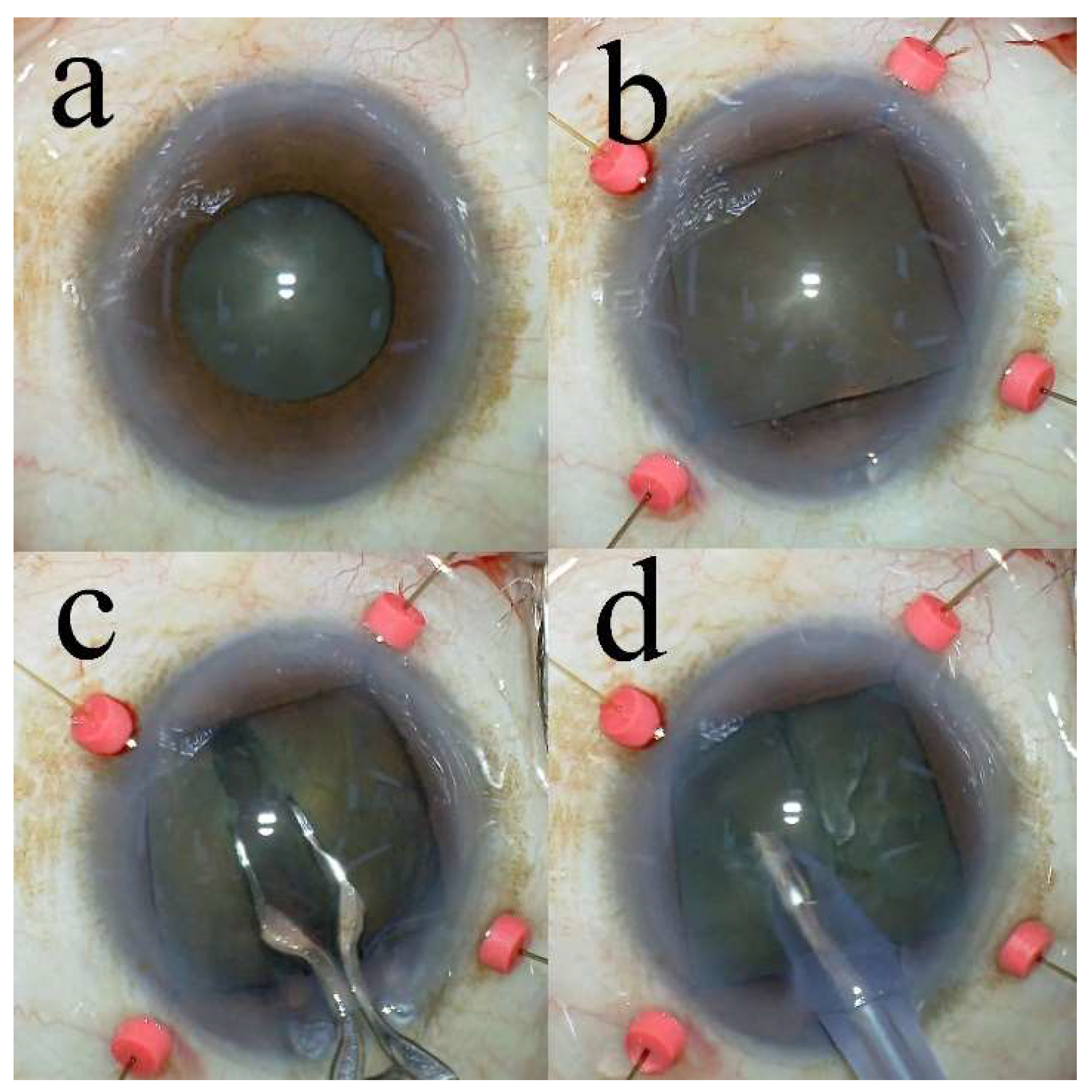
Figure 1. The phacoemulsification procedure. (a) The pupil diameter is 5.6 mm before installation of the iris hooks. (b) Four iris hooks are installed. (c) The eight-chop technique is performed by installing the iris hooks. (d) Phacoemulsification is performed by dividing the lens nucleus into eight sections.
Preprints 118802 g001
Table 1. Preoperative characteristics and intraoperative parameters.
Characteristic/Parameter Iris hooks group Control group p-Value
Number of eyes 65 65
Age (y) 76.8 ± 7.8 75.0 ± 6.2 .16a
Gender: Men 42 (65%) 16 (25%) <.01b
               Women 23 (35%) 49 (75%)
Diabetes mellitus 15 10 .27c
IFIS 10 0 <.01b
Preoperative pupil size (mm) 5.34 ± 0.53 7.62 ± 0.55 <.01d
Postoperative pupil size (mm) 6.37 ± 0.68 7.33 ± 0.65 <.01d
Iris hooking time (min) 3.3 ± 1.37 -
Operative time (min) 10.6 ± 2.32 4.6 ± 1.20 <.01d
Phaco time (s) 20.7 ± 6.4 16.2 ± 7.6 <.01d
Aspiration time (s) 101.1 ± 52.2 72.1 ± 18.6 <.01d
CDE 7.8 ± 2.44 7.0 ± 3.36 .16a
Volume of fluid used (mL) 38.0 ± 8.8 28.9 ± 9.2 <.01d
Values are expressed as mean ± standard deviation or numbers with percentages unless otherwise mentioned. a No significant differences were observed among the groups (unpaired t-test). b Significant differences were observed among the groups (chi-square test). c No significant differences were observed among the groups (chi-square test). d Significant differences were observed among the groups (unpaired t-test). IFIS, intraoperative floppy iris syndrome; CDE, cumulative dissipated energy.
Table 2. Pre- and postoperative best-corrected visual acuity and corneal endothelial cell density values.
Group/Parameter Preoperatively 7 weeks
postoperatively
19 weeks
postoperatively
p-Value
Iris hooks
BCVA (logMAR) (n = 65) 0.21 ± 0.33 −0.011 ± 0.083 −0.026 ± 0.078 <.01a, <.01a, <.01a
CECD (cells/mm2) (n = 37) 2503.6 ± 213.0 2437.4 ± 215.6 2448.8 ± 215.7 <.01a, .51b, <.01a
CECD loss (%) - 2.6 ± 4.3 2.1 ± 4.4 -
Control
BCVA (logMAR) (n = 65) 0.17 ± 0.29 −0.042 ± 0.074 −0.039 ± 0.075 <.01a, .42b, <.01a
CECD (cells/mm2) (n = 65) 2585.1 ± 236.2 2553.2 ± 254.6 2553.2 ± 246.0 <.01a, .99b, <.01a
CECD loss (%) - 1.3 ± 3.2 1.2 ± 3.2 -
p-Value .47c, .09c <.05d, <.05d .38c, <.05d
* Values represent mean ± standard deviation. p-Values in the right column are shown in the following order: preoperatively vs. 7 weeks postoperatively, 7 weeks postoperatively vs. 19 weeks postoperatively, and preoperatively vs. 19 weeks postoperatively. p-Values in the bottom row are shown in the order of BCVA, CECD. a Significant differences were observed between the groups (paired t-test). b No significant differences were observed between the groups (paired t-test). c No significant differences were observed between the groups (unpaired t-test). d Significant differences were observed between the groups (unpaired t-test). BCVA, best-corrected visual acuity; logMAR, logarithmic minimum angle of resolution; CECD, corneal endothelial cell density.
Table 3. Mean intraocular pressure and mean decrease in intraocular pressure over time.
Mean IOP (mmHg) ± SD (% decrease)
Examination Iris hooks group (n = 51) Control group (n = 65) p-Value
Preoperatively 14.5 ± 2.5 - 14.0 ± 2.2 - .33a
7 weeks
postoperatively
13.0 ± 2.7 (10.1 ± 15.4) 11.8 ± 2.3 (15.6 ± 12.3) <.01b <.01c <.01d
19 weeks
postoperatively
12.7 ± 2.6 (12.0 ± 11.8) 11.5 ± 2.4 (18.1 ± 9.7) <.01b <.01 c <.01d
IOP, intraocular pressure; SD, standard deviation. a No significant differences were observed between the groups (unpaired t-test). b Significant differences were observed between the groups (unpaired t-test). c Significant differences were observed between the preoperative and respective time values in the iris hooks group (paired t-test). d Significant differences were observed between the preoperative and respective time values in the control group (paired t-test).
Table 4. Mean intraocular pressure and mean decrease in the groups with preoperative intraocular pressure levels above and below 15 mmHg of the iris hooks and control groups.
Iris hooks group
Examination IOP above 15 mmHg group (n = 26) p-Value IOP below 15 mmHg group (n = 25) p-Value
Preoperatively 16.5 ± 1.4 - 12.5 ± 1.6 -
7 weeks postoperatively 14.6 ± 2.0 (11.3 ± 10.8) <.01a 11.3 ± 2.4 (8.9 ± 19.1) <.01a
19 weeks postoperatively 14.4 ± 1.9 (12.3 ± 11.2) <.01a 11.0 ± 2.0 (11.6 ± 12.7) <.01a
Control group
Examination IOP above 15 mmHg group (n = 27) p-Value IOP below 15 mmHg group (n = 38) p-Value
Preoperatively 16.0 ± 0.9 12.7 ± 1.3
7 weeks postoperatively 12.8 ± 2.0 (20.4 ± 12.1) <.01a 10.8 ± 1.9 (14.9 ± 13.0) <.01a
19 weeks postoperatively 12.8 ± 1.8 (20.2 ± 10.7) <.01a 10.6 ± 1.8 (16.7 ± 9.6) <.01a
Values are expressed as mean ± standard deviation or numbers with percentages unless otherwise mentioned. a Significant differences were observed between the preoperative and respective time values (paired t-test). IOP, intraocular pressure.
Disclaimer/Publisher’s Note: The statements, opinions and data contained in all publications are solely those of the individual author(s) and contributor(s) and not of MDPI and/or the editor(s). MDPI and/or the editor(s) disclaim responsibility for any injury to people or property resulting from any ideas, methods, instructions or products referred to in the content.
Copyright: This open access article is published under a Creative Commons CC BY 4.0 license, which permit the free download, distribution, and reuse, provided that the author and preprint are cited in any reuse.
Prerpints.org logo

Preprints.org is a free preprint server supported by MDPI in Basel, Switzerland.

Subscribe

© 2024 MDPI (Basel, Switzerland) unless otherwise stated