Preprint
Article

Fungi as Indicators of Indoor Air Quality: Their Effects on Human Health and Treatment

Altmetrics

Downloads

122

Views

37

Comments

0

This version is not peer-reviewed

Submitted:

23 September 2024

Posted:

24 September 2024

You are already at the latest version

Alerts
Abstract
Indoor air quality (IAQ) is a critical determinant of human health, with fungal contamination posing significant risks. In 2023, a total of 160 samples were collected from 40 selected rooms. These samples were cultured on Sabouraud Dextrose Agar (SDA) medium using a portable air sampler and incubated at 28°C for 7 days. Initial identification of fungal isolates was performed based on their morphology. Suspected isolates underwent further analysis using internal transcribed spacer (ITS) rDNA analysis. The results revealed 11 species across 8 genera in various rooms. The most prevalent fungi were Aspergillus niger (183 CFU) , Penicillium chrysogenum (139 CFU) and Cladosporium halotolerans (135 CFU). On the other hand, Curvularia hawaiiensis and A. ustus were rarely isolated (10 and 19 respectively). Effective treatment involves a combination of preventive measures, such as controlling humidity and ensuring proper ventilation, alongside targeted interventions like antifungal treatments and thorough cleaning. Continuous monitoring and management of indoor environments are essential to reduce fungal contamination, enhance air quality, and protect human health from the harmful effects of airborne fungi. This abstract underscore the importance of addressing indoor fungal contamination to promote safer, healthier living and working spaces.
Keywords: 
Subject: Biology and Life Sciences  -   Biology and Biotechnology

1. Introduction

Indoor air quality is a crucial determinant of environmental health, with poor air quality significantly impacting human well-being. Fungal contamination is a key factor influencing IAQ, as certain fungi can proliferate in indoor environments and pose various health risks. Common indoor fungi, including Aspergillus, Penicillium, Cladosporium, and Candida, are frequently detected in air samples from homes, offices, hospitals, and even in residential furniture (Sham et al., 2021). These fungi thrive in environments with elevated moisture levels, often found in areas with poor ventilation or high humidity. Dampness resulting from leaks, inadequate ventilation, or high humidity can create ideal conditions for fungal growth. This is particularly problematic in homes and offices, where these conditions can be exacerbated by the presence of carpets, upholstered furniture, and other materials that can retain moisture and fungal spores (Sánchez Espinosa et al., 2024). Indoor air typically contains only 10–30% of the fungal spore concentration found in outdoor environments, indicating that additional contamination sources indoors are minimal (WHO, 2020). Fungal diversity in indoor air is typically measured in colony-forming units (CFU), which indicates the concentration of viable fungal spores present. Studies have shown that CFU concentrations can vary widely, ranging from 20 to over 1300 CFU/m³ depending on factors such as building maintenance, ventilation, and moisture levels (Sánchez Espinosa et al., 2022). For instance, high CFU levels of Aspergillus and Penicillium species are often linked to damp conditions and inadequate ventilation, which can exacerbate respiratory problems and allergic reactions (Tang, 2015). Factors such as temperature, moisture, relative humidity, insulation, air circulation systems, and duct maintenance are key in regulating indoor air quality and influencing the presence of biological contaminants. Indoor environments generally have different bacterial concentrations and types compared to outdoor air, partly due to the lack of ultraviolet light that can kill airborne microorganisms outdoors (Bragoszewska et al., 2018). A concentration of 10^3 microorganisms/m³ is commonly considered a general safety threshold for indoor air quality (WHO, 2020; American Air and Water, 2020).
In medical centers and communities, especially in neonatal units, the presence of fungi is a major concern. The compromised immune systems of patients, coupled with the high humidity and ventilation systems in these critical areas, can lead to an increased risk of fungal infections and complications (Sánchez Espinosa et al., 2022). (Navale et al., 2021; Al Hallak et al., 2023). Commonly isolated fungi in these environments include Aspergillus niger, and Penicillium chrysogenum. These fungi are known to exacerbate respiratory issues and allergies. They produce mycotoxins such as aflatoxins and ochratoxin A, which can be detrimental to human health even at very low concentrations. Ingestion or inhalation of these toxins can lead to severe health problems.
Fungi in indoor air pose significant human health risks, particularly in environments where moisture levels are high. These microscopic organisms, including molds and yeasts, thrive in damp, warm conditions and can become airborne, leading to indoor air contamination. When inhaled, fungal spores can trigger allergic reactions, asthma, and other respiratory problems, particularly in individuals with weakened immune systems, the elderly, and young children. Some fungi can produce mycotoxins, which are toxic compounds that can lead to more severe health issues, including neurological symptoms and immune suppression (Chawla et al., 2023). Reducing fungal contamination and enhancing air quality requires a combination of preventive and treatment measures. Effective prevention includes managing moisture levels, improving ventilation, and addressing any leaks or water damage promptly. Regular cleaning is also essential in maintaining a clean and dry indoor environment. When fungal growth is detected, treatment options such as antifungal agents, air purifiers, and fungicidal cleaning products should be utilized. In more severe cases, professional remediation may be necessary to eliminate the contamination entirely. Evidence suggests that integrating these strategies with ongoing air quality monitoring can lead to significant improvements in health outcomes and reduce the risk of illnesses associated with fungal exposure (Sánchez Espinosa et al., 2022). The aim of this study was to isolate indoor air fungi and identify them through molecular analysis to assess the indoor air quality in various rooms within the Faculty of Sciences at King Abdulaziz University, providing insights into the necessary measures to mitigate the adverse effects of indoor fungal contamination on human health, While also highlighting the importance of monitoring and managing indoor air quality, and identifying effective treatments to enhance human health.

2. Materials and Methods

2.1. Sampling Locations

The study, conducted in 2023 at the Faculty of Sciences, Microbiology Section, Department of Biological Sciences, King Abdulaziz University in Jeddah, Saudi Arabia, involved the collection of 160 samples. Airborne fungal samples were taken at 11 a.m. from 40 selected rooms, including 10 classrooms, 10 staff offices, 10 microbiology laboratories and 10 toilets, with each location sampled twice monthly over a two-month period. All rooms were equipped with a forced-air ventilation system and air conditioning.

2.2. Sampling Method

Airborne fungi were isolated using the impaction method with a portable air sampler (e.g., Spin air sampler, IUL micro, Spain). The sampler was positioned at a height of approximately 1.5 meters, simulating the breathing zone of occupants. For each location, air samples were collected for 5 minutes at a flow rate of 28.3 liters per minute (L/min) onto 90-mm Petri dishes containing SDA supplemented with chloramphenicol (50 μg/mL, QUELAB, USA) to inhibit bacterial growth (Tabatabaei et al., 2020). The internal part of the sampler was cleaned with 70% alcohol before each use.

2.3. Incubation and Identification

After sampling, the Petri dishes were sealed and transported to the laboratory for incubation. The plates were incubated at 25°C for 5-7 days in the dark. After incubation, fungal colonies concentrations were counted and recorded as colony-forming units (CFU) of air. Morphologically distinct colonies were subcultured onto fresh SDA plates to obtain pure cultures.

2.4. Microscopic Examination

After isolation, the identification of fungi was based on both macroscopic and microscopic characteristics. This included assessing colony color, texture, size, and the structure of reproductive features such as spores, conidia, and hyphae. For microscopic analysis, a small portion of each fungal colony was placed on a microscope slide, with a drop of lactophenol cotton blue added. The slide was then covered with a coverslip to create a thin, uniform layer. These prepared slides were examined under a light microscope at magnifications of 400x to 1000x to observe the detailed morphology of the fungal structures. Standard mycological keys and references were employed to compare these features with known fungal species. For accurate and updated fungal identification, the 2020 edition Pocket Guide to Mycological Diagnosis by De Aguiar et al. (2020) was used, providing contemporary methods and diagnostic criteria for reliable classification.

2.5. Molecular Analysis

The method for fungal genomic DNA extraction was conducted as follows Alghamdi et al. (2023). A sample was taken from a pure fungal colony grown on SDA and transferred into a 100 ml Erlenmeyer flask containing 20 ml of potato dextrose broth (PDB). This was then incubated at 28°C for 7 days. Following incubation, the broth was filtered through a 0.22µm sterilized nitrocellulose filter paper to collect the mycelia. The collected mycelia were washed several times with deionized sterilized water and stored at -20°C. The frozen mycelia were then mixed with liquid nitrogen and ground into a fine powder using a sterilized pestle and mortar. This powder was distributed into 1.5 ml Eppendorf centrifuge tubes and stored at -20°C.
The DNA extraction was performed separately for each fungus. Thirty mg of frozen, ground mycelia were lysed with the lysis buffer from the Gene JET Genomic DNA extraction kit (Thermo Scientific, USA, 10223). The samples were suspended in 500 µl of lysis buffer and incubated in a water bath at 37 °C for 60 minutes. The ITS region of ribosomal DNA (rDNA) was amplified using polymerase chain reaction (PCR). Internal transcribed spacer (ITS) rDNA including (CTTGGTCATTTAGGGAAGTAA) and ITS4 (TCCTCCGCTTATTGATATGC) in a thermal cycler (Esco Healthcare, Swift Max Pro, Malaysia). The 50 µl reaction mixture contained 3 µl of template DNA, 5 µl of each primer, 25 µl of Go Taq® Green Master Mix (Promega, USA), and 50 µl of nuclease-free water. The PCR procedure involved an initial denaturation at 96 °C for 1 minute, followed by 35 amplification cycles at 94 °C for 1 minute, 56 °C for 45 seconds, and 72 °C for 1 minute, concluding with a final extension at 72 °C for 6 minutes. A negative control was prepared by using DNA extract without any additional reagents.
The DNA loading dye was combined with each PCR product, and 20 µl of the mixture was loaded onto a 1.5% agarose gel. The gel was subjected to horizontal electrophoresis (Cleaver Scientific, UK) for 45 minutes at 130 volts and stained with ethidium bromide. A 20 µl DNA marker (100 bp, Invitrogen, USA) was included to help quantify and identify the PCR products. Bands were visualized using UV light (Gel Doc Imager, BioRad, USA). Clear bands were then excised and sent to Macrogen, South Korea, for purification and sequencing. Sequence identities were determined using the Basic Local Alignment Search Tool (BLAST) against general GenBank databases from the National Center for Biotechnology Information (NCBI). Sequence alignments and neighbor analyses were performed using MEGA-X software.

2.6. Data Analysis

Fungal concentrations (CFU/m) were calculated and statistically analyzed to determine the differences in fungal load between different locations. Descriptive statistics and analysis of variance (ANOVA) were used to compare the mean fungal counts across the sampling sites.

3. Results and Discussion

3.1. The Concentration and Diversity of Fungal Isolates from Indoor Air

The total count across all the studied rooms is 971 CFU, reflecting the presence and distribution of 8 fungal genera including Alternaria, Aspergillus, Cladosporium, Curvularia, Fusarium, Paecilomyces, Penicillium and Trichoderma shown in Table 1. Classrooms exhibit the highest concentration of fungal isolates, with a total of 400 CFU, which is significantly higher compared to other locations (p < 0.05). Staff offices follow with a significant concentration of 239 CFU, showing a notable difference from classrooms (p < 0.05). Toilets rank third with 171 CFU, significantly lower than classrooms (p < 0.05) and staff offices (p < 0.05). Microbiology laboratories have the lowest concentration at 161 CFU, which is significantly lower compared to classrooms (p < 0.05), staff offices (p < 0.05), and toilets (p < 0.05). The distribution of fungal isolates indicates that classrooms, with the highest human activity and potential dust accumulation, are the most conducive environment for fungal growth, followed by staff offices. Toilets, likely due to higher moisture levels, also support significant fungal presence. Laboratories, despite being controlled environments, still harbor a notable number of fungi. Also, classrooms exhibit the greatest fungal diversity, hosting all 11 identified fungal species. Staff offices also show significant diversity with 8 different fungal species. Toilets contain 9 fungal species. Microbiology laboratories, despite their controlled environments, have a diverse presence of 9 fungal types.
In our study, the concentration and diversity of fungal isolates from indoor air have been extensively studied, revealing a wide range of species and varying levels of concentration. Aspergillus, Penicillium and Cladosporium species were identified as among the most prevalent fungi, found abundantly in various environments. These findings align with studies by Navale et al. (2021) and Belizario et al. (2021). A review of research from 2005 to 2019 highlighted the presence of Aspergillus, Penicillium and Cladosporium species in the indoor air of critical hospital areas. Also, among the common fungi identified in indoor environments are Cladosporium halotolerans, Paecilomyces variotii, and Trichoderma harzianum, each with its own unique characteristics and implications for indoor air quality. Cladosporium species is often found in indoor environments, particularly in areas with high humidityStudies have shown that Cladosporium species, including C. halotolerans, can contribute significantly to indoor air spore concentrations, particularly in damp or water-damaged buildings. This species is known for its tolerance to salt, which allows it to thrive in conditions where other fungi might not survive. (Sánchez Espinosa et al., 2022). Paecilomyces variotii has been reported as another common indoor fungus, frequently isolated from air samples in various studies. This species is known for its ability to grow in a wide range of environmental conditions, including those with limited nutrients and varying temperatures (Lu et al., 2021). Trichoderma harzianum is a well-known indoor fungus, often found in soil and decaying organic matter. However, it can also colonize indoor environments, particularly in areas with high moisture levels. Trichoderma species are known for their antagonistic properties against other fungi, making them a common contaminant in buildings with water damage or mold issues (Polizzi et al., 2011). A study in Havana, Cuba, sampled indoor from 44 bedrooms during 2018 and 2019. Results indicated poor indoor air quality in 18 bedrooms, with concentrations of fungal propagules between 20 and 1330 CFU/m³. The most frequent genera identified were Cladosporium, Aspergillus, Penicillium, and Curvularia. Another study using qPCR found Aspergillus, Penicillium, and Paecilomyces variotii among the detected species, highlighting seasonal variations and the presence of fungal fragments in indoor air (Lu et al., 2021; Sánchez Espinosa et al., 2022).
In contrast, Curvularia sp. was more restricted in this study, primarily occurring outdoors rarely present indoors. This distribution underscores the adaptability of fungal species and their varied prevalence in different indoor settings. Curvularia species, is primarily associated with plants and soil, thriving in outdoor environments. While it can occasionally be detected indoors, it is not typically a dominant mold. Its spores may enter buildings through ventilation systems or open windows, but its lower prevalence indoors minimizes its impact compared to more common indoor fungi like Aspergillus and Penicillium species (Tang, 2015). Aspergillus ustus is another species that is rarely present in indoor air. It is primarily found in soil and decaying organic matter, making it more common outdoors. Although it can occasionally be detected indoors, its prevalence is significantly lower compared to other Aspergillus species. This limited presence indicates that A. ustus is not a predominant indoor mold, further emphasizing the variability in fungal species distribution between indoor and outdoor environments (Andersson et al., 2022).
PCR amplifications of DNA extracted from the 11 fungal species were performed using the ITS1/4 universal fungal primer pair. The sequencing data of the fungal strains were then aligned with sequences of closely related strains available in GenBank. The Table 2 presents the fungal accession numbers, closest related species, and their similarity percentages based on sequencing data. Alternaria alternata, with accession number OR533706.1, shows a 100% similarity to its closest related species. Aspergillus flavus (MT635198.1) has an 89.29% similarity, while A. niger (JX556221.1) has a 90% similarity. Aspergillus ustus (KC800599.1) exhibits an 88.83% similarity, and C. hawaiiensis (KY788103.1) has an 87.32% similarity. Fusarium proliferatum (MT466521.1) shows a 91.21% similarity, and P. variotii (MN547409.1) has an 88.57% similarity. Penicillium chrysogenum (OK510242.1) exhibits an 89.69% similarity, while P. citrinum (MH990629.1) shows a 91.24% similarity. Cladosporium halotolerans (MW412494.1) has a 93.07% similarity, and T. harzianum (MF108874.1) exhibits a 91.43% similarity. This data highlights the genetic relationships and diversity among the studied fungal isolates.
The provided phylogenetic tree illustrates the genetic relationships among various fungal isolates based on sequencing data as shown in Figure 1. Curvularia hawaiiensis (KY788103.1) stands as a distinct outgroup, indicating its unique genetic position. Alternaria alternata (OR533706.1) and C. halotolerans (MW412494.1) form a close cluster, showing a strong genetic similarity. Fusarium proliferatum (MT466521.1) and T. harzianum (MF108874.1) also cluster together, indicating a closer genetic relationship. Paecilomyces variotii (MN547409.1) is positioned separately, suggesting moderate similarity to the others. Penicillium species, including P. chrysogenum (OK510242.1) and P. citrinum (MH990629.1), show close genetic ties. Aspergillus species such as A. flavus (MT635198.1), A. niger (JX556221.1), and A. ustus (KC800599.1) form a tight cluster, highlighting their genetic relatedness. Similar studies have isolated A. flavus, A. niger, and A. ustus from indoor air environments in research conducted (Nafis et al., 2023; Espinosa et al., 2021).
This tree emphasizes the diversity and evolutionary relationships among the studied fungal isolates, providing insights into their genetic affiliations. All 11 fungal species in the current study belong to the phylum Ascomycota. Most Ascomycota mycelium, composed of hyphae or filaments, forms the vegetative body of fungi. Spores, dispersed through the air, contribute to the anemophilous microbiota. These spores can be pathogenic, causing severe symptoms in immunocompromised individuals or those on long-term antimicrobial treatments. Variations in fungal isolate quantities and identities across studies are influenced by factors like exposure locations, collection techniques, identification methods (both conventional and molecular), and environmental conditions such as temperature and humidity (Tian et al., 2024).

3.2. Human Health Risks Associated with Indoor Fungi

Aspergillus species, such as Aspergillus niger, are commonly found in indoor environments and can pose serious health hazards to human health. Aspergillus niger is a saprophytic fungus that thrives in damp and poorly ventilated areas, making it a common indoor contaminant. Inhalation of Aspergillus spores can cause a variety of respiratory issues, including allergic reactions, asthma exacerbations, and allergic bronchopulmonary aspergillosis (ABPA). For immunocompromised individuals, the risk is significantly higher as A. niger can cause invasive aspergillosis, a severe and potentially fatal infection that can spread from the lungs to other parts of the body. The symptoms of invasive aspergillosis include fever, chest pain, cough, and hemoptysis (coughing up blood), and the infection requires prompt medical treatment (Agarwal et al., 2024). Penicillium species, including Penicillium chrysogenum, are commonly found in indoor environments, where they can become a significant source of allergens. These fungi thrive in conditions of high humidity and poor ventilation, often contaminating indoor air and surfaces. The spores released by Penicillium can contribute to various health issues, particularly for individuals with pre-existing respiratory conditions or compromised immune systems. Exposure to Penicillium spores is known to exacerbate asthma and other respiratory conditions. Individuals with asthma may experience increased frequency and severity of symptoms such as coughing, wheezing, and shortness of breath when exposed to these fungal spores. This exacerbation occurs because the allergens from Penicillium can trigger inflammatory responses in the airways, leading to heightened asthma symptoms and potentially more frequent asthma attacks. In individuals with chronic respiratory conditions or weakened immune systems, prolonged exposure can also lead to more severe health complications (Xing et al., 2022). Moreover, Penicillium chrysogenum has been associated with other allergic reactions and respiratory conditions beyond asthma. Studies have shown that inhalation of Penicillium spores can induce hypersensitivity pneumonitis, a condition where the lungs become inflamed due to an allergic reaction to inhaled organic dust, including fungal spores (Al Hallak et al., 2023). Cladosporium halotolerans, pose significant health risks to humans. Exposure to these fungi can lead to respiratory problems, allergic reactions, and exacerbate conditions like asthma. Cladosporium species, although less common, can cause phaeohyphomycosis, a serious fungal infection, particularly in immunocompromised individuals (Zhou et al., 2023).

3.3. Monitoring and Controlling Indoor Air Quality

To manage indoor fungal contamination and maintain a healthy environment, it is essential to implement regular monitoring and control measures. Proper ventilation is a critical factor in reducing indoor mold growth. Ensuring adequate airflow can help prevent the accumulation of moisture, which is a key contributor to fungal proliferation. Regular maintenance of HVAC systems and the use of air purifiers can also improve indoor air quality by reducing the concentration of airborne spores (Chen et al., 2023).
Moisture control is another crucial aspect of managing indoor fungal contamination. Leaks, condensation, and high humidity levels create favorable conditions for mold growth. Addressing these issues promptly through repairs and dehumidification can help prevent fungal proliferation. In areas prone to moisture, such as bathrooms and kitchens, the use of exhaust fans and moisture-resistant materials can further reduce the risk of mold growth (Engel et al., 2024).
Regular cleaning and maintenance are essential for minimizing fungal contamination. Dust and organic matter provide a substrate for fungal growth, so routine cleaning of surfaces, carpets, and ventilation systems is necessary to reduce the availability of these materials. Using cleaning agents with antifungal properties can also help eliminate mold spores from surfaces. The diversity and concentration of these and other fungal species in indoor air highlight the complexity of indoor air quality management. Factors such as humidity, ventilation, building materials, and human activity all influence the types and levels of fungi present. This includes regular inspection and maintenance of HVAC systems, controlling humidity levels, and promptly addressing water damage to prevent the proliferation of fungi like Cladosporium halotolerans, Paecilomyces variotii, and Trichoderma harzianum (Loukou et al., 2024).
Monitoring indoor air quality is vital for early detection of fungal contamination. Air sampling and spore counts can provide valuable information about the types and concentrations of fungi present in indoor environments. This data can inform targeted interventions to address specific sources of contamination and improve overall air quality. The use of molecular techniques for pathogen identification has enhanced the accuracy of fungal monitoring, allowing for more precise identification and tracking of fungal species (Tang et al., 2015).

3.4. Airborne Fungi Treatment

Effective treatment of fungal infections requires an understanding of the specific pathogens involved, as A. niger, P. chrysogenum, and C. halotolerans each present unique challenges and necessitate targeted antifungal therapies.
For infections caused by A. niger and P. chrysogenum, the primary treatment strategies involve similar antifungal agents due to their overlapping efficacy against various molds. Voriconazole is often the treatment of choice for both fungi. This newer antifungal agent is effective against a broad spectrum of fungi by inhibiting fungal cytochrome P450 enzymes, disrupting ergosterol synthesis, and thereby compromising fungal cell membrane integrity. Amphotericin B is another option for these infections; it works by binding to ergosterol in the fungal cell membrane, causing membrane disruption. It is particularly used in severe cases or when voriconazole is not tolerated. Lipid formulations of amphotericin B may be preferred to minimize renal and hepatic toxicity. Both voriconazole and amphotericin B can have serious side effects and may interact with other medications, so careful monitoring for adverse effects and drug interactions is essential. Accurate diagnosis and susceptibility testing are crucial for guiding appropriate antifungal therapy (Avilés-Robles et al., 2016).
Cladosporium halotolerans, a less common cause of phaeohyphomycosis, requires specific treatment approaches. Itraconazole, an azole antifungal, is used for Cladosporium infections. It inhibits ergosterol synthesis in the fungal cell membrane, similar to voriconazole but with a broader spectrum. For refractory cases, especially those involving skin lesions, aminolevulinic acid photodynamic therapy (ALA-PDT) has proven effective. This treatment involves applying a photosensitizing agent followed by light exposure to target and destroy fungal cells. In cases where lesions are localized and accessible, surgical intervention can be effective, though it may not always be feasible, particularly for periorbital or other challenging locations (Zhou et al., 2023).
Each treatment approach must be tailored to the specific fungal pathogen and patient condition, with careful monitoring for side effects and drug interactions. For all these infections, a multidisciplinary approach involving infectious disease specialists is often beneficial to optimize outcomes.

4. Conclusion

Fungi in indoor air serve as critical indicators of air quality, with significant implications for human health. Exposure to airborne fungi can lead to respiratory issues, allergies, and more severe conditions in vulnerable populations. Effective management involves regular monitoring of indoor air quality, controlling moisture, and implementing proper ventilation. Treatment strategies, including antifungal medications and thorough cleaning, are essential for addressing fungal contamination. By maintaining a clean, dry indoor environment and employing targeted interventions, we can significantly reduce the health risks associated with indoor fungal exposure and promote better overall air quality.

Acknowledgments

This project was supported by the Deanship of Scientific Research (DSR) at King Abdulaziz University, Jeddah. The authors, therefore, acknowledge with thanks DSR.

References

  1. Agarwal: R., Muthu, V., & Sehgal, I.S. (2024). Clinical Manifestation and Treatment of Allergic Bronchopulmonary Aspergillosis. Semin Respir Crit Care Med, 45(01), 114-127.
  2. Al Hallak, M., Verdier, T., Bertron, A., Roques, C., & Bailly, J.-D. (2023). Fungal Contamination of Building Materials and the Aerosolization of Particles and Toxins in Indoor Air and Their Associated Risks to Health: A Review. Toxins, 15(3), 175. [CrossRef]
  3. Alghamdi, R.G., Zabermawi, N.M., Altihani, F.A., Bokhari, F.M., Makki, R.M., Hassoubah, S.A., Sharawi, Z.W., & Najjar, A.A. (2023). Diversity and Density of Fungi Isolated from Dried Fruits. Journal of Biochemical Technology, 14(4), 45-55.
  4. Andersson, M.; Varga, A.; Mikkola, R.; Vornanen-Winqvist, C.; Salo, J.; Kredics, L.; Kocsubé, S.; Salonen, H. Aspergillus Was the Dominant Genus Found during Diversity Tracking of Potentially Pathogenic Indoor Fungal Isolates. 2022. Pathogens. 10(2), 22-34.
  5. American Air and Water (2020) Mold, mold spores and indoor air quality. American Air and Water. Available at https:// www. ameri canai randw ater. com/ mold Accessed on: 8 June 2020.
  6. Avilés-Robles, M., Gómez-Ponce, C., Reséndiz-Sánchez, J., Rodríguez-Tovar, A.V., Ceballos-Bocanegra, A., & Martínez-Rivera, Á. (2016). Disseminated penicilliosis due to Penicillium chrysogenum in a pediatric patient with Henoch–Schönlein syndrome. International Journal of Infectious Diseases, 51, 78-80.
  7. Belizario, J.A., Lopes, L.G., & Pires, R.H. (2021). Fungi in the indoor air of critical hospital areas: A review. Aerobiologia, 37, 379-394.
  8. Chawla, H., Anand, P., Garg, K., Bhagat, N., Varmani, S.G., Bansal, T., McBain, A.J., & Marwah, R.G. (2023). A comprehensive review of microbial contamination in the indoor environment: Sources, sampling, health risks, and mitigation strategies. Frontiers in Public Health, 11(2), 128-133.
  9. Chen, X., Wang, Y., Wang, Y., Zhang, Y., Shen, Y., He, X., & Xiao, C. (2023). A natural moisture gradient affects soil fungal communities on the south shore of Hulun Lake, Inner Mongolia, China. Journal of Fungi, 9(5), 549. [CrossRef]
  10. De Aguiar, C., R. (2020). Pocket Guide to Mycological Diagnosis (1st ed.). Taylor and Francis. CRC Press.
  11. Engel, A., Simler-Williamson, A., Ravenscraft, A., Bittleston, L., & de Graaf, M.-A. (2024). Interactive effects of fungal community structure and soil moisture on Wyoming big sagebrush performance. Plant and Soil. [CrossRef]
  12. Espinosa, K.C.S., Chávez, M.A., Duarte-Escalante, E., Flores, T.I.R., Frías-De-León, M.G., & Reyes-Montes, M.R. (2021). Phylogenetic Identification, Diversity, and Richness of Aspergillus from Homes in Havana, Cuba. Microorganisms, 9(1), 115. [CrossRef]
  13. Kumar, P., Kausar, M.A., Singh, A.B., & Singh, R. (2021). Biological contaminants in the indoor air environment and their impacts on human health. Air Quality, Atmosphere & Health, 14, 1723–1736.
  14. Loukou, E., Jensen, N.F., Rohde, L., & Andersen, B. (2024). Damp buildings: Associated fungi and how to find them. Journal of Fungi, 10(2), 108. [CrossRef]
  15. Lu, R.; Pørneki, A.D.; Lindgreen, J.N.; Li, Y.; Madsen, A.M. Species of Fungi and Pollen in the PM1 and the Inhalable Fraction of Indoor Air in Homes. Atmosphere 2021, 12, 404. [CrossRef]
  16. Nafis, M.M.H., Quach, Z.M., Al-Shaarani, A.A.Q.A., Muafa, M.H.M., & Pecoraro, L. (2023). Pathogenicity of Aspergillus Airborne Fungal Species Collected from Indoor and Outdoor Public Areas in Tianjin, China. Pathogens, 12(9), 1154. [CrossRef]
  17. Navale, V., Vamkudoth, K.R., Ajmera, S. and Dhuria, V. (2021). Navale, V., Vamkudoth, K.R., Ajmera, S. and Dhuria, V. (2021). Aspergillus derived mycotoxins in food and the environment: Prevalence, detection, and toxicity. Toxicology Reports, 8, 1008-1030. [CrossRef]
  18. Polizzi, V., Adams, A., Picco, A.M., Adriaens, E., Lenoir, J., Van Peteghem, C., De Saeger, S., & De Kimpe, N. (2011). Influence of environmental conditions on production of volatiles by Trichoderma atroviride in relation with the sick building syndrome. Building and Environment, 46(4), 945-954.
  19. Sánchez Espinosa, K.C., Rodríguez Davydenko, S., Rojas Flores, T.I., Venero Fernández, S.J., & Almaguer, M. (2022). Indoor air quality and diversity of fungi inside and outside residences of children with a history of allergy in Cuba. Grana, 61(4), 284-295.
  20. Sánchez Espinosa, K.C., Rodríguez Davydenko, S., Rojas Flores, T.I., Fernández-González, M., & Almaguer, M. (2024). Xerophilic and cellulolytic fungi in the indoor air of houses in Havana. International Biodeterioration & Biodegradation, 188, 105730.
  21. Sham, N.M., Ahmad, N.I., Pahrol, M.A., & Leong, Y.-H. (2021). Fungus and mycotoxins studies in hospital environment: A scoping review. Building and Environment, 193, 107626.
  22. Tabatabaei, Z., Rafiee, A., Abbasi, A., Mehdizadeh, A., Morovati, R., & Hoseini, M. (2020). Investigation of fungal contamination in indoor air and on surfaces of traditional public baths in a historical city. Journal of Environmental Health Science and Engineering, 18(2), 925–932. [CrossRef]
  23. Tang, J.W. (2015). Investigating the airborne transmission pathway – different approaches with the same objectives. Indoor Air, 25(2), 119–124. [CrossRef]
  24. Tian, X.G., Bao, D.F., Karunarathna, S.C., Jayawardena, R.S., Hyde, K.D., Bhat, D.J., Luo, Z.L., Elgorban, A.M., Hongsanan, S., Rajeshkumar, K.C., Maharachchikumbura, S.S.N., Suwannarach, N., Dawoud, T.M., Lu, Y.Z., Han, J.J., Xiao, Y.P., Du, T.Y., Lu, L., Xu, R.F., Dai, D.Q., Liu, X.F., Liu, C., & Tibpromma, S. (2024). Taxonomy and phylogeny of ascomycetes associated with selected economically important monocotyledons in China and Thailand. Mycosphere, 15(1), 1–274.
  25. Xing, H., Wang, J., Sun, Y., & Wang, H. (2022). Recent Advances in the Allergic Cross-Reactivity between Fungi and Foods. Journal of Immunology Research, 2022, Article ID 7583400. [CrossRef]
  26. WHO (2020) Coronavirus disease (COVID-19) World Health Organization situation reports 142. Available on: https:// www. who. int/docs/ defau lt- source/ coron aviru se/ situa tion- repor ts Accessed on-10 June 2020.
  27. Zhou, H., Xia, X., Wang, Y., Ma, Y., Zhao, Y., Wang, P., Tang, C., & Wang, P. (2023). A rare case of refractory facial phaeohyphomycosis caused by Cladosporium halotolerans successfully treated with aminolevulinic acid photodynamic therapy. Photodiagnosis and Photodynamic Therapy, 42, 103347.
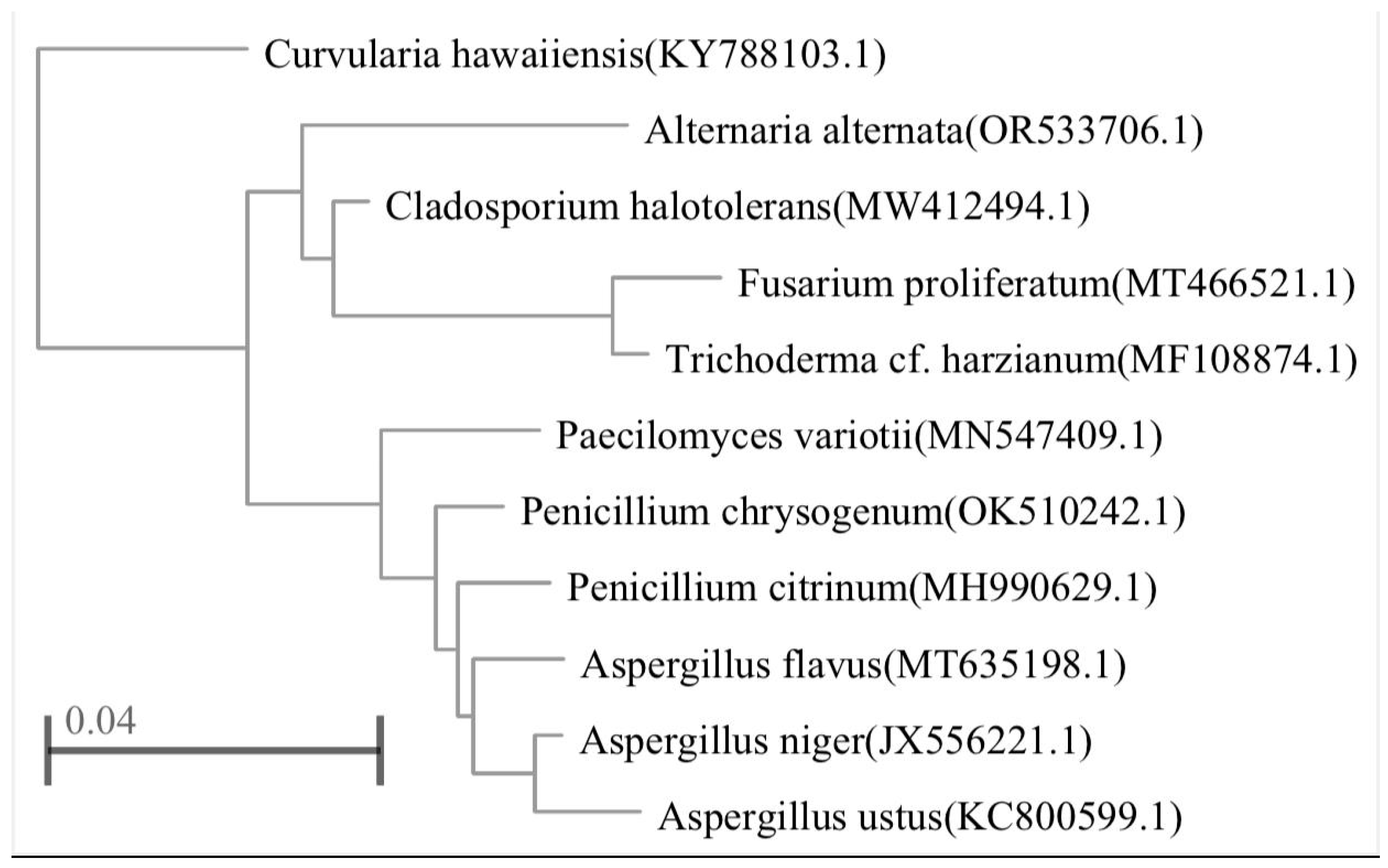
Figure 1. Dendrogram showing phylogenetic analysis based on the ITS region and NCBI GenBank database for 11 fungal species.
Preprints 119099 g001
Table 1. Fungal concentration in the air of the studied rooms (CFU).
No. Fungal isolates Classrooms Staff offices Microbiology laboratories Toilets Total(CFU)
1 Alternaria sp. 24 0 12 0 36
2 Aspergillus flavus 28 10 22 36 96
3 A. niger 80 40 38 25 183
4 A. ustus 19 0 0 0 19
5 Cladosporium sp. 64 29 20 22 135
6 Curvularia sp. 10 0 0 0 10
7 Fusarium sp. 0 22 0 3 25
8 Paecilomyces variotii 50 41 18 11 120
9 Penicillium sp.1 44 50 17 28 139
10 Penicillium sp.2 31 11 22 19 83
11 Trichoderma sp. 50 36 12 27 125
Total 400* 239* 161* 171* 971
* Significant difference at (p 0.05).
Table 2. List of fungal species accession numbers isolated from indoor air of the studied rooms and most closely related fungal species with their similarity percentage found in the NCBI website.
Fungal accession numbers Closest related species Similarity (%)
Alternaria alternate OR533706.1 100
Aspergillus flavus MT635198.1 89.29
A. niger JX556221.1 90
A. ustus KC800599.1 88.83
Curvularia hawaiiensis KY788103.1 87.32
Fusarium proliferatum MT466521.1 91.21
Paecilomyces variotii MN547409.1 88.57
Penicillium chrysogenum OK510242.1 89.69
P. citrinum MH990629.1 91.24
Cladosporium halotolerans MW412494.1 93.07
Trichoderma harzianum MF108874.1 91.43
Disclaimer/Publisher’s Note: The statements, opinions and data contained in all publications are solely those of the individual author(s) and contributor(s) and not of MDPI and/or the editor(s). MDPI and/or the editor(s) disclaim responsibility for any injury to people or property resulting from any ideas, methods, instructions or products referred to in the content.
Copyright: This open access article is published under a Creative Commons CC BY 4.0 license, which permit the free download, distribution, and reuse, provided that the author and preprint are cited in any reuse.
Prerpints.org logo

Preprints.org is a free preprint server supported by MDPI in Basel, Switzerland.

Subscribe

© 2024 MDPI (Basel, Switzerland) unless otherwise stated